radiology cardiovascular system dr. abeer kadem radiologist
TRANSCRIPT

RADIOLOGY
Cardiovascular System
Dr. Abeer KademRadiologist

Imaging Techniques:
1 -Plain Radiography:
*The standard plain films for evaluation of cardiac diseases are the PA view & Lateral chest film, the PA view must be sufficiently penetrated to see the shadow within the heart, eg. The double contour of the Lt. atrium & valve & pericardial calcification.
*It provides limited information's about the Heart.
*It provides limited information's about the effect of the cardiac diseases on the lungs & pleural cavities.
*We should assess the following points:
a- Heart (shape & size). b- Great vessels (size, shape), Aortic arch (normally
located to the Lt. of the Trachea, we should exclude the signs of coarctation of aorta).
c- If there is any calcification. d- The main point is the examination of the Lung field
for altered blood flow & if there is any evidence of heart failure.
** Note :Look for any thoracic abnormality (such as Pectus Excavatum).
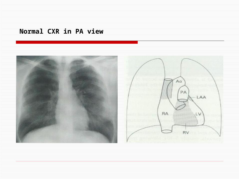
Normal CXR in PA view
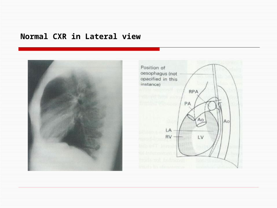
Normal CXR in Lateral view

2- Echocardiography(Cardiac US) :
*It is the major or basic imaging technique used in cardiology.
*It gives important informations about the Morphology& Function of the heart.
*It is an excellent technique to look for:
a- Heart valves. b- Chamber morphology & volume.
c- Determining the ventricular wall thickness. d- Any intra-luminal mass.
3 basic techniques are used inEchocardiography, & they are :
a) M-mode :
*It is a continuous scan over a period of time (5-10 seconds), with pencil – beam of sound directed to the site of interest.
*It can demonstrate chamber dimensions, wall thickness, & valve movement (mainly for Lt. ventricular dimension in systole & diastole).

M-mode
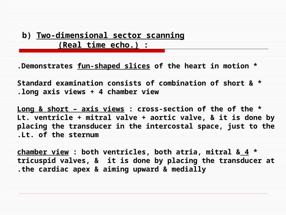
b) Two-dimensional sector scanning(Real time echo.) :
*Demonstrates fun-shaped slices of the heart in motion.
*Standard examination consists of combination of short & long axis views + 4 chamber view.
*Long & short – axis views : cross-section of the of the Lt. ventricle + mitral valve + aortic valve, & it is done by placing the transducer in the intercostal space, just to the Lt. of the sternum.
*4 chamber view : both ventricles, both atria, mitral & tricuspid valves, & it is done by placing the transducer at the cardiac apex & aiming upward & medially.
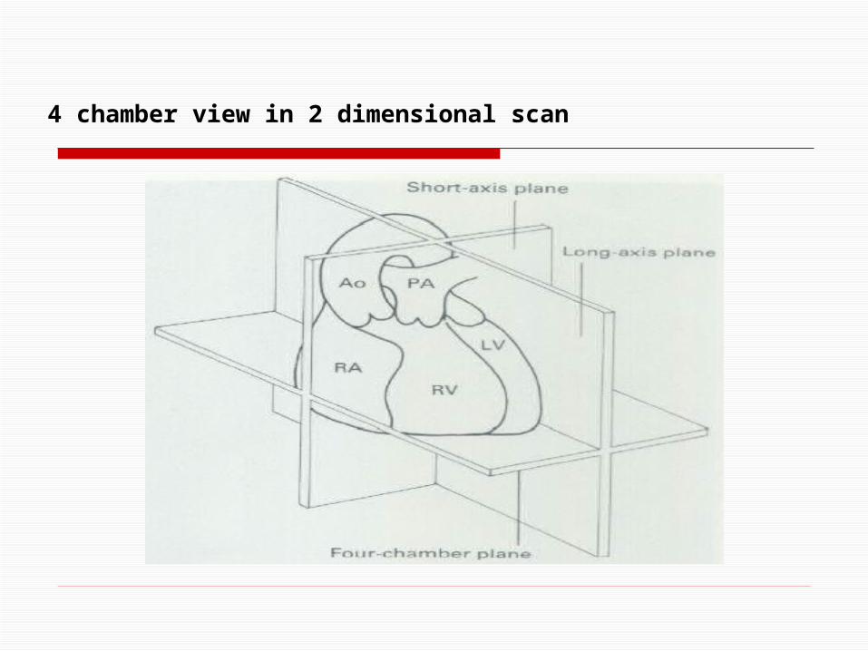
4 chamber view in 2 dimensional scan

Para-sternal long axis

Para-sternal short axis

Apical 4 chamber view

Para-sternal short axis(at Mitral valve level)

*Changing in the frequency of the sound waves are reflected from moving objects, this change depends on the velocity of the reflecting surface.
*RBCs are used as reflecting surface & the velocity of the blood flow can be measured.
c) Doppler echocardiography(Color, Pulse wave):

Doppler flow measurements are used to :
1 -Measure cardiac output or Lt. to Rt. shunt.
2 -Detect & quantify valvular regurgitation.
3 -Quantify pressure gradients across stenotic valves.
4 -Quantify flow.

3- Trans-Esophageal Echocardiography :
*By placing the U.S. probe in the esophagus immediately behind the Lt. atrium, so it will view the heart from behind.

Trans-Esophageal Echocardiography
(A = normal descending thoracic aorta)

RADIOLOGY
Cardiovascular System
Lec. II
Heart Diseases
*Evidence of heart diseases is given by:
1 -Size & shape of the heart.
2 -Pulmonary vessels, which provide information about the blood flow.
3 -The lungs, which may show pulmonary edema.
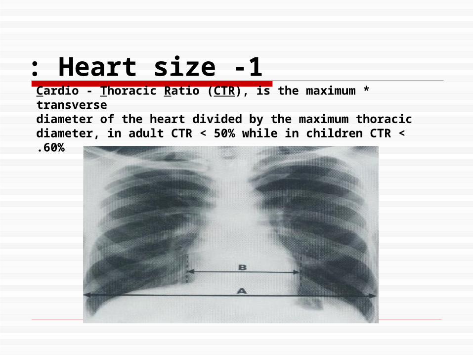
1 -Heart size : *Cardio - Thoracic Ratio (CTR), is the maximum transverse
diameter of the heart divided by the maximum thoracic diameter, in adult CTR < 50% while in children CTR < 60%.

*Comparing with previous films chest-x-ray films is often more useful.
-The transverse cardiac diameter varies with the phase of respiration & with cardiac cycle, so if the change in the cardiac size is < 1.5 cm; this is negligible because the heart size is affected by breathing & cardiac cycle.
*Overall increase in the heart size means: - Dilatation of more than one cardiac chamber.
- Pericardial effusion.
1 -Heart size :

2 -Chamber hypertrophy & dilatation:
a) Plain X-ray films:
*Pressure overload (as in : Hypertension, Aortic Stenosis, Pulmonary Stenosis), this will lead toventricular wall hypertrophy, & such change will produce little change in the external contour of the heart, until the ventricle fails.

*Volume overload (as in : Mitral Incompetence, Aortic Incompetence, Pulmonary Incompetence, Lt. to Rt. Shunt, & Damage of the heart muscle), this will lead to dilatation of the relevant ventricle, & this will cause an overall increase in the size of the heart (increase in the transverse cardiac
diameter.)
*Because enlargement of one ventricle affects the shape of the other, so it is only occasionally possible to get the classical feature Lt. or Rt. Ventricular enlargement.
a) Plain X-ray films:

-Lt. Ventricular enlargement, the cardiac apex is displaced downward & laterally.
Lt. Ventricular enlargement in a patient with Aortic Incompetence
a) Plain X-ray films:

-Rt. Ventricular enlargement, the cardiac apex is displaced upward (to the Lt. of diaphragm).
Rt. Ventricular enlargement in a patient with Primary Pulmonary Hypertension
a) Plain X-ray films:
Lt. Atrial Enlargement:
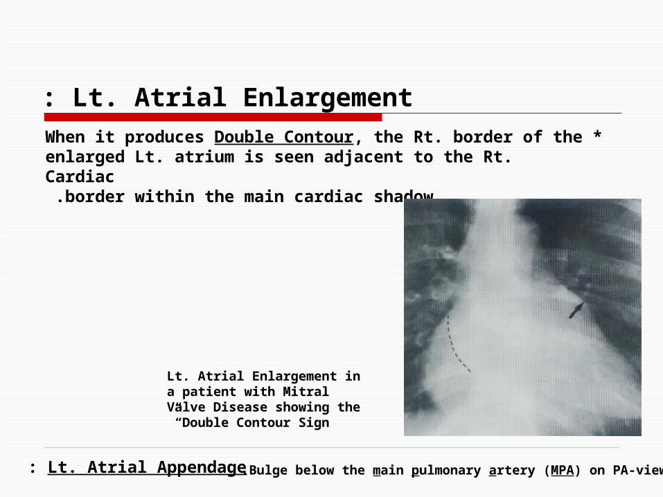
*When it produces Double Contour, the Rt. border of the enlarged Lt. atrium is seen adjacent to the Rt. Cardiac
border within the main cardiac shadow.
Lt. Atrial Appendage: Bulge below the main pulmonary artery (MPA) on PA-view.
Lt. Atrial Enlargement in a patient with Mitral Valve Disease showing the “Double Contour Sign”

Rt. Atrial Enlargement
*Will produce an increase of the Rt. cardiac border, & often accompanied by enlargement of Superior Vena Cava
( SVC.)
b) Echocardiography.
--------------------------------------------------------------------

Valve movement deformity &calcification
Plain X-ray films:
*Calcification is the only information could be obtained directly related to the morphology of the valve.
*Calcification is better seen by fluoroscopy.
*It occurs in mitral valve &/or aortic valve in rheumatic heart diseases; & if it occurs in aortic valve alone
( especially in adults )it is mainly congenital aortic stenosis.
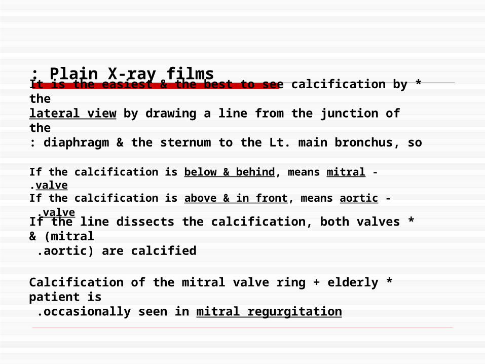
*It is the easiest & the best to see calcification by the lateral view by drawing a line from the junction of the
diaphragm & the sternum to the Lt. main bronchus, so:
-If the calcification is below & behind, means mitral valve. - If the calcification is above & in front, means aortic valve.
*If the line dissects the calcification, both valves (mitral& aortic) are calcified.
*Calcification of the mitral valve ring + elderly patient is occasionally seen in mitral regurgitation.
Plain X-ray films:
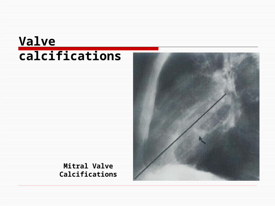
Valve calcifications
Mitral Valve Calcifications

Valve calcifications
Aortic Valve Calcifications

Ventricular Contractility
*General uniform decrease contractility in valvular disorder, congenital cardiomyopathy, & multi-vessel coronary artery
diseases.
*If there is focal decrease in contractility +/- dilatation in IHD.
*Increase contractility of the Lt. ventricle will cause hypertrophy as in aortic stenosis, HTN, & hypertrophic
obstructive cardiomyopathy (HOCM).

Pericardial Diseases
*20 – 50 ml of pericardial fluid is diagnosed by echo.
*Needle aspiration is needed to insure the nature of the fluid.
*CT scan & MRI can show the pericardial effusion; but more important is to measure the thickness of the pericardium
where thickness of the pericardium by echo. is poor.
*Unusual to diagnose pericardial effusion by plain-X-ray because the patient may have pericardial effusion to
cause a life-threatening tamponade; but only mild heart enlargement with otherwise normal contour.

*Marked increase or decrease in the transverse diameter of the cardiac shadow within one or two weeks + No
pulmonary edema is virtually diagnostic of pericardial effusion.
*Marked increase in the cardiac size + no specific chamber + normal pulmonary vasculature (flask
shape) (& the outline of the heart become very sharp) is diagnostic of pericardial effusion.
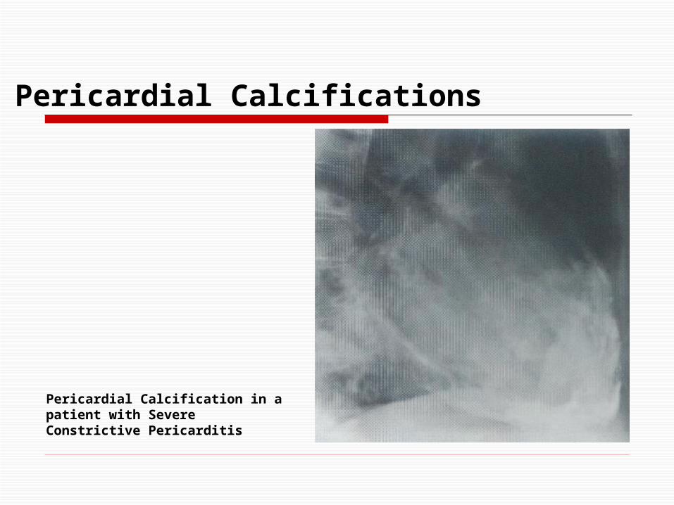
*Pericardial calcification is seen in 50% of patient within constrictive pericarditis, which is usually due to TB or
Coxsackie's virus infection. *Best seen on lateral CXR, along the anterior & inferior
surface, & it may possible on frontal CXR. *Usually the calcification is an important sign for
constrictive pericarditis.

Pericardial Effusion
Pericardial Effusion due to Viral Pericarditis

Pericardial Effusion
Congestive Cardiomyopathy, this appearance usually confused with Pericardial Effusion

Pericardial Effusion
Large Pericardial Effusion on an apical 4-chamber view echocardiogram
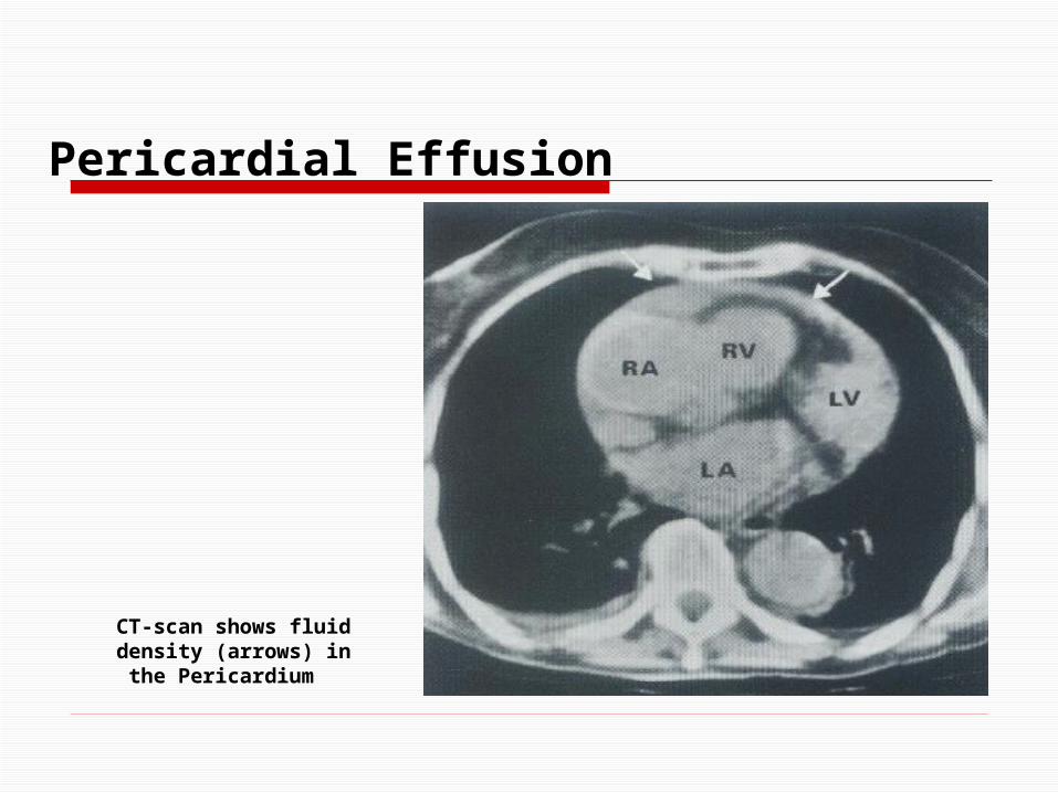
CT-scan shows fluid density (arrows) in the Pericardium
Pericardial Effusion

Pericardial Calcifications
Pericardial Calcification in a patient with Severe Constrictive Pericarditis
Pericardial Calcifications
Pericardial Calcification in a patient with Severe Constrictive Pericarditis

Pulmonary Vessels
*It is not possible to measure the diameter of the MPA from the plain film (usually subjective); but if there are variable
degrees of bulging, means enlarged MPA.
*Assessment of the hilar pulmonary arteries is more objective & the diameter of the Rt. lower lobe artery at its
mid-point (normally 9 – 16 mm).
*The size of pulmonary vessels with the lung reflects the pulmonary blood flow.
*Increase pulmonary blood flow is seen in ASD, VSD& , PDA, & all of these will lead to Systemic to Pulmonary (Lt.
to Rt. shunt) & these will to increase pulmonary blood flow.

Pulmonary Vessels
*Hemodynamically significant Lt. to Rt. shunt is (2/1 ratio or more) & this will produce CXR findings; if less ratio
there will be no CXR findings & all the pulmonary vessels will (from the MPA to the periphery of the lung) will be
enlarged, & this is called "Pulmonary Plethora."
*There is good correlation between the size of the vessel on CXR & degree of the shunt.
*Decrease pulmonary blood flow, all the vessels are small
" Pulmonary Oligemia."
*The commonest cause of decrease pulmonary blood flow is TOF & pulmonary stenosis.

*Obstruction of the Rt. ventricle outflow + VSD will lead to Rt. to Lt. shunt.
*Pulmonary stenosis will cause oligemia only is severe cases & babies or very young children.
Pulmonary Vessels

Pulmonary Arterial Hypertension
*The pressure in the pulmonary artery depends on:
1 -Cardiac output.
2 -Pulmonary vascular resistance.

Pulmonary Arterial Hypertension
*Conditions that cause significant pulmonary arterial hypertension all increase the resistance of blood flow
through the lungs, examples:
1 -Various lung diseases (cor pulmonale). 2 -Pulmonary embolism.
3 -Pulmonary arterial narrowing in response to mitral valve diseases or Lt. to Rt. shunt.
4 -Idiopathic pulmonary hypertension.

Pulmonary Arterial Hypertension
*By CXR: There will be enlargement of the mean pulmonary artery + the hilar pulmonary artery, vessels within the lung tissue are normal or small.
*Eisenmenger's syndrome:
Greatly raised pulmonary artery resistance in associationwith ASD, VSD, & PDA leading to reverse shunt (i.e. : Rt. to Lt. shunt).

Pulmonary Arterial Hypertension
*The cause of pulmonary arterial hypertension may be visible on the CXR as cor pulmonale & mitral valve
diseases.
Pulmonary Arterial Hypertension due to ASD & Eisenmenger's
syndrome

Pulmonary Venous Hypertension
*The commonest causes of pulmonary venous hypertension are:
1 -Mitral valve diseases. 2 -Lt. ventricular failure.
*In normal upright person (by CXR) the lower zone vessels
are larger than the upper zone.
*In pulmonary venous hypertension the upper zone vessels are enlarged.
*In severe cases, the upper zone vessels become larger
than that of the lower zone, & eventually Pulmonary Edema will supervene & may obscure the blood vessels.

Pulmonary Venous Hypertension
Pulmonary Venous Hypertension in a patient with Mitral Valve
Disease

Aorta
* Aortic dilatation of the ascending aorta is due to: 1 -Aneurysm.
2 -Aortic regurgitation or aortic stenosis. 3 -Systemic hypertension.
*The two common causes of descending aortic aneurysm are:
1 -Atheroma. 2 -Aortic dissection. (Also, there is a rare cause as
previous trauma following decelerating injury.)

Aorta *By CXR:
1 -The diagnosis of aortic aneurysm may be obvious, but substantial dilatation may be needed before the bulge of
Rt. mediastinal border can be recognized.
2 -Atheromatous aneurysm invariably shows calcification of their walls.
*CT scan with IVCM or CT angiography or MRA are very useful to assess the aneurysm.
Note:
IVCM = I.V. Contrast Media.MRA = Magnatic Resonance Angiography.

Dissecting Aortic Aneurysm
It is important to know the extent of the dissecting aneurysm as those involving the ascending aorta are treated surgically & those confined to the descending aorta are treated with hypotensive drugs.
*By CXR: Two congenital aortic anomalies can be seen, & they are:
1 -Coarctation of Aorta. 2 -Rt. sided aortic arch, in association with TOF,
Pulmonary Atresia, & Truncus Arteriosus, or it also can be isolated with no clinical significance.

Dissecting Aortic Aneurysm
Trans-Esophageal Echocardiogram showing the True (T) & False (F)
lumina in the descending aorta

Dissecting Aortic Aneurysm
CT-scan showing the displaced intima (arrows) separating the
true & false luminae in the ascending & descending aorta

Heart Failure *The plain X-ray findings include the followings:
a) Enlarged cardiac shadow +/- specific chamber
enlargement. b) Evidence of pulmonary venous hypertension
( enlargement of the vessels in the upper zone.) c) Evidence of pulmonary edema.
d) Pleural Effusion : It is usually bilateral, often larger on the Rt. than on the Lt. side; but if it is unilateral it is
almost always on the Rt. side.
Note: Acute Lt. ventricular failure, small effusion is seen at the costo-phrenic angle, running up the lateral chest wall; (this fluid may, in fact, be edema of the lungs rather than true pleural effusion).

Heart Failure
Congestive Heart Failure with bilateral Pleural Effusion

Valvular Heart Diseases
Mitral Valve Diseases
They Include:
1 -Mitral Stenosis ( MS )2 -Mitral Regurgitation ( MR )

Mitral Stenosis ( MS )
1 )By CXR:
The pathophysiological findings are:
*Lt. atrial enlargement + normal cardiac size . *Mitral calcification .
*Pulmonary venous hypertension . *Pulmonary edema.
*Pulmonary arterial hypertension, will lead to enlarged cardiac size (Rt. ventricle is enlarged).

Mitral Stenosis ( MS )
Plain X-ray of Mitral Stenosis, showing enlarged Lt. atrium as a double contour at the Rt. heart border (curved arrow), & enlarged
Lt. atrial appendage (straight arrow)

Mitral Regurgitation (MR)
The pathophysiological findings are:
1 )By CXR:
*Lt. atrium & Lt. ventricle are enlarged, so cardiac size will be enlarged in its Lt. ventricular configuration.
*Pulmonary venous hypertension.
*Pulmonary edema.
Note: Lt. atrial enlargement & pulmonary venous hypertension are the important signs of MR, which differs from MS by Lt. ventricular enlargement.

Valvular Heart Diseases
Aortic Valve Diseases
They Include:
1 -Aortic Stenosis ( AS )2 -Aortic Regurgitation ( AR )

Aortic Stenosis ( AS )
The pathophysiological findings are:
1 )By CXR:
*Aortic valve calcification. *Post stenotic dilatation of the ascending aorta (the
major feature.) *Lt. ventricular enlargement (Late feature).
*Increase pulmonary venous pressure (Late feature).
-Both late features will lead to Lt. ventricular failure.

Aortic Stenosis ( AS )
Aortic Stenosis ( AS ) showing post-
stenotic dilatation of the aorta (arrows)

Aortic Regurgitation (AR)
The pathophysiological findings are:
1 )By CXR:
*Dilatation of the ascending aorta.
*Increase in the cardiac size due to enlarged Lt. ventricle, & this occurs in the early course of
the disease.

Lt. Atrial Myxoma& Other Intra-cardiac Masses
*Intracardiac tumors are extremely rare.
*Lt. atrial myxoma is the most frequently encountered, it is a benign tumor which arises from:
a) Interatrial septum. b) Lt. atrial walls.
*As it enlarges, it becomes pedunculated to float in the Lt. atrial cavity, & therefore it will interfere with mitral valve
function & mimic MS or MR in both ways (clinically & by CXR.)
*It can be differentiated from other intra-cardiac masses by MRI & Echo., & the only differential Dx is Lt. atrial
thrombus in patient with Rheumatic MS.

Lt. Atrial Myxoma& Other Intra-cardiac Masses
Lt. Atrial Myxoma shown by 2-dimentional echocardiography – modified apical 4-
chamber view

Congenital Heart Diseases
A- Lt. to Rt. Shunt (as in ASD, VSD, & PDA):
*When the shunt is 2/1 or more, the following CXR findings will be seen:
a) Enlarged cardiac size (cardiomegally). b) Enlarged Mean Pulmonary Artery (MPA), hilar pulmonary
arteries. c) Pulmonary plethora.
*Absent of Lt. ventricular enlargement in the presence of increase
pulmonary flow is mainly indicate ASD.

Congenital Heart Diseases
VSD in a child
Congenital Heart Diseases
B- Pulmonary Stenosis (PS):
* BY CXR : there will be:
a) Normal heart size. b) Enlargement of MPA Lt. pulmonary artery
( Post-Stenotic Dilatation.)

C- Coarctation Of Aorta (COA):
Congenital Heart Diseases
*It is an abnormal aortic arch due to presence of narrowing, just distal to the origin of the Lt. subclavian
artery.
*CXR findings include: a) Indentation of the aortic arch.
b) Dilatation above (dilatation of the Lt. subclavian artery)& , below (post-stenotic dilatation).
c) Enlarged cardiac size & ascending aorta due to long standing hypertension.
d) Rib notching is due to enlargement of the intercostal arteries which act as a collateral, & there will small cortical indentation
on the inferior margin of the posterior halves of the ribs from the 3rd or 4th rib, & downward.
Note : COA itself can be seen by angiography or MRI.

Congenital Heart Diseases
Rib notching in COA

Congenital Heart Diseases
Angiogram showing COA (arrow)

Congenital Heart Diseases
Abnormal Aortic knuckle (arrow)
D- Tetralogy Of Fallot (TOF):
Congenital Heart Diseases
& *they are: a) VSD.
b) Rt. ventricular outflow obstruction (valvular or subvalvular.)
c) Rt. ventricular hypertrophy. d) Overriding of aorta over the VSD.

*BY CXR:
a) 50% of patients have normal CXR. b) Upturned cardiac apex.
c) Pulmonary bay at the region of the MPA, gives the Boot-Shaped Heart.
d) Oligemia of the lung. e) 25% Rt. sided aortic arch.
Congenital Heart Diseases
D- Tetralogy Of Fallot (TOF):

Thank You