raltegravir: m basisof its mechanismof action · and does not dissociate after 3’ processing, ......
TRANSCRIPT

November 24, 2009 5EUROPEAN JOURNAL OF MEDICAL RESEARCH
AbstractIntegration of the HIV-1 viral DNA generated by re-verse transcription of the RNA genome into the hostcell chromosomes is a key step of viral replication, cat-alyzed by the viral integrase. In October 2007, the firstintegrase inhibitor, raltegravir, was approved for clini-cal use under the name of IsentressTM. The results ofthe various clinical trials that have evaluated raltegravirhave been very encouraging with regard to the im-munological and virological efficacy and tolerance.However, as observed for other anti-retrovirals, specif-ic resistance mutations have been identified in patientsfailing to respond to treatment with raltegravir. Al-though knowledge of the integrase structural biologyremains fragmentary, the structures and modeling dataavailable might provide relevant clues on the origin ofthe emergence of these resistance mutations. In thisreview, we describe the mechanism of action of thisdrug and the main data relating to its use in vivo, to-gether with recent structural data important to our un-derstanding of the origin of viral resistance.Key words: HIV-1 integrase, raltegravir, isentress, resis-tance, molecular modelingAbbreviations: ARV = antiretroviral; CCD = catalyticcore domain; hDNA = host DNA; HIV = Human im-munodeficiency virus; IN = integrase; INI = integraseinhibitor; INSTI = integrase strand transfer inhibitor;LTR = long terminal repeat; PIC = preintegrationcomplex; PR = protease; RT = reverse transcriptase;tDNA = target DNA
INTRODUCTION
HIV replication is driven by a molecular engine con-sisting of three viral enzymes: reverse transcriptase(RT), protease (PR) and integrase (IN). Integrase cat-alyzes the covalent insertion of the viral DNA pro-duced by reverse transcription of the RNA into thechromosomes of infected cells. Once integrated, theprovirus persists in the host cell and serves as a tem-plate for the transcription of viral genes and replica-tion of the viral genome, leading to the production ofnew viruses. Due to its key function in the viral life cy-cle, IN is an attractive target for antiretroviral drugs(ARVs) and has thus been the object of intensive phar-macological research over the last 20 years. Since theend of the 1990s, several inhibitors with genuine an-tiviral activity have been identified and developed. Sev-eral of these compounds, including raltegravir (Isen-tressTM) and elvitegravir in particular, have shown great
promise, ensuring the rapid recognition of integraseinhibitors (INIs) as an important new class in the arse-nal of antiretroviral drugs (ARVs) [42]. Raltegravir wasapproved for clinical use in October 2007, followingthe demonstration of a rapid, potent and sustained an-tiretroviral effect in patients with advanced HIV-1 in-fection. It is well tolerated and, due to its mechanismof action, is likely to be active against viruses resistantto other class of antiretroviral drugs, such as nucleo-sides, nucleotides and non nucleosides reverse tran-scriptase inhibitors, protease and entry inhibitors.However as with other antivirals, resistance mutations,located in the integrase gene of replicating viruses andpreventing the establishment of specific interactionsbetween the inhibitor and its integrase target, rapidlyemerge associated with a reduced susceptibility to thedrug. In this review, we focus on the mechanism of ac-tion of raltegravir in vitro and in vivo and we presentthe structural data that shed light on the molecular ba-sis of its inhibitory potency and on the origin of theemergence of resistance.
1. INTEGRASE BIOCHEMISTRY
CCaattaa ll yy tt ii cc aacc tt ii vv ii ttyy . Virological data have demonstratedthat the precursor of the integrated genome, orprovirus, is the linear viral DNA produced by reversetranscription of the RNA genome [11]. Two reactionsare required for the covalent insertion of the viralgenome. First, integrase binds to short sequences lo-cated at either end of the viral long terminal repeat(LTR) and catalyzes an endonucleolytic cleavage, in areaction known as 3’ processing, removing a dinu-cleotide at either end of both 3’ LTRs, leading to theexposure of a conserved CA sequence. Integration sen-su stricto, or strand transfer, then occurs through attackof the phosphodiester backbone in target DNA by the3’ hydroxyl groups of the processed DNA (Fig. 1 A).Strand transfer takes place concomitantly for both ex-tremities, with a five-base gap between insertionpoints. In vivo, these two reactions are spatially andtemporally separated and energetically independent: 3’processing takes place in the cytoplasm of infectedcells, whereas strand transfer occurs in the nucleus.Both reactions are one-step transesterification reac-tions with no covalent intermediates between integraseand the DNA [14].
Cellular enzymes are responsible for cleaving theprotruding 5’ ends of the viral DNA that remain unat-tached during strand transfer and repairing flanking
Eur J Med Res (2009) 14(Suppl. III): 5-16 © I. Holzapfel Publishers 2009
RALTEGRAVIR: MOLECULAR BASIS OF ITS MECHANISM OF ACTION
Jean-François Mouscadet and Luba Tchertanov
LBPA, CNRS, Ecole Normale Supérieure de Cachan, France

gaps, thereby completing the integration process [10].The final product is a covalently inserted viral genome,colinear with cellular genes, with a short (e.g. 5 bp) du-plication on either side, the length of which is a hall-mark of the retrovirus concerned. It is possible to re-produce the whole integration process in vitro, usingshort DNA fragments or oligonucleotides mimickingthe sequence of the ends of the LTR in the presenceof recombinant integrase [14]. In terms of specificity,only the terminal 5’CA is strictly required for 3’ pro-cessing. The mutation of this dinucleotide completelyabolishes the reaction, whereas the requirements con-cerning the adjacent sequences are less stringent [33].It is intrinsically difficult to demonstrate the specificityof the enzyme for the viral DNA due to its ability tobind specific and non specific DNA sequences simulta-neously. Nevertheless, recent advances have led to thedevelopment of an assay faithfully reproducing fullyconcerted integration in vitro [60]. In vitro, a third re-action, known as disintegration, may be observed inwhich the reverse strand transfer process occurs [21].Unlike 3’ processing and strand transfer, which dependon the integrity of the enzyme, disintegration may becatalyzed by the isolated catalytic core domain contain-ing the active site. There is no experimental evidenceto suggest that disintegration occurs in vivo, but phar-macological approaches involving the stabilization ofintegrase on the strand transfer intermediate might fa-vor this reverse reaction, thereby decreasing the effi-ciency of integration.
Integrase functions in a multimeric form, as shownby the complementation of inactive proteins observedin virions [31]. Dimers formed at either end of the vi-ral DNA molecule are responsible for 3'-processing ac-tivity [41]. Pairs of dimers bring together the two endsof the viral DNA, leading to the formation of atetramer (a dimer-of-dimers), the active form requiredfor concerted integration [34]. During its catalytic cy-cle, IN must bind simultaneously to the viral DNA(vDNA) and the target DNA (tDNA). Current knowl-edge of the organization of this tetramer on the DNAis based exclusively on models constructed from partialstructural and biochemical data (see below), which mayprovide a platform for the rational design of new in-hibitors.
CCaatt iioonn ii cc cc oo ffaacc tt oorr .. All integrase activities strictly re-quire the presence of a metallic cationic cofactor,which is coordinated by two residues of the catalytictriad (D64, D116 and E152 for HIV-1 IN) [3]. The cat-alytic cation may be either Mn2+-or Mg2+ in vitro, butMg2+ is the cofactor required in vivo and Mg2+-depen-dent activities also reproduce physiological activitymore faithfully in vitro. IN displays non specific nucle-ase activity in the presence of Mn2+, and the Mg2+ en-zyme is much less tolerant of sequence variations atthe ends of the LTR than the Mn2+ enzyme [33]. Sev-eral mutations are known to have no effect on IN ac-tivity in Mn2+-dependent assays, whereas they do affectIN activity in Mg-dependent assays. For example, mu-tations of the HHCC domain known to be detrimentalfor the virus in vivo alter 3’processing in vitro in thepresence of Mg2+, but not in the presence of Mn2+
[56]. In addition, factors promoting integrase multi-
merization, such as Zn2+, also specifically stimulate theMg2+-dependent activity of the enzyme, consistentwith the multimeric nature of the functional enzyme[57]. These differences between cofactor activities haveresulted in pharmacological discrepancies, as some ear-ly IN inhibitors identified on the basis of Mn2+-depen-dent assays were not active against the Mg2+ enzyme.Based on a model of another phosphatidyl transferase,the 3’-5’ exonuclease of E. coli DNA polymerase I [7],it was suggested early on that the retroviral integrasemight contain two metal cation cofactors. The 3Dstructures of avian sarcoma virus integrase and theTn5 transposase alone or in complex with DNA haveprovided structure-based evidence for a two-metal ac-tive site structure for retroviral integrases [13, 65].These considerations eventually led to the incorpora-tion of Mg2+-chelating groups into the rational designof IN inhibitors. Such groups are present in all effec-tive IN inhibitors, including raltegravir [40].
2. DEVELOPMENT OF INTEGRASE INHIBITORS
MMeecc hhaannii ssmmss oo ff iinnhh iibb ii tt ii oonn .. In terms of pharmacologi-cal development, two screening strategies have beenconsidered for the development of IN inhibitors: onebased on the free, unbound protein and the other onthe preformed IN•viral DNA (vDNA) complex. Bothapproaches were demonstrated to be feasible, with theidentification of inhibitors of either 3’ processing,blocking the binding of IN to viral DNA, or strandtransfer, targeting the IN•vDNA complex. Since theearly 1990s, a number of compounds inhibiting one orother of these reactions have been identified in vitro[83]. However, the complex resulting from the associa-tion of integrase with viral DNA whether isolatedfrom infected cells as a pre-integration complex (PIC),or reconstituted in vitro, is highly stable, keeping thecomplex together for long enough after the 3'-process-ing reaction for subsequent integration to occur [55].This complex has an intrinsically slow catalytic activityand does not dissociate after 3’ processing, limitingmultiple turnover [86]. This weak catalytic activity isnot detrimental in host cells, because a single integra-tion event is sufficient for overall function, but itmakes it difficult to develop competitive inhibitors offree IN. For these reasons, the Merck team lead by DrD. Hazuda suggested in the mid 1990s that the PICwould be a more suitable target for inhibitors. This hy-pothesis proved to be correct, particularly given thatPIC formation probably occurs within a capsid that isnot fully dissociated, thus precluding easy access tofree IN [1].
The design of new assays for screening ligands ofthe IN•vDNA complex eventually led to the identifica-tion of the first strand transfer inhibitors, L-731, 988and L-708, 906 at the turn of the century [43]. Thesecompounds compete with the target DNA by bindingto the IN•vDNA complex. They recognize a specificsite close to the catalytic triad, which opens following achange in conformation induced by the binding and 3’processing of the viral DNA [32]. The first selectiveinhibitors of strand transfer to be identified were α, γ-diketoacids (DKAs) [43]. Such compounds based onthe β-ketoenol fragment efficiently chelate the Mg2+
EUROPEAN JOURNAL OF MEDICAL RESEARCH6 November 24, 2009

cation required for the activity of integrase and theiroverall affinity for the target depends on their sur-rounding substituent groups (Fig. 1 B) [53, 90].
The prerequisites for a specific strand transfer in-hibitor include the presence of a chemical group in-
cluding the heteroatoms, nitrogen or oxygen, capableof binding two divalent cations and a hydrophobicaromatic part of the molecule likely to bind and stabi-lize the IN•vDNA complex, forming an active phar-macophore responsible for the activity of all strandtransfer inhibitors [4, 53] (Fig. 2). Compounds withthese properties selectively target and bind to theIN•vDNA complex, close to the 3' end of the donorDNA, thereby inhibiting target DNA binding, result-ing in selective inhibition of the strand transfer reac-tion with no significant effect on the 3'-processing re-action [78]. They therefore act as IN•DNA interfacialinhibitors, and are known as integrase strand transferinhibitors (INSTIs).
The replacement of the carboxylate group by itstetrazolium bioisostere led to the development of 5-CITEP (Fig. 2) and its analog, S-1360. Despite theweak activity of these molecules against integrase, thestructure of the integrase/5-CITEP complex has beendetermined, making it possible to construct a model ofthe structure of the inhibitor pharmacophore boundto the active site metal cation [36]. Modifications to theα, γ-diketoacid part of the molecule initially led to thereplacement of this group by 8-hydroxy quinoline, toincrease antiviral activity and to overcome pharmaco-logical limits, such as serum protein binding [98]. Com-pounds from this family, such as Merck L870, 812 (Fig.2), have potent antiviral activity, providing the proof-of-concept for INSTI activity in vivo despite their toxi-city in vivo [44]. The L870, 812 series of compoundswas not developed further, but the dihydroquinolineJTK303/GS9137 (Fig. 2) derived from quinolone an-tibiotics was used for further drug development and isnow at the advanced clinical development stage, underthe name of elvitegravir [80].
DDeevv ee ll ooppmmeenntt oo ff rraa ll tt eeggrraavvii rr .. The discovery of ralte-gravir stemmed from investigations of a series ofHCV polymerase inhibitors. The architecture of thecatalytic site and the arrangement of the metal cationsare very similar in integrase and the HCV NS5b RNA-dependent RNA polymerase. These similarities led theMerck team to test HCV polymerase inhibitors origi-
EUROPEAN JOURNAL OF MEDICAL RESEARCHNovember 24, 2009 7
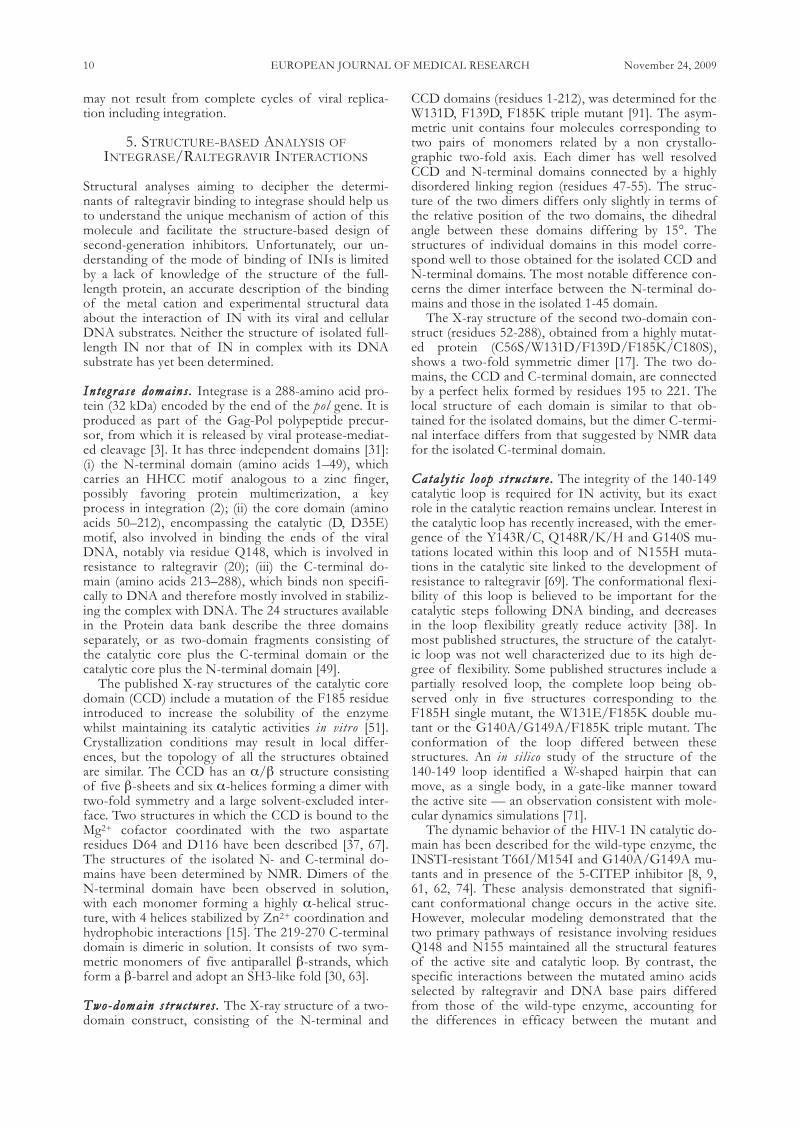
Fig. 1. Strand transfer reaction and proposed mechanism ofIN inhibition by INSTIs adapted from (53). A) A trans-esteri-fication reaction involving a nucleophilic attack on the 3’ hy-droxyl group of the two newly processed 3’ viral DNA endson the phosphodiester backbone of the host DNA. The hostDNA and viral DNA are shown in blue and red, respectively;the yellow arrow indicates the scissile phosphodiester. B) INstrand transfer inhibitors may chelate the two metal ions inthe catalytic site, thereby blocking the binding of host DNA.
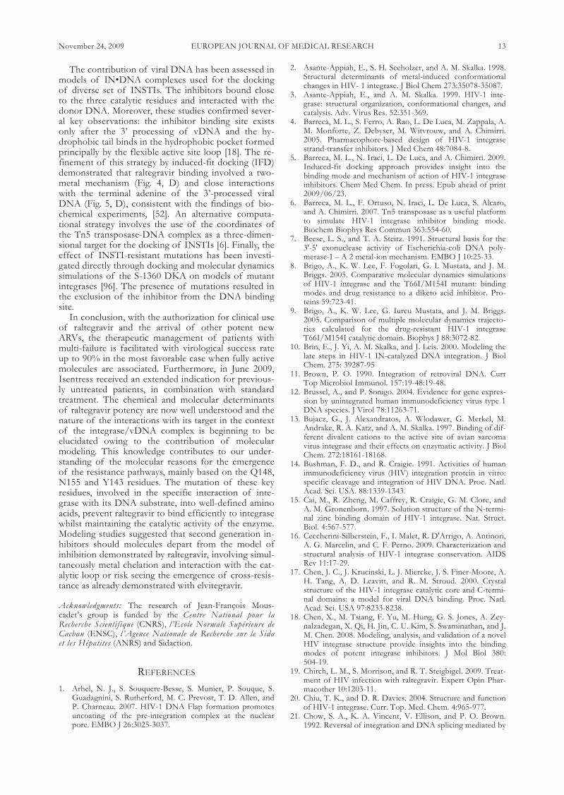
Fig. 2. Integrase strand transfer inhibitors.

nally designed as drug-compliant DKA replacements[89]. This led to the identification of a compound withactivity in the enzymatic assay, which was further opti-mized in cell culture [76, 89]. Raltegravir is a potent in-hibitor of the replication of HIV-1 and HIV-2 in vitro[70, 79]. It is more than 1000 times more selective forintegrase than for other phosphatidyl transferases, suchas HIV-1 RNAseH and human polymerases. It has anIC50 of 2 to 7nM for the inhibition of recombinantIN-mediated strand transfer in vitro and an IC95 of0.019 and 0.031 µM in 10% FBS and 50 % NHS, re-spectively, in a cell-based assay [70, 89]. Due to itsmode of action, it is independent of HIV-1 tropism(CCR5 and CXCR4) and active against viruses resistantto other classes of antiretroviral drugs, such as nucleo-side reverse transcriptase inhibitors, protease inhibitors,fusion and entry inhibitors [39].
3. ANTIVIRAL POTENCY OF RALTEGRAVIR
AAnn tt ii vv ii rr aa ll aa cc tt ii vv ii tt yy ii nn vv ii vv oo .. Phase II and III trialsdemonstrated a remarkable potency of combinationsof raltegravir and other ARVs in treatment-experi-enced patients [24, 39, 88]. The first phase II assay wasa dose-ranging study in patients with documented re-sistance to at least one drug in each of the three classesof ARVs. This population had considerable experienceof treatment and a very high level of drug resistance.There was an approximate 2.0 log copies/ml drop inplasma HIV RNA levels by week 24 in the raltegravirgroup, versus only 0.35 log with optimized therapyalone plus placebo, with no significant difference in vi-ral efficacy between the three dosage groups studied(200, 400, 600 mg) [39]. For the subsequent double-blind phase III BENCHMARK I and II studies, inwhich 699 patients with considerable experience oftreatment were enrolled, the combined analysis at 48weeks showed that 72.3% and 62.1% of raltegravir-treated patients had HIV RNA levels of less than 400and 50 copies/ml, respectively, whereas such levelswere found in only 37.1% and 32.9%, respectively, ofthe patients in the placebo group.
The 48-week results recently obtained for the phaseIII STARTMRK study comparing raltegravir-basedand efavirenz-based combination regimens as initialtreatment demonstrated that raltegravir suppressedHIV replication more rapidly than efavirenz, this rapidviral decay being of unknown origin [58]. Moreover,preliminary results from a non inferiority study of theuse of raltegravir to replace enfuvirtide in patients in-tolerant to enfuvirtide have shown raltegravir to be vi-rologically effective for sustained periods, with goodtolerance for up to 48 weeks. Conversely, theSWITCHMRK 1 and 2 trials, designed to examine thebenefit of replacing a protease inhibitor with ralte-gravir, suggested that the raltegravir combinationmight not inhibit HIV replication more efficiently. Insituations of resistance due to prior treatment failure,switching to raltegravir amounts to monotherapy, withthe rapid selection of raltegravir-resistant HIV strains,as the genetic barrier to raltegravir is easily overcome.Nevertheless, these results suggest that raltegravir is animportant additional drug for the initial treatment ofHIV-1 infection.
SSaaff ee ttyy .. Preclinical studies of toxicity by repeated ad-ministration, genotoxicity and toxic effects on develop-ment have been conducted with raltegravir, in mice,rats, dogs and rabbits. No mutagenic or teratogenic ef-fect was observed. The effects observed at levels ex-ceeding actual exposure levels revealed no likelihood ofa clinical risk in humans [89]. Raltegravir is well tolerat-ed and adverse events are rare. Most frequent drug-re-lated clinical events, such as diarrhea, nausea, headacheand fatigue, were moderate and transient [48]. Labora-tory abnormalities included an increase in serum lipid,aminotransferase and creatinine concentrations. In-creases in creatinine phosphokinase levels, althoughnot statistically significant, led to a cautious recom-mendation not to use raltegravir concomitantly withother drugs known to increase these levels. In phase IIand phase III trials, the frequency of clinical and labo-ratory adverse events was similar in the raltegravir andplacebo groups. In the STARTMRK trial, significantlyfewer drug-related clinical adverse events occurred inpatients on raltegravir than in those on efavirenz [58].The BENCHMRK trial suggested a small increase ofthe risk of cancer in the raltegravir arm, with a relativerisk of 1.5, but a recent analysis of all the available dataconcluded that the relative risk was actually less than 1[19].
PPhhaarrmmaacc ookkiinnee tt ii cc ss .. Raltegravir is administered orallyand is rapidly absorbed. Its absolute bioavailability hasyet to be determined, but the administration of 400 mgper day results in steady-state levels of the drug in thebody within two days, as demonstrated by pharmacoki-netics studies. About 83% of the raltegravir ingestedbinds to plasma proteins. Animal studies have shownraltegravir penetrate the stomach, liver, small intestine,kidney and bladder effectively, but have suggested thatpenetration into the brain is limited. Considerable in-tra- and interindividual variability was observed. Ralte-gravir is a substrate, but not an inhibitor of P-glyco-protein (Pgp). There is currently no evidence to sug-gest that inhibitors or inducers of Pgp could affect ral-tegravir, but this property may affect its absorption[23]. It could also account for the limited diffusion ofthis drug into the central nervous system. No effect ofage or sex has been identified in studies of the phar-macokinetics of raltegravir (no data are available forchildren) [46]. The half-life of raltegravir in the body isabout nine hours, with an initial phase of rapid elimi-nation lasting about 1 hour. At steady state, a slight in-crease in residual concentrations of the drug is ob-served, but with no effect on the maximum concentra-tion, making it possible to administer raltegravir twicedaily.
Raltegravir is mostly metabolized in the liver,through glucuronidation by uridine diphosphate-glu-curonolsy-transferase 1A1 (UGT1A1) to generate asingle metabolite, M2. Raltegravir is neither a substratenor an inhibitor of the cytochrome P450 enzymes,consistent with a lack of interaction with drugs metab-olized by P450 isoenzymes, including protease in-hibitors. It does not inhibit either UGT1A1 or 2B7 anddoes not induce CYP34A. As raltegravir is mostly me-tabolized by UGT1A1, it should be used with cautionwhen co-administered with strong inducers of
EUROPEAN JOURNAL OF MEDICAL RESEARCH8 November 24, 2009

UGT1A1, such as rifampicin. This antibiotic has beenshown to reduce plasma concentrations of raltegravir,although its impact on the efficacy of raltegravir is un-known. A mutation of the UGT1A1 gene resulting inthe production of an inactive enzyme has been identi-fied. Two studies have shown in the concentration ofraltegravir to be higher in patients with a homozygousmutant genotype. This genotype seems to be an impor-tant factor in interindividual variability, but its clinical relevance, in terms of efficacy and toxicity, is unknown (http://www.emea.europa.edu). Finally,atazana vir, a protease inhibitor affecting glucuroni-dation, decreases the formation of raltegravir glu-curonide and induces a moderate increase in raltegravirconcentration [39, 47].
RRee ss iiss ttaannccee tt oo rraa ll tt eegg rraa vviirr .. As with other antiretroviraldrugs, resistance to INI emerges through the selectionof mutations in the integrase gene affecting the sus-ceptibility of the virus to INI. More than 40 mutationshave been specifically associated with resistance to INSTIs in vitro and in vivo [16]. Resistance to ralte-gravir in vivo has been associated with 14 mutations, todifferent degrees, but the virologic failure observedduring the BENCHMRK trials was unambiguously as-sociated with two principal independent genetic path-ways involving primary mutations of residues N155(N155H) and Q148 (Q148K/R/H) [24, 88]. Thesemutations were not detected in the various studies onintegrase polymorphism in INI-naive patients, con-firming their likely role in conferring resistance to thisclass of drugs. Secondary mutations increasing the fit-ness of the resistant viruses were identified in bothpathways. In particular, the G140S mutation rescues areplication defect resulting from the primary mutationQ148H [27]. Phenotypic analysis showed that the pres-ence of the mutation at position 148 together with oneor more secondary mutations resulted in greater resis-tance to RAL than observed for viruses carrying themutation N155H. Clonal analysis of the viral popula-tions in 11 patients with treatment failure on raltegravirshowed that no viral clone simultaneously carried mu-tations in position 148 and 155, demonstrating the in-dependence and exclusivity of the two main pathways.Moreover, a switch of resistance profile from residue155 to residue 148 mutations may occur due to thehigher level of resistance to raltegravir conferred bythe pathways associated with residue 148 mutation andthe greater instability of the pathways associated withresidue 155 [68].
A small number of mutations involving residuesE92, E157 and Y143 might constitute another pathwayof resistance. There is some debate about whether thefirst two of these mutations are true primary mutationsfor RAL resistance, whereas the Y143 mutation hasbeen shown to confer a real decrease in susceptibilityto the inhibitor [85]. Y143R/C/H mutations occur lessfrequently and later than the other two mutations [25].
The major IN mutations E92Q, Q148K/R/H,N155H and E157Q are highly conserved and subjectto similar genetic barriers between subtypes B andCRF02_AG. However, the CRFO2_AG subtype has astronger genetic barrier to the acquisition of mutationsof residue G140 than subtype B [66]. Another showed
that treatment failure on raltegravir occurred morerapidly in patients infected with non B subtype viruses,indicating a possible impact of non B-associated poly-morphisms on the genetic barrier to raltegravir [85].
4. FATE OF NON INTEGRATED VIRAL GENOMES
A productive HIV-1 replication in T4 lymphocytes de-pends on the activation and multiplication of thesecells. HIV-1 can enter resting T cells, but in absence ofcell activation the fate of the viral genome is uncertain.Replication may abort during the reverse transcriptionstep or be blocked before integration [75, 94, 97]. Ithas been suggested that incoming HIV-1 subviral com-plexes may concentrate in the centrosome, in whichthey may remain in a stable state for several weeks [87].Thus, HIV-1 may persist in quiescent cells as a long-lived, centrosome-associated, preintegration intermedi-ate [95]. Upon cell activation, viral replication may re-sume, leading to viral gene expression [93] and provid-ing a possible explanation for the unusual decay kinet-ics of viral load during raltegravir treatment [72]. Thismay also account for the faster decay kinetics observedwith raltegravir than with efavirenz.
In the absence of integration, the linear viral DNAis circularized, probably by non-homologous end join-ing [54, 59] to yield circular forms that do not supportviral replication but that might persist in the nucleusfor an undetermined period of time [77]. This circular-ization of viral genomes is in fact one reason for theactivity of raltegravir. Indeed, it prevents the genomesfrom being integrated when the inhibitor, non-cova-lently bound to the PIC, is eventually released from itsbinding site. Accordingly, the residence time of ralte-gravir onto its target was found to be a determinant ofits inhibitory potency and is dramatically decreased bythe presence of the primary resistance mutations.
In the presence of strand transfer inhibitors, such asraltegravir or elvitegravir, an accumulation of 2-LTRcircular forms is observed. The current consensus isthat these forms do not play a significant role in viralreplication, although non integrated DNA largely ex-ceeds integrated forms in resting T cells duringHAART [22]. However, the production of the viralNef and Tat proteins has been demonstrated [92] andit has been suggested in various studies that these cir-cular species may be transcribed during HIV-1 infec-tion, so we cannot completely rule out a functional roleof these circles in viral replication [12]. In addition,certain integrase mutants unable to mediate integrationremain competent for replication in permissive cells,such as CEM MT4 cells, albeit with low efficiency, sug-gesting the direct involvement of the circles or an inte-grase-independent integration mechanism based on re-combination, for example [73].
In any case, unlike other ARVs, INSTIs do notcause the complete disappearance of the viral genomefrom infected cells. Instead, they merely preventgenome integration. The fate of the circular speciesduring treatment with INSTIs remains to be deter-mined. A recent study demonstrated that the intensifi-cation of raltegravir treatment over a 12-week perioddid not decrease low-level plasma viremia in patientson HAART. This finding suggests that residual viremia
EUROPEAN JOURNAL OF MEDICAL RESEARCHNovember 24, 2009 9
may not result from complete cycles of viral replica-tion including integration.
5. STRUCTURE-BASED ANALYSIS OFINTEGRASE/RALTEGRAVIR INTERACTIONS
Structural analyses aiming to decipher the determi-nants of raltegravir binding to integrase should help usto understand the unique mechanism of action of thismolecule and facilitate the structure-based design ofsecond-generation inhibitors. Unfortunately, our un-derstanding of the mode of binding of INIs is limitedby a lack of knowledge of the structure of the full-length protein, an accurate description of the bindingof the metal cation and experimental structural dataabout the interaction of IN with its viral and cellularDNA substrates. Neither the structure of isolated full-length IN nor that of IN in complex with its DNAsubstrate has yet been determined.
IInntteegg rraass ee ddoommaaiinnss .. Integrase is a 288-amino acid pro-tein (32 kDa) encoded by the end of the pol gene. It isproduced as part of the Gag-Pol polypeptide precur-sor, from which it is released by viral protease-mediat-ed cleavage [3]. It has three independent domains [31]:(i) the N-terminal domain (amino acids 1–49), whichcarries an HHCC motif analogous to a zinc finger,possibly favoring protein multimerization, a keyprocess in integration (2); (ii) the core domain (aminoacids 50–212), encompassing the catalytic (D, D35E)motif, also involved in binding the ends of the viralDNA, notably via residue Q148, which is involved inresistance to raltegravir (20); (iii) the C-terminal do-main (amino acids 213–288), which binds non specifi-cally to DNA and therefore mostly involved in stabiliz-ing the complex with DNA. The 24 structures availablein the Protein data bank describe the three domainsseparately, or as two-domain fragments consisting ofthe catalytic core plus the C-terminal domain or thecatalytic core plus the N-terminal domain [49].
The published X-ray structures of the catalytic coredomain (CCD) include a mutation of the F185 residueintroduced to increase the solubility of the enzymewhilst maintaining its catalytic activities in vitro [51].Crystallization conditions may result in local differ-ences, but the topology of all the structures obtainedare similar. The CCD has an α/β structure consistingof five β-sheets and six α-helices forming a dimer withtwo-fold symmetry and a large solvent-excluded inter-face. Two structures in which the CCD is bound to theMg2+ cofactor coordinated with the two aspartateresidues D64 and D116 have been described [37, 67].The structures of the isolated N- and C-terminal do-mains have been determined by NMR. Dimers of theN-terminal domain have been observed in solution,with each monomer forming a highly α-helical struc-ture, with 4 helices stabilized by Zn2+ coordination andhydrophobic interactions [15]. The 219-270 C-terminaldomain is dimeric in solution. It consists of two sym-metric monomers of five antiparallel β-strands, whichform a β-barrel and adopt an SH3-like fold [30, 63].
TTwwoo--ddoommaaiinn ss ttrruucc ttuurr ee ss .. The X-ray structure of a two-domain construct, consisting of the N-terminal and
CCD domains (residues 1-212), was determined for theW131D, F139D, F185K triple mutant [91]. The asym-metric unit contains four molecules corresponding totwo pairs of monomers related by a non crystallo-graphic two-fold axis. Each dimer has well resolvedCCD and N-terminal domains connected by a highlydisordered linking region (residues 47-55). The struc-ture of the two dimers differs only slightly in terms ofthe relative position of the two domains, the dihedralangle between these domains differing by 15°. Thestructures of individual domains in this model corre-spond well to those obtained for the isolated CCD andN-terminal domains. The most notable difference con-cerns the dimer interface between the N-terminal do-mains and those in the isolated 1-45 domain.
The X-ray structure of the second two-domain con-struct (residues 52-288), obtained from a highly mutat-ed protein (C56S/W131D/F139D/F185K/C180S),shows a two-fold symmetric dimer [17]. The two do-mains, the CCD and C-terminal domain, are connectedby a perfect helix formed by residues 195 to 221. Thelocal structure of each domain is similar to that ob-tained for the isolated domains, but the dimer C-termi-nal interface differs from that suggested by NMR datafor the isolated C-terminal domain.
CCaattaa ll yy tt ii cc ll oooopp ss tt rruucc tt uurree .. The integrity of the 140-149catalytic loop is required for IN activity, but its exactrole in the catalytic reaction remains unclear. Interest inthe catalytic loop has recently increased, with the emer-gence of the Y143R/C, Q148R/K/H and G140S mu-tations located within this loop and of N155H muta-tions in the catalytic site linked to the development ofresistance to raltegravir [69]. The conformational flexi-bility of this loop is believed to be important for thecatalytic steps following DNA binding, and decreasesin the loop flexibility greatly reduce activity [38]. Inmost published structures, the structure of the catalyt-ic loop was not well characterized due to its high de-gree of flexibility. Some published structures include apartially resolved loop, the complete loop being ob-served only in five structures corresponding to theF185H single mutant, the W131E/F185K double mu-tant or the G140A/G149A/F185K triple mutant. Theconformation of the loop differed between thesestructures. An in silico study of the structure of the140-149 loop identified a W-shaped hairpin that canmove, as a single body, in a gate-like manner towardthe active site — an observation consistent with mole-cular dynamics simulations [71].
The dynamic behavior of the HIV-1 IN catalytic do-main has been described for the wild-type enzyme, theINSTI-resistant T66I/M154I and G140A/G149A mu-tants and in presence of the 5-CITEP inhibitor [8, 9,61, 62, 74]. These analysis demonstrated that signifi-cant conformational change occurs in the active site.However, molecular modeling demonstrated that thetwo primary pathways of resistance involving residuesQ148 and N155 maintained all the structural featuresof the active site and catalytic loop. By contrast, thespecific interactions between the mutated amino acidsselected by raltegravir and DNA base pairs differedfrom those of the wild-type enzyme, accounting forthe differences in efficacy between the mutant and
EUROPEAN JOURNAL OF MEDICAL RESEARCH10 November 24, 2009

wild-type integrases in vitro [71]. Together with theo-retical studies that have predicted that the Q146, Q148,and N144 residues of the loop form a DNA bindingsite [29], this result suggest that raltegravir acts bycompeting with DNA for residues N155 and/or Q148.In order to thwart the inhibitory effect, the virus mayhave to select mutations that maintain the integrity ofIN structure while allowing alternative modes of DNArecognition.
TThheeoo rr ee tt ii ccaa ll mmooddee ll ss .. In the absence of complete andaccurate experimental data, computational methodshave become a key tool for probing the interactions ofintegrase with inhibitors and substrates. Fragmenteddata concerning the structure of HIV-1 IN have beenused to construct models to improve our understand-ing of inhibitor binding to the target. Theoretical mod-els of both the dimer and tetramer states have beenconstructed. De Luca and coworkers described adimeric model of the full-length IN/viral DNA com-plex with two Mg2+ cations in the active site, consistentwith cross-linking data indicating that the Q148 andY143 residues interact with viral DNA [26, 33]. Themolecular docking method has also been used to inves-tigate further the interactions of the HIV-1 IN dimerwith viral DNA before the 3' processing reaction [45].
Most theoretical models consider a tetrameric IN aloneor in complex with either viral DNA or viral DNA/target DNA [35 and references therein]. The influenceof metal ions on IN•DNA complexes has been ex-plored in a tetramer model constructed by homologymodeling and MD simulations [18]. It was found thatmetal cations could potentially influence the locationof the viral DNA on IN. Full-length models of theHIV-1 IN tetramer in complex with both viral and tar-get DNAs have been constructed with either one ortwo Mg2+ ions in the active site, to ensure consistencywith biochemical experimental findings.
MMooddeess oo ff rraa ll tt ee ggrraavvii rr ii nn ttee rraacc tt ii oonn ww iitt hh IINN.. The mole-cular docking of different DKAs onto the catalyticcore domain identified two unique binding areas withinthe active site, including either the conserved D64-D116-E152 motif or the flexible loop region formedby amino acid residues 140-149, and confirmed thatthe mechanism of inhibition by DKAs involves metalchelation by the β-ketoenol group [82]. A comparativeresidue interaction analysis (CoRIA) was recently per-formed [28], allowing evaluation of the non bondedinteraction energies of the inhibitors with individualactive site residues and an assessment of the correla-tion with biological activity, leading to the identifica-tion of crucial residues and characterization of interac-tions between the ligand and receptor. The modelssuggest that Asp64, Thr66, Val77, Asp116, Glu152 andLys159 are the key residues influencing the binding ofligands with the integrase. The docking of raltegravirand analogs onto Mg2+-complexed IN demonstratedthe establishment of direct interactions between ralte-gravir and the three catalytic residues D64, D116, andE152, and with residues T66, E92, Y143, Q148, andN155 [84] (Fig. 4, C and Fig. 5, C). This result wasagain consistent with the findings of clinical experi-mental resistance profiling and provided a rational forthe involvement of E92 and Y143residues in resis-tance.
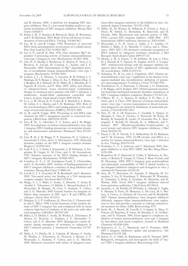
A single crystal structure of the IN core domainco-crystallized with an INSTI has been obtained with5CITEP [36]. The inhibitor is located between the ac-tive site residues D64, D116 and E152 (Fig. 3, A). TwoH-bonds are formed between the tetrazolium moietyand the K165 and K159 residues involved in DNAbinding [50]. The other contacts are the T66 residueimplicated in resistance to diketoacids in vitro and theN155, Y143 and Q148 residues involved in raltegravirresistance in vivo. Although obtained in the absence ofviral DNA it is assumed that the interactions between5-CITEP and IN observed in this structure at leastpartly mimic the contacts between IN and DNA (Fig.3, B), justifying the use of the integrase CCD•5CITEPcomplex as a surrogate platform for docking simula-tions [81]. This model was used to study the mode of binding of raltegravir [64]. Two conformations of raltegravir, differing in the nature of the inter-acting residues and the method of Mg2 chelation, were obtained (Fig. 4, A and B). However, this com-pound was systematically located in the vicinity of theY143, N155 and Q148 residues (Fig. 5, A and B),thereby confirming the role of these three aminoacids.
EUROPEAN JOURNAL OF MEDICAL RESEARCHNovember 24, 2009 11
A
B
Fig. 3. 5CITEP binding to the HIV-1 integrase. A) X-raystructure of 1QS4 (36). B) In silico modeling (81)(Reproduc-tion authorized). The 5-CITEP inhibitor is shown in similarorientations; H-bonds are indicated by dashed lines, cyan inA and white in B.

EUROPEAN JOURNAL OF MEDICAL RESEARCH12 November 24, 2009
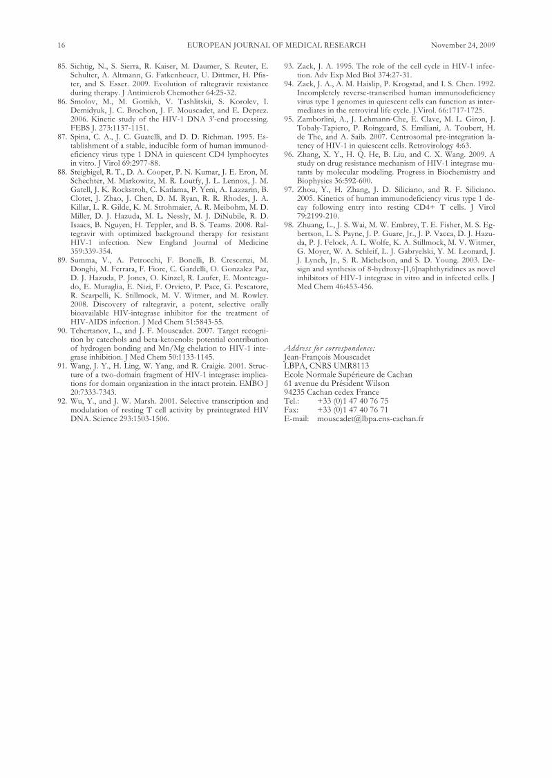
Fig. 4. Modeling of Mg binding by raltegravir.Top: Two different modes of Mg coordination,A- Mg chelation by oxadiazole-2-carboxylate(in E-conformation) and B - Mg chelation bythe β-keto enol group of carbonylamino-1-N-alkyl-5-hydroxypyrimidione (in Z-conforma-tion) (docked on the IN/5-CITEP complex byAutodock 4.0) (64). Bottom: C - Mg chelationby the β-keto enol group (docked on the 1BL3crystal structure by GOLD 3.2) (84) and D -Two-metal coordination by raltegravir (in-duced-fit docking on the IN•DNA com-plex)(5). The chelating centers, O and N, andMg cations are shown in red, blue and magenta,respectively.
A B
C D
Fig. 5. Raltegravir target binding: A) and B) Two different conformations obtained by inhibitor docking on the 5CITEP•INcomplex by Autodock 4.0 (64). Inhibitors and residues in close contact are indicated as sticks and the protein is shown as a sec-ondary structure cartoon. 5CITEP marking the terminal portion of 3’-processed viral DNA, the catalytic triad (D64, D116,E152) and interacting key residues are shown as gray, orange and yellow carbon backbone representations, respectively. C) Thebest docking (GOLD 3.2) of the inhibitor on the 1BL3 IN crystal structure (84). Inhibitor (magenta) and residues in close con-tact (green) are indicated as sticks; D) Inhibitor induced-fit docking on the IN•DNA complex (5). Two positions of the in-hibitor, 1 (orange) and 2 (green); residues in close contact are indicated as sticks and the protein is shown as a secondary struc-ture cartoon. The images are reproduced from the corresponding papers with kind permission of the authors.
The contribution of viral DNA has been assessed inmodels of IN•DNA complexes used for the dockingof diverse set of INSTIs. The inhibitors bound closeto the three catalytic residues and interacted with thedonor DNA. Moreover, these studies confirmed sever-al key observations: the inhibitor binding site existsonly after the 3’ processing of vDNA and the hy-drophobic tail binds in the hydrophobic pocket formedprincipally by the flexible active site loop [18]. The re-finement of this strategy by induced-fit docking (IFD)demonstrated that raltegravir binding involved a two-metal mechanism (Fig. 4, D) and close interactionswith the terminal adenine of the 3’-processed viralDNA (Fig. 5, D), consistent with the findings of bio-chemical experiments, [52]. An alternative computa-tional strategy involves the use of the coordinates ofthe Tn5 transposase-DNA complex as a three-dimen-sional target for the docking of INSTIs [6]. Finally, theeffect of INSTI-resistant mutations has been investi-gated directly through docking and molecular dynamicssimulations of the S-1360 DKA on models of mutantintegrases [96]. The presence of mutations resulted inthe exclusion of the inhibitor from the DNA bindingsite.
In conclusion, with the authorization for clinical useof raltegravir and the arrival of other potent newARVs, the therapeutic management of patients withmulti-failure is facilitated with virological success rateup to 90% in the most favorable case when fully activemolecules are associated. Furthermore, in June 2009,Isentress received an extended indication for previous-ly untreated patients, in combination with standardtreatment. The chemical and molecular determinantsof raltegravir potency are now well understood and thenature of the interactions with its target in the contextof the integrase/vDNA complex is beginning to beelucidated owing to the contribution of molecularmodeling. This knowledge contributes to our under-standing of the molecular reasons for the emergenceof the resistance pathways, mainly based on the Q148,N155 and Y143 residues. The mutation of these keyresidues, involved in the specific interaction of inte-grase with its DNA substrate, into well-defined aminoacids, prevent raltegravir to bind efficiently to integrasewhilst maintaining the catalytic activity of the enzyme.Modeling studies suggested that second generation in-hibitors should molecules depart from the model ofinhibition demonstrated by raltegravir, involving simul-taneously metal chelation and interaction with the cat-alytic loop or risk seeing the emergence of cross-resis-tance as already demonstrated with elvitegravir.
Acknowledgments: The research of Jean-François Mous-cadet’s group is funded by the Centre National pour laRecherche Scientifique (CNRS), l’Ecole Normale Supérieure deCachan (ENSC), l’Agence Nationale de Recherche sur le Sidaet les Hépatites (ANRS) and Sidaction.
REFERENCES
1. Arhel, N. J., S. Souquere-Besse, S. Munier, P. Souque, S.Guadagnini, S. Rutherford, M. C. Prevost, T. D. Allen, andP. Charneau. 2007. HIV-1 DNA Flap formation promotesuncoating of the pre-integration complex at the nuclearpore. EMBO J 26:3025-3037.
2. Asante-Appiah, E., S. H. Seeholzer, and A. M. Skalka. 1998.Structural determinants of metal-induced conformationalchanges in HIV- 1 integrase. J Biol Chem 273:35078-35087.
3. Asante-Appiah, E., and A. M. Skalka. 1999. HIV-1 inte-grase: structural organization, conformational changes, andcatalysis. Adv. Virus Res. 52:351-369.
4. Barreca, M. L., S. Ferro, A. Rao, L. De Luca, M. Zappala, A.M. Monforte, Z. Debyser, M. Witvrouw, and A. Chimirri.2005. Pharmacophore-based design of HIV-1 integrasestrand-transfer inhibitors. J Med Chem 48:7084-8.
5. Barreca, M. L., N. Iraci, L. De Luca, and A. Chimirri. 2009.Induced-fit docking approach provides insight into thebinding mode and mechanism of action of HIV-1 integraseinhibitors. Chem Med Chem. In press. Epub ahead of print2009/06/23.
6. Barreca, M. L., F. Ortuso, N. Iraci, L. De Luca, S. Alcaro,and A. Chimirri. 2007. Tn5 transposase as a useful platformto simulate HIV-1 integrase inhibitor binding mode.Biochem Biophys Res Commun 363:554-60.
7. Beese, L. S., and T. A. Steitz. 1991. Structural basis for the3'-5' exonuclease activity of Escherichia-coli DNA poly-merase-I – A 2 metal-ion mechanism. EMBO J 10:25-33.
8. Brigo, A., K. W. Lee, F. Fogolari, G. I. Mustata, and J. M.Briggs. 2005. Comparative molecular dynamics simulationsof HIV-1 integrase and the T66I/M154I mutant: bindingmodes and drug resistance to a diketo acid inhibitor. Pro-teins 59:723-41.
9. Brigo, A., K. W. Lee, G. Iurcu Mustata, and J. M. Briggs.2005. Comparison of multiple molecular dynamics trajecto-ries calculated for the drug-resistant HIV-1 integraseT66I/M154I catalytic domain. Biophys J 88:3072-82.
10. Brin, E., J. Yi, A. M. Skalka, and J. Leis. 2000. Modeling thelate steps in HIV-1 IN-catalyzed DNA integration. J BiolChem. 275: 39287-95
11. Brown, P. O. 1990. Integration of retroviral DNA. CurrTop Microbiol Immunol. 157:19-48:19-48.
12. Brussel, A., and P. Sonigo. 2004. Evidence for gene expres-sion by unintegrated human immunodeficiency virus type 1DNA species. J Virol 78:11263-71.
13. Bujacz, G., J. Alexandratos, A. Wlodawer, G. Merkel, M.Andrake, R. A. Katz, and A. M. Skalka. 1997. Binding of dif-ferent divalent cations to the active site of avian sarcomavirus integrase and their effects on enzymatic activity. J BiolChem. 272:18161-18168.
14. Bushman, F. D., and R. Craigie. 1991. Activities of humanimmunodeficiency virus (HIV) integration protein in vitro:specific cleavage and integration of HIV DNA. Proc. Natl.Acad. Sci. USA. 88:1339-1343.
15. Cai, M., R. Zheng, M. Caffrey, R. Craigie, G. M. Clore, andA. M. Gronenborn. 1997. Solution structure of the N-termi-nal zinc binding domain of HIV-1 integrase. Nat. Struct.Biol. 4:567-577.
16. Ceccherini-Silberstein, F., I. Malet, R. D'Arrigo, A. Antinori,A. G. Marcelin, and C. F. Perno. 2009. Characterization andstructural analysis of HIV-1 integrase conservation. AIDSRev 11:17-29.
17. Chen, J. C., J. Krucinski, L. J. Miercke, J. S. Finer-Moore, A.H. Tang, A. D. Leavitt, and R. M. Stroud. 2000. Crystalstructure of the HIV-1 integrase catalytic core and C-termi-nal domains: a model for viral DNA binding. Proc. Natl.Acad. Sci. USA 97:8233-8238.
18. Chen, X., M. Tsiang, F. Yu, M. Hung, G. S. Jones, A. Zey-nalzadegan, X. Qi, H. Jin, C. U. Kim, S. Swaminathan, and J.M. Chen. 2008. Modeling, analysis, and validation of a novelHIV integrase structure provide insights into the bindingmodes of potent integrase inhibitors. J Mol Biol 380:504-19.
19. Chirch, L. M., S. Morrison, and R. T. Steigbigel. 2009. Treat-ment of HIV infection with raltegravir. Expert Opin Phar-macother 10:1203-11.
20. Chiu, T. K., and D. R. Davies. 2004. Structure and functionof HIV-1 integrase. Curr. Top. Med. Chem. 4:965-977.
21. Chow, S. A., K. A. Vincent, V. Ellison, and P. O. Brown.1992. Reversal of integration and DNA splicing mediated by
EUROPEAN JOURNAL OF MEDICAL RESEARCHNovember 24, 2009 13

integrase of human immunodeficiency virus. Science 255:723-726.
22. Chun, T. W., L. Stuyver, S. B. Mizell, L. A. Ehler, J. A. Mi-can, M. Baseler, A. L. Lloyd, M. A. Nowak, and A. S. Fauci.1997. Presence of an inducible HIV-1 latent reservoir duringhighly active antiretroviral therapy. Proc Natl Acad Sci USA94:13193-13197.
23. Cocohoba, J., and B. J. Dong. 2008. Raltegravir: the firstHIV integrase inhibitor. Clin Ther 30:1747-65.
24. Cooper, D. A., R. T. Steigbigel, J. M. Gatell, J. K. Rock-stroh, C. Katlama, P. Yeni, A. Lazzarin, B. Clotet, P. N. Ku-mar, J. E. Eron, M. Schechter, M. Markowitz, M. R. Loutfy,J. L. Lennox, J. Zhao, J. Chen, D. M. Ryan, R. R. Rhodes, J.A. Killar, L. R. Gilde, K. M. Strohmaier, A. R. Meibohm, M.D. Miller, D. J. Hazuda, M. L. Nessly, M. J. DiNubile, R. D.Isaacs, H. Teppler, B. Y. Nguyen, and B. S. Teams. 2008.Subgroup and resistance analyses of raltegravir for resistantHIV-1 infection. New England Journal of Medicine359:355-365.
25. Croxtall, J. D., and S. J. Keam. 2009. Raltegravir: a review ofits use in the management of HIV infection in treatment-ex-perienced patients. Drugs 69:1059-75.
26. De Luca, L., A. Pedretti, G. Vistoli, M. L. Barreca, L. Villa,P. Monforte, and A. Chimirri. 2003. Analysis of the full-length integrase-DNA complex by a modified approach forDNA docking. Biochem Biophys Res Commun 310:1083-8.
27. Delelis, O., I. Malet, L. Na, L. Tchertanov, V. Calvez, A. G.Marcelin, F. Subra, E. Deprez, and J. F. Mouscadet. 2009.The G140S mutation in HIV integrases from raltegravir-re-sistant patients rescues catalytic defect due to the resistanceQ148H mutation. Nucleic Acids Res 37:1193-201.
28. Dhaked, D. K., J. Verma, A. Saran, and E. C. Coutinho.2009. Exploring the binding of HIV-1 integrase inhibitorsby comparative residue interaction analysis (CoRIA). J MolModel 15:233-45.
29. Dolan, J., A. Chen, I. T. Weber, R. W. Harrison, and J. Leis.2009. Defining the DNA substrate binding sites on HIV-1integrase. J Mol Biol 385:568-79.
30. Eijkelenboom, A. P., R. A. Lutzke, R. Boelens, R. H. Plas-terk, R. Kaptein, and K. Hard. 1995. The DNA-binding do-main of HIV-1 integrase has an SH3-like fold. Nat StructBiol 2:807-810.
31. Engelman, A., F. D. Bushman, and R. Craigie. 1993. Identi-fication of discrete functional domains of HIV-1 integraseand their organization within an active multimeric complex.EMBO J 12:3269-3275.
32. Espeseth, A. S., P. Felock, A. Wolfe, M. Witmer, J. Grobler,N. Anthony, M. Egbertson, J. Y. Melamed, S. Young, T.Hamill, J. L. Cole, and D. J. Hazuda. 2000. HIV-1 integraseinhibitors that compete with the target DNA substrate de-fine a unique strand transfer conformation for integrase.Proc Natl Acad Sci USA 97:11244-11249.
33. Esposito, D., and R. Craigie. 1998. Sequence specificity ofviral end DNA binding by HIV-1 integrase reveals criticalregions for protein-DNA interaction. EMBO J. 17:5832-5843.
34. Faure, A., C. Calmels, C. Desjobert, M. Castroviejo, A. Cau-mont-Sarcos, L. Tarrago-Litvak, S. Litvak, and V. Parissi.2005. HIV-1 integrase crosslinked oligomers are active invitro. Nucleic Acids Res. 33:977-986.
35. Fenollar-Ferrer, C., V. Carnevale, S. Raugei, and P. Carloni.2008. HIV-1 integrase-DNA interactions investigated bymolecular modelling. Computational and MathematicalMethods in Medicine 9:231-243.
36. Goldgur, Y., R. Craigie, G. H. Cohen, T. Fujiwara, T. Yoshi-naga, T. Fujishita, H. Sugimoto, T. Endo, H. Murai, and D.R. Davies. 1999. Structure of the HIV-1 integrase catalyticdomain complexed with an inhibitor: a platform for antiviraldrug design. Proc Natl Acad Sci USA 96:13040-13043.
37. Goldgur, Y., F. Dyda, A. B. Hickman, T. M. Jenkins, R.Craigie, and D. R. Davies. 1998. Three new structures of thecore domain of HIV-1 integrase: an active site that bindsmagnesium. Proc Natl Acad Sci USA 95:9150-9154.
38. Greenwald, J., V. Le, S. L. Butler, F. D. Bushman, and S.
Choe. 1999. The mobility of an HIV-1 integrase active siteloop is correlated with catalytic activity. Biochemistry38:8892-8898.
39. Grinsztejn, B., B. Y. Nguyen, C. Katlama, J. M. Gatell, A.Lazzarin, D. Vittecoq, C. J. Gonzalez, J. Chen, C. M. Har-vey, and R. D. Isaacs. 2007. Safety and efficacy of the HIV-1integrase inhibitor raltegravir (MK-0518) in treatment-expe-rienced patients with multidrug-resistant virus: a phase IIrandomised controlled trial. Lancet 369:1261-1269.
40. Grobler, J. A., K. Stillmock, B. Hu, M. Witmer, P. Felock,A. S. Espeseth, A. Wolfe, M. Egbertson, M. Bourgeois, J.Melamed, J. S. Wai, S. Young, J. Vacca, and D. J. Hazuda.2002. Diketo acid inhibitor mechanism and HIV-1 integrase:implications for metal binding in the active site of phospho-transferase enzymes. Proc Natl Acad Sci USA 99:6661-6666.
41. Guiot, E., K. Carayon, O. Delelis, F. Simon, P. Tauc, E. Zu-bin, M. Gottikh, J. F. Mouscadet, J. C. Brochon, and E. De-prez. 2006. Relationship between the oligomeric status ofHIV-1 integrase on DNA and enzymatic activity. J BiolChem 281:22707-19.
42. Hazuda, D., M. Iwamoto, and L. Wenning. 2009. Emergingpharmacology: inhibitors of human immunodeficiency virusintegration. Annu Rev Pharmacol Toxicol 49:377-94.
43. Hazuda, D. J., P. Felock, M. Witmer, A. Wolfe, K. Still-mock, J. A. Grobler, A. Espeseth, L. Gabryelski, W. Schleif,C. Blau, and M. D. Miller. 2000. Inhibitors of strand transferthat prevent integration and inhibit HIV-1 replication incells. Science 287:646-650.
44. Hazuda, D. J., S. D. Young, J. P. Guare, N. J. Anthony, R.P. Gomez, J. S. Wai, J. P. Vacca, L. Handt, S. L. Motzel, H.J. Klein, G. Dornadula, R. M. Danovich, M. V. Witmer, K.A. Wilson, L. Tussey, W. A. Schleif, L. S. Gabryelski, L. Jin,M. D. Miller, D. R. Casimiro, E. A. Emini, and J. W. Shiver.2004. Integrase inhibitors and cellular immunity suppressretroviral replication in Rhesus macaques. Science.
45. Hu, J. P., X. Q. Gong, J. G. Su, W. Z. Chen, and C. X.Wang. 2008. Study on the molecular mechanism of inhibit-ing HIV-1 integrase by EBR28 peptide via molecular model-ing approach. Biophys Chem 132:69-80.
46. Iwamoto, M., K. Kassahun, M. D. Troyer, W. D. Hanley, P.Lu, A. Rhoton, A. S. Petry, K. Ghosh, E. Mangin, E. P. De-Noia, L. A. Wenning, J. A. Stone, K. M. Gottesdiener, and J.A. Wagner. 2008. Lack of a pharmacokinetic effect of ralte-gravir on midazolam: in vitro/in vivo correlation. J ClinPharmacol 48:209-14.
47. Iwamoto, M., L. A. Wenning, G. C. Mistry, A. S. Petry, S. Y.Liou, K. Ghosh, S. Breidinger, N. Azrolan, M. J. Gutierrez,W. E. Bridson, J. A. Stone, K. M. Gottesdiener, and J. A.Wagner. 2008. Atazanavir modestly increases plasma levelsof raltegravir in healthy subjects. Clin Infect Dis 47:137-40.
48. Iwamoto, M., L. A. Wenning, A. S. Petry, M. Laethem, M.De Smet, J. T. Kost, S. A. Merschman, K. M. Strohmaier, S.Ramael, K. C. Lasseter, J. A. Stone, K. M. Gottesdiener, andJ. A. Wagner. 2008. Safety, tolerability, and pharmacokinet-ics of raltegravir after single and multiple doses in healthysubjects. Clin Pharmacol Ther 83:293-9.
49. Jaskolski, M., J. N. Alexandratos, G. Bujacz, and A. Wlo-dawer. 2009. Piecing together the structure of retroviral in-tegrase, an important target in AIDS therapy. FEBS J276:2926-46.
50. Jenkins, T. M., D. Esposito, A. Engelman, and R. Craigie.1997. Critical contacts between HIV-1 integrase and viralDNA identified by structure-based analysis and photo-crosslinking. EMBO J. 16:6849-6859.
51. Jenkins, T. M., A. B. Hickman, F. Dyda, R. Ghirlando, D. R.Davies, and R. Craigie. 1995. Catalytic domain of human im-munodeficiency virus type 1 integrase: identification of asoluble mutant by systematic replacement of hydrophobicresidues. Proc Natl Acad Sci USA 92:6057-6061.
52. Johnson, A. A., C. Marchand, S. S. Patil, R. Costi, S. R. Di,T. R. Burke, Jr., and Y. Pommier. 2007. Probing HIV-1 inte-grase inhibitor binding sites with position-specific integrase-DNA cross-linking assays. Mol Pharmacol 71:893-901.
53. Kawasuji, T., M. Fuji, T. Yoshinaga, A. Sato, T. Fujiwara,
EUROPEAN JOURNAL OF MEDICAL RESEARCH14 November 24, 2009
and R. Kiyama. 2006. A platform for designing HIV inte-grase inhibitors. Part 2: a two-metal binding model as a po-tential mechanism of HIV integrase inhibitors. Bioorg MedChem 14:8420-9.
54. Kilzer, J. M., T. Stracker, B. Beitzel, K. Meek, M. Weitzman,and F. D. Bushman. 2003. Roles of host cell factors in circu-larization of retroviral DNA. Virology 314:460-467.
55. Lee, M. S., and R. Craigie. 1994. Protection of retroviralDNA from autointegration: involvement of a cellular factor.Proc Natl Acad Sci USA 91:9823-9827.
56. Lee, S. P., and M. K. Han. 1996. Zinc stimulates Mg2+-de-pendent 3'-processing activity of human immunodeficiencyvirus type 1 integrase in vitro. Biochemistry 35:3837-3844.
57. Leh, H., P. Brodin, J. Bischerour, E. Deprez, P. Tauc, J. C.Brochon, E. LeCam, D. Coulaud, C. Auclair, and J. F.Mouscadet. 2000. Determinants of Mg2+-dependent activi-ties of recombinant human immunodeficiency virus type 1integrase. Biochemistry 39:9285-9294.
58. Lennox, J. L., E. Dejesus, A. Lazzarin, R. B. Pollard, J. V.Madruga, D. S. Berger, J. Zhao, X. Xu, A. Williams-Diaz, A.J. Rodgers, R. J. Barnard, M. D. Miller, M. J. Dinubile, B. Y.Nguyen, R. Leavitt, and P. Sklar. 2009. Safety and efficacyof raltegravir-based versus efavirenz-based combinationtherapy in treatment-naive patients with HIV-1 infection: amulticentre, double-blind randomised controlled trial.Lancet. In press. Epub ahead of print 2009/08/04.
59. Li, L., J. M. Olvera, K. E. Yoder, R. S. Mitchell, S. L. Butler,M. Lieber, S. L. Martin, and F. D. Bushman. 2001. Role ofthe non-homologous DNA end joining pathway in the earlysteps of retroviral infection. EMBO J. 20:3272-3281.
60. Li, M., and R. Craigie. 2005. Processing of viral DNA endschannels the HIV-1 integration reaction to concerted inte-gration. J.Biol.Chem. 280:29334-29339.
61. Lins, R. D., A. Adesokan, T. A. Soares, and J. M. Briggs.2000. Investigations on human immunodeficiency virus type1 integrase/DNA binding interactions via molecular dynam-ics and electrostatics calculations. Pharmacol Ther 85:123-131.
62. Lins, R. D., J. M. Briggs, T. P. Straatsma, H. A. Carlson, J.Greenwald, S. Choe, and J. A. McCammon. 1999. Moleculardynamics studies on the HIV-1 integrase catalytic domain.Biophys J 76:2999-3011.
63. Lodi, P. J., J. A. Ernst, J. Kuszewski, A. B. Hickman, A. En-gelman, R. Craigie, G. M. Clore, and A. M. Gronenborn.1995. Solution structure of the DNA binding domain ofHIV-1 integrase. Biochemistry 34:9826-9833.
64. Loizidou, E. Z., C. D. Zeinalipour-Yazdi, T. Christofides,and L. G. Kostrikis. 2009. Analysis of binding parameters ofHIV-1 integrase inhibitors: correlates of drug inhibition andresistance. Bioorg Med Chem 17:4806-18.
65. Lovell, S., I. Y. Goryshin, W. R. Reznikoff, and I. Rayment.2002. Two-metal active site binding of a Tn5 transposasesynaptic complex. Nat Struct Biol 9:278-81.
66. Maiga, A. I., I. Malet, C. Soulie, A. Derache, V. Koita, B.Amellal, L. Tchertanov, O. Delelis, L. Morand-Joubert, J. F.Mouscadet, R. Murphy, M. Cisse, C. Katlama, V. Calvez,and A. G. Marcelin. 2009. Genetic barriers for integrase in-hibitor drug resistance in HIV type-1 B and CRF02_AGsubtypes. Antivir Ther 14:123-9.
67. Maignan, S., J. P. Guilloteau, Q. Zhou-Liu, C. Clement-mel-la, and V. Mikol. 1998. Crystal structures of the catalytic do-main of HIV-1 integrase free and complexed with its metalcofactor: high level of similarity of the active site with otherviral integrases. J.Mol.Biol. 282:359-368.
68. Malet, I., O. Delelis, C. Soulie, M. Wirden, L. Tchertanov, P.Mottaz, G. Peytavin, C. Katlama, J. F. Mouscadet, V.Calvez, and A. G. Marcelin. 2009. Quasispecies variant dy-namics during emergence of resistance to raltegravir inHIV-1-infected patients. J Antimicrob Chemother 63:795-804.
69. Malet, I., O. Delelis, M. A. Valantin, B. Montes, C. Soulie,M. Wirden, L. Tchertanov, G. Peytavin, J. Reynes, J. F.Mouscadet, C. Katlama, V. Calvez, and A. G. Marcelin.2008. Mutations associated with failure of raltegravir treat-
ment affect integrase sensitivity to the inhibitor in vitro. An-timicrob Agents Chemother. 52:1351-1358.
70. Miller, M., M. Witmer, K. Stillmock, P. Felock, L. Ecto, J.Flynn, W. Schleif, G. Dornadula, R. Danovich, and D.Hazuda. 2006. Biochemical and antiviral activity of MK-0518, a potent HIV integrase inhibitor. AIDS 2006 - XVIInternational AIDS Conference Abstract no. THAA0302.
71. Mouscadet, J. F., R. Arora, J. Andre, J. C. Lambry, O.Delelis, I. Malet, A. G. Marcelin, V. Calvez, and L. Tcher-tanov. 2009. HIV-1 IN alternative molecular recognition ofDNA induced by raltegravir resistance mutations. J MolRecognit. Epub ahead of print 2009/07/23.
72. Murray, J. M., S. Emery, A. D. Kelleher, M. Law, J. Chen,D. J. Hazuda, B. Y. Nguyen, H. Teppler, and D. A. Cooper.2007. Antiretroviral therapy with the integrase inhibitor ral-tegravir alters decay kinetics of HIV, significantly reducingthe second phase. AIDS 21:2315-21.
73. Nakajima, N., R. Lu, and A. Engelman. 2001. Human im-munodeficiency virus type 1 replication in the absence of in-tegrase-mediated dna recombination: definition of permis-sive and nonpermissive T-cell lines. J Virol 75:7944-7955.
74. Nunthaboot, N., S. Pianwanit, V. Parasuk, J. O. Ebalunode,J. M. Briggs, and S. Kokpol. 2007. Hybrid quantum mechani-cal/molecular mechanical molecular dynamics simulations ofHIV-1 integrase/inhibitor complexes. Biophys J 93:3613-26.
75. O'Brien, W. A., A. Namazi, H. Kalhor, S. H. Mao, J. A.Zack, and I. S. Chen. 1994. Kinetics of human immunodefi-ciency virus type 1 reverse transcription in blood mononu-clear phagocytes are slowed by limitations of nucleotide pre-cursors. J Virol 68:1258-1263.
76. Pace, P., M. E. Di Francesco, C. Gardelli, S. Harper, E.Muraglia, E. Nizi, F. Orvieto, A. Petrocchi, M. Poma, M.Rowley, R. Scarpelli, R. Laufer, O. Gonzalez Paz, E. Mon-teagudo, F. Bonelli, D. Hazuda, K. A. Stillmock, and V.Summa. 2007. Dihydroxypyrimidine-4-carboxamides asnovel potent and selective HIV integrase inhibitors. J MedChem 50:2225-39.
77. Pauza, C. D., P. Trivedi, T. S. McKechnie, D. D. Richman,and F. M. Graziano. 1994. 2-LTR circular viral DNA as amarker for human immunodeficiency virus type 1 infectionin vivo. Virology 205:470-478.
78. Pommier, Y., A. A. Johnson, and C. Marchand. 2005. Inte-grase inhibitors to treat HIV/AIDS. Nat Rev Drug Discov4:236-248.
79. Roquebert, B., F. Damond, G. Collin, S. Matheron, G. Pey-tavin, A. Benard, P. Campa, G. Chene, F. Brun-Vezinet, andD. Descamps. 2008. HIV-2 integrase gene polymorphismand phenotypic susceptibility of HIV-2 clinical isolates tothe integrase inhibitors raltegravir and elvitegravir in vitro. JAntimicrob Chemother 62:914-20.
80. Sato, M., T. Motomura, H. Aramaki, T. Matsuda, M. Ya-mashita, Y. Ito, H. Kawakami, Y. Matsuzaki, W. Watanabe,K. Yamataka, S. Ikeda, E. Kodama, M. Matsuoka, and H.Shinkai. 2006. Novel HIV-1 integrase inhibitors derivedfrom quinolone antibiotics. J Med Chem 49:1506-8.
81. Savarino, A., M. Pistello, D. D'Ostilio, E. Zabogli, F. Taglia,F. Mancini, S. Ferro, D. Matteucci, L. De Luca, M. L. Bar-reca, A. Ciervo, A. Chimirri, M. Ciccozzi, and M. Bendinelli.2007. Human immunodeficiency virus integrase inhibitorsefficiently suppress feline immunodeficiency virus replica-tion in vitro and provide a rationale to redesign antiretrovi-ral treatment for feline AIDS. Retrovirology 4:79.
82. Sechi, M., A. Bacchi, M. Carcelli, C. Compari, E. Duce, E.Fisicaro, D. Rogolino, P. Gates, M. Derudas, L. Q. Al-Maw-sawi, and N. Neamati. 2006. From ligand to complexes: in-hibition of human immunodeficiency virus type 1 integraseby beta-diketo acid metal complexes. Journal of MedicinalChemistry 49:4248-4260.
83. Semenova, E. A., C. Marchand, and Y. Pommier. 2008.HIV-1 integrase inhibitors: update and perspectives. AdvPharmacol 56:199-228.
84. Serrao, E., S. Odde, K. Ramkumar, and N. Neamati. 2009.Raltegravir, elvitegravir, and metoogravir: the birth of "me-too" HIV-1 integrase inhibitors. Retrovirology 6:25.
EUROPEAN JOURNAL OF MEDICAL RESEARCHNovember 24, 2009 15
85. Sichtig, N., S. Sierra, R. Kaiser, M. Daumer, S. Reuter, E.Schulter, A. Altmann, G. Fatkenheuer, U. Dittmer, H. Pfis-ter, and S. Esser. 2009. Evolution of raltegravir resistanceduring therapy. J Antimicrob Chemother 64:25-32.
86. Smolov, M., M. Gottikh, V. Tashlitskii, S. Korolev, I.Demidyuk, J. C. Brochon, J. F. Mouscadet, and E. Deprez.2006. Kinetic study of the HIV-1 DNA 3'-end processing.FEBS J. 273:1137-1151.
87. Spina, C. A., J. C. Guatelli, and D. D. Richman. 1995. Es-tablishment of a stable, inducible form of human immunod-eficiency virus type 1 DNA in quiescent CD4 lymphocytesin vitro. J Virol 69:2977-88.
88. Steigbigel, R. T., D. A. Cooper, P. N. Kumar, J. E. Eron, M.Schechter, M. Markowitz, M. R. Loutfy, J. L. Lennox, J. M.Gatell, J. K. Rockstroh, C. Katlama, P. Yeni, A. Lazzarin, B.Clotet, J. Zhao, J. Chen, D. M. Ryan, R. R. Rhodes, J. A.Killar, L. R. Gilde, K. M. Strohmaier, A. R. Meibohm, M. D.Miller, D. J. Hazuda, M. L. Nessly, M. J. DiNubile, R. D.Isaacs, B. Nguyen, H. Teppler, and B. S. Teams. 2008. Ral-tegravir with optimized background therapy for resistantHIV-1 infection. New England Journal of Medicine359:339-354.
89. Summa, V., A. Petrocchi, F. Bonelli, B. Crescenzi, M.Donghi, M. Ferrara, F. Fiore, C. Gardelli, O. Gonzalez Paz,D. J. Hazuda, P. Jones, O. Kinzel, R. Laufer, E. Monteagu-do, E. Muraglia, E. Nizi, F. Orvieto, P. Pace, G. Pescatore,R. Scarpelli, K. Stillmock, M. V. Witmer, and M. Rowley.2008. Discovery of raltegravir, a potent, selective orallybioavailable HIV-integrase inhibitor for the treatment ofHIV-AIDS infection. J Med Chem 51:5843-55.
90. Tchertanov, L., and J. F. Mouscadet. 2007. Target recogni-tion by catechols and beta-ketoenols: potential contributionof hydrogen bonding and Mn/Mg chelation to HIV-1 inte-grase inhibition. J Med Chem 50:1133-1145.
91. Wang, J. Y., H. Ling, W. Yang, and R. Craigie. 2001. Struc-ture of a two-domain fragment of HIV-1 integrase: implica-tions for domain organization in the intact protein. EMBO J20:7333-7343.
92. Wu, Y., and J. W. Marsh. 2001. Selective transcription andmodulation of resting T cell activity by preintegrated HIVDNA. Science 293:1503-1506.
93. Zack, J. A. 1995. The role of the cell cycle in HIV-1 infec-tion. Adv Exp Med Biol 374:27-31.
94. Zack, J. A., A. M. Haislip, P. Krogstad, and I. S. Chen. 1992.Incompletely reverse-transcribed human immunodeficiencyvirus type 1 genomes in quiescent cells can function as inter-mediates in the retroviral life cycle. J.Virol. 66:1717-1725.
95. Zamborlini, A., J. Lehmann-Che, E. Clave, M. L. Giron, J.Tobaly-Tapiero, P. Roingeard, S. Emiliani, A. Toubert, H.de The, and A. Saib. 2007. Centrosomal pre-integration la-tency of HIV-1 in quiescent cells. Retrovirology 4:63.
96. Zhang, X. Y., H. Q. He, B. Liu, and C. X. Wang. 2009. Astudy on drug resistance mechanism of HIV-1 integrase mu-tants by molecular modeling. Progress in Biochemistry andBiophysics 36:592-600.
97. Zhou, Y., H. Zhang, J. D. Siliciano, and R. F. Siliciano.2005. Kinetics of human immunodeficiency virus type 1 de-cay following entry into resting CD4+ T cells. J Virol79:2199-210.
98. Zhuang, L., J. S. Wai, M. W. Embrey, T. E. Fisher, M. S. Eg-bertson, L. S. Payne, J. P. Guare, Jr., J. P. Vacca, D. J. Hazu-da, P. J. Felock, A. L. Wolfe, K. A. Stillmock, M. V. Witmer,G. Moyer, W. A. Schleif, L. J. Gabryelski, Y. M. Leonard, J.J. Lynch, Jr., S. R. Michelson, and S. D. Young. 2003. De-sign and synthesis of 8-hydroxy-[1,6]naphthyridines as novelinhibitors of HIV-1 integrase in vitro and in infected cells. JMed Chem 46:453-456.
Address for correspondence: Jean-François MouscadetLBPA, CNRS UMR8113Ecole Normale Supérieure de Cachan61 avenue du Président Wilson94235 Cachan cedex FranceTel.: +33 (0)1 47 40 76 75Fax: +33 (0)1 47 40 76 71E-mail: [email protected]
EUROPEAN JOURNAL OF MEDICAL RESEARCH16 November 24, 2009