research article a novel method for intraoral access to
TRANSCRIPT

Research ArticleA Novel Method for Intraoral Access to the Superior Head ofthe Human Lateral Pterygoid Muscle
Aleli Tôrres Oliveira,1 Anderson Aparecido Camilo,2 Paulo Roberto Valle Bahia,3
Antonio Carlos Pires Carvalho,3 Marcos Fabio DosSantos,4,5
Jorge Vicente Lopes da Silva,2 and André Antonio Monteiro1
1 Orofacial Pain and Masticatory Dysfunction Clinic, Dental Clinic Department, School of Dentistry,Federal University of Rio de Janeiro, Avenida Carlos Chagas Filho 373, Centro de Ciencias da Saude, Bloco K, 2∘ Andar,Sala 56, Ilha da Cidade Universitaria, 21941-902 Rio de Janeiro, RJ, Brazil
2 Renato Archer Information Technology Center (CTI), Rodovia Dom Pedro I (SP - 65) Km 143,6, Bairro Amarais,13069-901 Campinas, SP, Brazil
3 Radiology Department, School of Medicine, Federal University of Rio de Janeiro, Avenida Carlos Chagas Filho 373,Ilha da Cidade Universitaria, 21941-902 Rio de Janeiro, RJ, Brazil
4 Cellular Morphogenesis Laboratory, Biomedical Sciences Institute, Federal University of Rio de Janeiro,Instituto de Ciencias Biomedicas, CCS, Bloco F, Ilha da Cidade Universitaria, 21949-590 Rio de Janeiro, RJ, Brazil
5 Health Sciences Center, Grande Rio University, 1160 Bairro 25 de Agosto, Duque de Caxias, 25071-200 Rio de Janeiro, RJ, Brazil
Correspondence should be addressed to Aleli Torres Oliveira; [email protected]
Received 4 April 2014; Accepted 3 May 2014; Published 18 May 2014
Academic Editor: Marılia Gerhardt de Oliveira
Copyright © 2014 Aleli Torres Oliveira et al. This is an open access article distributed under the Creative Commons AttributionLicense, which permits unrestricted use, distribution, and reproduction in any medium, provided the original work is properlycited.
Background.Theuncoordinated activity of the superior and inferior parts of the lateral pterygoidmuscle (LPM) has been suggestedto be one of the causes of temporomandibular joint (TMJ) disc displacement. A therapy for this muscle disorder is the injection ofbotulinum toxin (BTX), of the LPM. However, there is a potential risk of side effects with the injection guide methods currentlyavailable. In addition, they do not permit appropriate differentiation between the two bellies of themuscle. Herein, a novelmethod ispresented to provide intraoral access to the superior head of the human LPMwith maximal control andminimal hazards.Methods.Computational tomography along with digital imaging software programs and rapid prototyping techniques were used to createa rapid prototyped guide to orient BTX injections in the superior LPM. Results. The method proved to be feasible and reliable.Furthermore, when tested in one volunteer it allowed precise access to the upper head of LPM, without producing side effects.Conclusions.The prototyped guide presented in this paper is a novel tool that provides intraoral access to the superior head of theLPM. Further studies will be necessary to test the efficacy and validate this method in a larger cohort of subjects.
1. Introduction
Functional mandibular movements are the result of pre-cisely coordinated muscle contractions, regulated by a highlyrefined neurologic system, and with the participation ofother craniomandibular structures, including the temporo-mandibular joints (TMJ), ligaments, and teeth. In thiscomplex arrangement, the mandibular elevator (masseter,temporal, and medial pterygoid) and depressor muscles(lateral pterygoid and supra- and infrahyoid muscles) play
an integrated role in mastication and TMJ biomechanics.The lateral pterygoid muscle (LPM) is responsible for threemandibular movements: (1) laterality, caused by unilateralcontraction, (2) protrusion, as a result of simultaneousbilateral contraction, and (3) full mouth opening, whenits bilateral contraction follows the action of the supra-and infrahyoid muscles. The LPM is anatomically dividedin two parts or bellies that coordinate the kinematics ofthe disc-condyle complex [1]. The superior LPM originatesfrom the infratemporal surface and infratemporal crest of
Hindawi Publishing CorporationBioMed Research InternationalVolume 2014, Article ID 432635, 8 pageshttp://dx.doi.org/10.1155/2014/432635

2 BioMed Research International
the greater wing of the sphenoid bone and is inserted intothe anteromedial aspect of the TMJ capsule and disc; theinferior LPM originates from the lateral surface of the lateralpterygoid plate and is inserted into the pterygoid fovea onthe mandibular condyle [2–4]. Due to its complex anatomyand physiology, the human LPM plays an important rolein the coordination of the disc-condyle complex motionandmandibularmovements. Furthermore, an uncoordinatedfunction of both parts or hyperactivity of its superior bellywould contribute to articular disc instability, leading to thevery common anterior disc displacement disorders [1, 5–7].Finally, this muscle has been implicated in other conditions,such as myositis and oromandibular dystonia [6, 8].
Botulinum toxin (BTX) has emerged as a promisingtherapy to treat myositis as well as oromandibular dystonia.BTX is a potent neurotoxin that acts by producing temporarychemical denervation of skeletal muscles. It has the proprietyof inhibiting the release of acetylcholine at the presynapticjunction, producing a transient and dose-dependent reduc-tion of muscle activity, without creating systemic effects [1].So far, few studies have examined the results of BTX whenapplied to the human LPM. In the most common technique,the procedure is guided by electromyography (EMG)using anintra- or extraoral access [1, 9–11].However, it does not permitdifferentiation between the superior and inferior heads of theLPM. Consequently, the BTX injection may be delivered inboth parts of the muscle or only in the inferior LPM. A moreinvasive approach is the injection guided by arthroscopy[10]. Nonetheless, the reduced space and lack of visibilityto the target area makes the differentiation of the superiorand inferior parts of the LPM virtually impossible. Moreover,the risk of severe injury or trauma is considered high, dueto the close anatomical relation between the LPM and theneurovascular structures located in the infratemporal fossa(ITF) [1, 12]. Therefore, the use of an apparatus for injectionguidance to the LPM, with accuracy to target its upperhead, would minimize the risks and optimize the results. Theadvent of modern imaging and rapid prototyping techniques,using computed tomography (CT) [13–16] made possible thedevelopment of such device, a customized injection guide tothe superior head of the LPM.
2. Methods
Virtual modeling was applied, using medical imaging soft-ware applications and modeling techniques, such as com-puter aided design (CAD) that generates STereoLithography(STL) file format. Those techniques were employed to printan injection guide (made of polyamide polymer) in a rapidprototyping machine, to orient the intraoral injection to thesuperior head of the LPM. In order to test the reliablility ofsuch device, a volunteer took part in the study. All proceduresadopted were conducted in accordance with the bioethicalrules for studies involving human beings of the World Medi-cal Association (WMA)—Declaration of Helsinki (1990) andthe volunteer gave written informed consent prior to thestudy. The subject recruited had been previously diagnosedwith anterior TMJ disc displacement with reduction, on theleft side. However, she was not under treatment for her
disorder. Her clinical evaluation revealed the presence ofreciprocal click and mandibular deviation during the mouthopening. Throughout her clinical exam, she complained ofmild pain (3 out of 10 in the visual analogue scale, VAS). Shehad no history of systemic disorders, recent orofacial surgery,trauma, or CT contraindications.
After the clinical examination, dental impressions wereobtainedwith the irreversible hydrocolloidHydrogum (Zher-mack, Badia Polesine, Italy) and a provisional guide wasconstructed, using autopolymerizing acrylic resin, JET (Art.Classico, Sao Paulo, Brazil), to keep the subject’s mandibleslightly opened and deflected to the left side. The provisionalguide was constructed to provide ample access to the targetregion (the left upper LPM) during the CT scan. This wasalso found to be the most appropriate position to access theLPM from an intraoral approach. The provisional appliancewas used as a template to the final device modeling andfabrication. In order to obtain satisfactory contrast duringthe image processing, the provisional guide was filled witha mix of barium sulfate with acrylic resin (1 : 2), following theprotocol described in a previous study [17].
With the appliance inserted in the mouth, the volunteerwas scanned in a six detectors computed tomograph (PhilipsBrilliance).The protocol was standardized with the followingparameters: Kvp = 120, mA = 250, section thickness = 0,8,reconstruction interval = 0,4, matrix size = 512 × 512, andfield of view = 200mm. All data was saved in a DigitalImaging Communications in Medicine (DICOM) format[18]. The images were imported into InVesalius 3.0 beta3(CTI, Brazil), an open source software (GNU GPL2) usedfor reconstruction of computed tomography and magneticresonance (MR) images [19]. This software generates a 3Dreconstruction from a sequence of 2D DICOM files. It usesfour windows to view and manipulate the 3D model in theaxial, sagittal, and coronal planes. In addition, this programprovides filters that are preset for density, thereby creating asemiautomatic segmentation (Figure 1).TheHounsfield scale[12] was further applied to edit, attribute values, and eraseimperfections, but maintaining the original anatomy.
The first challenge was to separate the provisional guide,from the teeth, since artifacts were created during the CTscan, owing to the presence of metallic dental restorations.This semiautomatic separation of soft and hard tissues wasachieved with a manual segmentation tool. The result wasa 3D provisional guide (Figure 2), which was saved ina STereoLithography (STL) format. STL is a standard fileformat for rapid prototyping machines composed of a meshof triangles and normal vectors.
The second challenge was the segmentation of the upperLPM, themost complex phase of the project, since themuscledensity is close to the density of the adjoining soft tissue,mak-ing their individualization difficult on the image obtained.Therefore, a thorough analysis of the anatomical componentsof the ITF, specially the upper head of the LPM in the sagittal,coronal, and transverse planes, was critical to build a 3Dmodel of the muscle. Based on a previous report, the averagelength of the upper LPM is approximately 30mm [20]. After-wards, the hard tissue and the 3D virtual model of the defini-tive guide were designed. The latter was imported into the

BioMed Research International 3
S
A
(a)
L
S
(b)
L
S
(c)
L
S
(d)
Figure 1: Sagittal (a), coronal (b), and axial (c) CT slices when imported into InVesalius.This software generates 3D reconstructions (d) from2D DICOM CT or MR images.
STLmanipulation softwareMagics STL 17.0 (Materialise, Bel-gium) (Figures 3(a) and 3(b)) andfine adjustmentsweremadeto enable its perfect fit to the dental arches of the volunteer.Those adjustments were necessary due to differences in thelong axis angulation of the superior and inferior teeth. Subse-quently, a small lumen was designed with the diameter of theneedle to guide to the upper LPM. After measuring the size,thickness, and shape of the needle and syringe, a small circu-lar tunnel-shaped structure was constructed (Figure 4(a)).
Considering that the permanent guide would be madeof polyamide, a nonmedical grade material, it was prudentto isolate the internal part of the passage that gives accessto the LPM, in order to avoid the undesirable transferenceof this component from the oral mucosa to the ITF by theneedle, which could lead to inflammatory reaction in theLPM or other structures in the ITF. This third challenge wassolved with the use of the software SolidWorks (DassaultSystemes SA, France) to design a plug with rounded edges,made of stainless steel, to be inserted into the tube projectedto target the superior LPM. All measures were meticulouslychecked to prevent gaps between the syringe/needle and thesmall passage just designed, which could interfere in the finalposition of the needle. Subsequently, the needle trajectoryfrom the intraoral mucosa to the superior portion of the LPMwas virtually trained.The infratemporal crest of the sphenoidbone was chosen to be the anatomical region of reference
to access the superior LPM, since this is the origin of theupper LPM. After analyzing the CT images, it was decidedto direct the needle to the region near the transition betweenthemiddle part and posterior two-thirds of the infratemporalcrest. This is the area where the upper LPM has its greatestvolume. A safety margin was intentionally left, retreating theneedle around 2-3mm from the infratemporal crest.
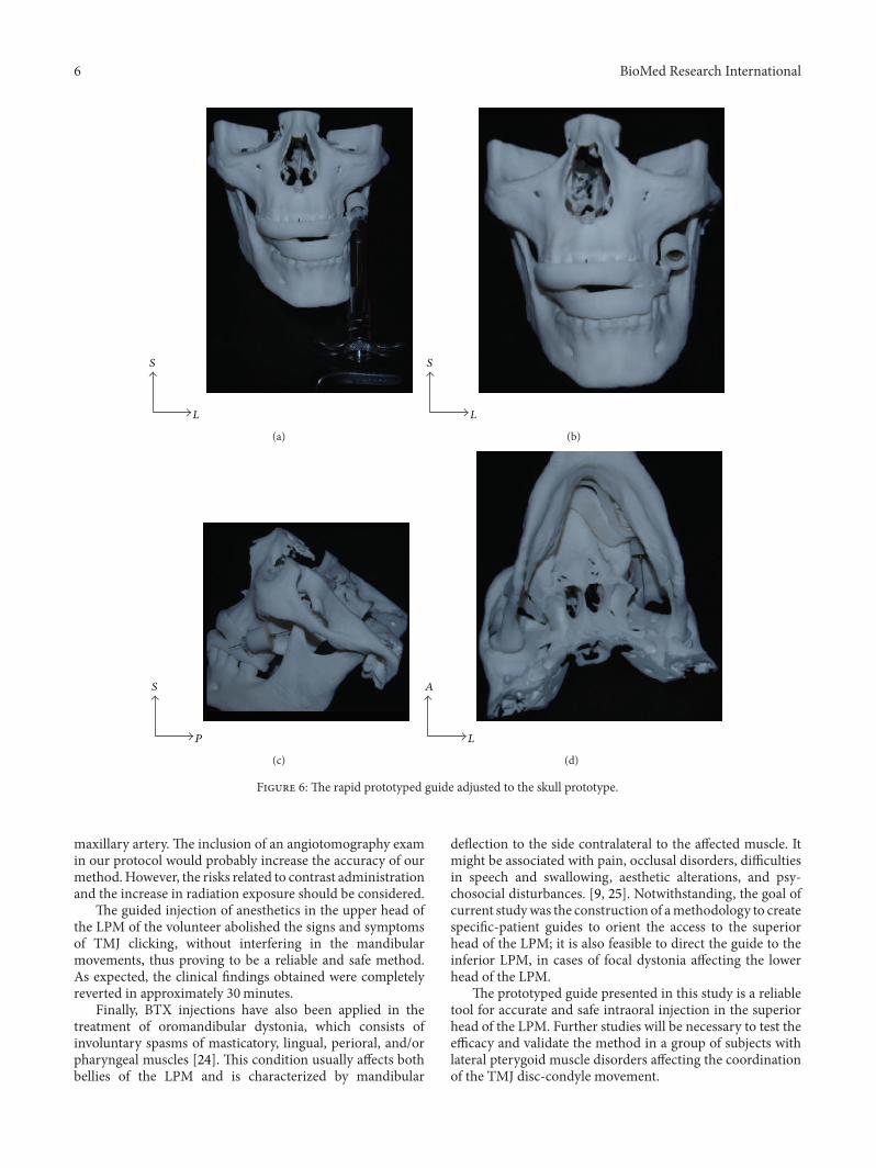
Finally, the image of the small circular guide previouslyprojected from the program SolidWorks (Dassault SystemesSA, France) was imported and aligned with the needle,using the program Magics STL 17.0 (Materialise, Belgium)(Figure 4(b)). In the last phase, all the components of thedefinitive guide were virtually connected (Figure 5). Uponsuccessful completion of the project, the prototypes of theskull and mandible, as well as the guide for BTX appli-cation, were prepared using selective laser sintering (SLS)technology. The unit used to manufacture the device and thebiomodel was a Sinterstation 2000 (3D systems, USA). Theupper LPM injection guide was then adjusted to the skullprototype and the accuracy of the needle directionwas tested.To preliminarily access the clinical reliability of the guide,2 cc of lidocaine hydrochloride 2%, without vasoconstrictor(Lidostesin DENTSPLY, Brazil), was infiltrated in the leftsuperior head of the LPM of the volunteer with a 27G×13/8 (0.4 × 35mm) dental needle (Terumo, Tokyo, Japan)mounted on a carpule syringe (Figures 6(a)–6(d)).

4 BioMed Research International
(a) (b)
S
A
(c) (d)
Figure 2: ((a)–(d)) The construction of a 3D virtual copy of the provisional guide (orange). (a) 3D reconstruction of the CT images. Noticethat the mandible is slighted opened and deflected to the left side. (b) 3D model of the provisional guide (orange) articulated to the dentalarches. (c) Provisional guide extracted. (d) Detailed view of the provisional guide adjusted to the upper and lower teeth.
3. Results and Conclusion
A novel method was developed to produce an injectionguide for intraoral access to the superior head of the humanLPM, using medical image processing programs and rapidprototyping technology. The guide presented in the currentstudy, resembles the surgical guides commonly used fordental implant placement [21]. Upon the manipulation ofthe 3D virtual images obtained from a CT scan and theidentification of the infratemporal crest of the greater wing ofthe sphenoid bone, the most consistent anatomical referenceto the upper head of the LPM, the layout of the final devicewas projected.The injection guide wasmeticulously designedto fit the upper and lower dental arches of our volunteer. Thedevice also contained a narrow passage to orient the needlethat was directed to the superior LPM.
The efficacy of BTX application in the LPM to treatanterior TMJ disc displacements has been reported in theliterature. However, the LPM access is challenging and thereare risks associated with the procedure [1, 4, 6–8]. The goalof such therapy is to abolish spasm of superior head of LPMthat prevents the disc from passively retruding with the disc-condyle complex to the TMJ fossa in the mandibular restposition. Instead, the traction provided by the superior headwould retain the disc anteriorly, while the condyles wouldbe pushed up and backwards by the powerful mandibular
elevator muscles, resulting in uncoordinated disc-condylekinematics. The thin upper ligament of the bilaminar zone,composed of elastin, would not support the tension providedby the upper LPM spasm, resulting in disc dislocation andclicking sound during its reduction to normal disc-condylerelationship.
Up to now, EMG is the most common technique appliedto acces the human lateral pterygoidmuscle in vivo. Notwith-standing it permits the localization of the LPM; it doesnot provide the proper differentiation between the superiorand inferior parts of the muscle [8]. This characteristic isextremely relevant given that BTX injection in the inferiorhead of the LPM is invariably associated with temporarylimitation of the lateral mandibular movement to the con-tralateral side [1] and jaw deflection duringmaximummouthopening. Although significant adverse events have not beenlargely described, there is a potential risk when the LPMBTX injection is carried out under EMG guidance, sincethe anatomical structures are not clearly identified. In fact,even the inferior LPM palpation is difficult and it has notbeen recommended in the clinical routine examination fortemporomandibular disorders (TMDs), since there is a highpossibility of false-positive findings [22].The use of a patient-specific guide to orient the direction of the needle during theBTX injection in the LPM allows the proper differentiationbetween the superior and inferior LPM. Thus, with this

BioMed Research International 5
S
A
(a) (b)
Figure 3: After the segmentation of the superior LPM (green), osseous tissues, and teeth, a 3D virtual model of the permanent guide (orange)was designed and imported into the image processing software Magics STL 17.0. The region close to the junction between the middle partand the posterior two-thirds of the upper LPM was chosen to be the target area (a) and the 3D model of the definitive guide was adjusted toperfectly fit the dental arches of our subject.
S
A
(a)
S
A
(b)
Figure 4: A small tube was virtually built and adapted to the 3D model of the definitive guide to provide access to the superior LPM (a) anda protection was designed in stainless steel, in order to isolate the inner part of this structure (b).
S
A
Figure 5:The final project of the upper LPM guide (green) with thevirtual needle directed to the upper LPM (green).
technique it is possible to selectively target the superior LPM,avoiding discomforting or any potential side effect relatedto the inferior LPM BTX injection. Arthroscopy is anothertechnique commonly applied to guide LPM BTX injection.Nevertheless, it can be considered a more invasive procedurewhen compared to EMG [10].
Among the structures located in the ITF, the maxillaryartery is one of the most important and it should beavoided during LPM injections. A comprehensive knowledgeof its variations and its relations to the LPM is highlyrecommended when planning the procedure. This artery isdivided in three portions. Although, in some cases its secondpart passes deep to the muscle [23], in the most commontopographical relationship, it is superficial to the LPM [12],which increases the chance of intravascular injections.There-fore, based on the regional anatomy it is possible to designthe access to the superior LPM, preventing traumas to the
6 BioMed Research International
S
L
(a)
S
L
(b)
S
P
(c)
A
L
(d)
Figure 6: The rapid prototyped guide adjusted to the skull prototype.
maxillary artery. The inclusion of an angiotomography examin our protocol would probably increase the accuracy of ourmethod.However, the risks related to contrast administrationand the increase in radiation exposure should be considered.
The guided injection of anesthetics in the upper head ofthe LPM of the volunteer abolished the signs and symptomsof TMJ clicking, without interfering in the mandibularmovements, thus proving to be a reliable and safe method.As expected, the clinical findings obtained were completelyreverted in approximately 30minutes.
Finally, BTX injections have also been applied in thetreatment of oromandibular dystonia, which consists ofinvoluntary spasms of masticatory, lingual, perioral, and/orpharyngeal muscles [24]. This condition usually affects bothbellies of the LPM and is characterized by mandibular
deflection to the side contralateral to the affected muscle. Itmight be associated with pain, occlusal disorders, difficultiesin speech and swallowing, aesthetic alterations, and psy-chosocial disturbances. [9, 25]. Notwithstanding, the goal ofcurrent studywas the construction of amethodology to createspecific-patient guides to orient the access to the superiorhead of the LPM; it is also feasible to direct the guide to theinferior LPM, in cases of focal dystonia affecting the lowerhead of the LPM.
The prototyped guide presented in this study is a reliabletool for accurate and safe intraoral injection in the superiorhead of the LPM. Further studies will be necessary to test theefficacy and validate the method in a group of subjects withlateral pterygoid muscle disorders affecting the coordinationof the TMJ disc-condyle movement.

BioMed Research International 7
Abbreviations
BTX: Botulinum toxinCAD: Computer aided designCT: Computed tomographyDICOM: Digital Imaging Communications in
MedicineEMG: ElectromyographyGNU GPL2: General Public License, version 2.0ITF: Infratemporal fossaLPM: Lateral pterygoid muscleMR: Magnetic resonanceSTL: STereoLithographyTMD: Temporomandibular disordersTMJ: Temporomandibular jointVAS: Visual analogue scaleV3: Mandibular nerve (third branch of the
fifth cranial nerve)WMA: World Medical Association3D: Three-dimensional.
Conflict of Interests
The authors declare that there is no conflict of interestsregarding the publication of this paper. All authors of thispaper declare that this research was conducted in the absenceof any commercial or financial relationships that could beconstrued as a potential conflict of interests.
Authors’ Contribution
Aleli Torres Oliveira conceived, designed, and performedthe clinical procedures; coordinated the study; and alsodrafted the paper. Anderson Aparecido Camilo analyzed thedata, projected the stereolithographic guide, and drafted thepaper. Paulo Roberto Valle Bahia and Antonio Carlos PiresCarvalho drafted the paper and supervised the radiologicalexam. Jorge Vicente Lopes da Silva participated in the designof the study and drafted the paper. Marcos Fabio DosSantosand Andre Antonio Monteira conceived and designed thestudy and drafted the paper. All authors read and approvedthe final version of this document.
Acknowledgments
Dr. Marcos Fabio DosSantos was supported by the ConselhoNacional de Desenvolvimento Cientıfico e Tecnologico (CNPq),Brazil. The authors acknowledge the technologists from theRadiology Department of the Federal University of Riode Janeiro, RJ, Brazil and from the Centro de Tecnologiada Informacao Renato Archer (CTI), Campinas, SP, Brazil.Finally, the authors thank Dr. Marcia Provenzano for herinestimable contribution during the elaboration of the studyand for all her scientific and technical support along theexperiments and Claudio Eduardo Bernardino de Oliveirafor his enormous assistance during the construction of theprototyped guide.
References
[1] M. Bakke, E. Møller, L. M. Werdelin, T. Dalager, N. Kitai, andS. Kreiborg, “Treatment of severe temporomandibular jointclicking with botulinum toxin in the lateral pterygoid musclein two cases of anterior disc displacement,” Oral Surgery, OralMedicine, Oral Pathology, Oral Radiology and Endodontology,vol. 100, no. 6, pp. 693–700, 2005.
[2] J. P. Okeson and W. E. Bell, Bell’s Orofacial Pains: the ClinicalManagement of Orofacial Pain, Quintessence Publishing Com-pany, Chicago, Ill, USA, 2005.
[3] L. C. Naidoo, “Lateral pterygoid muscle and its relationship tothe meniscus of the temporomandibular joint,” Oral Surgery,Oral Medicine, Oral Pathology, Oral Radiology and Endodontol-ogy, vol. 82, pp. 4–9, 1996.
[4] M. Q. Wang, C. Y. Yan, and Y. P. Yuan, “Is the superior bellyof the lateral pterygoid primarily a stabilizer? An EMG study,”Journal of Oral Rehabilitation, vol. 28, no. 6, pp. 507–510, 2001.
[5] G.M.Murray,M. Bhutada, C. C. Peck, I. Phanachet, D. Sae-Lee,and T. Whittle, “The human lateral pterygoid muscle,” Archivesof Oral Biology, vol. 52, no. 4, pp. 377–380, 2007.
[6] A. Arinci, E. Guven,M. Yazar, K. Basaran, and B. Keklik, “Effectof injection of botulinum toxin on lateral pterygoidmuscle usedtogether with the arthroscopy in patients with anterior diskdisplacement of the temporomandibular joint,” Kulak BurunBogaz Ihtisas Dergisi, vol. 19, no. 3, pp. 122–129, 2009.
[7] S. Fujita, T. Iizuka, and W. Dauber, “Variation of heads ofLateral Pterygoid Muscle and morphology of articular disc ofhuman temporomandibular joint—anatomical and histologicalanalysis,” Journal of Oral Rehabilitation, vol. 28, no. 6, pp. 560–571, 2001.
[8] E. Møller, T. Dalager, A. Fuglsang-Frederiksen, S. Prytz, L.Werdelin, andM. Bakke, “Focal dystonia of the lateral pterygoidmuscles treated with botulinum toxin,” Journal of Oral Rehabil-itation, vol. 29, no. 9, p. 879, 2002.
[9] E. Møller, M. Bakke, T. Dalager, and L. M. Werdelin, “Oro-mandibular dystonia involving the lateral pterygoid muscles:four cases with different complexity,”Movement Disorders, vol.22, no. 6, pp. 785–790, 2007.
[10] P. Martos-Dıaz, F.-J. Rodrıguez-Campo, R. Bances-Del Castilloet al., “Lateral pterygoid muscle dystonia. A new technique fortreatment with botulinum toxin guided by electromyographyand arthroscopy,”MedicinaOral, Patologia Oral y Cirugia Bucal,vol. 16, no. 1, Article ID 16797, pp. e96–e99, 2011.
[11] A. S. Emara, M. I. Faramawey, M. A. Hassaan, and M. M.Hakam, “Botulinum toxin injection for management of tem-poromandibular joint clicking,” International Journal of Oraland Maxillofacial Surgery, vol. 42, pp. 759–764, 2013.
[12] G. R. Isolan, R. Rowe, and O. Al-Mefty, “Microanatomy andsurgical approaches to the infratemporal fossa: an Anaglyphicthree-dimensional stereoscopic printing study,” Skull Base, vol.17, no. 5, pp. 285–302, 2007.
[13] D. N. Silva, M. Gerhardt de Oliveira, E. Meurer, M. I. Meurer,J. V. Lopes da Silva, and A. Santa-Barbara, “Dimensional errorin selective laser sintering and 3D-printing of models forcraniomaxillary anatomy reconstruction,” Journal of Cranio-Maxillofacial Surgery, vol. 36, no. 8, pp. 443–449, 2008.
[14] M. C. Goiato, M. R. Santos, A. A. Pesqueira, A. Moreno, D. M.Dos Santos, and M. F. Haddad, “Prototyping for surgical andprosthetic treatment,” Journal of Craniofacial Surgery, vol. 22,no. 3, pp. 914–917, 2011.

8 BioMed Research International
[15] V. N. Viegas, V. Dutra, R.M. Pagnoncelli, andM. G. de Oliveira,“Transference of virtual planning and planning over biomedicalprototypes for dental implant placement using guided surgery,”Clinical Oral Implants Research, vol. 21, no. 3, pp. 290–295, 2010.
[16] G. A. di Giacomo, J. V. da Silva, A. M. da Silva, G. H.Paschoal, P. R. Cury, and G. Szarf, “Accuracy and complicationsof computer-designed selective laser sintering surgical guidesfor flapless dental implant placement and immediate definitiveprosthesis installation,” Journal of Periodontology, vol. 83, no. 4,pp. 410–419, 2012.
[17] C. H. Basten and J. C. Kois, “The use of barium sulfate forimplant templates,” The Journal of Prosthetic Dentistry, vol. 76,pp. 451–454, 1996.
[18] National Electrical Manufacturers Association. DICOM—Digital Imaging and Communications in Medicine. Part 1.Introduction and Overview, 2013.
[19] Renato Archer Information Technology Center—CTI. InVesal-ius 3. Open source software for reconstruction of computedtomography and magnetic ressonance images, 2013.
[20] L. C. Naidoo and R. P. Juniper, “Morphometric analysis of theinsertion of the upper head of the lateral pterygoid muscle,”Oral Surgery, OralMedicine, Oral Pathology, Oral Radiology andEndodontology, vol. 83, pp. 441–446, 1997.
[21] M. Vercruyssen, R. Jacobs, N. van Assche, and D. van Steen-berghe, “The use of CT scan based planning for oral rehabilita-tion by means of implants and its transfer to the surgical field: acritical review on accuracy,” Journal of Oral Rehabilitation, vol.35, no. 6, pp. 454–474, 2008.
[22] U. Stratmann, K.Mokrys, U.Meyer et al., “Clinical anatomy andpalpability of the inferior lateral pterygoid muscle,”The Journalof prosthetic dentistry, vol. 83, no. 5, pp. 548–554, 2000.
[23] S. Verma, M. Fasil, and M. Murugan, “Unique variation in thecourse of maxillary artery in infratemporal fossa: a case report,”Surgical and Radiologic Anatomy, 2013.
[24] A. Michelotti, R. Silva, S. Paduano, R. Cimino, and M. Farella,“Oromandibular dystonia and hormonal factors: twelve yearsfollow-up of a case report,” Journal of Oral Rehabilitation, vol.36, no. 12, pp. 916–921, 2009.
[25] R. A. Mendes and L. G. Upton, “Management of dystonia of thelateral pterygoid muscle with botulinum toxin A,” British Jour-nal of Oral andMaxillofacial Surgery, vol. 47, no. 6, pp. 481–483,2009.

Submit your manuscripts athttp://www.hindawi.com
Stem CellsInternational
Hindawi Publishing Corporationhttp://www.hindawi.com Volume 2014
Hindawi Publishing Corporationhttp://www.hindawi.com Volume 2014
MEDIATORSINFLAMMATION
of
Hindawi Publishing Corporationhttp://www.hindawi.com Volume 2014
Behavioural Neurology
EndocrinologyInternational Journal of
Hindawi Publishing Corporationhttp://www.hindawi.com Volume 2014
Hindawi Publishing Corporationhttp://www.hindawi.com Volume 2014
Disease Markers
Hindawi Publishing Corporationhttp://www.hindawi.com Volume 2014
BioMed Research International
OncologyJournal of
Hindawi Publishing Corporationhttp://www.hindawi.com Volume 2014
Hindawi Publishing Corporationhttp://www.hindawi.com Volume 2014
Oxidative Medicine and Cellular Longevity
Hindawi Publishing Corporationhttp://www.hindawi.com Volume 2014
PPAR Research
The Scientific World JournalHindawi Publishing Corporation http://www.hindawi.com Volume 2014
Immunology ResearchHindawi Publishing Corporationhttp://www.hindawi.com Volume 2014
Journal of
ObesityJournal of
Hindawi Publishing Corporationhttp://www.hindawi.com Volume 2014
Hindawi Publishing Corporationhttp://www.hindawi.com Volume 2014
Computational and Mathematical Methods in Medicine
OphthalmologyJournal of
Hindawi Publishing Corporationhttp://www.hindawi.com Volume 2014
Diabetes ResearchJournal of
Hindawi Publishing Corporationhttp://www.hindawi.com Volume 2014
Hindawi Publishing Corporationhttp://www.hindawi.com Volume 2014
Research and TreatmentAIDS
Hindawi Publishing Corporationhttp://www.hindawi.com Volume 2014
Gastroenterology Research and Practice
Hindawi Publishing Corporationhttp://www.hindawi.com Volume 2014
Parkinson’s Disease
Evidence-Based Complementary and Alternative Medicine
Volume 2014Hindawi Publishing Corporationhttp://www.hindawi.com