review - genetics
TRANSCRIPT

Copyright � 2008 by the Genetics Society of AmericaDOI: 10.1534/genetics.108.098244
Review
Stem Cells, Their Niches and the Systemic Environment: An Aging Network
Daniela Drummond-Barbosa1
Department of Cell and Developmental Biology, Vanderbilt University Medical Center, Nashville, Tennessee 37232
ABSTRACT
Stem cells have a fascinating biology and offer great prospects for therapeutic applications, stimulatingintense research on what controls their properties and behavior. Although there have been significantadvances in our understanding of how local microenvironments, or niches, control the maintenance andactivity of stem cells, it is much less well understood how stem cells sense and respond to variable external,physiological, or tissue environments. This review focuses on the multidirectional interactions amongstem cells, niches, tissues, and the systemic environment and on potential ideas for how changes in thisnetwork of communication may relate to the aging process.
IN spite of their small numbers and considerablestealth, stem cells have a tremendous impact on the
biology of multicellular organisms. Endowed with self-renewal ability and multipotency, stem cells maintaintissues that undergo rapid turnover, regenerate dam-aged tissue, and ensure optimal tissue and organfunction. Many layers of regulation in response to local,systemic, and environmental factors govern stem cellbehavior (Drummond-Barbosa 2005; Morrison andSpradling 2008). Evidence from several systems alsosuggests that stem cell functional output is alteredduring aging (Rando 2006; Jones 2007; Sharpless andDepinho 2007), although causal relationships andmolecular underpinnings are poorly understood. Toeffectively wield these double-edged swords for thera-peutic interventions, itwillbeessential to furtherexploretheir regulation and function using the combined powerof multiple model organisms.
STEM CELLS RECEIVE LOCAL NICHE SIGNALS
Although the theoretical existence of a specializedmicroenvironment, or niche, that controls stem cellactivity was proposed 3 decades ago (Schofield 1978), astrong experimental basis for the niche concept camefrom more recent studies of Drosophila melanogastergermline stem cells (GSCs). In the anterior region ofthe ovary, each germarium houses two to three GSCsanchored via E-cadherin-containing adherens junctionsto somatic cap cells and in close proximity to terminalfilament cells (Figure 1A). Bone morphogenetic pro-tein (BMP) signals are expressed in the somatic niche
and act directly on GSCs to repress differentiation andmaintain stem cell fate (Wong et al. 2005). Janus kinase–signal transducer and activator of transcription (JAK–STAT) signaling controls BMP signal production in theniche (Lopez-Onieva et al. 2008; Wang et al. 2008). Atthe tip of the testis, five to nine GSCs, each surroundedby a pair of somatic cyst stem cells, maintain closecontact with a dome-shaped apical hub composed oftightly packed somatic cells (Figure 1B). The hub-produced ligand Unpaired (Upd) stimulates JAK–STATsignaling in both stem cell types, which is required fortheir maintenance (Wong et al. 2005). Surprisingly,ectopic JAK–STAT activation in somatic but not germcells is sufficient to induce overproliferation of GSCsand somatic stem cells. Zfh-1, a transcriptional repressorinduced by JAK–STAT signaling, is required in somaticstem cells for their maintenance, and its forced somaticexpression induces their overproliferation and, non-cellautonomously, that of GSCs (Leatherman and Dinardo
2008). Analogously, the germarium contains a popula-tion of escort stem cells that are in close association withGSCs and require JAK–STAT signaling for their pro-liferation and for GSC maintenance (Decotto andSpradling 2005). BMP signaling is also directly requiredfor GSC maintenance and bam repression in the testis(Wong et al. 2005).
There have also been advances toward defining stemcell niches in other systems (Morrison and Spradling
2008). In Caenorhabditis elegans adult hermaphrodites,germ cells in the distal regions of each gonad arm definea self-renewing population blocked from entry intomeiosis via their Notch-mediated interaction with so-matic distal tip cells (Wong et al. 2005). Transplantationassays have demonstrated the existence of GSCs (orspermatogonial stem cells) in the mammalian testis1Author e-mail: [email protected]
Genetics 180: 1787–1797 (December 2008)

(Figure 1C) (Wong et al. 2005). Large somatic Sertolicells closely associate with GSCs and other undifferen-tiated spermatogonia next to the basement membranein the seminiferous tubules. Sertoli cells produce glialcell-line-derived neurotrophic factor (GDNF), which isrequired for GSC maintenance (Wong et al. 2005).Recent studies revealed that undifferentiated spermato-gonia are preferentially localized to regions adjacent tothe vascular network and associated interstitial cellsunderlying the basement membrane (Yoshida et al.2007), suggesting a vascular niche for GSCs. Neuralstem cells (NSCs) reside in the subventricular zone(SVZ; Figure 1D) and in the subgranular zone (SGZ;Figure 1E) of the hippocampus and, in both regions,neural precursors are astrocytes that self-renew andgenerate neuroblasts (Riquelme et al. 2008). Lineage-tracing studies demonstrated that NSCs in the SVZ areheterogeneous (Merkle et al. 2007). Astrocytes are alsokey components of the NSC niche, which includesblood vessels, a basal lamina, and axon terminals(Riquelme et al. 2008). Recently, a pinwheel nichearchitecture of cerebrospinal-fluid-contacting ependy-mal cells encircling clustered apical surfaces of astro-cytes that also extend long basal processes to bloodvessels has been described (Mirzadeh et al. 2008).
In other cases, the niche remains undefined. Satellitecells are sandwiched between the basal lamina and themuscle fiber (Figure 1F) and generate myogenic pre-cursor cells (Morrison and Spradling 2008). Notchsignaling is required for maintaining satellite stem cells,and lineage tracing and transplantation studies haveshown that one-tenth of satellite cells behave as stemcells. Planar division frequently results in two stem cells,while apical–basal division results in one stem cell andone committed daughter (Morrison and Spradling
2008). In the mammalian skin, different types of stemcells reside in different microenvironments (Figure1G). Epidermal stem cells reside in the basal layer ofthe epidermis, while hair follicle stem cells reside in thebulge at the base of the permanent portion of thefollicle (Morrison and Spradling 2008). Wnt andBMP signaling play critical roles in regulating bulgestem cells during hair follicle growth cycles (Morrison
and Spradling 2008). Melanocyte stem cells, whose
Figure 1.—Stem cell niches. (A) Drosophila germarium il-lustrating GSCs in their niche, formed by cap cells, terminalfilament cells, and escort stem cells (top left). Escort stem celland GSC progeny are shown in light green and pink, respectively.Confocal images (top right and bottom) showing germariafrom control, decapentaplegic (dpp) mutant, and dpp-overexpressingfemales. dpp encodes a BMP signal. GSCs (arrowheads) arelost in dpp mutants. Arrows point to differentiating germ-cellcysts. Increased numbers of GSC-like cells (arrowhead) resultfrom high Dpp levels. Bar, 10 mm. Confocal images (top rightand bottom) were reproduced from Figure 1D and Figure 4, Aand G, in Xie and Spradling (1998). (B) Male DrosophilaGSCs in their niche, comprising the hub and somatic stemcells. (C) Seminiferous epithelium in the mammalian testis.GSCs and their progeny (pink) are closely associated with Ser-toli cells, and GSCs reside in proximity to the vasculature andinterstitial cells. (D) The subventricular zone showing astro-cytes that function both as NSCs and as niche components.NSCs are closely associated with ependymal cells, blood ves-sels, a specialized basal lamina, and axon terminals. (E)
The subgranular zone depicting NSCs in close associationwith blood vessels. In D and E, NSC progeny are shown inpink. (F) Satellite stem cell (red) in the mammalian muscle.Satellite stem cells and committed satellite cells (pink ovals)reside sandwiched between the muscle fiber and the basallamina. The depicted satellite stem cell has recently dividedto produce one stem cell and one committed daughter. (G)Mammalian hair follicle and part of epidermis. Hair folliclestem cells reside in the bulge (bulge stem cells), and separatepopulations of stem cells reside in the basal layer of the epi-dermis and in the sebaceous gland (SG). (H) HSC in thebone marrow. HSCs reside in close proximity to the innerbone surface and to specialized blood vessels.
1788 D. Drummond-Barbosa

progeny produce melanin that is transferred to hairs,are located near hair follicle stem cells and respond toSteel/Kit signaling (Nishimura et al. 2002). Most hema-topoietic stem cells (HSCs) reside in the bone marrow ator near the inner bone surface (endosteum) (Figure 1H)and although osteoblasts lining the endosteum appear toplay a niche role, many HSCs localize adjacent tospecialized blood vessels (sinusoids) (Morrison andSpradling 2008).
STEM CELLS RESPOND TO SYSTEMIC SIGNALS
It seems intuitively obvious that stem cells mustrespond to environmental and systemic signals to adjustcell production to varying demands. Several examplesillustrate the responsiveness of stem cells to tissue-extrinsic factors, although the underlying mechanismsremain poorly understood. A well-studied example isthe response of ovarian stem cells to diet and insulinsignals in Drosophila. On a protein-rich diet, GSCs andfollicle stem cells (and their descendants) have highdivision rates, whereas on a protein-poor diet, theserates are reduced; this process requires insulin signaling(Drummond-Barbosa and Spradling 2001). Brain-derived insulin-like peptides have been shown bygenetic mosaic analysis to directly stimulate GSCs tocontrol their proliferation (Lafever and Drummond-Barbosa 2005). In contrast, daf-2/insulin receptormutations in C. elegans have been reported not to affectproliferation of germ cells in a wild-type background,although this conclusion was based on comparisons ofthe total number of phosphohistone H3 (a mitoticmarker)-positive cells per gonad arm (Pinkston et al.2006). Intriguingly, insulin signaling has an additional,separate role in controlling GSC maintenance via theniche in Drosophila (H. J. Hsu and D. Drummond-Barbosa, unpublished results). Notch signaling con-trols cap cell number (Ward et al. 2006; Song et al.2007). Insulin-like peptides regulate Notch signaling tomaintain cap cell numbers, and they also promotecap cell–GSC association at least in part via E-cadherin(H. J. Hsu and D. Drummond-Barbosa, unpublishedresults). This illustrates the profound impact that thesystemic environment can have on both stem cell activityand the niche that controls stem cell fate.
NSCs also sense and respond to injury or physiolog-ical changes. For example, in adult rodents, focalcerebral ischemia leads to increased proliferation ofneural progenitors in the SVZ and SGZ, includingthose far from the area of infarction (Jin et al. 2001;Zhang et al. 2001; Arvidsson et al. 2002; Parent et al.2002). Insulin-like growth factor-1 (IGF-1) is a keydiffusible factor mediating this response because anti-IGF-1 antibodies significantly inhibit ischemia-inducedproliferation in the SVZ and SGZ in vivo (Yan et al.2006). Low estradiol levels increase the number ofnewborn neurons in the dorsal region of the SVZ
following stroke injury, and this effect requires theestrogen receptors a and b (Dubal et al. 2006; Suzuki
et al. 2007). It is unclear, however, what cells are thedirect IGF-1 or estradiol targets. Effects of androgenson normal adult neurogenesis in the dentate gyrushave also been reported, although this is due toincreased cell survival (Spritzer and Galea 2007).Moderate estrogen levels stimulate cell proliferationand neurogenesis in the female rat hippocampus, andprogesterone antagonizes this effect (Tanapat et al.1999, 2005). Pregnancy stimulates the proliferation ofSVZ NSCs via prolactin (Shingo et al. 2003). Pregnancyand postpartum also influence neurogenesis in thedentate gyrus, although specific effects on NSCs havenot been directly examined. Low thyroid hormonelevels lead to reduced proliferation of neural precur-sors in the SVZ (Lemkine et al. 2005). The location ofNSCs near blood vessels is conducive to exposure tosystemic signals. NSCs are also near axon terminals,and neuronal activity or signals from neuroblasts caninfluence NSC activity (Riquelme et al. 2008). Never-theless, dissecting the network of direct and indirectinputs into NSCs and their contributions to neuro-genesis will require the manipulation of gene functionin specific cell types followed by the analyses of in-dividual steps of neurogenesis.
Pregnancy and lactation in mice influence the haircycle because estrogens and prolactins inhibit anageninduction in telogen and catagen induction in anagen(Paus et al. 2008), although it is unclear whether stemcells are directly affected. Insulin acts as a major growthfactor for human hair follicle by inducing IGF-1. IGF-1and IGF-2 double knock-out and IGF-1 receptor knock-out mice have epidermal hypoplasia and reduced hairfollicle number (Liu et al. 1993), while transgenic miceexpressing IGF-1 in the hair follicle have alteredfollicular proliferation and differentiation and abnor-mal hair growth cycle (Weger and Schlake 2005). Itremains unclear, however, which cells are directlycontrolled by insulin or IGF-1 or how these signals areintegrated with other local factors controlling hairfollicle biology.
Other stem cell systems also respond to systemicsignals. Follicle-stimulating hormone promotes GDNFexpression, which controls GSCs in the testis (Tadokoro
et al. 2002). Growth hormone stimulates NSC andmammary stem cell proliferation, and testosterone in-duces increased satellite cell numbers (Sinha-Hikim et al.2003). SVZ cell proliferation requires thyroid hormoneand its a-receptor (Lemkine et al. 2005). Estrogencontrols mammary stem cell proliferation via paracrinesignals (Lamarca and Rosen 2008). In many of theseexamples, it remains to be demonstrated that stem cellnumbers or activity are affected (as opposed to those ofsubsequent progenitors). Nevertheless, they likely repre-sent the ‘‘tip of the iceberg’’ of systemic influences onstem cells.
Review 1789
STEM CELLS RESPOND TO THESURROUNDING TISSUE
The aforementioned examples show that systemicsignals can influence stem cells, but each tissue itselfalso has the capacity to modulate the activity of its stemcells. As mentioned above, NSCs proliferate at higherrates in response to focal ischemia. Similarly, in mam-malian testes depleted of differentiating germ cells,GSC proliferation is vastly increased (Tadokoro et al.2002). Satellite stem cells are activated in response toinjury to generate myoblasts that differentiate intomuscle (Luo et al. 2005). Delta is induced within 24 hrof a muscle injury and activates Notch to induce satellitecell proliferation and increased premyoblast numbers.Which specific satellite cell subpopulations requireDelta or Notch remains to be determined. Satellite cellsrespond to exercise-induced physiological stimuli in asimilar manner (Adams et al. 2002; Parise et al. 2008).
Not only stem cell activity level but also stem cell be-havior changes according to the needs of the surround-ing tissue. Lineage-tracing studies have shown that hairfollicle bulge stem cells do not normally contribute tothe epidermis; however, after epidermal injury, bulgestem cells respond rapidly to generate short-lived cellsthat migrate from the bulge to the epidermis and towardthe center of the wound (Figure 2) (Ito et al. 2005).
Hair follicles are formed from epidermal cells in therepaired region, and lineage analysis shows that theseepidermal cells are not derived from hair follicle stemcells. Wnt signaling regulates this process becauseinhibiting Wnt abrogates new hair follicle formation,while overexpressing Wnt increases new hair folliclenumber (Ito et al. 2007). Analogous flexibility of stemcell progeny fate is also apparent in the brain. Normally,SVZ NSCs generate new interneurons that populate theolfactory bulb; however, upon damage to the mousecerebral cortex, neurogenesis increases and neuroblastsmigrate to the site of injury (Magavi et al. 2000; Parent
et al. 2002; Goings et al. 2004; Sundholm-Peters et al.2005). In the Drosophila ovary, each GSC normallydivides asymmetrically to self-renew and to form a moredifferentiated cystoblast; upon GSC loss, however, theremaining GSC can divide symmetrically to generate twoGSCs (Xie and Spradling 2000). The plasticity of stemcell behavior hinted at by the findings above is fascinatingand holds great therapeutic promise. How is tissuedamage sensed by stem cells to modulate cell production?How is the fate of their daughters altered? Do stem cellsactively redirect the fate of their progeny? What are thecellular and molecular mechanisms involved? Could wemodulate this process for therapeutic purposes? Theseare some key questions to be addressed.
STEM CELLS INFLUENCE THESURROUNDING TISSUE
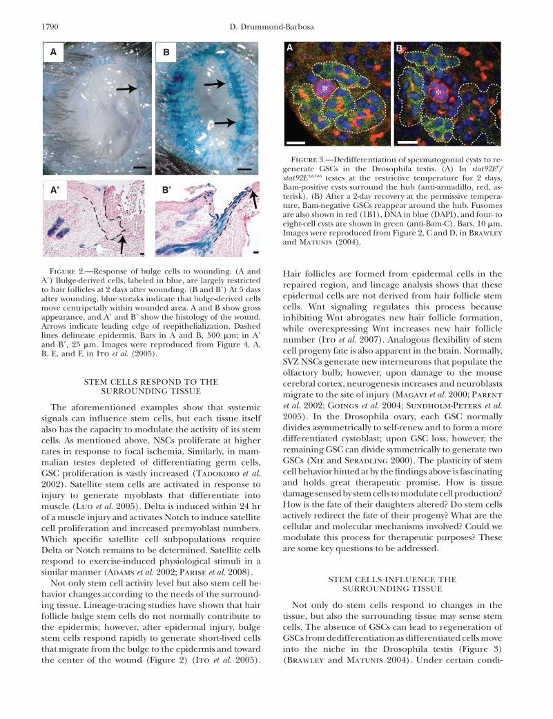
Not only do stem cells respond to changes in thetissue, but also the surrounding tissue may sense stemcells. The absence of GSCs can lead to regeneration ofGSCs from dedifferentiation as differentiated cells moveinto the niche in the Drosophila testis (Figure 3)(Brawley and Matunis 2004). Under certain condi-
Figure 3.—Dedifferentiation of spermatogonial cysts to re-generate GSCs in the Drosophila testis. (A) In stat92EF/stat92E 06346 testes at the restrictive temperature for 2 days,Bam-positive cysts surround the hub (anti-armadillo, red, as-terisk). (B) After a 2-day recovery at the permissive tempera-ture, Bam-negative GSCs reappear around the hub. Fusomesare also shown in red (1B1), DNA in blue (DAPI), and four- toeight-cell cysts are shown in green (anti-Bam-C). Bars, 10 mm.Images were reproduced from Figure 2, C and D, in Brawley
and Matunis (2004).
Figure 2.—Response of bulge cells to wounding. (A andA9) Bulge-derived cells, labeled in blue, are largely restrictedto hair follicles at 2 days after wounding. (B and B9) At 5 daysafter wounding, blue streaks indicate that bulge-derived cellsmove centripetally within wounded area. A and B show grossappearance, and A9 and B9 show the histology of the wound.Arrows indicate leading edge of reepithelialization. Dashedlines delineate epidermis. Bars in A and B, 500 mm; in A9and B9, 25 mm. Images were reproduced from Figure 4, A,B, E, and F, in Ito et al. (2005).
1790 D. Drummond-Barbosa

tions, dedifferentiation also occurs in females (Kai andSpradling 2004). An empty niche can also affect othercell types within a tissue. Forced differentiation of GSCsin Drosophila females results in gradual changes in theniche surroundings, bringing follicle cell progenitorsinto the vacant niche. These incoming somatic cellsreceive BMP signals and divide within the invaded niche(Kai and Spradling 2003). In agametic Drosophilatestis, cyst cells, which are normally quiescent, continueto proliferate and adopt the hub cell fate, suggesting thateither germ cells restrict cyst cell proliferation and fateor cyst cells become exposed to signals not normallyavailable to them in the absence of germ cells (Gonczy
and Dinardo 1996). In mutant mice that lack tail hairfollicles (and bulge stem cells), tail wound reepithelial-ization is delayed, but eventually occurs from thesurrounding epidermis (Langton et al. 2008). In mousetestes depleted of germ cells (including GSCs) bybusulfan treatment, GDNF expression increases four-fold (Ryu et al. 2006). Finally, somatic stem cells in theDrosophila testis give rise not only to cyst cells, but also tohub cells, showing how a stem cell can directly impact itsown niche (Voog et al. 2008).
Are stem cells passive in the communication with thetissue, merely adjusting their activity levels according totissue demands? Do stem cells influence their tissuesstrictly by virtue of the progeny that they produce or thesignals normally destined for them that become avail-able to other cells upon their absence? Or is it possiblethat stem cells have a more direct/active role in thetissue where they reside, potentially via secreted ormembrane-bound factors? In fact, intestinal stem cellsin Drosophila express Delta and, when they divide togenerate a stem cell and an enteroblast, Delta is rapidlydownregulated in the enteroblast. The intestinal stemcell Delta signal activates Notch in the enteroblast, andstem cell Delta levels determine whether the enteroblastdifferentiates into an enterocyte (high Delta) or enter-oendocrine cell (low Delta) (Ohlstein and Spradling
2007). Transcriptome analyses revealed that bulge stemcells express many secreted or membrane-bound fac-tors, including known signaling molecules such asEphrinB1 and Fgf1 (Morris et al. 2004; Tumbar et al.2004). In the HSC transcriptome, signaling molecules,including secreted ligands, are conspicuously present(Ivanova et al. 2002). Microarray analysis of DrosophilaGSCs show expression of secreted factors (Kai et al.2005), and Delta has been proposed to function in GSCs(Ward et al. 2006). Moreover, as described below, GSCsnegatively modulate life span in C. elegans (Arantes-Oliveira et al. 2002). Thus, perhaps more effort shouldbe put into studying how stem cells influence theirsurrounding cells, tissues, and organs, not just in termsof cell production for tissue maintenance or repair, butalso in terms of active signaling mechanisms that maytrigger chain reactions that impact organismal physiol-ogy as a whole.
STEM CELL FUNCTION DECLINES WITH AGING
Stem cell number or activity decreases during organ-ismal aging. As discussed below, the cause/consequencerelationship between changes in stem cell number orfunction and aging is unclear. First, however, I willdescribe the correlation between organismal aging andstem cell aging and review the potential mechanismsinvolved in stem cell decline. GSCs proliferate moreslowly and are gradually lost in aging Drosophila femalesand males ( Jones 2007). The numbers of HSCs, NSCs,or satellite cells do not appear to decline with age, buttheir activity becomes compromised (Geiger and Van
Zant 2002; Conboy et al. 2003; Hattiangady andShetty 2008). In contrast, impaired melanocyte stemcell self-renewal underlies hair graying (Sharpless andDepinho 2007), while numbers of GSCs are significantlydecreased in older mouse testes (Ryu et al. 2006; Zhang
et al. 2006). Although the mechanisms involved arepoorly understood, it is clear that both intrinsic andextrinsic factors contribute to the changes exhibited bystem cells with aging, as discussed below.
Some evidence suggests that stem cells age intrinsi-cally. Overexpression of superoxide dismutase, anantioxidant enzyme, in Drosophila GSCs extends theirlife span (Pan et al. 2007). Colonies arising in host testesfrom GSCs derived from 2-year-old mouse donors withatrophied testes are smaller than those of younger mice,suggesting intrinsic aging of GSCs (Zhang et al. 2006).Another study, however, showed no age-dependentdifference in colony size (Ryu et al. 2006). Dependingon the strain of mice, HSCs from old individuals are at asignificant disadvantage relative to young HSCs incompetitive repopulation or serial transplantation as-says (Geiger and Van Zant 2002). Several mechanismshave been proposed to explain the intrinsic aging ofstem cells, including exhaustion of their proliferativepotential, telomere shortening, accumulation of DNAdamage, or epigenetic alterations (Rando 2006).
Aging of the niche has also been proposed to con-tribute to the decline in stem cell function with age. InDrosophila ovaries, the number of cap cells decreasesand BMP signaling from the niche to GSCs is impairedwith age, while the E-cadherin-mediated associationbetween GSCs and cap cells weakens. Increasing nicheexpression of BMP signals, strengthening the GSC–nicheassociation, or expressing superoxide dismutase in theniche can prolong GSC life span and increase theirproliferation rates (Pan et al. 2007). In older Drosophilatestes, hub cells display lower levels of E-cadherin and ofUpd, whereas Upd overexpression in the niche main-tains GSCs in aging males (Boyle et al. 2007). Similarly,in aging mouse testes, expression of GDNF in Sertoli cellsis markedly reduced (Ryu et al. 2006) and GSCs can beconsecutively transplanted to the testes of young malesfor extended periods of time (Ogawa et al. 2003; Ryu
et al. 2006), underscoring a major role of aging of the
Review 1791

niche in GSC function decline. Old HSCs are also able toreconstitute blood in serial-transplantation experiments,although their effectiveness varies with the mouse strain(Geiger and Van Zant 2002; Warren and Rossi 2008).Finally, in the SGZ, there is a correlation between de-creased neurogenesis and a decline in vascular niches(Hattiangady and Shetty 2008), although causalityhas not been tested.
Given the remarkable ability of stem cells to sense andrespond to external stimuli, it is not surprising that thephysiological changes that result from aging can alsoimpact stem cells and their niches. As discussed above,insulin-like peptides control cap cell number via Notchsignaling in the niche, and they also control howeffectively cap cells associate with GSCs in Drosophila(H. J. Hsu and D. Drummond-Barbosa, unpublishedresults). As females age, systemic insulin-signaling levelsdecline (H. J. Hsu and D. Drummond-Barbosa, un-published results). Remarkably, excess insulin-like pep-tides suppress the normal process of GSC loss thatoccurs with age, while a poor diet enhances GSC loss,demonstrating how changes in systemic signals canpotentially impact the GSC niche during the agingprocess (H. J. Hsu and D. Drummond-Barbosa, un-published results). Several reports suggest that insulinsecretion or sensitivity decline in aging humans (Shimizu
et al. 1996; Chang et al. 2006a,b; Szoke et al. 2008); it ispossible that this also impacts certain stem cell popula-tions. In old mice, muscle regeneration is impaired dueto insufficient levels of Notch signaling in satellite cells,and forced activation of Notch is sufficient to restoreefficient regeneration of older muscle (Conboy et al. 2003).Intriguingly, exposure of old muscle to the systemic en-vironment of a young mouse in heterochronic para-biosis is sufficient to restore Notch signaling andefficient satellite cell activation in older muscle (Figure4) (Conboy et al. 2005), suggesting potential parallelswith the Drosophila ovary.
GERMLINE STEM CELLS, INSULIN SIGNALING,AND LONGEVITY ARE CONNECTED
As reviewed above, changes in intrinsic factors, theniche microenvironment, or the systemic environmentcan lead to an aged phenotype of stem cells. Conversely,changes in stem cell function may also have an impacton the physiological status of an individual, possiblycreating positive or negative feedback loops duringaging. The best-known example is the reported negativeeffect that GSCs exert on longevity in C. elegans.
Ablation of the germline leads to life-span extensionin C. elegans, and this effect requires DAF-16/FOXO, atranscriptional factor that acts as a negative downstreameffector of the insulin pathway, and a nuclear hormonereceptor, DAF-12 (Guarente and Kenyon 2000). Al-though lack of germline due to mutations in the earlyacting genes tudor, germ cell-less, or oskar does not extend
life span in Drosophila (Barnes et al. 2006), another studysuggests that the effect of the germline on longevity maybe evolutionarily conserved (Flatt et al. 2008). Loss ofgerm cells due to forced bam expression leads to life-span extension and also to alterations in systemic insulinsignaling, suggesting active communication betweenthe germline and other tissues (Flatt et al. 2008).
Caloric restriction also leads to increased longevity inmany organisms, possibly due in part to reduced in-sulin/IGF signaling (Guarente and Kenyon 2000).Mutation of the daf-2/insulin receptor gene or of otherinsulin pathway components doubles the life span of C.elegans hermaphrodites (Guarente and Kenyon 2000).In Drosophila, mutation of dinr/insulin receptor orchico/insulin receptor substrate or ablation of insulin-producing cells also extends life span (Giannakou andPartridge 2007). Recent evidence suggests that this isalso the case in mammals (Bartke 2000; Baba et al.2005; Taguchi et al. 2007). A prospective follow-upstudy in humans found that, in women, reduced insulinsignaling appears to correlate with lower body heightand improved old age survival (van Heemst et al. 2005).
Reproduction is also tightly linked to diet andphysiological status (Gong 2002; Loucks 2007; Pinelli
and Tagliabue 2007). Despite this apparent intersec-
Figure 4.—Exposure to a young systemic environment re-stores muscle stem cell activation and regeneration in agedmice. (A and A9) In isochronic parabiosis between two agedmice, muscles do not regenerate well after hind-limb injury,showing prominent fibrosis 5 days later. (B and B9) In con-trast, heterochronic parabiosis between young and aged micesignificantly enhances the regeneration of muscle in the oldpartner. Nascent myotubes can be recognized because theymaintain centrally located nuclei and express embryonic my-osin heavy chain (eMHC). A and B show hematoxylin and eo-sin staining. A9 and B9 show eMHC (red) and Hoechst dye(blue, nuclei). Images were reproduced from part of Figure1A in Conboy et al. (2005).
1792 D. Drummond-Barbosa

tion, however, several lines of evidence suggest that theextension of life span in insulin pathway mutants is notsimply due to effects on reproduction. Ablation ofsomatic gonad precursors along with the germline doesnot increase life span in C. elegans (Guarente andKenyon 2000). There is also a temporal separation ofrequirements of daf-2/insulin receptor for reproduc-tion and longevity. On the basis of RNA interference(RNAi) treatment, reduction in insulin signaling duringadulthood is required and sufficient for extending lifespan. In contrast, initiating daf-2 RNAi treatment inhatchlings, but not in adults, impairs reproduction(Dillin et al. 2002). One caveat of these experiments,however, is that the effectiveness of the RNAi treatmentin the germline (or whatever tissue requires daf-2 forreproduction) may be lower in adults relative to younglarvae; therefore, the possibility that daf-2 functions inadults to control reproduction cannot be ruled out.Nevertheless, these results clearly show that it is possibleto create conditions that separate extension of life spanfrom impairment of reproductive function. In Drosoph-ila, effects of dFOXO overexpression on longevity couldalso be uncoupled from reproduction (Hwangbo et al.2004; Giannakou et al. 2007). Honeybee queens arelong lived and fertile, whereas workers are short livedand sterile (Corona et al. 2007). Not only can longevityand reproduction be uncoupled, but also the effect ofdiet on longevity does not stem solely from what isingested. In Drosophila, yeast odorants can reverse thelife extension afforded by a restricted diet, whereasimpairment of olfaction due to the Orb83 mutationresults in life-span extension in the absence of dietaryrestriction (Libert et al. 2007).
Intriguingly, GSCs are responsible for the effects ofthe germline on longevity in C. elegans because genet-ically blocking the production of differentiated germcells does not extend life span, whereas forcing GSCs todifferentiate does, and GSC overproliferation shortenslife span (Arantes-Oliveira et al. 2002). These findingssuggest that GSCs communicate with other cells/tissuesto control aging. In fact, a screen for genes required forthe extended longevity of animals lacking a germlineresulted in the identification of kri-1, which controlsDAF-16 nuclear localization in the intestine likely inresponse to a lipophilic hormone (Berman and Kenyon
2006). Thus, GSCs may control longevity independentlyof their role in gamete production per se. Moreover, partof the effect of diet and insulin signaling on longevitymay occur via the modulation of GSCs/germ cells,which in turn may produce secreted/humoral factors.As described above, insulin signaling controls Drosoph-ila female GSCs, and the GSC loss and decreased activityobserved with aging may at least partially be explainedby reduced insulin signaling (Lafever and Drummond-Barbosa 2005; H. J. Hsu and D. Drummond-Barbosa,unpublished results). Although this has not beenaddressed experimentally, it is conceivable that part of
the effects of reduced insulin signaling in extending lifespan in Drosophila result from lower GSC numbers, andthat the natural decrease in insulin signaling over timerepresents a feedback mechanism to prolong life spanonce the peak period of reproduction is past. Tounderstand the potential role of GSCs on life-spancontrol will require that many questions be addressed.Are the germline effects on life span specific to GSCs inDrosophila? Is a complete absence of GSCs necessary foran impact on longevity or would less drastic decreases inGSC numbers be sufficient for an effect? How wellconserved is the role of the germline or GSCs acrossspecies, including humans (in which males, but notfemales, have GSCs)? How are the effects of GSCs in-tegrated with those of other factors controlling longevity?
HOW DO SOMATIC STEM CELLSIMPACT LONGEVITY?
Declines in stem cell function and/or number areclearly associated with aging, but causal relationshipsare largely unknown. In other words, does aging lead tostem cell decline, or does stem cell decline lead toaging? A less often asked question is, would stem celldecline always promote aging, as our intuitions suggest,or could stem cell decline in some instances counteractthe aging process? As discussed above, GSCs in C.elegans, and possibly in Drosophila, exert a negativeeffect on longevity. However, unlike many other stemcell types, GSCs are required for reproduction, a processnot essential for survival of a particular individual. Animportant question, therefore, is whether the negativeeffects of GSCs on longevity are a peculiarity of thegermline, or whether they could reflect a functionshared by some other stem cell types.
One possibility is that, with the exception of GSCs,decreases in stem cell function lead to reduced longev-ity. Intrinsic aging of stem cells (Figure 5A), their niches(Figure 5B), or the systemic environment (Figure 5C)would result in decreased stem cell function, impairedtissue function, and further systemic aging. For exam-ple, HSC activity is required for blood cell production,and blood cells have immune functions, carry oxygen totissues, and enable blood clotting; declines in any ofthese functions would be expected to decrease longevity(Geiger and Van Zant 2002). Another possibility,albeit counterintuitive, is that small decreases in stemcell function may actually promote longevity. Excessivestem cell activity may result in uncontrolled prolifera-tion as tumors, and this would clearly tend to shortenlife span. For example, ectopic Wnt signaling inducedby overexpression of the Wnt1 ligand or of an activatedform of b-catenin, which is highly oncogenic, increasesprogenitor cell numbers and activity in mouse mam-mary epithelium (Liu et al. 2004). A variant of thisgeneral possibility is that factors that increase stem cellfunction may have a deleterious effect on longevity.
Review 1793

Ames Dwarf mice are long lived and have low levels ofcirculating growth hormone and IGF-1 (Sun et al. 2005);however, they have a local increase in IGF-1 in thehippocampus and an increase in neurogenesis, suggest-ing that there could be mechanisms to circumvent thispotential problem. But could stem cells perhaps havemore subtle or indirect negative effects on longevity?
For example, could maintenance of young-equivalentlevels of stem cell function in older individuals not onlyincrease the risk of cancer, as has been proposed(Sharpless and Depinho 2007), but also overtax anolder physiological environment to sustain a high rateof cell production and elevated tissue performance inmultiple tissues? In that case, the decrease in stem cellfunction in response to an older physiological environ-ment would actually serve as a negative feedbackmechanism and slow down the aging of the organism.Alternatively, whether or not the effect of decreasedstem cell function would positively or negatively impactlongevity may vary with the stem cell type (Figure 5D).Further research will be required to determine thecausal relationships between physiological changes andstem cell function during aging and to determine whatthe impact of those changes on organismal longevity isfor different types of stem cells. As discussed above,multiple mechanisms contribute to stem cell aging.Perhaps because different stem cell types have distinctspecific requirements for their maintenance and func-tion, they also appear to have different susceptibility tointrinsic or extrinsic aging. It is therefore very likely thatnot all stem cells age simultaneously or in a similarfashion—some may simply respond to an aged environ-ment, others may intrinsically age and thereby poten-tially contribute to the age-induced physiologicalchanges, or a combination of these factors (Figure5D). Given the complexity of the network of interac-tions (Rando 2006), the large number of stem cell typessupporting tissues with diverse functions, very fine,tissue-specific genetic manipulation will be required toaddress whether or not the more global physiologicalchanges that stem cells respond to during aging (stemcell extrinsic aging) originate from intrinsic changes insubsets of stem cells (intrinsic stem cell aging).
CLOSING THOUGHTS
Stem cells participate in a web of multidirectionalinteractions with their niches and tissues of residence,the systemic environment, and external factors. Stemcells not only respond to multiple stimuli, but also havean impact on the organism; it is therefore important toconsider each stem cell interaction in both directions.By producing cells, stem cells influence tissue activityand function, which in turn determines both the level ofdemand for resources and the generation of beneficialcells or factors. But stem cells may also be active beyondtheir role in the production of cells, perhaps via thesecretion of factors. A detailed cellular and molecularmap of all the interactions for each type of stem cell,combined with tools for manipulating particular inter-actions and measuring aging parameters, will helpreveal the relationships between various stem cell typesand the aging process. Are stem cells major determi-nants of aging because of their role in maintaining
Figure 5.—Models of how stem cell function affects and isaffected during aging. (A) Intrinsic stem cell aging may be apredominant factor leading to aging of the organism. (B) Ag-ing of the niches may significantly contribute to aging by af-fecting stem cell activity. (C) Aging of the systemic environmentmay drive aging of stem cells. (D) Complex interactions arelikely to drive aging. It is possible that different stem cells ageat different rates and via different mechanisms and that the im-pact that their aging has on the aging of the organism as a wholevaries according to their specific function. Different shapes rep-resent distinct types of stem cells and niches. Gray color repre-sents aging of stem cells, niches, and the systemic environment.See text for details.
1794 D. Drummond-Barbosa

tissue function? If this is the case, organisms age as aconsequence of stem cell function decline resultingfrom changes in intrinsic factors or in the niche. Or ismaintenance of high levels of stem cell function overtime deleterious because of elevated cancer risk or anincreased demand for resources imposed by excessivetissue activity? In this case, reductions in stem cellactivity and/or numbers, at least in some tissues (suchas the germline and, perhaps, additional tissues), couldprolong life. Or is stem cell function not a majordeterminant of aging, but some of the changes involvedin aging also happen to affect niche/stem cell activity,showing a simple correlation instead of a causal relation-ship? Or, as seems more logical, is there a combination ofall possibilities above, with the particular role of stemcells in aging varying according to the particular stemcell type or the function of the tissue they support?
I am grateful for anonymous reviewer comments. This work wassupported by National Institutes of Health grant no. GM 069875 andby American Cancer Society grant no. RSG-07-182-01-DDC.
LITERATURE CITED
Adams, G. R., V. J. Caiozzo, F. Haddad and K. M. Baldwin,2002 Cellular and molecular responses to increased skeletalmuscle loading after irradiation. Am. J. Physiol. Cell Physiol.283: C1182–C1195.
Arantes-Oliveira, N., J. Apfeld, A. Dillin and C. Kenyon,2002 Regulation of life-span by germ-line stem cells in Caeno-rhabditis elegans. Science 295: 502–505.
Arvidsson, A., T. Collin, D. Kirik, Z. Kokaia and O. Lindvall,2002 Neuronal replacement from endogenous precursors inthe adult brain after stroke. Nat. Med. 8: 963–970.
Baba, T., T. Shimizu, Y. Suzuki, M. Ogawara, K. Isono et al.,2005 Estrogen, insulin, and dietary signals cooperatively regu-late longevity signals to enhance resistance to oxidative stressin mice. J. Biol. Chem. 280: 16417–16426.
Barnes, A. I., J. M. Boone, J. Jacobson, L. Partridge and T. Chapman,2006 No extension of lifespan by ablation of germ line in Dro-sophila. Proc. Biol. Sci. 273: 939–947.
Bartke, A., 2000 Delayed aging in Ames dwarf mice. Relationshipsto endocrine function and body size. Results Probl. Cell Differ.29: 181–202.
Berman, J. R., and C. Kenyon, 2006 Germ-cell loss extends C. ele-gans life span through regulation of DAF-16 by kri-1 and lipo-philic-hormone signaling. Cell 124: 1055–1068.
Boyle, M., C. Wong, M. Rocha and D. L. Jones, 2007 Decline inself-renewal factors contributes to aging of the stem cell nichein the Drosophila testis. Cell Stem Cell 1: 470–478.
Brawley, C., and E. Matunis, 2004 Regeneration of male germlinestem cells by spermatogonial dedifferentiation in vivo. Science304: 1331–1334.
Chang, A. M., M. J. Smith, C. J. Bloem, A. T. Galecki, J. B. Halter
et al., 2006a Limitation of the homeostasis model assessment topredict insulin resistance and beta-cell dysfunction in older peo-ple. J. Clin. Endocrinol. Metab. 91: 629–634.
Chang, A. M., M. J. Smith, A. T. Galecki, C. J. Bloem and J. B. Halter,2006b Impaired beta-cell function in human aging: response tonicotinic acid-induced insulin resistance. J. Clin. Endocrinol.Metab. 91: 3303–3309.
Conboy, I. M., M. J. Conboy, G. M. Smythe and T. A. Rando,2003 Notch-mediated restoration of regenerative potential toaged muscle. Science 302: 1575–1577.
Conboy, I. M., M. J. Conboy, A. J. Wagers, E. R. Girma, I. L. Weiss-
man et al., 2005 Rejuvenation of aged progenitor cells by expo-sure to a young systemic environment. Nature 433: 760–764.
Corona, M., R. A. Velarde, S. Remolina, A. Moran-Lauter, Y.Wang et al., 2007 Vitellogenin, juvenile hormone, insulin sig-
naling, and queen honey bee longevity. Proc. Natl. Acad. Sci.USA 104: 7128–7133.
Decotto, E., and A. C. Spradling, 2005 The Drosophila ovarianand testis stem cell niches: similar somatic stem cells and signals.Dev. Cell 9: 501–510.
Dillin, A., D. K. Crawford and C. Kenyon, 2002 Timing require-ments for insulin/IGF-1 signaling in C. elegans. Science 298:830–834.
Drummond-Barbosa, D., 2005 Regulation of stem cell populations,pp. 67–98 in Encyclopedia of Molecular Cell Biology and MolecularMedicine, edited by R. A. Meyers. Wiley-VCH, Weinheim, Germany.
Drummond-Barbosa, D., and A. C. Spradling, 2001 Stem cells andtheir progeny respond to nutritional changes during Drosophilaoogenesis. Dev. Biol. 231: 265–278.
Dubal, D. B., S. W. Rau, P. J. Shughrue, H. Zhu, J. Yu et al.,2006 Differential modulation of estrogen receptors (ERs) inischemic brain injury: a role for ERalpha in estradiol-mediatedprotection against delayed cell death. Endocrinology 147:3076–3084.
Flatt, T., K. J. Min, C. D’Alterio, E. Villa-Cuesta, J. Cumbers et al.,2008 Drosophila germ-line modulation of insulin signaling andlifespan. Proc. Natl. Acad. Sci. USA 105: 6368–6373.
Geiger, H., and G. Van Zant, 2002 The aging of lympho-hemato-poietic stem cells. Nat. Immunol. 3: 329–333.
Giannakou, M. E., and L. Partridge, 2007 Role of insulin-like sig-nalling in Drosophila lifespan. Trends Biochem. Sci. 32: 180–188.
Giannakou, M. E., M. Goss, J. Jacobson, G. Vinti, S. J. Leevers et al.,2007 Dynamics of the action of dFOXO on adult mortality inDrosophila. Aging Cell 6: 429–438.
Goings, G. E., V. Sahni and F. G. Szele, 2004 Migration patterns ofsubventricular zone cells in adult mice change after cerebral cor-tex injury. Brain Res. 996: 213–226.
Gonczy, P., and S. DiNardo, 1996 The germ line regulates somaticcyst cell proliferation and fate during Drosophila spermatogen-esis. Development 122: 2437–2447.
Gong, J. G., 2002 Influence of metabolic hormones and nutritionon ovarian follicle development in cattle: practical implications.Domest. Anim. Endocrinol. 23: 229–241.
Guarente, L., and C. Kenyon, 2000 Genetic pathways that regulateageing in model organisms. Nature 408: 255–262.
Hattiangady, B., and A. K. Shetty, 2008 Aging does not alter thenumber or phenotype of putative stem/progenitor cells in theneurogenic region of the hippocampus. Neurobiol. Aging 29:129–147.
Hwangbo, D. S., B. Gershman, M. P. Tu, M. Palmer and M. Tatar,2004 Drosophila dFOXO controls lifespan and regulates insu-lin signalling in brain and fat body. Nature 429: 562–566.
Ito, M., Y. Liu, Z. Yang, J. Nguyen, F. Liang et al., 2005 Stem cells inthe hair follicle bulge contribute to wound repair but not to ho-meostasis of the epidermis. Nat. Med. 11: 1351–1354.
Ito, M., Z. Yang, T. Andl, C. Cui, N. Kim et al., 2007 Wnt-dependentde novo hair follicle regeneration in adult mouse skin afterwounding. Nature 447: 316–320.
Ivanova, N. B., J. T. Dimos, C. Schaniel, J. A. Hackney, K. A. Moore
et al., 2002 A stem cell molecular signature. Science 298: 601–604.Jin, K., M. Minami, J. Q. Lan, X. O. Mao, S. Batteur et al.,
2001 Neurogenesis in dentate subgranular zone and rostralsubventricular zone after focal cerebral ischemia in the rat. Proc.Natl. Acad. Sci. USA 98: 4710–4715.
Jones, D. L., 2007 Aging and the germ line: where mortality and im-mortality meet. Stem Cell Rev. 3: 192–200.
Kai, T., and A. Spradling, 2003 An empty Drosophila stem cellniche reactivates the proliferation of ectopic cells. Proc. NatlAcad. Sci. USA 100: 4633–4638.
Kai, T., and A. Spradling, 2004 Differentiating germ cells can re-vert into functional stem cells in Drosophila melanogaster ova-ries. Nature 428: 564–569.
Kai, T., D. Williams and A. C. Spradling, 2005 The expression pro-file of purified Drosophila germline stem cells. Dev. Biol. 283:486–502.
LaFever, L., and D. Drummond-Barbosa, 2005 Direct control ofgermline stem cell division and cyst growth by neural insulinin Drosophila. Science 309: 1071–1073.
Review 1795

Lamarca, H. L., and J. M. Rosen, 2008 Hormones and mammarycell fate: What will I become when I grow up? Endocrinology149: 4317–4321.
Langton, A. K., S. E. Herrick and D. J. Headon, 2008 An extendedepidermal response heals cutaneous wounds in the absence of ahair follicle stem cell contribution. J. Invest. Dermatol. 128:1311–1318.
Leatherman, J. L., and S. Dinardo, 2008 Zfh-1 controls somaticstem cell self-renewal in the Drosophila testis and nonautono-mously influences germline stem cell self-renewal. Cell Stem Cell3: 44–54.
Lemkine, G. F., A. Raj, G. Alfama, N. Turque, Z. Hassani et al.,2005 Adult neural stem cell cycling in vivo requires thyroid hor-mone and its alpha receptor. FASEB J. 19: 863–865.
Libert, S., J. Zwiener, X. Chu, W. Vanvoorhies, G. Roman et al.,2007 Regulation of Drosophila life span by olfaction andfood-derived odors. Science 315: 1133–1137.
Liu, B. Y., S. P. McDermott, S. S. Khwaja and C. M. Alexander,2004 The transforming activity of Wnt effectors correlates withtheir ability to induce the accumulation of mammary progenitorcells. Proc. Natl. Acad. Sci. USA 101: 4158–4163.
Liu, J. P., J. Baker, A. S. Perkins, E. J. Robertson and A. Efstratiadis,1993 Mice carrying null mutations of the genes encoding insu-lin-like growth factor I (Igf-1) and type 1 IGF receptor (Igf1r).Cell 75: 59–72.
Lopez-Onieva, L., A. Fernandez-Minan and A. Gonzalez-Reyes,2008 Jak/Stat signalling in niche support cells regulates dpptranscription to control germline stem cell maintenance in theDrosophila ovary. Development 135: 533–540.
Loucks, A. B., 2007 Energy availability and infertility. Curr. Opin.Endocrinol. Diabetes Obes. 14: 470–474.
Luo, D., V. M. Renault and T. A. Rando, 2005 The regulation ofNotch signaling in muscle stem cell activation and postnatal myo-genesis. Semin. Cell Dev. Biol. 16: 612–622.
Magavi, S. S., B. R. Leavitt and J. D. Macklis, 2000 Induction ofneurogenesis in the neocortex of adult mice. Nature 405: 951–955.
Merkle, F. T., Z. Mirzadeh and A. Alvarez-Buylla, 2007 Mosaicorganization of neural stem cells in the adult brain. Science317: 381–384.
Mirzadeh, Z., F. T. Merkle, M. Soriano-Navarro, J. M. Garcia-Verdugo and A. Alvarez-Buylla, 2008 Neural stem cells con-fer unique pinwheel architecture to the ventricular surface inneurogenic regions of the adult brain. Cell Stem Cell 3: 265–278.
Morris, R. J., Y. Liu, L. Marles, Z. Yang, C. Trempus et al.,2004 Capturing and profiling adult hair follicle stem cells.Nat. Biotechnol. 22: 411–417.
Morrison, S. J., and A. C. Spradling, 2008 Stem cells and niches:mechanisms that promote stem cell maintenance throughoutlife. Cell 132: 598–611.
Nishimura, E. K., S. A. Jordan, H. Oshima, H. Yoshida, M. Osawa
et al., 2002 Dominant role of the niche in melanocyte stem-cellfate determination. Nature 416: 854–860.
Ogawa, T., M. Ohmura, Y. Yumura, H. Sawada and Y. Kubota,2003 Expansion of murine spermatogonial stem cells throughserial transplantation. Biol. Reprod. 68: 316–322.
Ohlstein, B., and A. Spradling, 2007 Multipotent Drosophila in-testinal stem cells specify daughter cell fates by differential notchsignaling. Science 315: 988–992.
Pan, L., S. Chen, C. Weng, G. Call, D. Zhu et al., 2007 Stem cellaging is controlled both intrinsically and extrinsically in the Dro-sophila ovary. Cell Stem Cell 1: 458–469.
Parent, J. M., Z. S. Vexler, C. Gong, N. Derugin and D. M.Ferriero, 2002 Rat forebrain neurogenesis and striatal neuronreplacement after focal stroke. Ann. Neurol. 52: 802–813.
Parise, G., I. W. McKinnell and M. A. Rudnicki, 2008 Muscle sat-ellite cell and atypical myogenic progenitor response followingexercise. Muscle Nerve 37: 611–619.
Paus, R., P. Arck and S. Tiede, 2008 (Neuro-)endocrinology of ep-ithelial hair follicle stem cells. Mol. Cell. Endocrinol. 288: 38–51.
Pinelli, G., and A. Tagliabue, 2007 Nutrition and fertility. MinervaGastroenterol. Dietol. 53: 375–382.
Pinkston, J. M., D. Garigan, M. Hansen and C. Kenyon,2006 Mutations that increase the life span of C. elegans inhibittumor growth. Science 313: 971–975.
Rando, T. A., 2006 Stem cells, ageing and the quest for immortality.Nature 441: 1080–1086.
Riquelme, P. A., E. Drapeau and F. Doetsch, 2008 Brain micro-ecologies: neural stem cell niches in the adult mammalian brain.Philos. Trans. R. Soc. Lond. B Biol. Sci. 363: 123–137.
Ryu, B. Y., K. E. Orwig, J. M. Oatley, M. R. Avarbock andR. L. Brinster, 2006 Effects of aging and niche microenviron-ment on spermatogonial stem cell self-renewal. Stem Cells 24:1505–1511.
Schofield, R., 1978 The relationship between the spleen colony-forming cell and the haemopoietic stem cell. Blood Cells 4:7–25.
Sharpless, N. E., and R. A. DePinho, 2007 How stem cells ageand why this makes us grow old. Nat. Rev. Mol. Cell Biol. 8:703–713.
Shimizu, M., S. Kawazu, S. Tomono, T. Ohno, T. Utsugi et al.,1996 Age-related alteration of pancreatic beta-cell function. In-creased proinsulin and proinsulin-to-insulin molar ratio in el-derly, but not in obese, subjects without glucose intolerance.Diabetes Care 19: 8–11.
Shingo, T., C. Gregg, E. Enwere, H. Fujikawa, R. Hassam et al.,2003 Pregnancy-stimulated neurogenesis in the adult femaleforebrain mediated by prolactin. Science 299: 117–120.
Sinha-Hikim, I., S. M. Roth, M. I. Lee and S. Bhasin,2003 Testosterone-induced muscle hypertrophy is associatedwith an increase in satellite cell number in healthy, youngmen. Am. J. Physiol. Endocrinol. Metab. 285: E197–E205.
Song, X., G. B. Call, D. Kirilly and T. Xie, 2007 Notch signalingcontrols germline stem cell niche formation in the Drosophilaovary. Development 134: 1071–1080.
Spritzer, M. D., and L. A. Galea, 2007 Testosterone and dihy-drotestosterone, but not estradiol, enhance survival of newhippocampal neurons in adult male rats. Dev. Neurobiol. 67:1321–1333.
Sun, L. Y., M. S. Evans, J. Hsieh, J. Panici and A. Bartke,2005 Increased neurogenesis in dentate gyrus of long-livedAmes dwarf mice. Endocrinology 146: 1138–1144.
Sundholm-Peters, N. L., H. K. Yang, G. E. Goings, A. S. Walker andF. G. Szele, 2005 Subventricular zone neuroblasts emigrate to-ward cortical lesions. J. Neuropathol. Exp. Neurol. 64: 1089–1100.
Suzuki, S., L. M. Gerhold, M. Bottner, S. W. Rau, C. Dela Cruz
et al., 2007 Estradiol enhances neurogenesis following ischemicstroke through estrogen receptors alpha and beta. J. Comp. Neu-rol. 500: 1064–1075.
Szoke, E., M. Z. Shrayyef, S. Messing, H. J. Woerle, T. W. van
Haeften et al., 2008 Effect of aging on glucose homeostasis:accelerated deterioration of beta-cell function in individualswith impaired glucose tolerance. Diabetes Care 31: 539–543.
Tadokoro, Y., K. Yomogida, H. Ohta, A. Tohda and Y. Nishimune,2002 Homeostatic regulation of germinal stem cell prolifera-tion by the GDNF/FSH pathway. Mech. Dev. 113: 29–39.
Taguchi, A., L. M. Wartschow and M. F. White, 2007 Brain IRS2signaling coordinates life span and nutrient homeostasis. Science317: 369–372.
Tanapat, P., N. B. Hastings, A. J. Reeves and E. Gould,1999 Estrogen stimulates a transient increase in the numberof new neurons in the dentate gyrus of the adult female rat.J. Neurosci. 19: 5792–5801.
Tanapat, P., N. B. Hastings and E. Gould, 2005 Ovarian steroidsinfluence cell proliferation in the dentate gyrus of the adultfemale rat in a dose- and time-dependent manner. J. Comp. Neu-rol. 481: 252–265.
Tumbar, T., G. Guasch, V. Greco, C. Blanpain, W. E. Lowry et al.,2004 Defining the epithelial stem cell niche in skin. Science303: 359–363.
van Heemst, D., M. Beekman, S. P. Mooijaart, B. T. Heijmans, B. W.Brandt et al., 2005 Reduced insulin/IGF-1 signalling and hu-man longevity. Aging Cell 4: 79–85.
Voog, J., C. D’Alterio and D. L. Jones, 2008 Multipotent somaticstem cells contribute to the stem cell niche in the Drosophila tes-tis. Nature 454: 1132–1136.
Wang, L., Z. Li and Y. Cai, 2008 The JAK/STAT pathway positivelyregulates DPP signaling in the Drosophila germline stem cellniche. J. Cell Biol. 180: 721–728.
1796 D. Drummond-Barbosa

Ward, E. J., H. R. Shcherbata, S. H. Reynolds, K. A. Fischer, S. D.Hatfield et al., 2006 Stem cells signal to the niche throughthe Notch pathway in the Drosophila ovary. Curr. Biol. 16: 2352–2358.
Warren, L. A., and D. J. Rossi, 2008 Stem cells and aging in the he-matopoietic system. Mech Ageing Dev. (in press).
Weger, N., and T. Schlake, 2005 Igf-I signalling controls the hairgrowth cycle and the differentiation of hair shafts. J Invest Der-matol 125: 873–882.
Wong, M. D., Z. Jin and T. Xie, 2005 Molecular mechanisms ofgermline stem cell regulation. Annu Rev Genet 39: 173–195.
Xie, T., and A. C. Spradling, 1998 decapentaplegic is essential for themaintenance and division of germline stem cells in the Drosoph-ila ovary. Cell 94: 251–260.
Xie, T., and A. C. Spradling, 2000 A niche maintaining germ linestem cells in the Drosophila ovary. Science 290: 328–330.
Yan, Y. P., K. A. Sailor, R. Vemugantiand R. J. Dempsey, 2006 Insulin-like growth factor-1 is an endogenous mediator of focal ischemia-inducedneuralprogenitorproliferation.Eur. J.Neurosci.24:45–54.
Yoshida, S., M. Sukeno and Y. Nabeshima, 2007 A vasculature-associated niche for undifferentiated spermatogonia in themouse testis. Science 317: 1722–1726.
Zhang, R.L.,Z.G.Zhang,L.ZhangandM.Chopp,2001 Proliferationand differentiation of progenitor cells in the cortex and the subven-tricular zone in the adult rat after focal cerebral ischemia. Neurosci-ence 105: 33–41.
Zhang, X., K. T. Ebata, B. Robaire and M. C. Nagano, 2006 Agingof male germ line stem cells in mice. Biol. Reprod. 74: 119–124.
Communicating editor: A. Spradling
Review 1797