roxburgh, c. s., shia, j., vakiani, e., daniel, t. and
TRANSCRIPT
Roxburgh, C. S., Shia, J., Vakiani, E., Daniel, T. and Weiser, M. R. (2018)
Potential immune priming of the tumor microenvironment with FOLFOX
chemotherapy in locally advanced rectal cancer. OncoImmunology, 7(6),
e1435227.(doi:10.1080/2162402x.2018.1435227)
This is the author’s final accepted version.
There may be differences between this version and the published version.
You are advised to consult the publisher’s version if you wish to cite from
it.
http://eprints.gla.ac.uk/156794/
Deposited on: 01 February 2018
Enlighten – Research publications by members of the University of Glasgow
http://eprints.gla.ac.uk
brought to you by COREView metadata, citation and similar papers at core.ac.uk
provided by Enlighten
1
Potential immune priming of the tumor microenvironment with FOLFOX
chemotherapy in locally advanced rectal cancer
Campbell S. Roxburgha,b, Jinru Shiac, Efsevia Vakianic, Tanisha Danielb, and Martin R. Weisera
aDepartment of Surgery, Memorial Sloan Kettering Cancer Center, New York, NY 10065; bInstitute of
Cancer Sciences, College of Medical, Veterinary and Life Sciences, University of Glasgow, Glasgow,
G12 8QQ United Kingdom; cDepartment of Pathology, Memorial Sloan Kettering Cancer Center, New
York, NY 10065
E-mail addresses: [email protected], [email protected], [email protected],
[email protected], [email protected]
CONTACT: Martin R. Weiser, MD, Colorectal Service, Department of Surgery, Memorial Sloan
Kettering Cancer Center, 1275 York Avenue, New York, NY 10065. Email: [email protected].
2
ABSTRACT
Strategies to enhance tumor immunogenicity may expand the role of immunotherapy beyond the
mismatch repair-deficient subtype. In this pilot study, biopsies were performed at baseline and after
four cycles of FOLFOX in eight patients receiving neoadjuvant chemotherapy for stage II/III locally
advanced rectal cancer. Immunostaining was performed for T cell subsets (CD3+, CD8+, CD45RO+);
macrophages (CD163+); T regulatory cells (FOXP3+); and expression of MHC class I, PD-1 and PD-L1.
Changes in cell number or intensity were quantified and correlated with treatment response.
Pretreatment patterns of immune infiltrates were mixed and did not correlate with treatment
response. Posttreatment increases in T cell infiltrates (CD3+, CD8+ and CD45RO+) and MHC-I
expression were observed in five patients. CD163+ cell numbers increased in four patients. FOXP3+
cells numbers increased in two patients, decreased in two other patients and remained unchanged
in three patients. PD-1 scores increased in seven patients, and PD-L1 scores increased in four
patients. Changes in tumor T cell responses did not correlate with treatment response. Changes in
FOXP3+ cells were associated with treatment response in some patients: two patients with increases
in FOXP3+ cells had poor responses, whereas the patient with the greatest reduction in FOXP3+ cells
had a complete response. The patient with a complete clinical response had a much higher increase
in MHC-I expression than other patients. These results suggest that chemotherapy can increase
immune activity in the tumor microenvironment and could potentially be utilized to prime immune
responses prior to immunomodulatory treatments.
KEYWORDS: Locally advanced rectal cancer; Neoadjuvant therapy; Chemotherapy; Immune priming;
Immunotherapy; T cells
3
Introduction
Immunotherapies have become routine in treatment of various cancers, such as melanoma, where
checkpoint blockade of CTLA-4 and PD-1 T-cell inhibitory molecules helps lengthen progression-free
survival and overall survival.1 T-cell regulatory (Treg) receptor PD-1 and its ligand PD-L1 are
expressed in most cancers,2 and together they can dampen the antitumor immune response.
Tumors thought to be susceptible to anti-PD-1 include those with high mutational load and
neoantigen burden 3 or with evidence of an immune-active tumor microenvironment (CD3/CD8+
infiltrates and MHC class I expression).4,5 Intratumoral PD-1/PD-L1 expression and presence of T
cells within the tumor microenvironment are thought to represent the most useful indicator that
anti- PD-1 therapy may be effective.
To date, the role of immunotherapy in colorectal cancer (CRC) has been limited to the mismatch
repair-deficient (dMMR) subtype. dMMR tumors are strongly immunogenic, with high numbers of
tumor-infiltrating lymphocytes thought to be responding to neoantigens brought about by the high
mutational burden.6 Compared with mismatch repair-proficient (pMMR) CRC, dMMR tumors also
express higher levels PD-1/PD-L1.7,8 Phase II trials have shown that anti-PD-1/PD-L1 agents help
lengthen progression-free and overall survival in patients with stage IV dMMR CRC.9,10 However,
since dMMR tumors account for only 25%, 12% and 4% of stage II, III and IV CRCs, respectively,
immunotherapy is a treatment option in only a small subset of CRCs.
The majority of pMMR CRCs exhibit low numbers of T cells in the tumor microenvironment, along
with low levels of PD-1/PD-L1 expression; therefore, checkpoint blockade immunotherapy is not
thought to be effective.11 Strategies aimed at enhancing immunogenicity in pMMR CRC may allow
broader use of immunotherapy. Along this line of reasoning, conventional cytotoxic chemotherapy
and radiotherapy (RT) are thought to result in immunogenic cell death, with T cell infiltrates
4
responding to release of antigen from damaged cells.12 Such treatments may synergize with
immunotherapies, enhancing treatment response. Trials are currently under way employing
combinations of immunotherapies with immune-priming strategies in gastroesophageal, head and
neck, and lung cancers (NCT02872116, NCT02764593 and NCT02944396 on
www.ClinicalTrials.gov).13
Observational data suggest that RT enhances T cell infiltration in locally advanced rectal cancer, and
preclinical models suggest that cytotoxic chemotherapy may have similar effects.14-18 However, the
effect of cytotoxic chemotherapy on intratumoral immune responses in patients with CRC has not
previously been studied. In this prospective pilot study, we investigated whether conventional
cytotoxic chemotherapy can induce immune priming in pMMR locally advanced rectal cancer.
Results
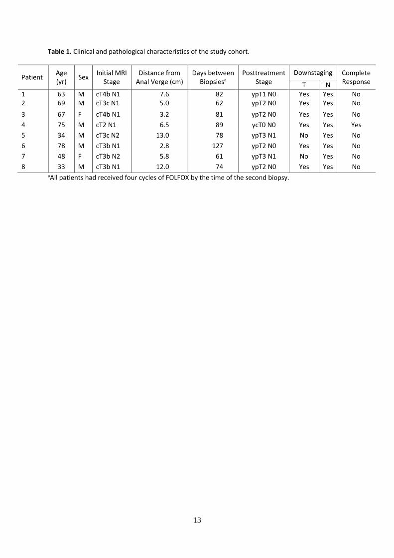
Matched biopsies taken before and after four cycles of induction FOLFOX chemotherapy (folinic acid,
fluorouracil and oxaliplatin) were collected in eight patients: six men and two women, ages 33-78
years. The clinical and pathological characteristics of the cohort are listed in Table 1. All eight
patients had clinical stage III (node-positive) disease; two had cT4 tumors, five had cT3 tumors, and
one had a cT2 tumor. The distance of the tumor from the anal verge was <5 cm in three patients, 5-
10 cm in three other patients and >10 cm in two patients.
Pretreatment MMR status was available for seven patients; all seven had retained expression of
MMR proteins. The number of days between the index biopsy and completion of four cycles of
FOLFOX ranged from 61 to 127 days (mean 82 days). By the completion of neoadjuvant treatment,
six patients had downstaging of T stage and all eight patients had downstaging of N stage. Two
5
patients had a poor clinical response, five patients had an incomplete response, and one patient had
a complete response, based on digital exam, endoscopy and MRI.
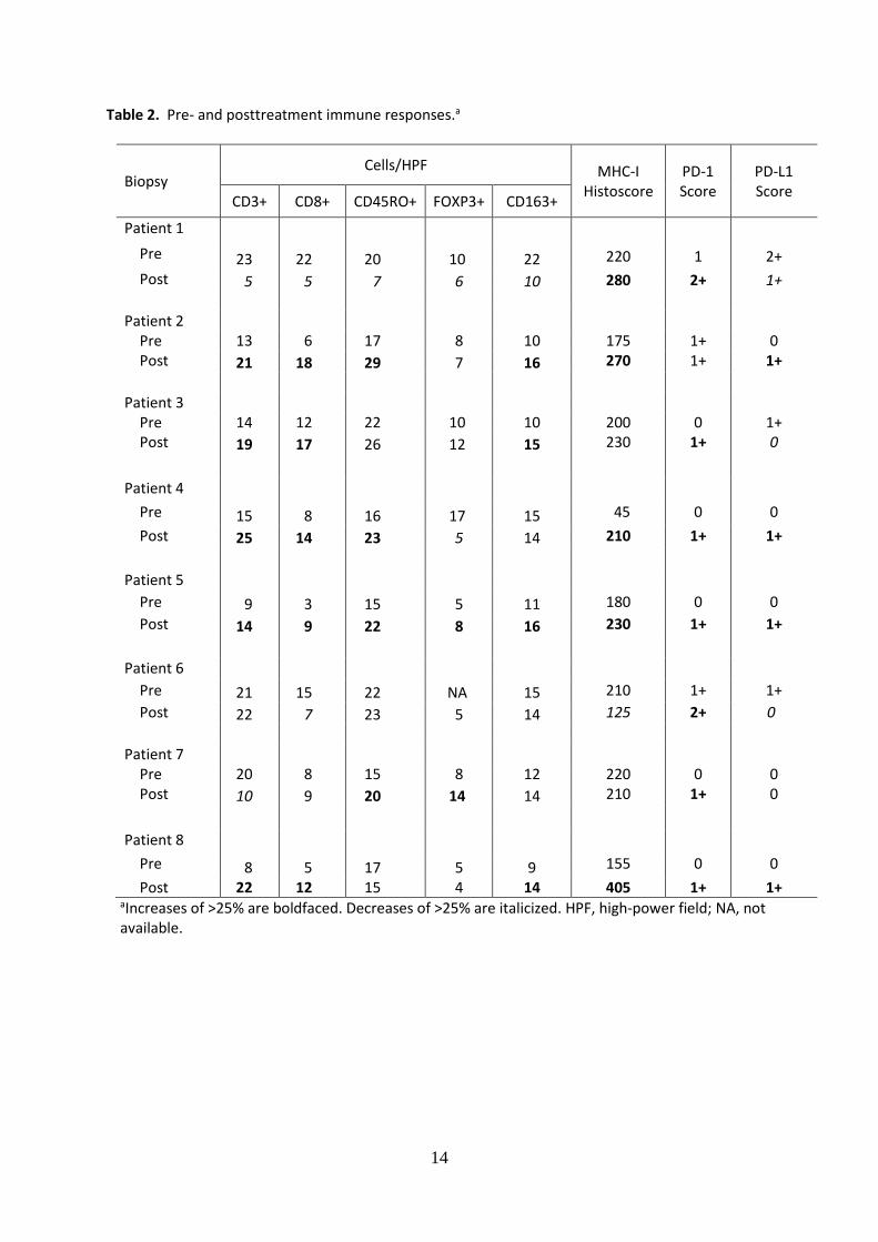
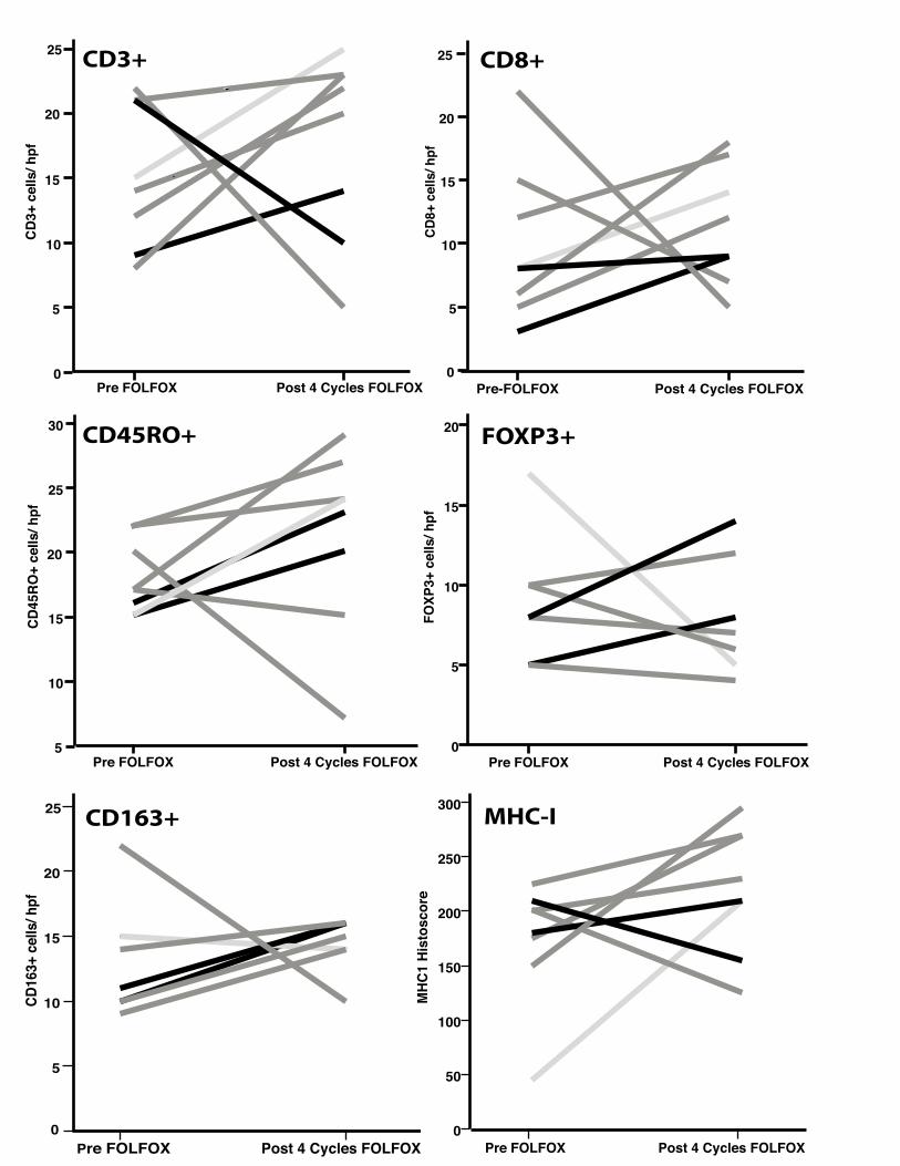
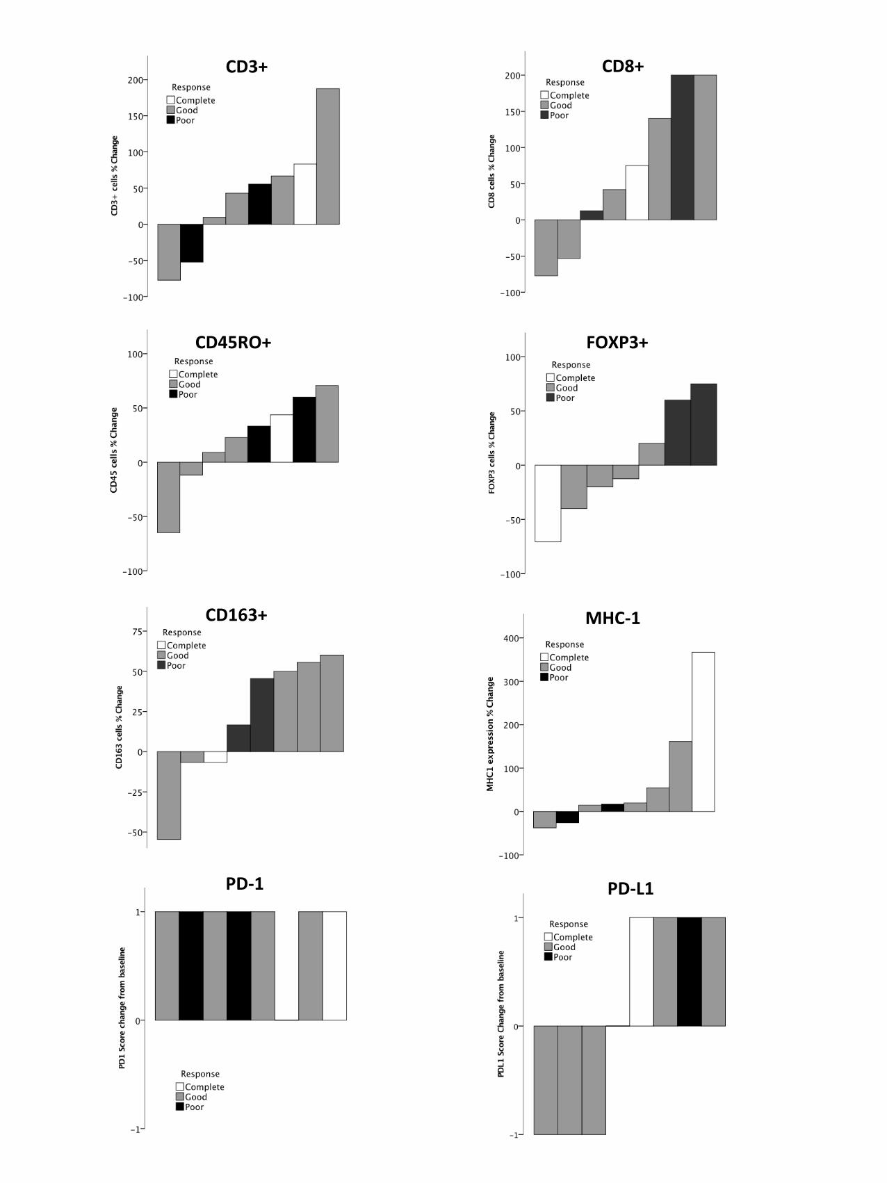
The data on pre- and posttreatment tumor immune responses are shown in Table 2 and Figure 1.
Pretreatment patterns of immune infiltrates were mixed and did not appear to correlate with
responses to treatment. CD3+ and CD8+ infiltrates increased in five patients, CD45RO+ infiltrates
increased in four patients, and CD163+ cells increased in four patients. The MHC-I histoscore
increased in five patients, the PD-1 score increased in seven patients, and the PD-L1 score increased
in four patients. FOXP3+ infiltrates increased in two patients, decreased in two other patients and
remained unchanged in three patients (pretreatment FOXP3+ data was not available for one of the
eight patients). The two patients with increases in FOXP3+ cells had poor responses to treatment,
whereas the patient with the greatest reduction in FOXP3+ cells had a complete clinical response
(Fig. 2). The patient with a complete response had a much higher increase in MHC-I expression
within the tumor than the other patients.
Discussion
The findings of our pilot study suggest that FOLFOX chemotherapy is capable of inducing changes in
the immune contexture within the tumor microenvironment, such as increases in T cells (CD3+,
CD8+ and CD45RO+) and in MHC-I and PD-1 expression. These changes are similar to those
observed in preclinical models after chemoradiotherapy (CRT) secondary to immunogenic cell death
with antigen release, increased MHC-I and cross-presentation to T cells.15-19 In rectal cancer patients
treated with CRT, higher numbers of CD4+/CD8+ cells in resected specimens than in pretreatment
biopsies have been reported.14,20 CRT also induces systemic immune changes, including reductions
in circulating Tregs and myeloid-derived suppressor cells.21 This immunomodulation may account
for the reported abscopal effects on distant metastases after RT to the primary tumor, potentially
mediated by cross-priming of T cells following tumor cell death.12, 18,19
6
Our results are consistent with previous reports of association between changes in Tregs and
treatment response. We observed that tumors in which FOXP3+ Tregs increased had a poor
treatment response. The role of Tregs in CRC is not clear, but they are known to harbor
immunosuppressive activity, dampening T cell responses.22 Low numbers of FOXP3+ Tregs in
posttreatment resection specimens have been reported to be associated with greater treatment
response after CRT.23 No strong relationship between pretreatment Tregs and treatment response
has been observed, suggesting that the type of immune response generated by RT is more
important.24
The patient with a complete clinical response had the largest increase in MHC-I expression. High
MHC-I expression reflects increased antigen presentation and is considered a measure of an
immune-active microenvironment; it may therefore indicate ongoing immunogenic cell death in
response to FOLFOX. Further work is required to test this hypothesis and to determine whether
MHC-I has potential as an early biomarker of response during chemotherapy treatment.
We found no appreciable relationship between T cell changes and treatment response, even for
tumors with a 200% increase in T cell numbers. Further work is required to determine whether the
effects of these T cell responses are mitigated by the presence of inhibitory immune checkpoints
(e.g., PD-1 and CTLA-4) or other T cell suppressors (myeloid-derived suppressor cells or Tregs).
No biomarkers of response to immunotherapies (e.g., anti-CTLA-4 and anti-PD-1/ PDL1) are
available, but pretreatment evidence of an immune-active tumor microenvironment (e.g., high T
cells and MHC-I expression) is considered important.3-5 Where such features are absent, strategies to
prime the microenvironment in order to induce immune responses are sought. FOLFOX may be
capable of achieving these desirable effects in a proportion of patients. Sequencing of
7
chemotherapy and RT to optimize immunological sequelae is likely to be critical to successful
utilization of immunotherapy and requires further investigation. The relative ease with which tumor
biopsies were collected from outpatients in our study indicates that rectal tumors are ideal for
studying the scheduling of priming treatments.
In summary, the results of our pilot study indicate that FOLFOX chemotherapy in patients with
locally advanced rectal cancer was associated with increases in T cell infiltrates, MHC-I expression
and PD-1 expression. It is likely that the immunogenicity generated in response to chemotherapy
plays a role in treatment response, and evidence of this role may be more apparent in a larger study.
Our findings suggest that neoadjuvant chemotherapy can potentially be utilized to prime immune
responses prior to immunomodulatory treatments. These observations provide support for ongoing
work investigating the role of immune stimulation by conventional treatments in rectal cancer and
for future trials aimed at evaluating combinations of chemotherapy, RT, and immunotherapy.
Patients and Methods
Patients
The study population consisted of nonconsecutive patients seen over a 4-month period with locally
advanced rectal cancer clinically staged cT3/4 or N+ (American Joint Committee on Cancer stage II
and III) who received neoadjuvant therapy prior to planned rectal resection. As per National
Comprehensive Cancer Network guidelines, the patients received eight cycles of induction FOLFOX
prior to CRT.25 With approval from the institutional review board, we prospectively evaluated the
immune contexture of tumor biopsies taken at baseline and after four cycles of FOLFOX
chemotherapy. All patients were considered to have pMMR tumors based on pretreatment biopsy
immunohistochemistry (no loss of MLH1, PMS2, MSH2 or MSH6 protein expression).
Samples
8
Immunohistochemistry was performed on matched samples (biopsy at initial diagnosis and after
four cycles of FOLFOX) to grade infiltrates of T cells (CD3+, CD8+, CD45RO+), Tregs (FOXP3+) and
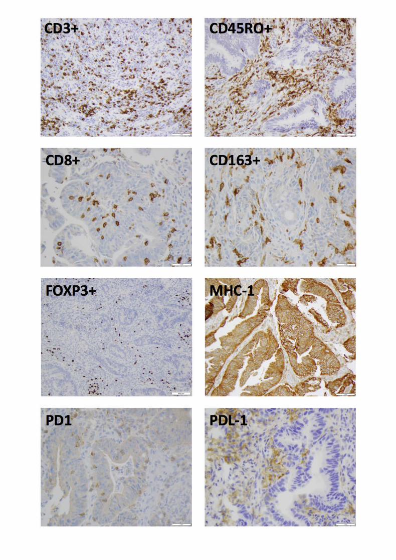
macrophages (CD163+) (Fig. 3). We also evaluated MHC-I (as a measure of antigen presentation)
and PD-1/PDL-1 expression. Sections of 4-μm thickness were obtained from paraffin-embedded
biopsy samples. After deparaffinization and rehydration, sections were incubated with primary
monoclonal antibodies recognizing CD3 (LN10, 1:100; Leica Biosystems), CD8 (SP57, ready to use;
Ventana), CD45RO (UCHL1, 1:200; Dako), FOXP3 (236A/E7, 1:500; Abcam), CD163 (MRQ-26, ready to
use; Cell Marque), MHC-I (A4; Thermo Fisher Scientific), PD-1 (NAT105, ready to use; Cell Marque),
or PD-L1 (E1L3N, 1:250; Cell Signaling Technology). We used the staining platforms BenchMark Ultra
from Roche (CD3 and CD163) and BenchMark Ultra from Ventana (CD8) and the detection systems
OptiView from Ventana (CD3, CD8, CD163 and PD-1) and Bond Polymer Refine from Leica
Biosystems (CD45RO, FOXP3, MHC-I and PD-L1).
Immune response
Immune cells expressing CD3+, CD8+, CD45RO+, FOXP3+ or CD163+ were quantified in biopsy
specimens using a cell counting approach. For each specimen, three high-power fields containing
both tumor stroma and epithelium were selected at random. Cell counts were averaged over the
three fields. MHC-I expression was assigned a modified histoscore obtained by multiplying the
percentage of cells expressing MHC-I by the intensity of staining, which was graded on a scale of 0 to
3. Histoscore values therefore ranged from 0 to 300. PD-1 and PD-L1 were assessed using a
previously described method.26 Briefly, tumor cells staining for PD-L1 and tumor-infiltrating
lymphocytes expressing PD-1 were graded on a scale from 0 to 2 (0, no staining; 1+, faint or weak
staining; 2+, moderate or strong staining). Increases or decreases of >25% in absolute cell counts or
antibody staining intensity, in addition to any point change in the PD-1/PD-L1 scores (0-2) in
posttreatment samples, were categorized as changes in the immune response. C.R. scored all the
slides, with dual scoring for three patients by J.S. to ensure consistency.
9
Staging and clinical response
Pretreatment staging was based on MRI data. Posttreatment staging was based on MRI/CT data in
patients with a complete clinical response or on resection specimen pathology in patients
undergoing surgery. A downward posttreatment change in T or N stage (e.g., T4 to T3 or N1 to N0)
was categorized as downstaging. Complete clinical response was defined as absence of clinical,
endoscopic or radiographic evidence of tumor at completion of neoadjuvant therapy. At Memorial
Sloan Kettering, such patients are offered the option of nonoperative, watch-and-wait management.
A reduction of <50% in tumor bulk was categorized as poor response, and a reduction of 50-99% was
categorized as a good but incomplete response.
Disclosure
None of the authors had a conflict of interest.
Acknowledgment
We gratefully acknowledge the editorial assistance of Arthur Gelmis.
Funding
NCI Cancer Center Support Grant P30 CA008748.
10
References 1. Larkin J, Chiarion-Sileni V, Gonzalez R, Grob JJ, Cowey CL, Lao CD, et al. Combined
Nivolumab and Ipilimumab or Monotherapy in Untreated Melanoma. The New England
journal of medicine 2015; 373:23-34.
2. Freeman GJ, Long AJ, Iwai Y, Bourque K, Chernova T, Nishimura H, et al. Engagement
of the PD-1 immunoinhibitory receptor by a novel B7 family member leads to negative
regulation of lymphocyte activation. The Journal of experimental medicine 2000;
192:1027-34.
3. Rizvi NA, Hellmann MD, Snyder A, Kvistborg P, Makarov V, Havel JJ, et al. Cancer
immunology. Mutational landscape determines sensitivity to PD-1 blockade in non-small
cell lung cancer. Science (New York, NY) 2015; 348:124-8.
4. Masucci GV, Cesano A, Hawtin R, Janetzki S, Zhang J, Kirsch I, et al. Validation of
biomarkers to predict response to immunotherapy in cancer: Volume I - pre-analytical
and analytical validation. Journal for immunotherapy of cancer 2016; 4:76.
5. Meng X, Huang Z, Teng F, Xing L, Yu J. Predictive biomarkers in PD-1/PD-L1
checkpoint blockade immunotherapy. Cancer treatment reviews 2015; 41:868-76.
6. Michael-Robinson JM, Biemer-Huttmann A, Purdie DM, Walsh MD, Simms LA, Biden
KG, et al. Tumour infiltrating lymphocytes and apoptosis are independent features in
colorectal cancer stratified according to microsatellite instability status. Gut 2001;
48:360-6.
7. Llosa NJ, Cruise M, Tam A, Wicks EC, Hechenbleikner EM, Taube JM, et al. The
vigorous immune microenvironment of microsatellite instable colon cancer is balanced
by multiple counter-inhibitory checkpoints. Cancer discovery 2015; 5:43-51.
8. Xiao Y, Freeman GJ. The microsatellite instable subset of colorectal cancer is a
particularly good candidate for checkpoint blockade immunotherapy. Cancer discovery
2015; 5:16-8.
9. Le DT, Uram JN, Wang H, Bartlett BR, Kemberling H, Eyring AD, et al. PD-1 Blockade
in Tumors with Mismatch-Repair Deficiency. The New England journal of medicine
2015; 372:2509-20.
10. Overman MJ, McDermott R, Leach JL, Lonardi S, Lenz HJ, Morse MA, et al. Nivolumab
in patients with metastatic DNA mismatch repair-deficient or microsatellite instability-
high colorectal cancer (CheckMate 142): an open-label, multicentre, phase 2 study. The
Lancet Oncology 2017; 18:1182-91.
11. Taube JM, Klein A, Brahmer JR, Xu H, Pan X, Kim JH, et al. Association of PD-1, PD-1
ligands, and other features of the tumor immune microenvironment with response to anti-
PD-1 therapy. Clinical cancer research : an official journal of the American Association
for Cancer Research 2014; 20:5064-74.
12. Shahabi V, Postow MA, Tuck D, Wolchok JD. Immune-priming of the tumor
microenvironment by radiotherapy: rationale for combination with immunotherapy to
improve anticancer efficacy. American journal of clinical oncology 2015; 38:90-7.
13. Lieu C, Bendell J, Powderly JD, Pishvaian MJ, Hochster H, Eckhardt SG, et al. Safety
and efficacy of MPDL3280A (anti-PDL1) in combination with bevacizumab and/or
chemotherapy in patients with locally advanced or metastatic solid tumors. Annals of
Oncology 2014; 25:iv361-iv.
14. Lim SH, Chua W, Cheng C, Descallar J, Ng W, Solomon M, et al. Effect of neoadjuvant
chemoradiation on tumor-infiltrating/associated lymphocytes in locally advanced rectal
cancers. Anticancer research 2014; 34:6505-13.
15. Green DR, Ferguson T, Zitvogel L, Kroemer G. Immunogenic and tolerogenic cell death.
Nature reviews Immunology 2009; 9:353-63.
11
16. Obeid M, Tesniere A, Panaretakis T, Tufi R, Joza N, van Endert P, et al. Ecto-calreticulin
in immunogenic chemotherapy. Immunological reviews 2007; 220:22-34.
17. Serrano A, Tanzarella S, Lionello I, Mendez R, Traversari C, Ruiz-Cabello F, et al.
Rexpression of HLA class I antigens and restoration of antigen-specific CTL response in
melanoma cells following 5-aza-2'-deoxycytidine treatment. International journal of
cancer 2001; 94:243-51.
18. Nowak AK, Lake RA, Marzo AL, Scott B, Heath WR, Collins EJ, et al. Induction of
tumor cell apoptosis in vivo increases tumor antigen cross-presentation, cross-priming
rather than cross-tolerizing host tumor-specific CD8 T cells. Journal of immunology
(Baltimore, Md : 1950) 2003; 170:4905-13.
19. Demaria S, Ng B, Devitt ML, Babb JS, Kawashima N, Liebes L, et al. Ionizing radiation
inhibition of distant untreated tumors (abscopal effect) is immune mediated. International
journal of radiation oncology, biology, physics 2004; 58:862-70.
20. Shinto E, Hase K, Hashiguchi Y, Sekizawa A, Ueno H, Shikina A, et al. CD8+ and
FOXP3+ tumor-infiltrating T cells before and after chemoradiotherapy for rectal cancer.
Annals of surgical oncology 2014; 21 Suppl 3:S414-21.
21. Napolitano M, D'Alterio C, Cardone E, Trotta AM, Pecori B, Rega D, et al. Peripheral
myeloid-derived suppressor and T regulatory PD-1 positive cells predict response to
neoadjuvant short-course radiotherapy in rectal cancer patients. Oncotarget 2015; 6:8261-
70.
22. Bates GJ, Fox SB, Han C, Leek RD, Garcia JF, Harris AL, et al. Quantification of
regulatory T cells enables the identification of high-risk breast cancer patients and those
at risk of late relapse. Journal of clinical oncology : official journal of the American
Society of Clinical Oncology 2006; 24:5373-80.
23. McCoy MJ, Hemmings C, Miller TJ, Austin SJ, Bulsara MK, Zeps N, et al. Low stromal
Foxp3+ regulatory T-cell density is associated with complete response to neoadjuvant
chemoradiotherapy in rectal cancer. British journal of cancer 2015; 113:1677-86.
24. McCoy MJ, Hemmings C, Anyaegbu CC, Austin SJ, Lee-Pullen TF, Miller TJ, et al.
Tumour-infiltrating regulatory T cell density before neoadjuvant chemoradiotherapy for
rectal cancer does not predict treatment response. Oncotarget 2017; 8:19803-13.
25. National Comprehensive Cancer Network. NCCN guidelines: rectal cancer.
www.nccn.org/professionals/physician-gls/pdf/rectal.pdf.
26. Lee LH, Cavalcanti MS, Segal NH, Hechtman JF, Weiser MR, Smith JJ, et al. Patterns
and prognostic relevance of PD-1 and PD-L1 expression in colorectal carcinoma. Modern
pathology : an official journal of the United States and Canadian Academy of Pathology,
Inc 2016; 29:1433-42.
12
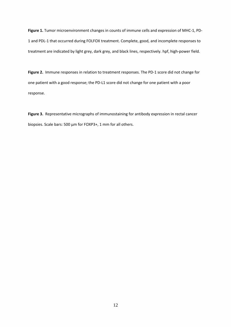
Figure 1. Tumor microenvironment changes in counts of immune cells and expression of MHC-1, PD-
1 and PDL-1 that occurred during FOLFOX treatment. Complete, good, and incomplete responses to
treatment are indicated by light grey, dark grey, and black lines, respectively. hpf, high-power field.
Figure 2. Immune responses in relation to treatment responses. The PD-1 score did not change for
one patient with a good response; the PD-L1 score did not change for one patient with a poor
response.
Figure 3. Representative micrographs of immunostaining for antibody expression in rectal cancer
biopsies. Scale bars: 500 µm for FOXP3+, 1 mm for all others.
13
Table 1. Clinical and pathological characteristics of the study cohort.
Patient Age (yr)
Sex Initial MRI
Stage Distance from
Anal Verge (cm) Days between
Biopsiesa Posttreatment
Stage Downstaging Complete
Response T N
1 63 M cT4b N1 7.6 82 ypT1 N0 Yes Yes No 2 69 M cT3c N1 5.0 62 ypT2 N0 Yes Yes No
3 67 F cT4b N1 3.2 81 ypT2 N0 Yes Yes No
4 75 M cT2 N1 6.5 89 ycT0 N0 Yes Yes Yes
5 34 M cT3c N2 13.0 78 ypT3 N1 No Yes No
6 78 M cT3b N1 2.8 127 ypT2 N0 Yes Yes No
7 48 F cT3b N2 5.8 61 ypT3 N1 No Yes No
8 33 M cT3b N1 12.0 74 ypT2 N0 Yes Yes No aAll patients had received four cycles of FOLFOX by the time of the second biopsy.
14
Table 2. Pre- and posttreatment immune responses.a
Biopsy Cells/HPF MHC-I
Histoscore PD-1 Score
PD-L1 Score
CD3+ CD8+ CD45RO+ FOXP3+ CD163+
Patient 1
Pre 23 22 20 10 22 220 1 2+
Post 5 5 7 6 10 280 2+ 1+
Patient 2
Pre 13 6 17 8 10 175 1+ 0 Post 21 18 29 7 16 270 1+ 1+
Patient 3
Pre 14 12 22 10 10 200 0 1+ Post 19 17 26 12 15 230 1+ 0
Patient 4
Pre 15 8 16 17 15 45 0 0
Post 25 14 23 5 14 210 1+ 1+
Patient 5
Pre 9 3 15 5 11 180 0 0
Post 14 9 22 8 16 230 1+ 1+
Patient 6
Pre 21 15 22 NA 15 210 1+ 1+
Post 22 7 23 5 14 125 2+ 0
Patient 7
Pre 20 8 15 8 12 220 0 0 Post 10 9 20 14 14 210 1+ 0
Patient 8
Pre 8 5 17 5 9 155 0 0
Post 22 12 15 4 14 405 1+ 1+ aIncreases of >25% are boldfaced. Decreases of >25% are italicized. HPF, high-power field; NA, not available.
Post 4 Cycles FOLFOXPre FOLFOX
CD3+
cel
ls/ h
pf25
20
15
10
5
0Post 4 Cycles FOLFOXPre-FOLFOX
CD8+
cel
ls/ h
pf
25
20
15
10
5
0
Post 4 Cycles FOLFOXPre FOLFOX
CD45
RO+
cells
/ hpf
30
25
20
15
10
5
CD3+ CD8+
CD45RO+
Post 4 Cycles FOLFOXPre FOLFOX
FOXP
3+ c
ells
/ hpf
20
15
10
5
0
FOXP3+
Post 4 Cycles FOLFOXPre FOLFOX
CD16
3+ c
ells
/ hpf
25
20
15
10
5
0
CD163+
Post 4 Cycles FOLFOXPre FOLFOX
MHC
1 Hi
stos
core
300
250
200
150
100
50
0
MHC-I
CD3+ CD8+
CD45RO+ FOXP3+
CD163+ MHC-1
PD-1 PD-L1