rubeomycin, a new anthracycline antibiotic complex i
TRANSCRIPT

938 THE JOURNAL OF ANTIBIOTICS AUG. 1981
RUBEOMYCIN, A NEW ANTHRACYCLINE ANTIBIOTIC COMPLEX
I. TAXONOMY OF PRODUCING ORGANISM, ISOLATION, CHARACTERIZATION
AND BIOLOGICAL ACTIVITIES OF RUBEOMYCIN A, A1, B AND B1
YASUO OGAWA, HIDEO SUGI, NOBORU FUJIKAWA and HIROYUKI MORI
Central Research Laboratory, Ishihara Sangyo Kaisha, Ltd., 3-1, 2-chome, Nishi-Shibukawa, Kusatsu-city, Shiga-ken, Japan
(Received for publication April 1, 1981)
A new antibiotic complex has been obtained from the cultures of an actinomycete, strain
FA-1 180, isolated from a soil sample collected at lake side of Biwa in Japan.
On the basis of taxonomic studies the producing microorganism is designated as Actino-
madura roseoviolacea var. biwakoensis nov. var. The antibiotic complex belongs to the class
of anthracycline glycoside antibiotics. All components form deep red fine needles on crystal-
lization; components are named rubeomycin A, A1, B and B1. These components exhibit
activity against Gram-positive bacteria as well as Yoshida sarcoma cell in vitro. These com-
ponents are also effective on P388 leukemia.
In the course of our screening program for new antibiotics produced by rare actinomycetes, we
found that the Actinomadura strain FA-1180, isolated from a soil sample collected at lake side of Biwa
in Japan, produced an anthracycline antibiotic
complex which inhibited the growth of Gram-
positive bacteria as well as Yoshida sarcoma cell
in vitro but not of Gram-negative bacteria, yeasts
and fungi. In addition to its in vitro activity the
antibiotic increased the survival of CDF1 mice
bearing P388 leukemia. The antibiotic complex
was isolated from the fermented mycelial cake
of strain FA-1180 and separated into four related
components named rubeomycin A, A1, B and B1.
On the basis of their physicochemical properties,
these antibiotics represent new anthracycline
derivatives and are related to carminomycinn
(Fig. 1).
In this paper, the taxonomy of the producing
strain, production, isolation, preliminary investi-
gation of physicochemical properties and biolog-
ical activities of the antibiotics are presented.
The chemical structure elucidation of these antibiotics will be reported in the next paper.
Taxonomy of Producing Organism
Most of the taxonomic studies on the culture were carried out in accordance with the methods
adopted by the International Streptomyces Project2). Additional media recommended by WAKSMAN3)
Fig. 1. Structures of rubeomycin A, A1, B and B1.
o OH
R
OH
014 0 011 0
~0, CH3
0~ N112
AandA1 Band B1
O OH
R: -C-CH3 -CH-CH3 R': (same in all components)
-CH-O-CH-CH3
CH2 CH1OH CHOH 6H,

939VOL. XXXIV NO. 8 THE JOURNAL OF ANTIBIOTICS
and NONOMURA et al.4) were also used. Observation of the cultures was made after cultivation for
3 weeks at 27 ~ 28°C unless otherwise stated. The cell-wall components were analyzed with the pure
cell-wall prepared in accordance with the method of YAMAGUCHI5). The cell-wall amino acids were
detected by amino acid autoanalyzer Yanagimoto LC-5, sugars were detected by GLC-Mass LKB-9000
as their corresponding trimethylsilyl derivatives and stereoisomers of 2,6-diaminopimelic acid were
determined by the paper chromatographic methods of HOARE and WORK6). The mycelium was cul-
tivated for 7 days at 35°C in the yeast extract-malt extract broth (ISP No. 1).
Morphological Characteristics
The aerial mycelia were well developed, long, straight to wavy and monopodially branched on
yeast extract - malt extract agar, oatmeal agar, oatmeal agar with added vitamin B mixture4) and
inorganic salts - starch agar with added vitamin B mixture (Fig. 2). The spore chains formed tightly
closed spirals with 2~5 turns; pseudosporangia were also formed (Fig. 3). Electron microscopy
revealed that the mature spores were about 0.8~1.1 ×0.6~1.0 µ, oval in shape, and with a smooth
surface.
Cell Wall Composition
The cell-wall of strain FA-1180 contains meso-diaminopimelic acid, glucose, mannose and madu-
rose7), but lacks glycine. The aforementioned cell-wall composition indicates the strain FA-1180 is
an actinomycete species of the cell-wall type III B8).
Cultural Characteristics
The cultural characteristics of strain FA-1180 are shown in Table 1. On most media, orange
Fig. 2. Aerial mycelium of strain FA-1180 (ISP - 4 V, 14 days).
20µ 10)1
Fig. 3. Electron micrograph of strain FA-1180 (ISP - 3 V, 14 days).
2µ I

940 THE JOURNAL OF ANTIBIOTICS AUG. 1981
Table 1. Cultural characteristics of strain FA-1 180.
Medium
Tryptone - yeast extract broth (ISP No. 1)
Yeast extract - malt extract agar (ISP No. 2)
Oat meal agar (ISP No. 3) and ISP No. 3 V*
Inorganic salts-starch agar (ISP No. 4)
ISP No. 4 V
Glycerol - asparagine agar (ISP No. 5)
ISP No. 5 V
Peptone - yeast extract - iron agar (ISP No. 6)
Tyrosine agar (ISP No. 7)
ISP No. 7 V
Characteristics
GR: fair, pale yellowish flaky growth on bottom of tube,
partially light orange to strong reddish orange ring on surface, in contact with glass
AM: restricted, white to pale pink
SP: slight, bright reddish orange
GR: good, remarkably raised, many folds
RC: strong orange to deep reddish orange, turning later into deep red
AM: fair, pink to purplish pink
SP: slight, strong reddish orange
GR: fair to somewhat restricted, flat, spreading
RC: light yellowish brown
AM: fair, powdery, white to pale pink, sometimes with drops formed on surface
SP: strong reddish orange
GR: restricted, penetrating into medium
RC: light yellowish brown
AM: scant, white
SP: none
GR: good, somewhat raised
RC: strong reddish orange
AM: fair to good, pale pink
SP: strong orange zone around growth
GR: scant
RC: light yellowish brown
AM: none
SP: none
GR: good, remarkably raised
RC: strong reddish orange to deep orange
AM: restricted, white to pale pink
SP: slight, bright orange
GR: fair, raised, many fine folds
RC: grayish brown to dark grayish brown
AM: none
SP: slight, deep red
GR: fair to somewhat restricted, flat
RC: dull red, turning later into deep red purple
AM: none
SP: none
GR: fair, flat
RC: deep red purple
AM: none
SP: deep red purple

941VOL. XXXIV NO. 8 THE JOURNAL OF ANTIBIOTICS
Table 1. (Continued)
Medium
Sucrose - nitrate agar (Waksman No. I )
Waksman No. 1 V
Glucose - asparagine agar (Waksman No. 2)
Waksman No. 2 V
Bouillon agar (Waksman No. 8)
Glucose -bouillon agar
Calcium malate agar
Calcium malate agar V
Glycerol - calcium malate agar
Glycerol - calcium malate agar V
Bouillon - gelatin stab
Characteristics
GR: fair
RC: light orange to strong reddish orange
AM: restricted, white to pale pink
SP: none or slight, reddish orange
GR: fair to good
RC: bright orange to deep red purple
AM: fair, pale pink to pale yellowish pink
SP: deep red purple
GR: scant
RC: pale yellow
AM: none
SP: none
GR: fair to good, somewhat raised
RC: bright reddish orange
AM: scant, white to pale pink
SP: bright orange
GR: fair
RC: dull red to grayish red
AM: scant, white
SP: dull red
GR: very good, remarkably raised, many folds
RC: dull red to dark red
AM: scant, white
SP: dull red
GR: scant or none
AM: none
SP: none
GR: somewhat restricted, flat, spreading
RC: vivid red purple
AM: none
SP: vivid red purple
GR: restricted to scant
RC: bright orange
AM: none
SP: none
GR: fair
RC: dull red
AM: none
SP: none or slight, dull red
GR: none

942 THE JOURNAL OF ANTIBIOTICS AUG. 1981
to red or violet vegetative growth developed moderately and aerial mycelia were colored white to pink.
The soluble pigments were orange to red or violet and behaved as pH indicators. The growth of
this strain was enhanced by the addition of a vitamin B mixture in media.
Physiological Characteristics
The physiological characteristics of strain FA-1180 and its profile of carbohydrate utilization are
shown in Table 2 and Table 3, respectively.
The microscopical and cultural studies and cell-wall analysis of FA-1180 indicate that this strain
belongs to the genus Actinomadura; it closely resembles Actinomadura roseoviolacea Nonomura &
Ohara. The comparison of cultural and physiological characteristics of strain FA-1180 with Actino-
madura roseoviolacea Nonomura & Ohara KCC A-0145 was made by simultaneous cultivation and is
shown in Table 4.
The differences, however, were not sufficient to designate strain FA-1180 as a new species and it
Table 1. (Continued)
Medium
Potato plug (Waksman No. 40)
Lofler's blood serum
Skim milk
Characteristics
GR: fair, somewhat raised
RC: strong reddish orange to dull purplish red
AM: restricted, white to grayish pink
SP: grayish pink to grayish red
GR: fair to somewhat restricted
RC: bright yellowish red, turning later into deep red purple
AM: none
SP: slight, deep red purple i
GR: fair to somewhat restricted, ring present, bright orange to bright reddish orange
AM: none
SP: slight, light orange to bright orange
Symbols: GR, growth. RC, reverse color. AM, aerial mycelium. SP, soluble pigment.
* V: vitamin B mixture added4)
Color names were assigned according to "Manual of Color Name", a manual published by Nippon
Shikisai Kenkyusho, Tokyo, Japan.
Table 2. Physiological properties of strain FA-1180.
Parameter observed I
Optimum temperature for growth
Liquefaction of gelatin
Hydrolysis of starch
Milk peptonization
Milk coagulation
Nitrate reduction
Production of H2S
Production of ammonia
Melanoid pigment
Results
35~40°C
negative
weakly positive
weakly positive
weakly positive
positive
negative
negative
negative
Table 3. Carbon utilization of strain FA-1180.
Carbon source
L-Arabinosc
D-Xylose
D-Glucose
D-Galactose
D-Fructose
L-Rhamno,c
D-Mannoe
D-Lactose
Maltose
Sucrose
Response Carbon source
Melibiose
Raffinose
Trehalose
Cellobiose
i-Inositol
D-Mannitol
D-Sorbitol
Salicin
Inulin
Response
-I-
Symbols:+ , utilization.± , doubtful utilization.
-, no utilization.

943VOL. XXXIV NO. 8 THE JOURNAL OF ANTIBIOTICS
was named Actinomadura roseoviolacea var. biwakoensis nov. var. This strain has been deposited
at the Institute for Fermentation, Osaka, Japan and Fermentation Research Institute, Agency of
Industrial Science and Technology, Japan, where it has been assigned the designations IFO 14092 and
FERM-P 5155, respectively.
Production and Isolation
The producing organism, A. roseoviolacea var. biwakoensis, was grown in submerged culture in
a 50-liter jar fermentor containing 35 liters of a medium consisting of 3 % glucose, 1 % corn steep
liquor, 1 % soy bean flour and 0.1 % K2HPO4. The fermentor was inoculated with 700 ml of the seed
culture which was prepared in 2-liter flasks each containing 350 ml of the same medium and incubated
on a reciprocal shaker at 35°C for 3~5 days. Incubation was continued at 35°C for 7 days with aera-
tion at 30 liters per minute and agitation at 250 rpm. The progress of fermentation was monitored by
determination of the mycelial volume and the optical density at 496 nm of acetone extracts of mycelium,
because the antibiotics were accumulated in the mycelium and not excreted in the filtrate under these
culture conditions.
The isolation of antibiotics was accomplished using the general procedure for anthracycline anti-
biotics, as shown in Fig. 4. The fermentation broth (100 liters) was centrifuged and the antibiotics
were extracted from the harvested mycelial cake (5 kg) three times with acetone (10 liters) at 50°C.
Table 4. Differences between strain FA-1180 and A. roseoviolacea KCC A-0145.
Medium
ISP No.1
ISP No. 4
ISP No. 5 V
ISP No. 6
Waksman No. 1 V
Waksman No. 7 V
Carbon utilization
Salicin
D-Sorbitol
Sucrose
i-Inositol
D-Mannitol
Hydrolysis of starch
Strain FA-1180
GR: fair, pale yellowish flaky growth on bottom of tube, partially
light orange to strong reddish orange ring on surface, in contact
with glass
SP: slight, bright reddish orange
GR: restricted, penetrating into medium
RC: light yellowish brown
AM: scant, white
AM: restricted, white to pale pink
SP: slight, deep red
GR: fair
RC: deep red purple
AM: fair, pale pink to pale yellowish pink
SP: none
weakly positive
A. roseoviolacea KCC A-0145
flaky growth on bottom of tube,
pale yellow
restricted, spreading
light reddish brown
fair, white to pale pink
none
none
scant
deep red purple
none
vivid red purple
negative
Symbols: Same as in Tables 1 and 3.

944 THE JOURNAL OF ANTIBIOTICS AUG. 1981
The extract was concentrated in vacuo to remove acetone and the concentrate extracted three times
with 1 liter of chloroform at pH 8.6. The chloroform layer was concentrated in vacuo to 1 liter. Then
the antibiotics were extracted three times with 0.1 N acetic acid (500 ml). The acetic acid layer was
subsequently adjusted to pH 8.6 with a sodium bicarbonate saturated solution and extracted three times
with 1 liter of chloroform. The combined extracts were dried with anhydrous sodium sulfate, concen-
trated in vacuo to a small volume, and then a large excess of n-hexane was added to precipitate the
antibiotics (1500 mg).
Further separation of the complex into its components was achieved by droplet countercurrent
chromatography (DCC) using chloroform - methanol - 0.1 N acetic acid (5: 5: 3) as the solvent system.
A DCC apparatus containing 300 separation tubes was used and separation was accomplished by the
down flow method. The effluents were monitored by silica gel TLC with a solvent system of chloro-
form - methanol - acetic acid (80: 20: 4). The fractions containing each component were pooled and
adjusted to pH 8.6 with the sodium bicarbonate saturated solution. The separated solvent layer was
washed with deionized water and dried with anhydrous sodium sulfate, concentrated in vacuo to a small
volume, and the components were crystallized using solvent systems chloroform - n-hexane for A
and A1, and chloroform for B and B,. Each component was obtained as deep red fine needle crystals.
Physicochemical Properties
All the components of rubeomycin were amphoteric and soluble in methanol, acetone, chloroform
and acidic water, but barely soluble or insoluble in ethyl ether, n-hexane, petroleum ether and water.
The physicochemical properties of the components are summarized in Table 5. The UV and visible
spectra of all the components were very similar (Figs. 5A~5D). The IR spectra were approximately
Fig. 4. Purification process of the antibiotics.
Fermentation broth (100 liters)
centrifuged
Supernatant Mycelial cake (5 kg)
I extracted with acetone (10 liters x 3 tiiiies) Filtrate Cake
I concentrated in vacuo to remove acetone Aqueous solution
I extracted with chloroform at p118.6 (1 liter x 3 times) , Aqueous layer Solvent layer
concentrated in vacuo to 1 liter extracted with 0.lN acetic acid
(500ml x 3 times) Acetic acid layer er Solvent layer
extracted with chloroform at pH 3.6 (1 1 iter x 3 tinies) Aqueous layer Solvent layer
dried with anhydroUs sodiurn sulfate concentrated in vac- to all volume
added n-hexane Precipitate (1500rig)
(FA-1180 complex) DCC with chlorofon:r-methanol -0.1 N acetic acid
(5:5:3) stationary phase :upper layer
mobile phase :lower layer
Pubeomycin Rubeomycin A Rubeomycin S1 Ruheonycin S (80 111q) (400 mg) (70mg) (i3'i'ip)

945VOL. XXXIV NO. 8 THE JOURNAL OF ANTIBIOTICS
the same for all the components, except for the absoption at 1720 cm-1, which indicates, in the spectra
of A and A1 components, the presence of a -COCH3 group in the hydroaromatic ring; this band is lack-
ing in the spectra of the B and B1 components (Figs. 6A and 6B). This consideration was supported
from the CMR spectra (Figs. 7A~7D). In the CMR spectra of all the components, the signals due
to the 33 carbon atoms are present.
Biological Properties
Antimicrobial Activities
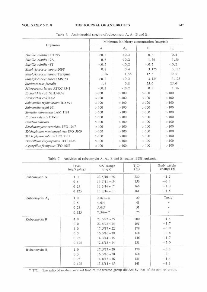
Antimicrobial activities of each component of rubeomycin are shown in Table 6. One loopful
of test organism suspension was streaked on agar plates containing two-fold dilutions of the antibiotic
acetic acid salt. The inoculated plates were incubated for 24 hours at 37°C for bacteria or 7 days
at 30°C for fungi and yeasts. Brain heart infusion agar (Nissui) for bacteria and SABOURAUD's agar
for fungi and yeasts were used. All the components showed selective antibacterial activity against
Gram-positive bacteria, A and A1 exhibiting higher activity (lower MIC) than B and B1.
Growth Inhibitory Activities against Yoshida Sarcoma Cell
The acetic acid salts of the antibiotics were dissolved in the Fisher's medium containing 20% of
horse serum; dilutions were made with the same medium. These media containing the antibiotics
were inoculated with 5 × 10⁴ cells/ml of Yoshida sarcoma and incubated for 2~3 days at 37°C. The
growth inhibitory activity of the A1 component was about 10 times larger than that of other components.
Table 5. Physicochemical properties of rubeomycin A, A1, B and B1.
Melting point ° C (dec.)
Elemental analysis (%)*
C :
H : N :
O: Molecular weight (FD-mass)
Molecular formula
?max nm (E1%1cm) in MeOH
[a]20D in CHCl3
Rf value on a:** silica gel b: TLC c:
A
155~160
F C
58.7 60.1
6.7 6.2 2.0 2.1
31.6 31.6
659
C33H41NO13
237 (595)
255 (447)
292 (141)
493 (250)
530 (174)
+120.6 c 0.199
0.57
0.05
0.31
Al
174~176
F C
58.7 60.1
5.6 6.2
1.9 2.1
31.6 659
C33H41NO13
237 (605)
255 (464)
292 (161)
493 (260)
530 (196)
+170.4 c 0.053
0.74
0.11 0.40
B
144~146
F C
59.7 59.1
6.7 6.5
1.7 2.1 - 31 .5
661
C33H43NO13
237 (562)
255 (453)
292 (133)
493 (239)
530 (173)
+118.0 c 0.128
0.34
0.02 0.23
B1
152154
F C 58.4 59.1
7.3 6.5
1.9 2.1
31.5
661
C33 H43N013
237 (582)
255 (466) 292 (137)
493 (251)
530 (182)
+151.0 C 0.090
0.47
0.04
0.27
* F: found, C: calculated.
** a: CHCl3 - MeOH - acetic acid , 80: 20: 4.
b: CHCl3 - MeOH, 10:1.
c: CHCl3 - MeOH - toluene, 7: 3: 3.

946 THE JOURNAL OF ANTIBIOTICS AUG. 1981
Antitumor Activities in Mice
The antitumor activities of the antibiotics were studied by prolongation of the median survival
time (MST) of mice bearing P388 leukemia. For testing P388 leukemia was implanted intraperitoneally
into CDF1 mice using 101 cells/animal. Twenty-four hours after the implantation of tumor cells,
Fig. 5A. UV spectrum of rubeomycin A.
MeOH/NaOH
~MeiiPl
Fig. 5B. UV spectrum of rubeomycin B.
X011/NaJH,.
Fig. 5C. UV spectrum of rubcomycin A1.
1
Fig. 5D. UV spectrum of rubeomycin B1.
~^eOH; I;aOH
1\
~'cr4
947VOL. XXXIV NO. 8 THE JOURNAL OF ANTIBIOTICS
Table 6. Antimicrobial spectra of rubeomycin A, A1, B and B1.
Organism
Bacillus subtilis PCI 219
Bacillus subtilis 17A
Bacillus subtilis 45T
Staphylococcus aureus 209P
Staphylococcus aureus Terajima Staphylococcus aureus MS353
Streptococcus faecalis
Micrococcus huteus ATCC 9341
Escherichia coli NIHJ-JC-2
Escherichia coli Keio
Salmonella typhimurium IID 971
Salmonella typhi 901
Serratia marcescens IAM 1184
Proteus vulgaris OX-19
Candida albicans
Saccharomyces cerevisiae IFO 1047 Trichophyton mentagrophytes IFO 5809
Trichophyton rubrum IFO 9185
Penicillium chrysogenum IFO 4626
Aspergillus fumigatus IFO 4057
Minimum inhibitory concentration (mcg/ml)
A
<0.2
0.8
< 0.2
0.8
1.56
<0.2
1.6
<0.2
> 100
> 100
> 100
> 100
> 100
100
> 100
> 100
> 100
> 100
> 100
-100
Al
;0.2
<0.2
<0.2
0.4
1.56
< 0.2
0.8
< 0.2
>100
>100
>100
> 100
>100
>100
>100
>100
>100
>100
>100
>100
B
0.8
1.56
<0.2
3.125
12.5
3.125
25.0
0.8
> 100
>l00
> 100
> 100
> 100
>100
>100
>100
> 100 >100
>100
>100
B1
0.4
1.56
< 0.2
3.125
12.5
3.125
25.0
1.56
>100
>100
>100
>100
>100
> 100
>100
>100
>100
<100
>100
>100
Table 7. Activities of rubeomycin A, A1, B and B1 against P388 leukemia.
Rubeomycin A
Rubeomycin A1
Rubeomycin B
Rubeomycin B1
Dose (mg/kg/day)
1.0
0.5
0.25
0.125
1.0
0.5
0.25
0.125
4.0
2.0
1.0 0.5
0.25
0.125
1.0
0.5 0.25
0.125
MST/range (days)
22.5/10~26
14.5/11~19 16.3/16~17
15.8/16~17
2.8/3~4
4.0/4
5.0/5
7.3/6~7
23.5/22~25
22,5/22~25 17.5/17~22
16.5/16~18
14.3/14~15
12.8/13~14
17.5/17~20 16.5/16~20
14.8/15~16
13.8/14~15
T/C* (%)
230
156
166
161
29
41
51
75
200 191
179
168 146
131
179
168
151
141
Body weight change (g)
-1 .3
-0 .7
+ 1.0
+ 1.5
Toxic
-1 .4
-1 .7
+-0 .9
+ 0.8
+ 1.7
+ 2.0
- 0.8
0
+1.4
+1.1
* T/C: The ratio of median survival time of the treated group divided by that of the control group.

948 THE JOURNAL OF ANTIBIOTICS AUG. 1981
Fig. 6A. Infrared absorption spectra of rubeomycin A and B (KBr).
Fig. 6B. Infrared absorption spectra of rubeomycin A1 and B1 (KBr).
Fig. 7A. CMR spectrum of rubeomycin A in CDCl3.
11

949VOL. XXXIV NO. 8 THE JOURNAL OF ANTIBIOTICS
graded doses of the antibiotics dissolved in saline were administered to mice intraperitoneally (0.1 ml
per 10 grams of body weight). The treatment was given once a day for 5 days.
The results are shown in Table 7. Rubeomycin A, B and Bl showed significant prolongation of
life with doses in the range of 0.1254.0 mg/kg/day, but the Al component was more toxic than the
others; therefore the prolongation of life with Al was not observed under the experimental conditions
used.
Fig. 7B. CMR spectrum of rubeomycin B in CDCI3 - CH3OH.
Fig. 7C. CMR spectrum of rubeomycin Al in CDCI3.

950 THE JOURNAL OF ANTIBIOTICS AUG. 1981
Acknowledgments
The authors wish to thank Dr. K. MUNAKATA and Dr. N. KATO of Nagoya University for their assistance
with CMR spectra and FD-mass spectra measurement and their helpful advice throughout those studies.
Thanks are also due to Dr. A. SEINO of Kaken Chemical Co., Ltd. for providing us with Actinomadura
roseoviolacea KCC A-0145.
References
1) BRAZHNIKOVA, M. G.; V. B. ZBARSKY, V. I. PONOMARENKO & N. P. POTAPOVA: Physical and chemical characteristics and structure of carminomycin, a new antitumor antibiotic. J. Antibiotics 27: 254~259,
1974 2) SHIRLING, E. B. & D. GOTTLIEB: Methods for Characterization of Streptoinyces species. Inter. J. Syst. Bacteriol. 16: 313~340, 1966 3) WAKSMAN, S. A.: The Actinomycetes. Vol. II. The Williams and Wilkins Co., Baltimore, 1961 4) NONOMURA, H. & Y. OHARA: Distribution of actinomycetes in soil. XI. Some new species of the genus Actinomadura Lechevalier et al. J. Ferment. Technol. 49: 904~912, 1971 5) YAMAGUCHI, T.: Comparison of the cell-wall composition of morphologically distinct actinomycetes. J. Bacteriol. 89: 444~453, 1965 6) HOARE, D. S. & E. WORK: The stereoisomers of a,s-diaminopimelic acid. 2. Their distribution in the bacterial order Actinomycetales and in certain Eubacteriales. Biochem. J. 65: 441~447, 1957
7) LECHEVALIER, M. P. & N. N. GERBER: The identity of madurose with 3-O-methyl-n-galactose. Carbohyd. Res. 13: 451~454, 1970 8) LECHEVALIER, M. P. & H. A. LECHEVALIER: Chemical composition of cell as a criterion in the classification of aerobic actinomycetes. Inter. J. Syst. Bacteriol. 20: 435~443, 1970
Fig. 7D. CMR spectrum of rubeomycin B1 in CDCl3; - CH3OH.