structure of blood vessels composed of three layers (tunics) tunica intima – composed of simple...
Post on 19-Dec-2015
221 views
TRANSCRIPT

Structure of Blood Vessels
Composed of three layers (tunics) Tunica intima – composed of simple squamous
epithelium Tunica media – sheets of smooth muscle
Contraction – vasoconstrictionRelaxation – vasodilation
Tunica externa – composed of connective tissue Lumen
Central blood-filled space of a vessel
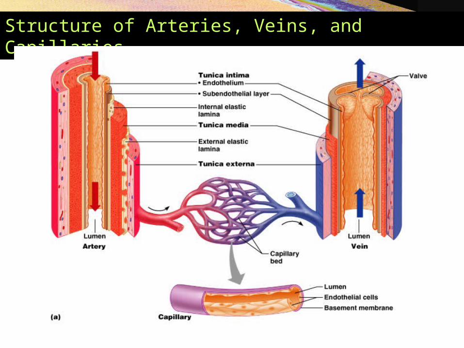
Structure of Arteries, Veins, and Capillaries
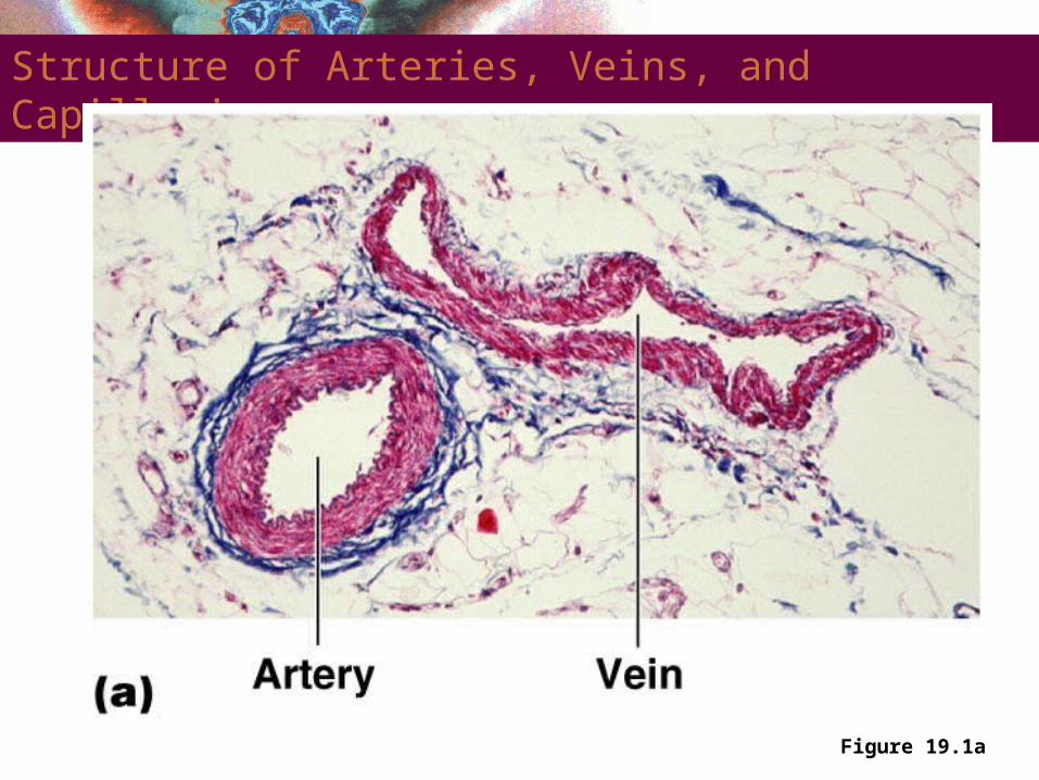
Structure of Arteries, Veins, and Capillaries
Figure 19.1a

Types of Blood Vessels
Arteries – carry blood away from the heart Capillaries – smallest blood vessels
The site of exchange of molecules between blood and tissue fluid
Veins – carry blood toward the heart
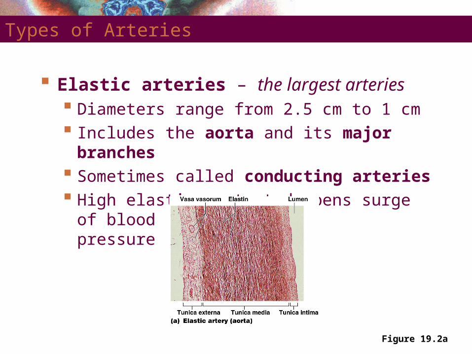
Types of Arteries
Elastic arteries – the largest arteries Diameters range from 2.5 cm to 1 cm Includes the aorta and its major branches Sometimes called conducting arteries High elastin content dampens surge of blood
pressure
Figure 19.2a
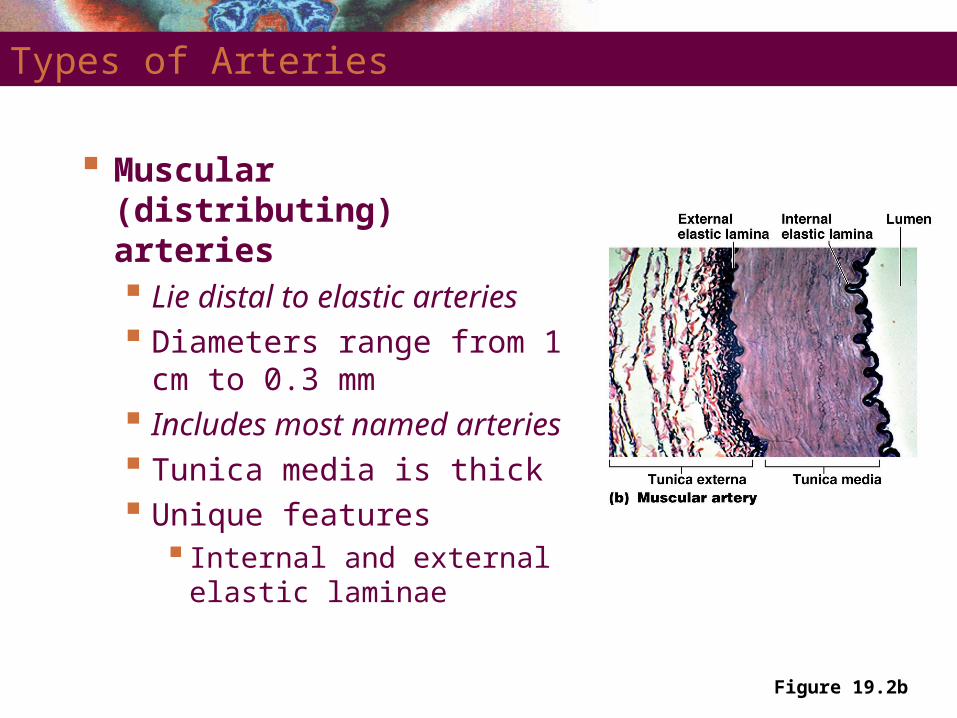
Types of Arteries
Muscular (distributing) arteries Lie distal to elastic arteries Diameters range from 1 cm
to 0.3 mm Includes most named
arteries Tunica media is thick Unique features
Internal and external elastic laminae
Figure 19.2b
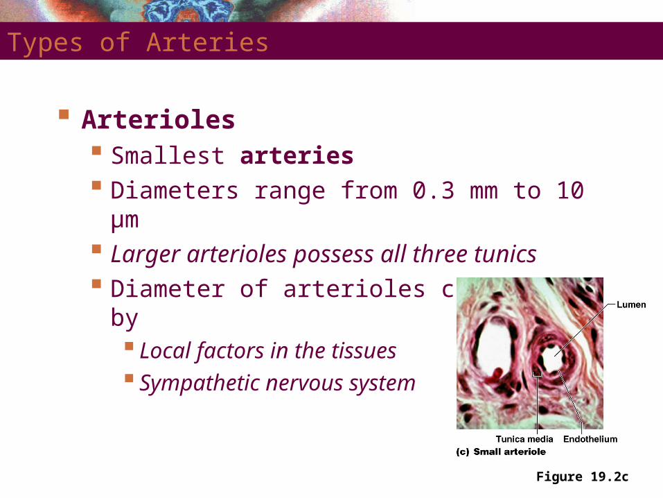
Figure 19.2c
Types of Arteries
Arterioles Smallest arteries Diameters range from 0.3 mm to 10 µm Larger arterioles possess all three tunics Diameter of arterioles controlled by
Local factors in the tissues Sympathetic nervous system

Capillaries
Smallest blood vessels Diameter from 8–10 µm
Red blood cells pass through single file
Site-specific functions of capillariesLungs – oxygen enters blood, carbon dioxide leaves Small intestines – receive digested nutrientsEndocrine glands – pick up hormonesKidneys – removal of nitrogenous wastes

RBCs in a Capillary
Figure 19.3
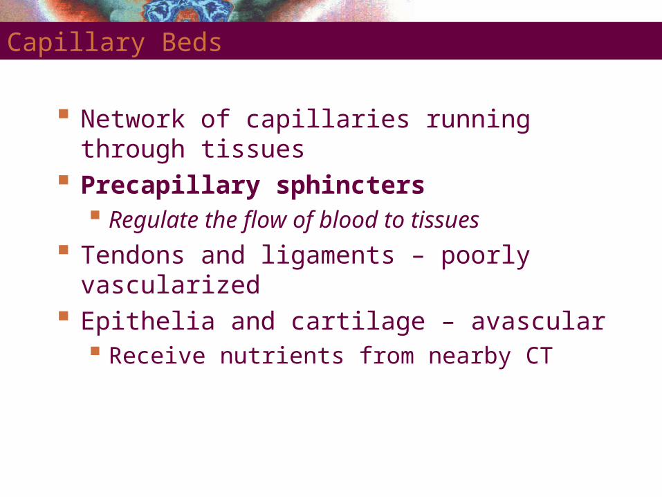
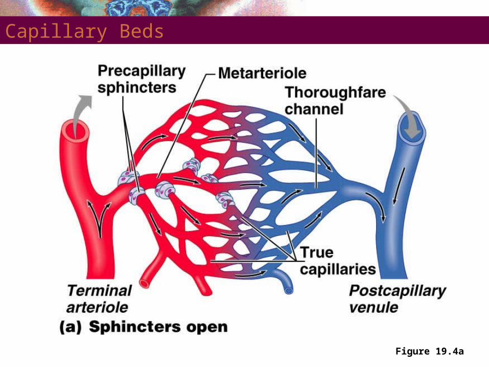
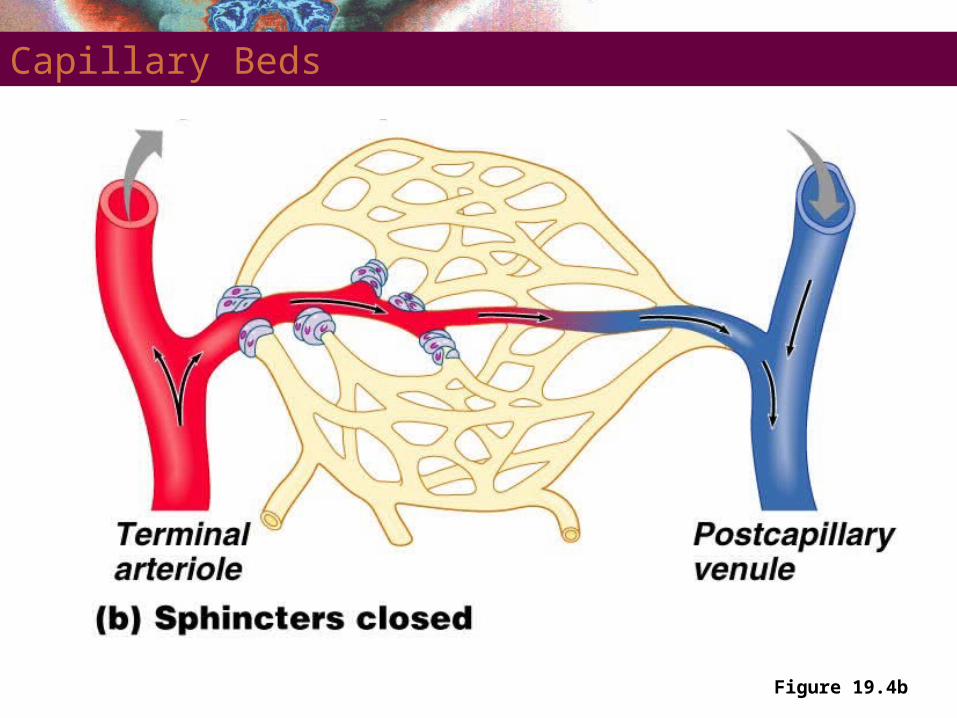
Capillary Beds
Network of capillaries running through tissues Precapillary sphincters
Regulate the flow of blood to tissues
Tendons and ligaments – poorly vascularized Epithelia and cartilage – avascular
Receive nutrients from nearby CT
Capillary Beds
Figure 19.4a
Capillary Beds
Figure 19.4b

Capillary Permeabillity
Endothelial cells – held together by tight junctions and desmosomes
Intercellular clefts – gaps of unjoined membrane Small molecules can enter and exit
Two types of capillary Continuous – most common Fenestrated – have pores
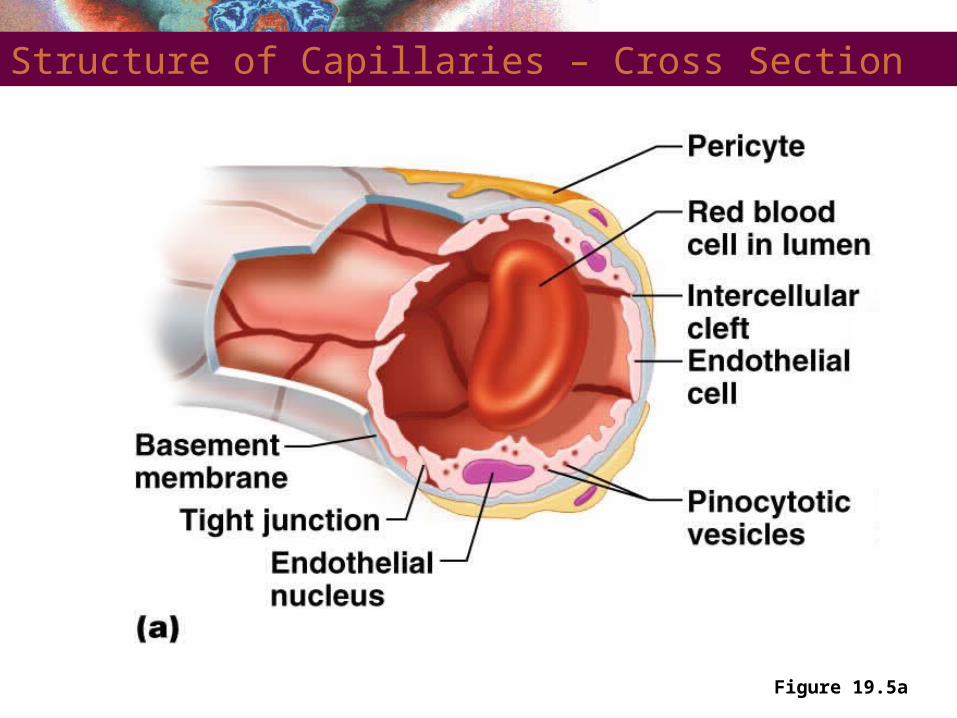
Structure of Capillaries – Cross Section
Figure 19.5a

Structure of Capillaries – Cross Section
Figure 19.5b
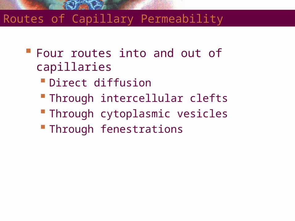
Routes of Capillary Permeability
Four routes into and out of capillaries Direct diffusion Through intercellular clefts Through cytoplasmic vesicles Through fenestrations

Low Permeability Capillaries
Blood-brain barrier Capillaries have complete tight junctions No intercellular clefts are present Vital molecules pass through
Highly selective transport mechanisms
Not a barrier against Oxygen, carbon dioxide, and some anesthetics
Sinusoids
Wide, leaky capillaries found in some organs Usually fenestrated Intercellular clefts are wide open
Occur in bone marrow and spleen Sinusoids have a large diameter and twisted course

Sinusoids
Figure 19.5c

Veins
Conduct blood from capillaries toward the heart Blood pressure is much lower than in arteries Smallest veins – called venules
Diameters from 8 – 100 µm Smallest venules – called postcapillary venules
Venules join to form veins Tunica externa is the thickest tunic in veins
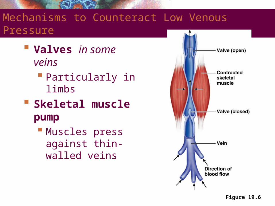
Figure 19.6
Mechanisms to Counteract Low Venous Pressure
Valves in some veins Particularly in limbs
Skeletal muscle pump Muscles press against
thin-walled veins

Vascular Anastomoses
Vessels interconnect to form vascular anastomoses Organs receive blood from more than one arterial
source
Neighboring arteries form arterial anastomoses Provide collateral channels
Veins anastomose more frequently than arteries
Vasa Vasorum
Tunica externa of large vessels have Tiny arteries, capillaries, and veins
Vasa vasorum vessels of vessels Nourish outer region of large vessels
Inner half of large vessels receive nutrients from luminal blood
Pulmonary Circulation
Pulmonary trunk leaves the right ventricle Divides into right and left pulmonary arteries
Superior and inferior pulmonary veins Carry oxygenated blood into the left atrium