study of ribosomes having modifications in the ... · study of ribosomes having modifications in...
TRANSCRIPT
Study of Ribosomes having Modifications in the Peptidyltransferase Center Using Non-
α-L-Amino Acids and Synthesis and Biological Evaluation of Topopyrones
by
Rumit Maini
A Dissertation Presented in Partial Fulfillment
of the Requirements for the Degree
Doctor of Philosophy
Approved July 2013 by the
Graduate Supervisory Committee:
Sidney M. Hecht, Chair
Ian Gould
Hao Yan
ARIZONA STATE UNIVERSITY
August 2013

i
ABSTRACT
The ribosome is a ribozyme and central to the biosynthesis of proteins in all
organisms. It has a strong bias against non-α-L-amino acids, such as α-D-amino acids and
β-amino acids. Additionally, the ribosome is only able to incorporate one amino acid in
response to one codon. It has been demonstrated that reengineering of the
peptidyltransferase center (PTC) of the ribosome enabled the incorporation of both α-D-
amino acids and β-amino acids into full length protein.
Described in Chapter 2 are five modified ribosomes having modifications in the
peptidyltrasnferase center in the 23S rRNA. These modified ribosomes successfully
incorporated five different β-amino acids (2.1 – 2.5) into E. coli dihydrofolate reductase
(DHFR). The second project (Chapter 3) focused on the study of the modified ribosomes
facilitating the incorporation of the dipeptide glycylphenylalanine (3.25) and fluorescent
dipeptidomimetic 3.26 into DHFR. These ribosomes also had modifications in the
peptidyltransferase center in the 23S rRNA of the 50S ribosomal subunit. The modified
DHFRs having β-amino acids 2.3 and 2.5, dipeptide glycylphenylalanine (3.25) and
dipeptidomimetic 3.26 were successfully characterized by the MALDI-MS analysis of
the peptide fragments produced by “in-gel” trypsin digestion of the modified proteins.
The fluorescent spectra of the dipeptidomimetic 3.26 and modified DHFR having
fluorescent dipeptidomimetic 3.26 were also measured.
The type I and II DNA topoisomerases have been firmly established as effective
molecular targets for many antitumor drugs. A “classical” topoisomerase I or II poison
acts by misaligning the free hydroxyl group of the sugar moiety of DNA and preventing
the reverse transesterfication reaction to religate DNA. There have been only two classes
ii
of compounds, saintopin and topopyrones, reported as dual topoisomerase I and II
poisons. Chapter 4 describes the synthesis and biological evaluation of topopyrones.
Compound 4.10, employed at 20 µM, was as efficient as 0.5 μM camptothecin, a potent
topoisomerase I poison, in stabilizing the covalent binary complex (~30%). When
compared with a known topoisomerase II poison, etoposide (at 0.5 uM), topopyorone
4.10 produced similar levels of stabilized DNA–enzyme binary complex (~34%) at 5 μM
concentration.

iii
ACKNOWLEDGEMENTS
I would like to extend my sincere gratitude to my research advisor Professor
Sidney M. Hecht who has been an outstanding mentor over the last five years. I am
extremely grateful to him for providing me with an opportunity to work and grow in a
stimulating research environment. I would also like to thank him for allowing me to
explore opportunities in the field of protein biochemistry along with synthetic organic
chemistry. I must thank Professors Seth Rose and Ian Gould for their guidance
throughout the years and help with the complexities of graduate school. I shall also
acknowledge and thank Professor Hao Yan for accepting my request to be on my
committee on such short notice.
I must extend my sincere gratitude to Dr. Larisa Dedkova for being an amazing
mentor, colleague and friend. I am grateful to her for training me as a proficient
biochemist. Dr. Simon J. Leiris was my first mentor in my graduate research and I am
thankful to him for imparting strong synthetic organic chemistry skills to me. I am
grateful to Dr. Shengxi Chen, Dr. Pablo Arce, Dr. Manikandidas M.M., and Dr. Rakesh
Paul for their continuous input regarding many complicated issues related to my research.
Thanks to Dr. Paul A. Zaleski for his work on the biological study of topoyrone
derivatives.
I extend my gratitude to Dr. Ryan Nangreave for being a great lab mate and an
amazing friend; research would not have been as much fun without you. I would also like
to thank two wonderful ladies, Dr. Jeanette Nangreave and Emma Nangreave for their
constant support over the years. I thank my dear friend, Trevor Bozeman, for his
continual assistance in and outside the lab and for introducing me to various aspects of
iv
the American culture. I would especially like to thank my dearest friend Caridad
Rodriguez, who has been a pillar in my life here in the US. I have learned a lot of from
her and I could not have done this without her constant support. I would like to thank
Basab Roy for giving me rides to lab on numerous occasions. My thanks also go to all the
current and former members of the Sidney Hecht group.
I would finally like to thank my parents and my sister for their unconditional love
and support. I am grateful for their patience and guidance and letting me follow my
dreams. I would not have achieved this without their encouragement. I would finally like
to dedicate this work to my nani (grandmother) and nana (late grandfather) who have
always showered me with their unconditional love.
v
TABLE OF CONTENTS
Page
LIST OF ABBREVIATIONS ........................................................................................ vii
LIST OF TABLES .......................................................................................................... x
LIST OF FIGURES ....................................................................................................... xii
LIST OF SCHEMES ..................................................................................................... xv
CHAPTER
1. THE RIBOSOME AND PROTEIN SYNTHESIS: GENERAL
INTRODUCTION .............................................................................................. 1
1.1. The ribosome ................................................................................. 1
1.2. The ribosome is a ribozyme ............................................................ 5
1.3 Biosynthesis of modified proteins……………………………….....6
1.4 Dihydrofolate reductase (DHFR)…………………………………..7
1.5 Modified ribosomes……………………………………………….11
2. INCORPORATION OF β-AMINO ACIDS INTO E. COLI DHFR
USING RIBOSOMES HAVING MODIFICATIONS IN THE
PEPTIDYLTRANSFERASE CENTER (PTC) ................................................ 14
2.1. Introduction.................................................................................. 14
2.2. Results ......................................................................................... 16
2.3. Discussion .................................................................................... 35
2.4. Experimental ................................................................................ 39

vi
CHAPTER Page
3. INCORPORATION OF A DIPEPTIDE AND DIPEPTIDOMIMETIC
INTO DHFR USING RIBOSOMES HAVING MODIFICATIONS
IN THE PEPTIDYLTRANSFERASE CENTER .............................................. 50
3.1. Introduction.................................................................................. 50
3.2. Results ......................................................................................... 56
3.3. Discussion .................................................................................... 73
3.4. Experimental ................................................................................ 78
4. SYNTHESIS AND BIOLOGICAL EVALUATION OF
TOPOPYRONES ............................................................................................. 83
4.1. Introduction.................................................................................. 83
4.2. Results ......................................................................................... 92
4.3. Discussion .................................................................................... 98
4.4. Experimental .............................................................................. 105
REFERENCES .................................................................................................. 121

vii
LIST OF ABBREVIATIONS
aaRS aminoacyl-tRNA synthetase
AMP adenosine-5'-monophosphate
ATP adenosine-5'-triphosphate
aq aqueous
bs broad singlet
BSA bovine serum albumin
13C NMR carbon-13 nuclear magnetic resonance spectroscopy
oC degrees Celsius
cat catalytic
cm centimeter
δ chemical shift (ppm)
d doublet
dec decomposition
DEAE diethylaminoethyl
DHF 7,8-dihydrofolate
DHFR dihydrofolate reductase
DIPEA diisopropylethylamine
DMAP dimethylaminopyridine
DMF dimethylformamide
DMSO dimethylsulfoxide
DNA deoxyribonucleic acid
DTT dithiothreitol
viii
E. coli Escherichia coli
EDTA ethylenedinitrilotetraacetic acid
ESI electrospray ionization
GDP guanosine-5'-diphosphate
GTP guanosine-5'-triphosphate
1H NMR proton nuclear magnetic resonance spectroscopy
HBTU O-benzotriazole-N,N,N ,N -tetramethyl-uronium-hexafluoro-phosphate
HPLC high performance liquid chromatography
Hz Hertz
IPTG isopropyl-β-D-thiogalactopyranoside
J coupling constant
L liter
M molar
m multiplet
MALDI-MS matrix assisted laser desorption ionization – mass spectrometry
MHz mega Hertz
mL milliliter
mM millimolar
mmol millimole(s)
mRNA messenger ribonucleic acid
uM micromolar
μmol micromole(s)
nm nanometer

ix
NADPH nicotinamide adenine dinucleotide phosphate
NMR nuclear magnetic resonance
pdCpA 5 -O-phosphoryl-2 -deoxycytidyl(3 5 )adenosine
PTC peptidyltransferase center
p-TsCl p-toluenesulfonyl chloride
pYRNA8 plasmid containing the gene encoding 74 nucleotide tRNACUA
Rf ratio of fronts
RNA ribonucleic acid
s singlet
SDS-PAGE sodium dodecyl sulfate polyacrylamide gel electrophoresis
t triplet
TBA tetrabutylammonium
TBAF.H2O tetrabutylammoniumfluoride monohydrate
TFA trifluoroacetic acid
THF 5,6,7,8-tetrahydrofolate
TLC thin layer chromatography
Topo topoisomerase
tRNA transfer RNA
UV ultraviolet
Val valine
x
LIST OF TABLES
Table Page
2.1 23S rRNA sequence modifications in the PTC for the clones used for S-30
preparations………………………………………………………………...............23
2.2 Incorporation of β-amino acids 2.1, 2.2 and 2.3 into position 10 of E. coli
DHFR by the use of S-30 systems having different modified ribosomes………….24
2.3 Incorporation of β-alanine analogues 2.2, 2.4 and 2.5 into position 18 of E. coli
DHFR by the use of S-30 systems having different modifications in the
PTC………………………………………………………………………………...25
2.4 MALDI mass spectrometry analysis of tryptic digests of wild-type and
modified DHFR samples…………………………………………………...............30
3.1 Characterization of selected clones sensitive to puromycin derivative 1.3 and
erythromycin……..………………………………………………………………...58
3.2 23S rRNA sequence modifications in the PTC for the clones used for S-30
preparations………………………………………………………………...............59
3.3 Incorporation of glycylphenylalanine (3.25) and dipeptidomimetic 3.26
into position 10 of E. coli DHFR by the use of S-30 systems having different
modified ribosomes………………………………………………………………...63
3.4 MALDI-MS analysis of tryptic digests of the wild-type and modified DHFRs
1and 2 samples……………………………………………………………………..66
4.1 Topoisomerase I-mediated DNA cleavage by CPT and topopyrones on a 3′-32
P
end labeled 23-base pair oligonucleotide substrate………………………………..97
xi
Table Page
4.2 Nitrocellulose filter binding of CPT or topopyrone stabilized
DNA–topoisomerase covalent binary complexes………………………………….98
xii
LIST OF FIGURES
Figure Page
1.1 Schematic diagram of a ribosome bound to an mRNA showing the A, P
and E-sites .............................................................................................................. 2
1.2 Strategy for the incorporation of unnatural amino acids into proteins in vitro .......... 8
1.3 The catalytic cycle of E. coli DHFR at pH 7 ......................................................... 11
1.4 Structures of puromycin and its derivatives ........................................................... 12
2.1 Structures of puromycin (1.1) and β-puromycin (1.2)............................................ 15
2.2 Structures of β-amino acids studied ...................................................................... 16
2.3 Preparation of β-aminoacyl-tRNACUAs.................................................................. 20
2.4 In vitro synthesis of DHFR utilizing β-aminoacyl-tRNACUAs to suppress an UAG
codon at position 49 of DHFR mRNA using wild-type ribosomes ........................ 21
2.5 In vitro synthesis of DHFR utilizing β-aminoacyl-tRNACUAs to suppress an UAG
codon at position 18 of DHFR mRNA using wild-type ribosomes ........................ 21
2.6 In vitro synthesis of DHFR utilizing tRNACUAs, activated with β-amino acids
2.1, 2.2 and 2.3, to suppress a UAG codon at position 10 of DHFR mRNA
using ribosomal clone 040329............................................................................... 24
2.7 In vitro synthesis of DHFR utilizing tRNACUAs, activated with β-amino acids
2.1 and 2.3, to suppress a UAG codon at position 10 of DHFR mRNA using
ribosomal clone 040217. ....................................................................................... 25

xiii
Figure Page
2.8 In vitro synthesis of DHFR utilizing tRNACUAs, activated with β-amino acids
2.2, 2.4 and 2.5, to suppress a UAG codon at position 18 of DHFR mRNA
using ribosomal clone 040329............................................................................... 27
2.9 In vitro synthesis of DHFR utilizing tRNACUAs, activated with β-amino acids
2.2, 2.4 and 2.5, to suppress a UAG codon at position 18 of DHFR mRNA
using ribosomal clone 0403x4............................................................................... 27
2.10 Relative suppression efficiencies of β-(p-bromophenyl)alanyl-tRNACUA at three
different positions (10, 18 and 49) in DHFR, using ribosomal clone 040217……..28
2.11 MALDI-MS of tryptic fragments of wild-type and modified DHFRs, the latter
having β-amino acid 2.5 in position 49…………………………………….............32
2.12 MALDI-MS of tryptic fragments of wild-type and modified DHFRs, the
latter having β-amino acid 2.3 in position 18……………………………………...34
3.1 General structure of β-turn motif…………………………………………………..51
3.2 Structures of dipeptide β-turn mimetics……………………………………………51
3.3 Structures of thioproline-based dipeptidomimetics………………………………..52
3.4 Some common peptide backbone modifications…………………………………..52
3.5 Structures of glycylphenylalanine (3.25) and dipeptidomimetic 3.26……………..56
3.6 Stucture of puromycin derivative 1.3………………………………………………56
3.7 Preparation of tRNACUAs activated with 3.25 and 3.26……………………………61
xiv
Figure Page
3.8 In vitro synthesis of DHFR utilizing glycylphenylalanyl-tRNACUA to suppress a
UAG codon at position 10 of DHFR mRNA using wild-type
ribosomes……………………………………………………………......................62
3.9 In vitro synthesis of DHFR utilizing glycylphenylalanyl-tRNACUA to suppress
a UAG codon at position 10 of DHFR mRNA using ribosomal clones 080337,
010326R5 and 010310R4 ..................................................................................... 64
3.10 In vitro synthesis of DHFR utilizing glycylphenylalanyl-tRNACUA and tRNACUA
activated with 3.26 to suppress a UAG codon at position 10 of DHFR mRNA
using ribosomal clone 010328R4 .......................................................................... 65
3.11 MALDI-MS of tryptic fragments of DHFR V10F and modified DHFRs
1 and 2 .................................................................................................................. 69
3.12 Fluorescence emission spectrum of dipeptidomimetic 3.26 .................................... 70
3.13 Fluorescence emission spectra of wild-type DHFR and modified DHFR
containing 3.26 following irradiation at 302 nm .................................................... 71
3.14 Fluorescence emission spectra of dipeptidomimetic 3.26 and the modified DHFR
having 3.26 ........................................................................................................... 72
4.1 Stabilization of DNA–topoisomerase I or DNA–topoisomerase II covalent
binary complex by topoisomerase I or II poisons .................................................. 84
4.2 Mechanism of DNA relaxation by topoisomerases ................................................ 85
4.3 Hydrolysis of camptothecin to its inactive carboxylate form. ................................ 88
4.4 Structures of topopyrones A to D, topopyrone derivatives, etoposide and
camptothecin ........................................................................................................ 91
xv
LIST OF SCHEMES
Scheme Page
1.1 Formation of peptide bond in the PTC .................................................................... 3
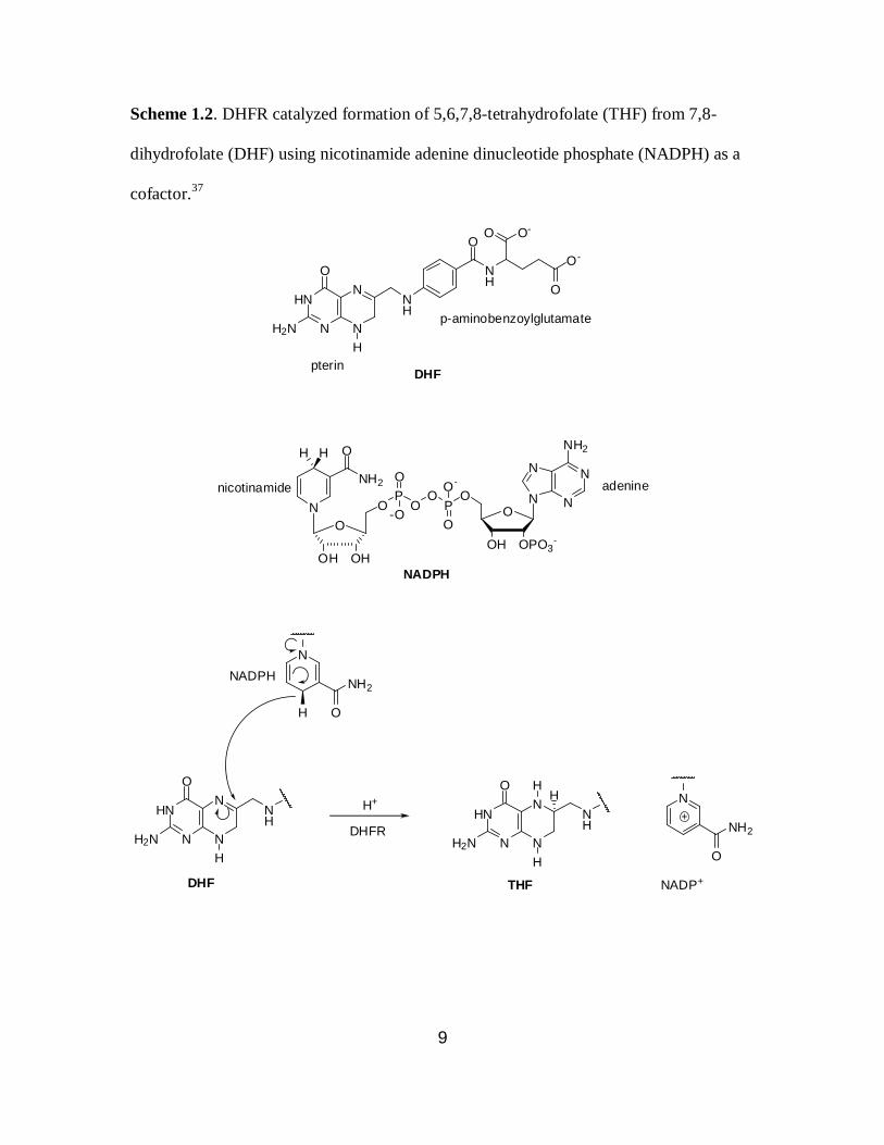
1.2 DHFR catalyzed formation of 5,6,7,8-tetrahydrofolate (THF) from 7,8-
dihydrofolate (DHF) ............................................................................................... 9
2.1 Preparation of N-pentenoyl-α-methyl-β-alanyl-tRNACUA. ..................................... 18
2.2 Preparation of N-pentenoyl-β,β-dimethyl-β-alanyl-tRNACUA ................................ 18
2.3 Preparation of N-pentenoyl-β-phenylalanyl-tRNACUA ........................................... 19
2.4 Preparation of N-pentenoyl-β-(p-bromophenyl)alanyl-tRNACUA ........................... 19
3.1 Proposed mechanism for the chromophore formation in GFP................................ 54
3.2 Preparation of glycylphenylalanyl-tRNACUA ......................................................... 60
3.3 Preparation of tRNACUA activated with the dipeptidomimetic ............................... 60
4.1 Synthesis of topopyrones 4.5 and 4.6. ................................................................... 94
4.2 Synthesis of topopyrones 4.7, 4.8 and 4.11 ........................................................... 95

1
Chapter 1
THE RIBOSOME AND PROTEIN SYNTHESIS: GENERAL INTRODUCTION
1.1. The ribosome
Proteins are one of the most versatile classes of polymers that nature synthesizes
to perform important biological functions. Scientists have long been intrigued by the
structural and functional diversity of proteins. Proteins are comprised of different amino
acid monomers, which are linked by peptide bonds. The cellular machinery which
synthesizes proteins, one amino acid at a time, is called the ribosome.1
The ribosome is made up of a complex array of ribosomal RNAs (rRNAs) and
proteins. It synthesizes proteins with high speed and excellent fidelity using messenger
RNAs (mRNAs) and transfer RNAs (tRNAs).2 The bacterial ribosome is comprised of
50S (large) and 30S (small) ribosomal subunits. The large subunit consists of 23S RNA,
5S RNA and approximately 30 proteins, whereas 16S RNA and 20 proteins together form
the 30S subunit.1 Each ribosomal subunit has three sites for tRNA binding, designated as
the A-site (site for aminoacyl-tRNA), P-site (site for peptidyl-tRNA) and E-site (site for
deacylated tRNA set to exit the ribosome) (Figure 1.1).3 In bacteria, the 30S ribosomal
subunit binds to the mRNA upstream from the start codon, and the 16S ribosomal RNA
base pairs with its complementary Shine–Dalgarno sequence in the bound mRNA.1 In
addition, the 30S ribosomal subunit binds to the anticodon stem-loop of tRNA and
contributes to the fidelity of protein synthesis by monitoring codon-anticodon base
pairing during the decoding process.3 The 50S ribosomal subunit is responsible for
catalyzing peptide bond formation.5 The α-amino moiety of the aminoacyl group of

2
Figure 1.1. Schematic diagram of a ribosome bound to an mRNA showing the A, P and
E-sites.4
tRNA, located in the A-site, mediates a nucleophilic attack on the carbonyl carbon of the
ester bond connecting the nascent peptide to the peptidyl-tRNA (Scheme 1.1a). As a
consequence, the growing polypeptide is transferred onto the tRNA occupying the A-site,
adding a new amino acid to the growing chain (Scheme 1.1b). This event triggers the
hydrolysis of GTP, followed by translocation of the mRNA relative to the ribosome. The
peptidyl-tRNA in the A-site and the deacylated tRNA in the P-site translocate to the P-
site and E-site, respectively (Scheme 1.1c). Following this event a new cycle begins, in
which the next aminoacyl-tRNA binds to mRNA in the A-site and the deacylated tRNA
dissociates from the E-site (Scheme 1.1d).

3
Scheme 1.1. Formation of a peptide bond in the PTC.1
O
O
NH
UUUmRNAAAA
AGAUCU
O
O
H2N
Aminoacyl SitePeptidyl Site
a.
OH
UUUmRNAAAA
AGAUCU
O
Aminoacyl SitePeptidyl Site
b.
O
NH
HN
peptidyltransferase
OH
UUUmRNAAAA
AGAUCU
Peptidyl SiteExit Site
c.
translocation
AUAUAU
Aminoacyl SitePeptidyl Site
AGAmRNAUCU
O
O
H2N
next cycle
dissociation of tRNA fromE-site
binding of new aminoacyl-tRNAat A-site
d.
OH
O
OH
peptide
peptide
O
O
NH
HN
O
OH
peptide
O
O
NH
HN
O
OH
peptide
OH
During mRNA translation, the overall error rates range from 6 x 10⁻⁴
to 5 x 10⁻³
which is a result of the net accumulation of errors in several steps, mainly transcription
(10-4
), aminoacyl-tRNA synthesis (10-4
) and ribosomal decoding (10-4
).6,7
The aminoacyl-
tRNA synthetase (aaRS) activates an amino acid with ATP to form an
aminoacyladenylate, followed by the transfer of the activated amino acid to its cognate
4
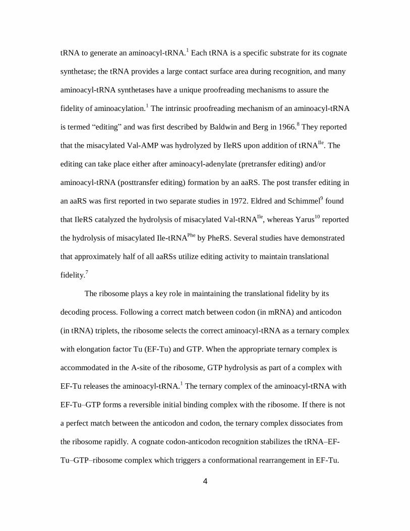
tRNA to generate an aminoacyl-tRNA.1 Each tRNA is a specific substrate for its cognate
synthetase; the tRNA provides a large contact surface area during recognition, and many
aminoacyl-tRNA synthetases have a unique proofreading mechanisms to assure the
fidelity of aminoacylation.1 The intrinsic proofreading mechanism of an aminoacyl-tRNA
is termed “editing” and was first described by Baldwin and Berg in 1966.8 They reported
that the misacylated Val-AMP was hydrolyzed by IleRS upon addition of tRNAIle
. The
editing can take place either after aminoacyl-adenylate (pretransfer editing) and/or
aminoacyl-tRNA (posttransfer editing) formation by an aaRS. The post transfer editing in
an aaRS was first reported in two separate studies in 1972. Eldred and Schimmel9 found
that IleRS catalyzed the hydrolysis of misacylated Val-tRNAIle
, whereas Yarus10
reported
the hydrolysis of misacylated Ile-tRNAPhe
by PheRS. Several studies have demonstrated
that approximately half of all aaRSs utilize editing activity to maintain translational
fidelity.7
The ribosome plays a key role in maintaining the translational fidelity by its
decoding process. Following a correct match between codon (in mRNA) and anticodon
(in tRNA) triplets, the ribosome selects the correct aminoacyl-tRNA as a ternary complex
with elongation factor Tu (EF-Tu) and GTP. When the appropriate ternary complex is
accommodated in the A-site of the ribosome, GTP hydrolysis as part of a complex with
EF-Tu releases the aminoacyl-tRNA.1 The ternary complex of the aminoacyl-tRNA with
EF-Tu–GTP forms a reversible initial binding complex with the ribosome. If there is not
a perfect match between the anticodon and codon, the ternary complex dissociates from
the ribosome rapidly. A cognate codon-anticodon recognition stabilizes the tRNA–EF-
Tu–GTP–ribosome complex which triggers a conformational rearrangement in EF-Tu.

5
This change in conformation of EF-Tu is accompanied by the hydrolysis of GTP to GDP;
the GDP bound EF-Tu loses its affinity for the aminoacyl-tRNA. After the release of the
aminoacyl-tRNA by EF-Tu, the 3'-end of the aminoacyl-tRNA moves into the PTC,
where it participates in peptide bond formation.11
1.2. The ribosome is a ribozyme
The catalytic center of the ribosome resides in the 23S RNA of the 50S ribosomal
subunit and is called the peptidyltransferase center (PTC).3 Enhancement of the rate of
peptide bond formation by the ribosome is at least ~105-fold as compared to uncatalyzed
peptide bond formation between two amino acids.12
Crick, in 1968, had proposed that the
23S rRNA might contain the catalytic activity of the ribosome and thus introduced the
idea of a ribosome being a ribozyme.13
A ribozyme (ribonucleic acid enzyme, also
called RNA enzyme or catalytic RNA) is an RNA molecule that catalyzes a chemical
reaction in a fashion similar to protein enzymes. Many natural ribozymes catalyze the
hydrolysis of one of their own phosphodiester bonds, or those in other RNAs.14
The first
ribozymes were discovered by Sidney Altman15
and Thomas Cech16
in separate studies;
in 1989, both of them received the Nobel prize in chemistry for this work. Following the
discovery of ribozymes, Crick’s hypothesis began to gain interest. Noller and co-workers
prepared large ribosomal subunit particles after increasingly vigorous deproteinization
and showed that the particles retained peptidyltransferase activity.17
In a separate study,
Watanabe and co-workers reported that the in vitro synthesized protein free 23S rRNA
also demonstrated peptidyltransferase activity.18
In 2000, Steitz published the 2.3 Å
atomic resolution structures of a 50S ribosomal subunit from the archeon H. marismortui
and its complexes with two aminoacyl-tRNA mimics.19
In this study, it was established

6
that there are no ribosomal protein side chain atoms in the vicinity of the peptide bond
being formed. They proposed that N3 of A2486 (A2451 in E. coli) participates in general
acid/base catalysis in the peptidyltransferase reaction.20
The availability of X-ray crystal
structures with improved resolution indicated that N3 of A2451 is not near enough to the
α-amino group of the amino acid to enable hydrogen bonding with this functionality.21
Substitution of the α-amino group of the aminoacyl moiety of the aminoacyl-tRNA with a
less reactive hydroxyl group resulted in a reaction rate of depsipeptide bond formation
which was pH independent.22
Subsequently, it was proposed that the 2' -OH group of the
sugar moiety of A76 of the peptidyl-tRNA mediates a concerted proton shuttling during
peptide bond formation.2,23
Weinger et al. substituted the 2'-OH group of A76 with H or F
and observed a ~106 fold reduction in the rate of the peptidyltransferase reaction.
23 In
contrast, Sprinzl et al.24
demonstrated that a peptidyl-tRNA having a 2'-deoxy sugar
moiety in A76 does not make the peptidyltransferase center inactive; instead the
peptidyltransferase reaction still proceeds at a reasonable rate. Therefore, this study
suggested that the 2'-OH group of the 3'-adenosine residue of peptidyl-tRNA may not be
required for the proton shuttling.
It is now established that the ribosome is a ribozyme which catalyzes the
peptidyltransferase reaction, but the role of N3 of A2451and 2'-OH group of the 3'-
adenosine residue of peptidyl-tRNA are still not clear.
1.3. Biosynthesis of modified proteins
The study of protein structure and function has been a central focus for the last
few decades. Engineering proteins to improve function or create novel functionalities has
applications in protein therapeutics,25
bio-imaging,26
protein folding and self-assembly,27
7
and organic synthesis.28
The incorporation of non-canonical amino acids into proteins
represents an important tool for protein engineering.29
The first report of the
incorporation of a non-canonical amino acid into protein was published by Cohen and
Cowie30
in 1957, wherein complete substitution of methionine with selenomethionine in
E. coli auxotrophic for methionine was achieved. Over the past decade, selenomethionine
incorporation into proteins has played an important role in protein crystallography.31
The major strategy for the site-specific incorporation of unnatural amino acids
into proteins has been via the suppression of a nonsense codon in mRNA with a tRNA
bearing an unnatural amino acid (Figure 1.2). Hecht and co-workers developed a key
methodology for preparing misacylated tRNAs, demonstrating that the chemically
synthesized aminoacylated pCpA could be ligated to an abbreviated tRNA using T4 RNA
ligase.32,33
Schultz and co-workers utilized tRNAs bearing unnatural amino acids for the
in vitro translation of full length proteins.34
To produce proteins bearing unnatural amino
acids at specific sites in vivo, orthogonal tRNA/tRNA synthetase pairs for unnatural
amino acids were later developed by the Schultz laboratory.35
For instance, a suppressor
tRNATyr
and a cognate tyrosyl-tRNA synthetase, derived from the archeal bacterium M.
jannasschii, was modified and introduced into E. coli to mediate in vivo incorporation of
O-methyl-L-tyrosine into dihydrofolate reductase (DHFR) in response to a UAG
nonsense codon.35
Such technological advances over the past three decades have
revolutionized protein engineering and provided access to proteins with novel
functions.29
1.4 Dihydrofolate redutase (DHFR)

8
Dihydrofolate reductase (DHFR) is an essential and ubiquitous enzyme required
for normal cellular metabolism in both prokaryotes and eukaryotes. The primary role of
DHFR is maintenance of the intracellular pools of 5,6,7,8-tetrahydrofolate (THF) and its
derivatives, which are required for the biosynthesis of purines, pyrimidines and several
modified protein
(2) deprotection
in vitrotranslation
COH
unnatural amino acid
ACC
unnatural amino acid
AUC
CUA
pET28b(+)-protein(TAG)
T7promoter
invit r
o
transcri
ption
UAGmRNA
TAG
(1) pdCpA-
unnatural amino acid,
T4 RNA ligase S-30 system havingprotein synthesis machinary
abbreviated suppressor tRNA
plasmid bearing a stop codon
Figure 1.2. Strategy for the incorporation of unnatural amino acids into proteins in
vitro.34
amino acids. The enzyme catalyzes the reduction of 7,8-dihydrofolate (DHF) to THF by
transferring the pro-R hydride of nicotinamide adenine dinucleotide phosphate (NADPH)
cofactor stereospecifically to the C6 atom of the pterin nucleus with simultaneous
protonation at N5 (Scheme 1.2).36,37
DHFR is the only source of THF in both prokaryotes
9
Scheme 1.2. DHFR catalyzed formation of 5,6,7,8-tetrahydrofolate (THF) from 7,8-
dihydrofolate (DHF) using nicotinamide adenine dinucleotide phosphate (NADPH) as a
cofactor.37
HN
N N
NNH
NH
OO O-
O-
O
O
H2N
H
pterin
p-aminobenzoylglutamate
DHF
O
OPO3-OH
OP
O
OO-
OP
O
O
-ON
O
OH OH
O
NH2N
NN
N
NH2
NADPH
nicotinamide adenine
HN
N N
NNH
O
H2N
H
H O
NH2
HN
N N
NNH
O
H2N
H
N
O
NH2
HH
NADPH
NADP+
DHFR
H+
DHF THF
N
HH

10
and eukaryotes. Several anticancer and antibiotic drugs target DHFR to block the
conversion of DHF to THF. Methotrexate is one of the most potent chemotherapeutic
drugs and DHFR is the major target of this drug.38
Another compound, trimethoprim, is
an important antibacterial agent as it binds to bacterial DHFR 105 times more strongly
than to vertebrate DHFRs.39
DHFRs from different organisms share low sequence homology but high
structural homology. The first crystal structure of a DHFR enzyme was published in
1978.40
E. coli DHFR is an 18 kD protein consisting of eight β-strands and four flanking
α-helices. It is divided into two structural subdomains: the adenosine binding subdomain
and the major subdomain. In E. coli DHFR, the adenosine moiety binds to the smaller
adenosine binding domain which is formed by the amino acid residues 38-88. Close to
100 amino acid residues from both the N and C termini make up the major subdomain.
This subdomain is dominated by three loops around the active site, which comprise 40%
to 50% of the major domain. The loops are defined as ‘Met20’ (residues 9-24), ‘F-G’
(residues 116-132) and ‘G-H’ (residues 142-150). The Met20 loop is present directly
above the active site, protecting it from solvent. The F-G and G-H loops play important
roles in stabilizing the Met20 loop via a network of hydrogen bonding interactions.
Benkovic and coworkers have reported the complete catalytic scheme for E. coli
DHFR (Figure 1.3).41
The most intriguing aspect of the DHFR catalytic mechanism is
that after the hydride transfer, product release does not follow directly. Instead, NADP+
is
first released followed by rebinding of NADPH, which is the rate-determining step. After
binding of NADPH, the product is released, leaving the enzyme primed for the next
round of catalysis. There is a synergistic interaction between the substrate and cofactor

11
binding sites, where the rate of release of NADP+ is increased in the presence of the
bound product and the rate of the release of the product is elevated in presence of bound
NADPH.
E:DHF:NADPH E:THF:NADP+
E:THFE:THF:NADPHE:NADPH
NADP+
NADPHTHF
DHF
220 s-1
0.03 s-1
200 s-15 uM-1 s-1
85 s-1
8 uM-1 s-1
2 uM-1 s-1
12.5 s-1
40 s-140 uM-1 s-1
Figure 1.3. The catalytic cycle of E. coli DHFR at pH 7.41
1.5. Modified ribosomes
Studying ribosome structure and function by mutagenesis at critical nucleotide
residues has been carried out for some time, yet studies of ribosome modifications to
enable incorporation of amino acids other than α-L-amino acids are non-existent.2,42
The
structural recognition of an aminoacyl-tRNA by the ribosome is crucial for the
incorporation of unnatural amino acids into proteins.43
Puromycin (1.1) is
an aminonucleoside antibiotic (Figure 1.4), whose structure resembles the 3'-end of the
aminoacyl-tRNA, rendering it a putative aminoacyl-tRNA mimic.44,45
It binds to the A-
site of the PTC and takes part in peptide bond formation with the nascent polypeptide
attached to the peptidyl-tRNA in the P-site, which leads to premature chain release.44,45
Puromycin and its derivatives having different amino acid side chains show different
12
ribosomal inhibition efficiencies.46,47
This suggests that the ribosomal A-site shows
specificity towards binding the different side chains of amino acids, at least at the level of
puromycin.
Hecht and co-workers have successfully demonstrated that the modifications at
multiple sites in the 23S rRNA can dramatically change the architecture of the ribosome
while still preserving essential ribosomal functions.48-51
The first report of modified
ribosomes able to utilize α-D-amino acids was published by Hecht and co-workers in
2003.48
Modifications in the PTC and helix 89 allowed the modified ribosomes to
incorporate α-D-methionine and α-D-phenylalanine into E. coli DHFR in moderate
yields.48,49
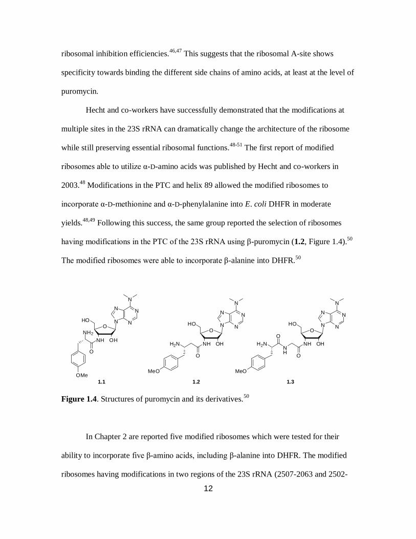
Following this success, the same group reported the selection of ribosomes
having modifications in the PTC of the 23S rRNA using β-puromycin (1.2, Figure 1.4).50
The modified ribosomes were able to incorporate β-alanine into DHFR.50
N
NN
N
N
O
OHNH
HO
O
NH2
OMe
1.1
N
NN
N
N
O
OHNH
HO
O
1.2
H2N
MeO
N
NN
N
N
O
OHNH
HO
O
NH
O
H2N
MeO
1.3
Figure 1.4. Structures of puromycin and its derivatives.50
In Chapter 2 are reported five modified ribosomes which were tested for their
ability to incorporate five β-amino acids, including β-alanine into DHFR. The modified
ribosomes having modifications in two regions of the 23S rRNA (2507-2063 and 2502-
13
2507 or 2496-2501) were selected using β-puromycin (1.2). Similar selection
experiments, which were done using the puromycin derivative 1.3 (Figure 1.4), led to the
selection of six modified ribosomes enabling the incorporation of a dipeptide and
dipeptidomimetic into DHFR (Chapter 3). These modified ribosomes also had
modifications in two regions of the 23S rRNA (2507-2063 and 2502-2507).

14
Chapter 2
INCORPORATION OF β-AMINO ACIDS INTO E. COLI DHFR USING RIBOSOMES
HAVING MODIFICATIONS IN THE PEPTIDYLTRANSFERASE CENTER (PTC)
2.1. Introduction
β-Amino acids are not found in ribosomally synthesized proteins or enzymes even
though they are present in cells.52
Synthetic peptide analogues having one or more β-
amino acids have been shown to have helical properties, some of which are very similar
to the overall structures of peptides formed from α-amino acids.53-56
Additionally, such
peptide analogues having β-amino acids often show enhanced resistance to proteolysis,
which make them ideal candidates to be included in the growing list of unnatural amino
acids that can be incorporated into proteins.57-60
The incorporation of β-amino acids into
proteins or large peptides can facilitate the creation of artificial protein-like
macromolecules with interesting properties. At present, all peptide analogues having β-
amino acids are prepared by synthetic or semi-synthetic methods which limits the size of
peptide analogues accessible and thus their use. The largest β-mimetic peptide reported to
date is only 17 amino acid long.59
The strong bias of the ribosome against β-amino acids
has limited their incorporation into proteins using in vitro or in vivo protein synthesis
methods.61,62
As discussed in the previous chapter, puromycin is a small molecule mimic of the
3'-end of aminoacyl-tRNA (Figure 2.1) and is a universal translation inhibitor. It binds at
the ribosomal A-site and readily accepts the peptide chain from the peptidyl-tRNA in the
P-site, thereby terminating protein synthesis. Analogues of puromycin, in which the O-
methyl-α-L-tyrosine moiety was replaced by either α-D-amino acids (D-alanine and 4-
15
methyl-D-phenylalanine) or β-amino acids (β-alanine and 4-methyl-β-L-phenylalanine),
were shown to inhibit protein translation weakly as compared to puromycin.47
This
suggests that the ribosome can recognize β-aminoacyl- and α-D-aminoacyl-tRNA
mimics.
N
NN
N
N
O
OHNH
HO
O
NH2
OMe
1.1
N
NN
N
N
O
OHNH
HO
O
1.2
H2N
MeO
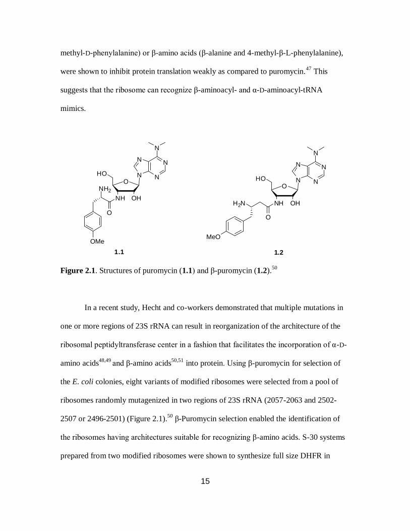
Figure 2.1. Structures of puromycin (1.1) and β-puromycin (1.2).50
In a recent study, Hecht and co-workers demonstrated that multiple mutations in
one or more regions of 23S rRNA can result in reorganization of the architecture of the
ribosomal peptidyltransferase center in a fashion that facilitates the incorporation of α-D-
amino acids48,49
and β-amino acids50,51
into protein. Using β-puromycin for selection of
the E. coli colonies, eight variants of modified ribosomes were selected from a pool of
ribosomes randomly mutagenized in two regions of 23S rRNA (2057-2063 and 2502-
2507 or 2496-2501) (Figure 2.1).50
β-Puromycin selection enabled the identification of
the ribosomes having architectures suitable for recognizing β-amino acids. S-30 systems
prepared from two modified ribosomes were shown to synthesize full size DHFR in

16
presence of β-alanyl-tRNACUA and DHFR mRNA with UAG codons.50
Based on UAG
codon suppression to afford full length protein, it was demonstrated that β-alanine was
incorporated into protein in moderate yields, but no direct evidence of β-alanine
incorporation into DHFR was reported. Wild-type DHFR produced using DHFR mRNA
and the modified ribosomes retained good activity, indicating that the ribosomes retained
their ability to discriminate individual α-amino acids accurately.
In this thesis, five β-amino acids (Figure 2.2), including β-alanine, were chosen
for their incorporation into DHFR. Five different types of modified ribosomes were
selected to facilitate their incorporation into DHFR. Two modified DHFRs were selected
for a larger scale synthesis and characterization by MALDI mass spectrometry after “in-
gel” trypsin digestion.
H2N
O
OHH2N OH
O
H2N OH
OH2N
O
OHH2N
O
OH
Br2.1 2.2 2.3
2.42.5
Figure 2.2. Structures of the β-amino acids studied.51
2.2. Results
Five β-amino acids were selected either with no side chains (2.1) or having
different side chains (2.2-2.5) at the α- or β-positions (Figure 2.2). β-Alanyl-tRNACUA50
was provided by Dr. Larisa Dedkova and N-pentenoyl-α-methyl-β-alanyl-pdCpA51
was
synthesized by Sandipan Roy Chowdhury. Additionally, three N-pentenoyl-β-aminoacyl-
pdCpA derivatives and four β-aminoacyl-tRNACUAs were prepared. Five different types
17
of modified ribosomes,50
selected by Dr. Larisa Dedkova, were used for S-30
preparations.
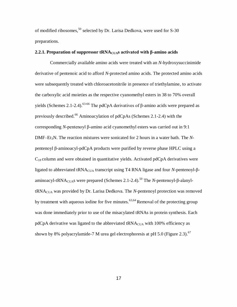
2.2.1. Preparation of suppressor tRNACUAs activated with β-amino acids
Commercially available amino acids were treated with an N-hydroxysuccinimide
derivative of pentenoic acid to afford N-protected amino acids. The protected amino acids
were subsequently treated with chloroacetonitrile in presence of triethylamine, to activate
the carboxylic acid moieties as the respective cyanomethyl esters in 38 to 70% overall
yields (Schemes 2.1-2.4).63-66
The pdCpA derivatives of β-amino acids were prepared as
previously described.66
Aminoacylation of pdCpAs (Schemes 2.1-2.4) with the
corresponding N-pentenoyl β-amino acid cyanomethyl esters was carried out in 9:1
DMF–Et3N. The reaction mixtures were sonicated for 2 hours in a water bath. The N-
pentenoyl β-aminoacyl-pdCpA products were purified by reverse phase HPLC using a
C18 column and were obtained in quantitative yields. Activated pdCpA derivatives were
ligated to abbreviated tRNACUA transcript using T4 RNA ligase and four N-pentenoyl-β-
aminoacyl-tRNACUAs were prepared (Schemes 2.1-2.4).50
The N-pentenoyl-β-alanyl-
tRNACUA was provided by Dr. Larisa Dedkova. The N-pentenoyl protection was removed
by treatment with aqueous iodine for five minutes.63,64
Removal of the protecting group
was done immediately prior to use of the misacylated tRNAs in protein synthesis. Each
pdCpA derivative was ligated to the abbreviated tRNACUA with 100% efficiency as
shown by 8% polyacrylamide-7 M urea gel electrophoresis at pH 5.0 (Figure 2.3).67
18
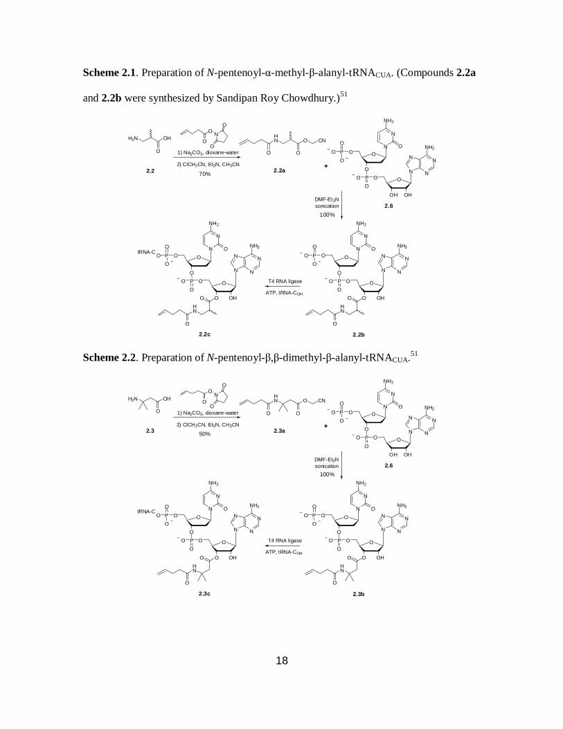
Scheme 2.1. Preparation of N-pentenoyl-α-methyl-β-alanyl-tRNACUA. (Compounds 2.2a
and 2.2b were synthesized by Sandipan Roy Chowdhury.)51
O
O
N
O
O
2) ClCH2CN, Et3N, CH3CN
1) Na2CO3, dioxane-water
N
NN
N
NH2
O
OHOH
OPO
O
O
N
N
NH2
O
O
OPO
O
O
+
N
NN
N
NH2
O
OHO
OPO
O
O
N
N
NH2
O
O
OPO
O
O
O
HN
O
N
NN
N
NH2
O
OHO
OPO
O
O
N
N
NH2
O
O
OPO
O
O
O
HN
O
tRNA-C
T4 RNA ligase
ATP, tRNA-COH
2.2
2.6
2.2c
DMF-Et3N
sonication
2.2b
OH
O
HN O
OO
CNH2N
2.2a70%
100%
Scheme 2.2. Preparation of N-pentenoyl-β,β-dimethyl-β-alanyl-tRNACUA.51
O
O
N
O
O
2) ClCH2CN, Et3N, CH3CN
1) Na2CO3, dioxane-water
N
NN
N
NH2
O
OHOH
OPO
O
O
N
N
NH2
O
O
OPO
O
O
+
N
NN
N
NH2
O
OHO
OPO
O
O
N
N
NH2
O
O
OPO
O
O
O
HN
O
N
NN
N
NH2
O
OHO
OPO
O
O
N
N
NH2
O
O
OPO
O
O
O
HN
O
tRNA-C
T4 RNA ligase
ATP, tRNA-COH
2.3
2.6
2.3c
DMF-Et3N
sonication
2.3b
OH
O
HN O
OO
CNH2N
2.3a50%
100%

19
Scheme 2.3. Preparation of N-pentenoyl-β-phenylalanyl-tRNACUA.51
O
O
N
O
O
2) ClCH2CN, Et3N, CH3CN
1) Na2CO3, dioxane-water
H2N
N
NN
N
NH2
O
OHOH
OPO
O
O
N
N
NH2
O
O
OPO
O
O
+
N
NN
N
NH2
O
OHO
OPO
O
O
N
N
NH2
O
O
OPO
O
O
O
HN
O
N
NN
N
NH2
O
OHO
OPO
O
O
N
N
NH2
O
O
OPO
O
O
O
HN
O
tRNA-C
T4 RNA ligase
ATP, tRNA-COH
2.4 2.4a
2.6
2.4c
sonication
2.4b
OH
O
HN O
OO
CN
40%
DMF-Et3N
100%
Scheme 2.4. Preparation of N-pentenoyl-β-(p-bromophenyl)alanyl-tRNACUA.51
O
O
N
O
O
2) ClCH2CN, Et3N, CH3CN
1) Na2CO3, dioxane-water
H2N
N
NN
N
NH2
O
OHOH
OPO
O
O
N
N
NH2
O
O
OPO
O
O
+
N
NN
N
NH2
O
OHO
OPO
O
O
N
N
NH2
O
O
OPO
O
O
O
HN
O
Br
N
NN
N
NH2
O
OHO
OPO
O
O
N
N
NH2
O
O
OPO
O
O
O
HN
O
Br
tRNA-C
T4 RNA ligase
ATP, tRNA-COH
2.52.5a
2.6
2.5c
DMF-Et3N
sonication
2.5b
OH
Br
O
HN O
Br
OO
CN
38%
100%

20
1 2 3 4 5
Figure 2.3. Preparation of β-aminoacyl-tRNACUAs. Lane 1, α-methyl-β-alanyl-tRNACUA;
lane 2, β,β-dimethyl-β-alanyl-tRNACUA; lane 3, β-phenylalanyl-tRNACUA; lane 4,
nonacylated abbreviated tRNACUA; lane 5, β-(p-bromophenyl)alanyl-tRNACUA.
2.2.2. In vitro translation using β-amino acids with wild-type ribosomes
As discussed previously in Section 2.1, wild-type ribosomes cannot mediate the
incorporation of β-amino acids into proteins. Therefore, it seemed logical to verify this
experimentally by the use of wild-type E. coli ribosomes. The ability of all five β-
aminoacyl-tRNACUAs to suppress a UAG codon at position 18 or 49 of DHFR mRNA
was compared with α-phenylalanyl-tRNACUA and α-threonyl-tRNACUA using the S-30
system prepared from the wild-type ribosome (Figures 2.4 and 2.5).
For position 49 of DHFR, the suppression efficiencies, relative to DHFR
synthesized from the wild-type gene, were 40% and 50% for α-phenylalanyl-tRNACUA
and α-threonyl-tRNACUA, respectively. As illustrated in Figure 2.4, the suppression
efficiencies for all five β-aminoacyl-tRNAs (0.4%-2.2%) were much lower than for α-
phenylalanyl-tRNACUA and α-threonyl-tRNACUA. Similar results were obtained when
position 18 of DHFR was evaluated (Figure 2.5). The incorporation yields of β-amino
acids into position 18 of DHFR were under 6% as compared to the wild-type DHFR
synthesis. In the same experiment, high levels of full length DHFR were obtained in
presence of α-phenylalanyl-tRNACUA (80%) and α-threonyl-tRNACUA (90%). These

21
results demonstrate that the wild-type ribosome cannot efficiently incorporate β-amino
acids into proteins.
1 2 3 4 5 6 7 8 9
100 0.7 40 0.5 50 0.4 2.2 0.5 0.7 % suppression
Figure 2.4. Translation of DHFR from wild-type (lane 1) and modified (lanes 2-9) (UAG
codon in position 49) mRNA in the presence of different suppressor tRNACUAs. Lane 2,
nonacylated tRNACUA; lane 3, tRNACUA activated with phenylalanine; lane 4, tRNACUA
activated with β-phenylalanine (2.4); lane 5, tRNACUA activated with threonine; lane 6,
tRNACUA activated with β-(p-bromophenyl)alanine (2.5); lane 7, tRNACUA activated with
β,β-dimethyl-β-alanine (2.3); lane 8, tRNACUA activated with α-methyl-β-alanine (2.2);
lane 9, tRNACUA activated with β-alanine (2.1). The suppression efficiency relative to
wild type is shown below each lane.51
1 2 3 4 5 6 7 8 9
100 80 3 4 90 3 6 2 2 % suppression
Figure 2.5. Translation of DHFR from wild-type (lane 1) and modified (lanes 2-9) (UAG
codon in position 18) mRNA in the presence of different suppressor tRNACUAs. Lane
2,tRNACUA activated with phenylalanine ; lane 3, nonacylated tRNACUA; lane 4,
← full length DHFR ← truncated at
position 48
← full length DHFR

22
tRNACUA activated with β-phenylalanine (2.4); lane 5, tRNACUA activated with
threonine;lane 6, tRNACUA activated with β-(p-bromophenyl)alanine (2.5); lane 7,
tRNACUA activated with β,β-dimethyl-β-alanine (2.3); lane 8, tRNACUA activated with α-
methyl-β-alanine (2.2); lane 9, tRNACUA activated with β-alanine (2.1). The suppression
efficiency relative to wild type is shown below each lane.51
2.2.3. In vitro translation using β-amino acids with modified ribosomes
To study the incorporation of β-amino acids into DHFR with modified ribosomes,
five S-30 systems were prepared from E. coli colonies harboring different modified
ribosomes. Their ability to individually synthesize full length DHFR in presence of β-
aminoacyl-tRNACUA and DHFR mRNA transcripts having a UAG codon was measured.
The nucleotide sequence of the ribosomes in the modified regions is compared to the
wild-type ribosome in Table 2.1. S-30 systems prepared from the modified ribosomes
produced different amounts of wild-type DHFR as compared to the wild-type ribosomal
system. Therefore, the suppression efficiencies were expressed relative to the suppression
obtained using α-threonyl-tRNA (Table 2.2) or α-phenylalanyl-tRNA (Table 2.3). As a
negative control, DHFR synthesis in the presence of nonacylated-tRNACUA was
evaluated. The amounts of DHFR produced by each system were quantified with a
phosphoimager, which monitored the incorporation of 35
S-methionine into DHFR.
Table 2.2 summarizes the incorporation yields of β-amino acids 2.1, 2.2 and 2.3
into DHFR at position 10, relative to α-threonine incorporation. Figure 2.6 illustrates the
synthesis of full length DHFR in presence of β-aminoacyl-tRNACUAs bearing β-amino

23
Table 2.1. 23S rRNA sequence modifications in the PTC for the clones used for S-30
preparations.51
Clone Sequence in 23S rRNA of modified ribosome
Region 1 Region 2
0403x2 2057AGCGUGA2063 2502UUACCG2507
0403x4 2057AGCGUGA2063 2502AGCCAG2507
040321 2057AGCGUGA2063 2502AGAUAA2507
040329 2057AGCGUGA2063 2502UGGCAG2507
040217 2057AGCGUGA2063 2496AUAGUU2501
Wild type 2057GAAAGAC2063 2496CACCUC2501
2502GAUGUC2507
acids 2.1, 2.2 and 2.3 using the S-30 system prepared from clone 040329. A racemic
mixture of 2.2 was deliberately employed for this study as to see any effect from either of
the isomers. The individual β-amino acids were incorporated with slight variations in
suppression yields (~2-fold) with the five S-30 preparations having modified ribosomes.
Among the three β-amino acids examined for the incorporation into positon 10 of DHFR,
the best results were obtained for 2.3 (~11% suppression yield) with S-30 systems
prepared from clones 040329 and 040217. Figure 2.7 illustrates the synthesis of full
length DHFR in presence of β-aminoacyl-tRNACUAs bearing β-amino acids 2.1 and 2.3
using the S-30 system prepared from clone 040217. All five S-30 preparations produced
very low levels of non-specific readthrough of the UAG codon in the absence of any
activated suppressor tRNA.
Table 2.3 summarizes the incorporation yields of β-amino acids 2.2, 2.4 and 2.5
into DHFR at position 18, relative to α-phenylalanine incorporation. These experiments
were done in a fashion similar to the experiments described above. The S-30 preparations

24
from clones 040329 and 0403x4 produced the best results for the three amino acids
tested.
Table 2.2. Incorporation of β-amino acids 2.1, 2.2 and 2.3 into position 10 of E. coli
DHFR by the use of S-30 systems having different modified ribosomes.51
Amino acids Suppression efficiency in different S-30 systems, having
modified ribosomes (%)a
040329 0403x2 0403x4 040321 040217
Thr 100 100 100 100 100
– 0.5 ± 0.3 2.1 ± 1.4 1.4 ± 0.7 0.9 ± 0.2 2.2 ± 1.0
2.1 8.2 ± 3.0 6.8 ± 3.2 8.1 ± 3.4 5.4 ± 1.3 9.9 ± 3.9
2.2 8.8 ± 1.0 3.8 ± 1.0 5.4 ± 1.9 not tested not tested
2.3 11b 7.9 ± 1.1 7.1 ± 3.6 5.6 ± 3.0 10.9 ± 4.4
a Each number represents the average of three independent experiments ± SD.
b tested in a single experiment.
1 2 3 4 5
1 100 5 6 11
% suppression
Figure 2.6. Translation of DHFR from a modified DHFR mRNA (UAG codon in
position 10) by the use of an S-30 system prepared from clone 040329 in the presence of
different suppressor tRNAs. Lane 1, nonacylated tRNACUA; lane 2, threonyl-tRNACUA;
lane 3, tRNACUA activated with α-methyl-β-alanine (2.2); lane 4, tRNACUA activated with
β-alanine (2.1); lane 5, tRNACUA activated with β,β-dimethyl-β-alanine (2.3).51
←full length DHFR

25
1 2 3 4
0.8 100 9.1 9.9
% suppression
Figure 2.7. Translation of DHFR from a modified DHFR mRNA (UAG codon in
position 10) by the use of an S-30 system prepared from clone 040217 in the presence of
different suppressor tRNAs. Lane 1, nonacylated tRNACUA; lane 2, threonyl-tRNACUA;
lane 3, tRNACUA activated with β-alanine (2.1); lane 4, tRNACUA activated with β,β-
dimethyl-β-alanine (2.3).51
Table 2.3. Incorporation of β-alanine analogues 2.2, 2.4 and 2.5 into position 18 of E.
coli DHFR by the use of S-30 systems having different modifications in the PTC.51
Amino acids Suppression efficiency in different S-30 systems, having
modified ribosomes (%)a
040329 0403x2 0403x4 040321 040217
Phe 100 100 100 100 100
– 1.5 ± 0.7 1.2 ± 0.4 2.4 ± 1.0 0.8 ± 0.4 1.0 ± 0.4
2.2 6.0 ± 0.5 1.5 ± 1.0 6.0 ± 1.8 1.3 ± 0.7 0.73 ± 0.2
2.4 18.4 ± 2.7 5.8 ± 1.3 13.2 ± 1.1 6.0 ± 1.8 7.8 ± 0.1
2.5 13.5 ± 1.5 5.3 ± 0.9 15.5 ± 2.0 7.6 ± 2.7 9.2 ± 0.4 aEach number represents the average of three independent experiments ± SD.
Figures 2.8 and 2.9 illustrate the incorporation of β-amino acids 2.2, 2.4 and 2.5
into DHFR at position 18 using the S-30 systems prepared from clones 040329 and
0403x4, respectively. The highest yield of full length DHFR (~18%) was obtained by the
combination of β-amino acid 2.4 and the S-30 system prepared from clone 040329. The
←full length DHFR

26
incorporation of 2.4 into DHFR gave yields between ~5.8% to ~18%, whereas 2.5
produced full length DHFR in yields ranging from ~5.3% to ~15%.
S-30 systems prepared from clones 0403x2 and 040321 did not perform as well as
the other three systems for the incorporation of β-amino acids into positions 10 or 18 of
DHFR. For position 10 of DHFR, S-30 systems prepared from clones 0403x2 and
040321 gave best results with β-amino acid 2.3; ~7.9% and ~5.6% suppression yields,
respectively (Table 2.2). In contrast, clone 040321 performed better (~7.6% suppression
yield with 2.5) than clone 0403x2 (~5.8% suppression yield with 2.4) for position 18 of
DHFR. As summarized in Table 2.3, it is evident that the five modified ribosomes did not
incorporate 2.2 efficiently into position 18 of DHFR as compared to β-amino acids 2.4
and 2.5. The suppression yields for β-amino acid 2.2 varied from ~0.7% (for clone
040217) to ~6% (for clone 040329) for position 18. For position 10, S-30 systems
prepared from three clones (040329, 0403x2 and 0403x4) were tested for the
incorporation of 2.2 into DHFR. The highest amount of full length DHFR having β-
amino acid 2.2 was produced by the S-30 system having clone 040329 (~8.8%
suppression yield, Table 2.2).
1 2 3 4 5
0.5 100 19.2 9.6 5.5
% suppression
Figure 2.8. Translation of DHFR from a modified DHFR mRNA (UAG codon in
position 18) by the use of an S-30 system prepared from clone 040329 in the presence of
←full length DHFR
27
different suppressor tRNAs. Lane 1, nonacylated tRNACUA; lane 2, phenylalanyl-
tRNACUA; lane 3, tRNACUA activated with β-phenylalanine (2.4); lane 4, tRNACUA
activated with β-(p-bromophenyl)-alanine (2.5); lane 5, tRNACUA activated with α-
methyl-β-alanine (2.2).51
1 2 3 4 5
100 4.5 13.2 16.4 7.5 % suppression
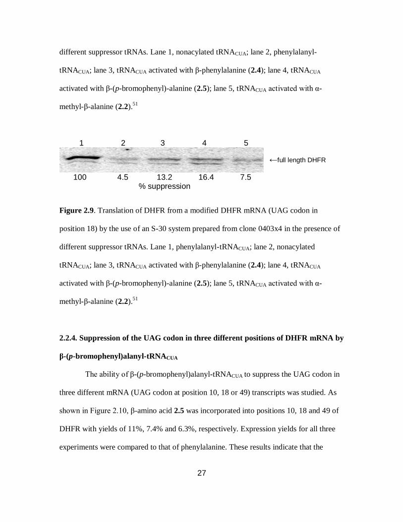
Figure 2.9. Translation of DHFR from a modified DHFR mRNA (UAG codon in
position 18) by the use of an S-30 system prepared from clone 0403x4 in the presence of
different suppressor tRNAs. Lane 1, phenylalanyl-tRNACUA; lane 2, nonacylated
tRNACUA; lane 3, tRNACUA activated with β-phenylalanine (2.4); lane 4, tRNACUA
activated with β-(p-bromophenyl)-alanine (2.5); lane 5, tRNACUA activated with α-
methyl-β-alanine (2.2).51
2.2.4. Suppression of the UAG codon in three different positions of DHFR mRNA by
β-(p-bromophenyl)alanyl-tRNACUA
The ability of β-(p-bromophenyl)alanyl-tRNACUA to suppress the UAG codon in
three different mRNA (UAG codon at position 10, 18 or 49) transcripts was studied. As
shown in Figure 2.10, β-amino acid 2.5 was incorporated into positions 10, 18 and 49 of
DHFR with yields of 11%, 7.4% and 6.3%, respectively. Expression yields for all three
experiments were compared to that of phenylalanine. These results indicate that the
←full length DHFR

28
modified ribosomes can incorporate β-amino acids at different positions in a protein with
similar efficiencies in an in vitro translation system.
1 2 3 4 5 6 7 8 9
100 11 0.01 100 7.4 1.0 100 6.3 1.1 % suppression
Figure 2.10. Relative suppression efficiencies of β-(p-bromophenyl)alanyl-tRNACUA into
three different positions (10, 18 and 49) of DHFR, using an S-30 system prepared from
clone 040217. Lanes 1, 4 and 7, L-phenylalanyl-tRNACUA; lanes 2, 5 and 8, β-(p-
bromophenyl)alanyl-tRNACUA; lanes 3, 6 and 9, unacylated tRNACUA. The suppression
efficiency relative to L-phenylalanine is shown below each lane. Lanes 1-3, modification
of position 10 in DHFR; lanes 4-6, modification of position 18 in DHFR; lanes 7-9,
modification of position 49 in DHFR.51
2.2.5. Characterization of β-amino acids introduced into DHFRs by MALDI mass
spectrometry analysis of the peptide fragments resulting from “in-gel” trypsin
digestion
Two modified DHFRs were synthesized on a larger scale for characterization by
MALDI-MS to provide a direct evidence of the incorporation of β-amino acids into
DHFR. The plasmid pETD49 and β-(p-bromophenyl)alanyl-tRNACUA were used to obtain
DHFR 1 modified at position 49 with β-amino acid 2.5. Similarly, pETD18 and β,β-
dimethyl-β-alanyl-tRNACUA were used to obtain DHFR 2 modified at position 18 with β-
←full length DHFR
←truncated at
position 48

29
amino acid 2.3. Three stages of purification were performed on both the modified DHFRs
including Ni-NTA chromatography, DEAE-Sephadex chromatography and SDS-
polyacrylamide gel electrophoresis. A previously reported “in-gel” trypsin digestion
protocol was used.68
Samples of the wild-type and modified DHFRs were treated with
trypsin to obtain peptide fragments which were subjected to MALDI-MS analysis. Table
2.4 summarizes the calculated and observed (in the MALDI mass spectrum) molecular
weights of the peptide fragments obtained from trypsin digestion of the wild-type and
Table 2.4. MALDI-MS analysis of tryptic digests of wild-type and modified DHFR
samples.51
Position Peptide sequence MALDI-MS analysis, molecular mass, Da
Wild-type Modified
DHFR 1
Modified
DHFR 2 Est MS Est MS Est MS
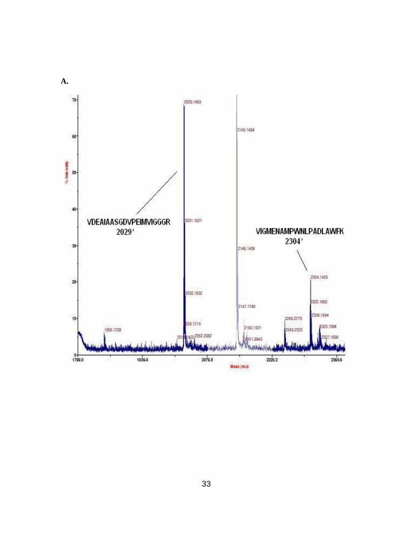
13-32 VIGMENAMPWNLPADLAWFK 2304 2304.1 2304 2303.6 2289 2289.9
34-44 NTLNKPVIMGR 1242 1242.8 1242 1242.6 1242 1242.8
45-57 HTWESIGRPLPGR 1506 1505.8 1644 1645.6 1506 1505.9
59-71 NIILSSQPGTDDR 1415 1415.8 1415 1415.6 1415 1415.8
72-76 VTWVK 632 632.5 632 634.3 632 632.4
77-98 VDEAIAASGDVPEIMVIGGGR 2028 2029.1 2028 2029.7 2028 2030.7
99-106 VYEQFLPK 1023 1023.7 1023 1023.5 1023 1023.6
Modified DHFR 1: β-alanine analogue 2.5 in position 49; modified DHFR 2: β-alanine
analogue 2.3 in position 18. Positions 18 and 49 are indicated in red. The values in bold
face reflect the presence of the respective β-amino acid.

30
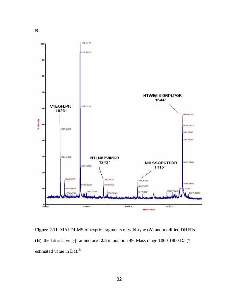
two modified DHFRs. For DHFR 1, the tryptic peptide encompassing amino acids 45-57
having β-amino acid 2.5 at position 49, was anticipated to have a molecular mass of 1644
Da. An ion peak was observed at m/z 1645.5 displaying the characteristic bromine
isotope pattern. This confirmed the presence of the anticipated peptide fragment (Figure
2.11B). Similarly, DHFR 2 produced an ion peak corresponding to m/z 2289.9, which
corresponds to peptide fragment (amino acids 13-32) having β-amino acid 2.3 at position
18 (Figure 2.12B). MALDI-MS analysis of the tryptic digest of wild-type DHFR did not
show ion peaks corresponding to m/z 1644 and 2289.9. The anticipated ion peaks for the
wild-type peptide fragments encompassing amino acids 45-57 with Ser49 at m/z 1506
(Figure 2.11A) or 13-32 with Asn18 at m/z 2304 (Figure 2.12A) were present. As listed
in Table 2.4, the remaining six peptide fragments obtained from wild-type DHFR
matched with those from the modified DHFRs 1 and 2. In replicate experiments, the N-
terminal fragment (containing a hexahistidine fusion peptide) and the (large) C-terminal
fragment were never observed.

31
A.
32
B.
Figure 2.11. MALDI-MS of tryptic fragments of wild-type (A) and modified DHFRs
(B), the latter having β-amino acid 2.5 in position 49. Mass range 1000-1800 Da (* =
estimated value in Da).51
33
A.

34
B.
Figure 2.12. MALDI-MS of tryptic fragments of wild-type (A) and modified DHFRs
(B), the latter having β-amino acid 2.3 in position 18. Mass range 1800-2500 Da. (* =
estimated value in Da).51

35
2.3. Discussion
In this thesis, four novel β-aminoacyl-tRNACUAs were prepared and the ability of
five β-aminoacyl-tRNACUAs to suppress the UAG codon in DHFR mRNA transcripts via
S-30 preparations from five different modified ribosomes (Table 2.1) was studied. The
preparations of the aminoacylated suppressor tRNAs involved four reactions starting
from commercially available β-amino acids. The protection by the amino group as N-
pentenoyl, followed by the activation of the carboxylic acid moiety as the cyanomethyl
ester, afforded the desired products in moderate yields (Schemes 2.1-2.4).
Coupling of the activated β-amino acids with pdCpA (2.6) gave the corresponding
pdCpA derivatives, activated with β-amino acids at either the 2'- or 3'-position, in
quantitative yields after reverse phase HPLC purification (Schemes 2.1-2.4).
Subsequently, all four β-aminoacyl-pdCpA derivatives were ligated to a 74-nucleotide
abbreviated tRNACUA by the use of T4 RNA ligase. Full length suppressor tRNACUAs
activated with the β-amino acids were obtained in quantitative yields as shown by
analysis on a 8% polyacrylamide-7 M urea gel electrophoresis at pH 5.0 (Figure 2.3).
Part of the 23S rRNA (nucleotides 2058 to 2609)42
forms the peptidyl transferase
center of the ribosome. Using β-puromycin for selection of colonies, five variants of
modified ribosomes were selected from a random pool of ribosomes having
modifications in two regions of 23S rRNA (2057-2063 and 2502-2507 or 2496-2501).
The selection experiments were carried out by Dr. Larisa Dedkova.50
The S-30
preparation protocols followed were as reported earlier.50
S-30 systems prepared from
five modified ribosomal clones were tested for their expression of the wild-type DHFR
relative to an S-30 system prepared from the wild-type ribosome. The expression yields

36
of DHFR from the wild-type gene using S-30 systems having different modified
ribosomes ranged from 22% to 77% relative to the S-30 system prepared from the wild-
type ribosome. Therefore, the suppression efficiencies of β-aminoacyl-tRNACUAs in
producing full length DHFR were compared to α-threonyl-tRNACUA (Table 2.2) or α-
phenylalanyl-tRNACUA (Table 2.3), also using the same modified ribosomes.
Three β-amino acids (2.3, 2.4 and 2.5, Figure 2.2) were chosen to study the effect
of different side chains at the β-position on incorporation yields. Amino acid 2.2 was
chosen to study the effect of substitution at α-position. A racemic mixture of 2.2 was
deliberately employed for this study to assure that any effect from either of the isomers
would be observed. The incorporation yields of β-amino acids into DHFR increased from
~10% to ~19% with increasing hydrophobic character of the side chains. The
incorporation of β-(p-bromophenyl)alanine into DHFR was slightly higher with three of
the five S-30 systems tested. The S-30 preparations from clones 040329 and 0403x2
incorporated β-phenylalanine better than its bromo derivative. In general, β-amino acid
2.2, having a methyl group at α-position, produced low levels of full length DHFR. The
selection procedure utilized β-puromycin in which the side chain of the amino acid
moiety is at the β-position. Therefore, it is perhaps not surprising that the selected S-30
systems incorporated β-amino acid 2.2 into DHFR less effectively than they incorporated
the other β-amino acids studied (Tables 2.2 and 2.3).
The variants of the ribosomes tested were modified in two regions of 23S rRNA,
nucleotides 2057-2063 (region 1) and nucleotides 2502-2507 or 2496-2501 (region 2).
All the clones had same sequence in region 1 but a variable sequence in region 2. The
wild-type sequence 2057GAAAGAC2063 in this region was changed to

37
2057AGCGUA2063 for all the clones. Nucleotide G2061 is the member of the hydrogen
bonding network that orients A2451 in the peptidyltransferase center.20
A G2061U
substitution was present in all of the clones and may be crucial for the steric factors
responsible for the acceptance of β-aminoacyl-tRNA in the active site. Nucleotide C2063
forms a non conventional base pair with A2450 which is located in the vicinity of
A2451.69
A C2063A substitution was found to be present in all five clones studied, which
would most likely disrupt the interaction with A2450. All adenosine nucleotides (A) were
substituted by either G or C, changing the hydrogen bond acceptor from a N-atom to an
O-atom.
In the second region, there is low sequence homology between the four clones
having modifications in region 2502-2507. Clones 0403x4 (2502AGCCAG2507) and
040329 (2502UGGCAG2507) have good homology in this region. Clones 040321 and
0403x2 have little homology to either of the clones mentioned above. One clone
(040217) had modification in a different region (2496-2502) altogether.
The region 2502-2507 is responsible for the stabilization of the 3'-ACC end of
aminoacyl-tRNA in the A-site. Nucleotide A76 of the aminoacyl-tRNA interacts with a
wobble base pair U2506-G2583 in the wild-type ribosome.20
For clones 0403x4, 040329,
040321 and 0403x2, nucleotide U2506 was substituted by either A or C. Nucleotide A76
of the peptidyl-tRNA in the P-site interacts with non Watson-Crick base pair C2501-
A2450 which is present in region 2496-2502.20
Clone 040217 had a C2501U substitution
in the second region. Therefore, the mutations in the second region of all the five clones
might perturb the interaction of 3'-end of the tRNAs with either the A-site or the P-site.
38
β-amino acid 2.5 was selected to study the suppression of UAG codon in
positions 10, 18 and 49 of the DHFR gene by β-aminoacyl-tRNACUA. S-30 preparations
from the modified ribosomal clone 040217 afforded the incorporation of 2.5 into position
10 in 11% yield relative to α-phenylalanine, whereas for positions 18 and 49, suppression
yields were 7.4% and 6.3%. This demonstrated that the modified ribosomes can achieve
incorporation of β-amino acids into different positions of a protein in reasonable yields.
To verify the incorporation of β-amino acids into DHFR by selected ribosomes,
direct evidence was sought. A widely used method, “in-gel” trypsin digestion followed
by MALDI-MS analysis68
of the peptide fragments, was performed on two modified
DHFRs. Large scale translation reactions were carried out to synthesize modified DHFRs
1 and 2. DHFR 1 had β-amino acid 2.5 substituted at position 49 and DHFR 2 had β-
amino acid 2.3 at position 18. Approximately 1 μg of each modified DHFR was obtained
after three stages of purification including Ni-NTA chromatography, DEAE-Sephadex
chromatography and SDS-polyacrylamide gel electrophoresis. A tryptic digest of wild-
type DHFR was used as a control for MALDI-MS analysis (Table 2.4). In case of the
modified DHFR 1, an ion peak at m/z 1645.6 showing a characteristic bromine isotope
pattern was observed (Figure 2.11B). The estimated molecular mass of the peptide
fragment encompassing amino acids 45-57 with β-(p-bromophenyl)alanine substituted at
position 49 is 1645 Da. Figure 2.11A shows the presence of an ion peak at m/z 1506,
reflecting the presence of wild-type peptide fragment with serine at position 49.
Similarly, MALDI-MS analysis of the tryptic digest from modified DHFR 2 gave an ion
peak at m/z 2289.9 (Figure 2.12B) and confirmed the incorporation of β-amino acid 2.3
into position 18 of DHFR (estimated mass is 2289 Da). The corresponding wild-type

39
peptide fragment gave the anticipated ion peak at m/z 2304 (Asn18) as seen from Figure
2.12A. For both the modified DHFRs, ion peaks for the other six peptide fragments
correlated with wild-type DHFR peptides. The ion peak corresponding to the N-terminal
fragment containing a hexahistidine fusion peptide was never seen. This may be a result
of aggregation of the peptide caused by histidine residues. The large C-terminal fragment
was also not observed for either wild-type or modified DHFRs.
2.4. Experimental
2.4.1. General materials and methods
Reagents and solvents for chemical synthesis were purchased from Aldrich Chemical
Co. or Sigma Chemical Co. and were used without further purification. Compounds 2.4
and 2.5 were purchased from Peptech Corporation and were used without further
purification. All reactions involving air- or moisture-sensitive reagents or intermediates
were performed under argon. Analytical TLC was performed using Silicycle silica gel 60
Å F254 plates (0.25 mm), and was visualized by UV irradiation (254 nm). Flash
chromatography was performed using Silicycle silica gel (40-60 mesh). 1H and
13C NMR
spectra were obtained using a Varian 400 MHz NMR spectrometer. Chemical shifts are
reported in parts per million (ppm, δ) referenced to the residual 1H of the solvent (CDCl3,
δ 7.26). 13
C NMR spectra were referenced to the residual 13
C resonance of the solvent
(CDCl3, δ 77.16). Splitting patterns are designated as follows: s, singlet; bs, broad singlet;
d, doublet; q, quartet; m, multiplet. High resolution mass spectra were obtained at the
Arizona State University CLAS High Resolution Mass Spectrometry Facility or the
Michigan State University Mass Spectrometry Facility.
40
Tris, acrylamide, bis-acrylamide, urea, ammonium persulfate, N,N,N',N'-
tetramethylenediamine (TEMED), dihydrofolic acid, glycerol, ampicillin, pyruvate
kinase, lysozyme, erythromycin, isopropyl- -D-thiogalactopyranoside (IPTG),
dithiothreitol (DTT) and 2-mercaptoethanol were purchased from Sigma Chemicals (St.
Louis, MO). 35
S-Methionine (10 µCi/µL) was obtained from Amersham (Pitscataway,
NJ). BL-21 (DE-3) competent cells and T4 RNA ligase were purchased from Promega
(Madison, WI). Plasmid MaxiKit (Life Science Products, Inc., Frederick, CO) and
GenEluteTM
HP plasmid miniprep kit (Sigma) were used for plasmid purification.
Phosphorimaging analysis was performed using a Molecular Dynamics 400E
PhosphoImager equipped with ImageQuant version 3.2 software. Ultraviolet and visible
spectral measurements were made using a Perkin-Elmer lambda 20 spectrophotometer.
2.4.2. Synthesis of N-4-Pentenoyl Cyanomethyl Ester β-Amino Acids.
HN O
OO
CN
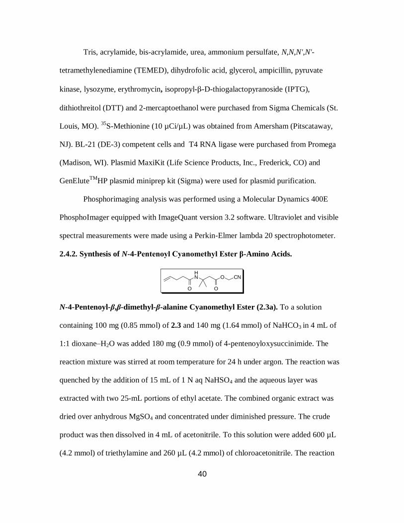
N-4-Pentenoyl-β,β-dimethyl-β-alanine Cyanomethyl Ester (2.3a). To a solution
containing 100 mg (0.85 mmol) of 2.3 and 140 mg (1.64 mmol) of NaHCO3 in 4 mL of
1:1 dioxane–H2O was added 180 mg (0.9 mmol) of 4-pentenoyloxysuccinimide. The
reaction mixture was stirred at room temperature for 24 h under argon. The reaction was
quenched by the addition of 15 mL of 1 N aq NaHSO4 and the aqueous layer was
extracted with two 25-mL portions of ethyl acetate. The combined organic extract was
dried over anhydrous MgSO4 and concentrated under diminished pressure. The crude
product was then dissolved in 4 mL of acetonitrile. To this solution were added 600 µL
(4.2 mmol) of triethylamine and 260 µL (4.2 mmol) of chloroacetonitrile. The reaction
41
mixture was stirred at room temperature for 24 h at which time 20 mL of ethyl acetate
was added. The organic layer was washed with 10 mL of 1 N aq NaHSO4 followed by 10
mL of brine, then dried over anhydrous MgSO4 and concentrated to dryness under
diminished pressure, affording a crude residue. The crude product was purified by flash
silica gel column chromatography (15 x 1 cm) using 1:1 ethyl acetate–hexanes for elution
to obtain 2.3a as colorless oil: yield 119 mg (50%); Rf 0.5 (1:1 ethyl acetate–hexanes); 1H
NMR (CDCl3) δ 1.38 (s, 6H), 2.16-2.20 (m, 2H), 2.29-2.34 (m, 2H), 2.92 (s, 2H), 4.66 (s,
2H), 4.95-5.05 (m, 2H), 5.66 (bs, 1H) and 5.73-5.83 (m, 1H); 13
C NMR (CDCl3) δ 27.5,
29.4, 36.3, 42.2, 47.9, 51.9, 114.4, 115.4, 137.0, 169.6 and 172.2; mass spectrum (APCI),
m/z 239.1398 (M+H)+ (C17H21NO3 requires m/z 239.1396).
HN O
OO
CN
N-4-Pentenoyl-β-S-phenylalanine Cyanomethyl Ester (2.4a). ). To a solution
containing 100 mg (0.6 mmol) of 2.4 and 105 mg (1.22 mmol) of NaHCO3 in 4 mL of 1:1
dioxane–H2O was added 135 mg (0.62 mmol) of 4-pentenoyloxysuccinimide. The
reaction mixture was stirred at room temperature for 24 h under argon. The reaction was
quenched by the addition of 15 mL of 1 N aq NaHSO4 and the aqueous layer was
extracted with two 25-mL portions of ethyl acetate. The combined organic extract was
dried over anhydrous MgSO4 and concentrated under diminished pressure. The crude
product was then dissolved in 4 mL of acetonitrile. To this solution were added 450 µL
(3.15 mmol) of triethylamine and 195 µL (3.15 mmol) of chloroacetonitrile. The reaction
mixture was stirred at room temperature for 24 h at which time 20 mL of ethyl acetate

42
was added. The organic layer was washed with 10 mL of 1 N aq NaHSO4 followed by 10
mL of brine, then dried over anhydrous MgSO4 and concentrated to dryness under
diminished pressure, affording a crude residue. The crude product was purified by flash
silica gel column chromatography (15 x 1 cm) using 1:1 ethyl acetate–hexanes for elution
to obtain 2.4a as colorless semi-solid: yield 114 mg (40%); Rf 0.31 (1:1 ethyl acetate–
hexanes); 1H NMR (CDCl3) δ 2.22-2.36 (m, 4H), 2.82-3.01 (m, 2H), 4.59 (d, 2H, J = 2.4
Hz), 4.95-5.04 (m, 2H), 5.39 (q, 1H, J = 11.6 and 6.4 Hz), 5.71-5.81 (m, 1H), 6.59 (d,
1H, J = 8.0 Hz) and 7.23-7.33 (m, 5H); 13
C NMR (CDCl3) δ 29.4, 35.6, 39.6, 48.5, 49.7,
114.2, 115.7, 126.4, 128.0, 128.9, 136.9, 139.8, 169.4 and 171.9; mass spectrum (APCI),
m/z 287.1516 (M+H)+ (C17H21NO3 requires m/z 286.1521).
HN O
Br
OO
CN
N-4-Pentenoyl-β-S-(p-bromophenyl)alanine Cyanomethyl Ester (2.5a). To a solution
containing 100 mg (0.41 mmol) of 2.5 and 70.0 mg (0.82 mmol) of NaHCO3 in 4 mL of
1:1 dioxane–H2O was added 90 mg (0.45 mmol) of 4-pentenoyloxysuccinimide. The
reaction mixture was stirred at room temperature for 24 h under argon. The reaction was
quenched by the addition of 15 mL of 1 N aq NaHSO4 and the aqueous layer was
extracted with two 25-mL portions of ethyl acetate. The combined organic extract was
dried over anhydrous MgSO4 and concentrated under diminished pressure. The crude
product was then dissolved in 4 mL of acetonitrile. To this solution were added 300 µL
(2.10 mmol) of triethylamine and 130 µL (2.10 mmol) of chloroacetonitrile. The reaction
mixture was stirred at room temperature for 24 h at which time 20 mL of ethyl acetate

43
was added. The organic layer was washed with 10 mL of 1 N aq NaHSO4 followed by 10
mL of brine, then dried over anhydrous MgSO4 and concentrated to dryness under
diminished pressure, affording a crude residue. The crude product was purified by flash
silica gel column chromatography (15 x 1 cm) using 1:1 ethyl acetate–hexanes for elution
to obtain 2.5a as colorless semi-solid: yield 140 mg (38%); Rf 0.33 (1:1 ethyl acetate–
hexanes); 1H NMR (CDCl3) δ 2.27-2.40 (m, 4H), 2.85-3.03 (m, 2H), 4.65 (s, 2H), 4.99-
5.07 (m, 2H), 5.39 (q, 1H, J = 6.8 Hz), 5.74-5.84 (m, 1H), 6.45 (d, 1H, J = 8.0 Hz), 7.15
(d, 2H, J = 8.0 Hz) and 7.45-7.47 (m, 2H); 13
C NMR (CDCl3) δ 29.4, 35.6, 39.2, 48.6,
49.0, 113.9, 115.9, 121.9, 128.1, 132.0, 136.7, 138.8, 169.3 and 171.8; mass spectrum
(APCI), m/z 365.0631 (M+H)+ (C17H20NO3Br requires m/z 365.0627).
2.4.3. Synthesis of β-aminoacyl-pdCpA derivatives.
N
NN
N
NH2
O
OHO
OPO
O
O
N
N
NH2
O
O
OPO
O
O_
O
HN
O
N-4-Pentenoyl-β,β-dimethyl-β-alanyl-pdCpA (2.3b). To a 1.5-mL Eppendorf tube
containing 5.3 mg (3.7 µmol) of the tris(tetrabutylammonium) salt of pdCpA was added
10 mg (42 µmol) of 2.3a dissolved in 100 µL of 9:1 DMF–Et3N. After 2 h of sonication,
the reaction mixture was purified by C18 reversed phase HPLC (250 x 10 mm) using a
gradient of 1%→65% acetonitrile in 50 mM ammonium acetate, pH 4.5, over a period of
45 min. The fractions eluting at 14.4 and 14.9 min were collected, combined and

44
lyophilized to afford 2.3b as a colorless solid: yield 3.6 mg (100%); mass spectrum (ESI),
m/z 816.2130 (M-H)- (C29H40N9O15P2 requires m/z 816.2125).
N
NN
N
NH2
O
OHO
OPO
O
O
N
N
NH2
O
O
OPO
O
O
O
HN
O
N-4-Pentenoyl-β-S-phenylalanyl-pdCpA (2.4b). To a 1.5-mL Eppendorf tube
containing 5.3 mg (3.7 µmol) of the tris(tetrabutylammonium) salt of pdCpA was added
10 mg (35 µmol) of 2.4a dissolved in 100 µL of 9:1 DMF–Et3N. After 2 h of sonication,
the reaction mixture was purified by C18 reversed phase HPLC (250 x 10 mm) using a
gradient of 1%→65% acetonitrile in 50 mM ammonium acetate, pH 4.5, over a period of
45 min. The fractions eluting at 17.4 and 18.5 min were collected, combined and
lyophilized to afford 2.4b as a colorless solid: yield 3.2 mg (100%); mass spectrum (ESI),
m/z 864.2126 (M-H)- (C33H40N9O15P2 requires m/z 864.2125).

45
N
NN
N
NH2
O
OHO
OPO
O
O
N
N
NH2
O
O
OPO
O
O
O
HN
O
Br
N-4-Pentenoyl-β-S-(p-bromophenyl)alanyl-pdCpA (2.5b).To a 1.5-mL Eppendorf tube
containing 5.3 mg (3.7 µmol) of the tris(tetrabutylammonium) salt of pdCpA was added
10 mg (27 µmol) of 2.5a dissolved in 100 µL of 9:1 DMF–Et3N. After 2 h of sonication,
the reaction mixture was purified by C18 reversed phase HPLC (250 x 10 mm) using a
gradient of 1%→65% acetonitrile in 50 mM ammonium acetate, pH 4.5, over a period of
45 min. The fractions eluting at 20.8 and 22.1 min were collected, combined and
lyophilized to afford 2.5b as colorless solid: yield 3.1 mg (100%); mass spectrum (ESI),
m/z 942.1234 (M-H)- (C33H39N9O15BrP2 requires m/z 942.1230).
2.4.4. Preparation of β-aminoacyl-tRNACUAs (2.2c, 2.3c, 2.4c and 2.5c). The activation
of suppressor tRNACUAs was carried out as described previously.50
Briefly, 100-µL
reaction mixtures of 100 mM Na Hepes, pH 7.5, contained 1.0 mM ATP, 15 mM MgCl2,
100 µg of suppressor tRNACUA-COH, 0.5 A260 unit of N-pentenoyl-protected -
aminoacyl-pdCpA, 15% DMSO, and 100 units of T4 RNA ligase. The reaction mixture
was incubated at 37 °C for 1 h, then quenched by the addition of 0.1 vol of 3 M NaOAc,
pH 5.2. The N-protected aminoacylated tRNA was precipitated with 3 vol of cold

46
ethanol. The efficiency of ligation was estimated by 8% polyarylamide–7 M urea gel
electrophoresis at pH 5.0 (Figure 2.3, Section 2.2).52
The N-pentenoyl-protected aminoacyl-tRNACUAs were deprotected by treatment
with 5 mM aqueous I2 at 25 °C for 15 min. The solution was centrifuged, and the
supernatant was adjusted to 0.3 M NaOAc and treated with 3 vol of cold ethanol to
precipitate the aminoacylated tRNA. The tRNA pellet was collected by centrifugation,
washed with 70% aq EtOH, air dried and dissolved in 50 µL of RNase free water.
2.4.5. Preparation of S-30 extracts from cells having modified ribosomes. Aliquots
(5-10 μL) from liquid stocks of E. coli BL-21(DE-3) cells, harboring plasmids with a
wild-type or modified rrnB gene, were placed on LB agar supplemented with 100 g/mL
of ampicillin and grown at 37 ºC for 16-18 h. One colony was picked from each agar
plate and transferred into 3 mL of LB medium supplemented with 100 g/mL of
ampicillin and 0.5 mM IPTG. The cultures were grown at 37 ºC for 3-6 h in a
thermostated shaker until OD600 ~ 0.15-0.3 was reached (about 3-5 h), diluted with LB
medium supplemented with 100 g/mL ampicillin, 1 mM IPTG and 3 µg/mL of
erythromycin (for selectively enhancing the modified ribosome fraction) until OD600 0.01
was reached, and then grown at 37 ºC for 12-18 h. The optimal concentration of the final
cultures was OD600 0.5-1.0. Cells were harvested by centrifugation (5000 × g, 4 ºC, 10
min), washed three times with S-30 buffer (1 mM Tris-OAc, pH 8.2, containing 1.4 mM
Mg(OAc)2, 6 mM KOAc and 0.1mM DTT) supplemented with β-mercaptoethanol (0.5
mL/L) and once with S-30 buffer having 0.05 mL/L β-mercaptoethanol. The weight of
the wet pellet was estimated and 1.27 mL of S-30 buffer was added to suspend each 1 g
of cells. The volume of the suspension was measured and used for estimating the amount
47
of other components. Pre-incubation mixture (0.3 mL) (0.29 M Tris, pH 8.2, containing 9
mM Mg(OAc)2, 13 mM ATP, 84 mM phosphoenol pyruvate, 4.4 mM DTT and 5 µM
amino acids mixture), 15 units of pyruvate kinase and 10 μg of lyzozyme were added per
1 mL of cell suspension and the resulting mixture was incubated at 37 ºC for 30 min. The
incubation mixture was then frozen at – 80 ºC (~ 30 min), melted (37 ºC, 30 min), and
again frozen and melted at room temperature (~ 30 min). Ethylene glycol tetraacetic acid
(EGTA) was then added to 2.5 mM final concentration and the cells were incubated at 37
ºC for 30 min and again frozen (– 80 ºC, 30 min). The frozen mixture was centrifuged
(15,000 × g, 4 ºC, 1 h) and the supernatant was stored in aliquots at – 80 ºC.
2.4.6. In vitro protein translation. Protein translation reactions were carried out in 12-
2000 L of incubation mixture containing 0.2-0.4 L/ L of S-30 system, 100 ng/ L of
plasmid, 35 mM Tris acetate, pH 7.4, 190 mM potassium glutamate, 30 mM ammonium
acetate, 2 mM DTT, 0.2 mg/mL total E. coli tRNA, 3.5% PEG 6000, 20 g/mL folinic
acid, 20 mM ATP and GTP, 5 mM CTP and UTP, 100 M amino acids mixture, 0.5
Ci/ L of 35
S-methionine and 1 μg/mL rifampicin. In the case of plasmids having a gene
with a TAG codon, a suppressor tRNA was added to a concentration of 0.3-0.5 g/ L
(for α-aminoacyl-tRNAs) and 0.6-1.0 g/ L (for β-aminoacyl-tRNAs). Reactions were
carried out at 37 C for 1 h and terminated by chilling on ice. Aliquots from in vitro
translation mixtures were analyzed by SDS-PAGE followed by quantification of the
radioactive bands by phosphorimager analysis.
2.4.7. Purification of DHFR. Samples of DHFR, prepared during in vitro translation,
were diluted with 50 mM Na phosphate, pH 8.0, containing 0.3 M NaCl and 0.1 mg/ml

48
BSA and applied to a 50- L Ni-NTA agarose column that had been equilibrated with the
same buffer. The column was washed with three 500- L portions of the same buffer and
DHFR was eluted by washing with 150 L of the same buffer also containing 250 mM
imidazole.
A 50- L column of DEAE-Sepharose was equilibrated with 500 L of 10 mM Na
phosphate, pH 7.4. Samples of DHFR purified by Ni-NTA chromatography were diluted
10-fold in the same buffer and applied to the resin. The column was washed with 500 L
of the same buffer. DHFR was eluted with a NaCl step gradient (0.1; 0.2; 0.3; 0.4; 0.5 M)
in 10 mM of Na phosphate, pH 7.4. The fractions were analyzed on a 12%
polyacrylamide gel, stained using Coomassie R-250, then destained in 20% ethanol–7%
acetic acid. The fractions containing DHFR were combined, dialyzed against 50 mM Na
phosphate, pH 7.4 and concentrated by ultrafiltration using a YM-10 filter (Amicon
Ultra, Millipore Corp, Billerica, MA).
2.4.8. “In gel” trypsin digestion. Samples to be digested in the gel were run in 3-4 lanes
of a 12% SDS-polyacrylamide gel, stained with Coomassie R-250 and destained until the
background was clear. That area of the gel having the DHFR was cut from the gel and
washed with 0.1 M ammonium bicarbonate (1 h, room temperature). The solution was
discarded and 0.1-0.2 mL of 0.1 M ammonium bicarbonate and 10-30 μL of 0.045 mM
DTT were added. Gel pieces were incubated at 60 ºC for 30 min, cooled to room
temperature and incubated at room temperature for 30 min in the dark after the addition
of 10-30 μL of 0.1 M iodoacetamide. Gel pieces were washed in 50% acetonitrile–0.1 M
ammonium bicarbonate until they became colorless. After discarding the solution, the gel
pieces were incubated in 0.1-0.2 mL of acetonitrile (10-20 min at room temperature) and,
49
after removal of solvent, were re-swelled in 50-100 μL of 25 mM ammonium bicarbonate
containing 0.02µg/µL trypsin. After incubation at 37 ºC for 4 h, the supernatant was
transferred to a new tube and the peptides were extracted with 60% acetonitrile–0.1% aq
TFA (20 min at room temperature). The combined fractions were dried and reconstituted
in 10 μL of 60% acetonitrile–0.1% aq TFA for each 1 μg of the protein (measured before
trypsin digestion). Approximately, 5 μL of this solution was mixed thoroughly with 2.5
μL of the matrix solution (saturated solution of dihydroxybenzoic acid in 60%
acetonitrile–0.1% aq TFA). A 2 μL aliqout from the resulting solution was deposited on
one of the wells in a 90-well MALDI-MS plate and dried.This was repeated twice and the
plate was subjected to MALDI-MS analysis.

50
Chapter 3
INCORPORATION OF A DIPEPTIDE AND DIPEPTIDOMIMETIC INTO DHFR
USING RIBOSOMES HAVING MODIFICATIONS IN THE
PEPTIDYLTRANSFERASE CENTER
3.1. Introduction
Peptidomimetics are compounds that spatially mimic a natural peptide or protein
and may retain the ability to interact with a biological target in a manner similar to the
natural counterpart. For biological and pharmaceutical studies, peptidomimetics offer
interesting advantages over physiologically active peptides. Peptidomimetics can solve
problems associated with natural peptides, e.g. stability against proteolysis and poor
bioavailability.70
Other properties, such as receptor selectivity or potency, can be
substantially improved with rational design of mimetics.71
Hence, peptidomimetics have
great potential in drug discovery.
3.1.1. β-Turn peptidomimetics
Reversel turns are commonly occurring structural features in globular proteins.72
β-turns are one of the most important reverse turn motifs and are often located on the
surface of proteins, most likely acting as recognition and antigenic sites.73
In a β-turn, a
tight loop is formed when the carbonyl oxygen of one residue (i) forms a hydrogen bond
with the amide proton of an amino acid (i+3) three residues away (Figure 3.1). Bicyclic
dipeptide mimetics (Figure 3.2) have been used extensively in peptides to generate β-turn
conformations.74,75
Recently, proline based dipeptidomimetics have been shown to be
capable of enhancing biological activities when incorporated into peptides76
or proteins.77
Hartley and co-workers synthesized analogues of an anti-HIV protein called RANTES
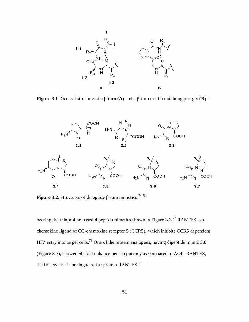
51
NH
O
NH
R2
O
NHR3
O
---
N NH
O
O
NH
O
R1
R2
R1
R4
i
i+1
i+2
i+3
A B-
--
Figure 3.1. General structure of a β-turn (A) and a β-turn motif containing pro-gly (B) .1
N
H2N
COOH
R
H
O
N
S
H2N
O COOH
N
N
NN
H2N
COOHR2
NO
H2N
N
OO
H2N
N
SO
H2N
NN
O
H2N COOH
3.1 3.2 3.3
3.4 3.5 3.6 3.7
H
R1 RCOOH
RCOOH
RCOOH
R
Figure 3.2. Structures of dipeptide β-turn mimetics.74,75
bearing the thioproline based dipeptidomimetics shown in Figure 3.3.77
RANTES is a
chemokine ligand of CC-chemokine receptor 5 (CCR5), which inhibits CCR5 dependent
HIV entry into target cells.78
One of the protein analogues, having dipeptide mimic 3.8
(Figure 3.3), showed 50-fold enhancement in potency as compared to AOP–RANTES,
the first synthetic analogue of the protein RANTES.77

52
S
N
X
HN
O
Y
O S
N
X
HN
O
Y
O
N
X
HN
O
Y
O
HO
N
X
HN
O
Y
O
HO
3.8 3.9 3.10 3.11
Figure 3.3. Structures of thioproline-based dipeptidomimetics.77
3.1.2. Peptide bond isosteres
The backbone of a peptide can be modified by isosteric or isoelectronic exchange
of structural elements in the peptide or by the introduction of additional fragments.79
A
few examples of peptide bond isosteres are shown in Figure 3.4. A p-cyanophenylalanine
(Cnf) containing an abbreviated protein fragment was ligated to a thioamide (3.12, Figure
3.4) labeled synthetic peptide to produce a full length protein, α-synuclein, using the
native chemical ligation technique.80
In this study, the Cnf/thioamide FRET pair was
studied to determine the N- and C-termini orientations in α-synuclein.
HN
S
N
O
HN
NH
O
O NH
OO
NNH
O
NH
O
thioamide 3.12 N-alkyl 3.13 retro-inverso 3.14
aza 3.15 alkyne 3.16 oxazole 3.17
N
N N
triazole 3.18
Figure 3.4. Some common peptide backbone modifications.79
53
Peptides exhibiting important biological activities have been modified by
incorporating N-methyl amino acids (3.13, Figure 3.4), which improve the
pharmacological properties of these peptides, such as metabolic stability, enhanced
potency and bioavailability.81
A good example of an N-methylated peptide is
cyclosporine A having seven N-methylated amino acids; it is one of the most successful
drugs used as immunosuppressant in organ transplantations.81
The introduction of a
triazole group (3.18, Figure 3.4) in the backbone of a peptide enabled the formation of β-
sheets in small peptides, where a turn was induced by the triazole moiety.82
3.1.3. Green fluorescent protein (GFP)
After the discovery of green fluorescent protein (GFP) from the jellyfish
Aequorea victoria in 1962 by Shimomura et al.,83
interest in GFP was limited to studies
of the luminescence of marine creatures. After its cloning in 199284
and its utilization as a
marker for gene expression in 1994,85
it became a widely used tool as a reporter for
structures and processes in molecular and cell biology.86
Amino acid residues in the
proteins belonging to the GFP family, fold into a β-barrel structure made up of 11 β-
sheets that enclose an internal distorted helix.87
In Aequorea victoria GFP, three amino
acid residues of the helix at positions 65-67 undergo a unique posttranslational
modification to form the chromophore (Scheme 3.1).88
The resulting chromophore is
located almost at the center of the barrel and therefore is well shielded from solvent
contacts.87
Amino acids Tyr66 and Gly67 are conserved among all natural GFP-like
proteins, whereas the amino acid residue at position 65 can vary.86

54
Scheme 3.1. Proposed mechanism for the chromophore formation in GFP.88
NH
NH
O
O
OH
O
NH
OH
Try66Gly67
Ser65
NH
NH+
O-
O
O
OH
HO
HN
NH
N
OH
O
O
OH
HO
HN
N
NO
O
OH
NH
HO
N
NO
O
OH
NH
HO
N
NO
O
O-
NH
HO
-H+, +H+
-H2O
-H+
+H+
3.19 3.20 3.21
3.223.233.24
O2
There has been a steady effort to develop new fluorescent proteins (FP) with
improved functions and novel characteristics. Diversity is mainly achieved by site-
directed mutagenesis which has produced a broad spectrum of GFP-like proteins.86
The
synthesis and spectral properties of FP chromophores have been studied in detail,89
yet
there is no report of the incorporation of a synthetic FP chromophore or a chromophore
mimic into the backbone of proteins using in vitro or in vivo techniques.
3.1.4. Biosynthesis of proteins having peptidomimetics
Synthetic and semi-synthetic strategies have enabled the incorporation of
peptidomimetics into peptides and proteins.76,77,80
However, the use of such strategies is
limited by the size of the polypeptide accessible or control over the site of insertion of

55
peptidomimetics into a protein. The incorporation of peptidomimetics into proteins using
in vitro or in vivo translation methods has not been reported to date. The architecture of
the wild-type ribosome is biased towards the incorporation of α-L-amino acids into
proteins.3,42
Engineering proteins with peptidomimetics, using in vitro or in vivo methods,
has the potential to open new doors for creating interesting biopolymers. Therapeutic
proteins having peptidomimetics can enhance the proteolytic and thermal stability of
those proteins, as well as increase potency, and improve target specificity and
bioavailability. The inability of the wild-type ribosome to incorporate peptidomimetics
into proteins, including the ones discussed earlier in this section, is the biggest challenge.
Reengineering the architecture of the peptidyltransferase center (PTC) of the ribosome
can enable the incorporation of peptidomimetics into proteins. Hecht and co-workers
have successfully demonstrated that the suitable mutations in some regions of the 23S
rRNA can afford modified ribosomes to facilitate the incorporation of non α-L-amino
acids into proteins.48-51
In this thesis, ribosomes having modifications in their 23S rRNA to enable the
incorporation of the dipeptide glycylphenylalanine (3.25) and dipeptidomimetic 3.26 into
DHFR are reported (Figure 3.5). Using puromycin derivative 1.3 (Figure 3.6) for the
selection of E. coli colonies, six variants of ribosomes having modifications in two
regions of the 23S rRNA (2057-2063 and 2502-2507) were selected. The selection
experiments were carried out by Dr. Larisa Dedkova in a fashion similar to that reported
earlier.50
The selected modified ribosomes were evaluated for the suppression of the
UAG codon at position 10 of DHFR mRNA by glycylphenylalanyl-tRNACUA (3.25b,
Scheme 3.2). Dipeptidomimetic 3.26 shown in Figure 3.5 is a fluorescent structural

56
analogue of the GFP chromophore 3.23 shown in Scheme 3.1. S-30 systems prepared
from two different modified ribosomes were used to study the incorporation of 3.26 into
position 10 of DHFR and the fluorescence spectra were obtained for the modified DHFR
at different protein concentrations.
H2N
HN
O
COOH
3.25
H2N
O
N
OH
O
OMe
3.26
Figure 3.5. Structures of glycylphenylalanine (3.25) and dipeptidomimetic 3.26.
N
NN
N
N
O
OHNH
HO
O
NH
O
H2N
MeO
1.3
Figure 3.6. Structure of puromycin derivative 1.3.
3.2. Results
For the selection of ribosomes able to incorporate both α-L-amino acids and
dipeptide or dipeptidomimetics, region 2057-2063 in the 23S rRNA was initially altered

57
to confer erythromycin resistance. Erythromycin resistance allowed the modified
ribosomes to grow at the expense of the wild-type ribosomes having little resistance to
the antibiotic. Five clones having different sequence in this 23S rRNA region were
identified and were further mutagenized in one of the three other regions (2582-2588,
2502-2507 and 2496-2501) of the 23S rRNA. The E. coli colonies harboring modified
ribosomal clones were grown in the presence of 100 μg/mL of the puromycin derivative
1.3 and plasmids from the colonies showing >50% inhibition of cell growth were isolated
and sequenced (Table 3.1). Four clones (010309, 010310, 010326 and 010328, Table 3.1)
were identified as mixtures of more than one clone as judged by the automatic DNA
sequencing and were transformed again into E. coli cells to separate the clones.
Clones having modifications in the region 2582-2588, or having the wild-type sequence
in the second region were not selected for S-30 preparations (Table 3.1). It was
demonstrated previously that modifications in the region 2582-2588 of the 23S rRNA
were responsible for low fidelity of translation.50
Of the remaining clones, nine mediated
successful preparation of S-30 systems for in vitro translation experiments (Table 3.2).
Eight of the clones shown in Table 3.2 were obtained after re-cloning the clone mixtures.
Three of the ribosomal clones (010309R9, 010326R6 and 010328R4) had modified
ribosomes with the same sequence (2057UGCGUGG2063 and 2502ACGAAG2507),
while other two ribosomal clones (010326R1 and 010328R2) also shared same sequence
in two modified regions (2057UGCGUGG2063 and 2502CUACAG2507). Therefore, six
different variants of modified ribosomes were selected using puromycin derivative 1.3.
The selection experiments were carried out by Dr. Larisa Dedkova.

58
Table 3.1. Characterization of selected clones sensitive to puromycin derivative 1.3 and
erythromycin.
Clones Puritya
Sequence in regions of mutagenesis Inhibition by
Region 1 Region 2 Puromycin
derivative 1.3 (%)
Erythro-
mycin (MIC,
μg/mL)
010120 single 2057TGCGTGG2063 2582CCGAATC2588 65.4 12.5–6.25
010309 mix 2057TGCGTGG2063 2502CTACGG2507*
63.2 12.5–6.25
010310 mix 2057TGCGTGG2063 2502CGCAAT2507*
52.9 12.5–6.25
010322 single 2057TGCGTGG2063 2502ACGAAG2507
77 12.5–6.25
010326 mix 2057TGCGTGG2063 2502CTACAG2507*
50.4 12.5–6.25
010328 mix 2057TGCGTGG2063 2502ACGAAG2507* 77 12.5–6.25
030240 single 2057TTGGTCC2063 2496AGTGGT2501 56 6.25–3.12
030247 single 2057TTGGTCC2063 2496CACCTC2501**
64 6.25–3.12
030248 single 2057TTGGTCC2063 2496CACCTC2501**
61 6.25–3.12
040228 single 2057AGCGTG2063 2496CACCTC2501**
51 6.25–3.12
040322 single 2057AGCGTG2063 2502CTGCTT2507 54 6.25–3.12
040329 single 2057AGCGTG2063 2502TGGCAG2507 54 6.25–3.12
040338 single 2057AGCGTG2063 2502ATCAGG2507 56 6.25–3.12
070307 single 2057ATTCCGG2063 2502GATGTC2507**
77 6.25–3.12
080118 single 2057AGTGAGA2063 2582ATGGGCT2588 66.5 25–12.5
080337 single 2057AGTGAGA2063 2502ATCCGA2507 51 25–12.5
Wild-
type
2057GAAAGAC2063 2496CACCTC2501
2502GATGTC2507
2582GATGTC2507
>1000 3.12–1.56
a Some colonies harbored more than one ribosomal clones.
* Most dominant sequence as
estimated by automatic sequencing. **
Same sequence as the wild-type ribosome in that
region.
S-30 extracts, prepared from cultures having different modified ribosomes were
tested for the incorporation of the dipeptide glycylphenylalanine (3.25) into position 10
of DHFR. S-30 systems having two different modified ribosomes were used for the
incorporation of the fluorescent dipeptidomimetic 3.26 into DHFR. Glycylphenylalanyl-
pdCpA (3.25a, Scheme 3.2) was synthesized by Dr. Rakesh Paul and the pdCpA

59
derivative 3.26a (Scheme 3.3) activated with dipeptidomimetic 3.26 was prepared by Dr.
Maninkandadas M. Madathil.
Table 3.2. 23S rRNA sequence modifications in the PTC for the clones used for S-30
preparations.
Clone Sequence in 23S rRNA of modified ribosomes
Region 1 (2057-2063) Region 2 (2502-2507)
010309R3 UGCGUGG ACGAAG
010326R6 UGCGUGG ACGAAG
010328R4 UGCGUGG ACGAAG
010310R4 UGCGUGG CGCACG
010326R5 UGCGUGG CUAUGU
010310R1 UGCGUGG CGCAAU
010328R2 UGCGUGG CUACAG
010326R1 UGCGUGG CUACAG
080337 AGUGAGA AUCCGA
Wild-type GAAAGAC GAUGUC
3.2.1. Preparation of suppressor tRNACUAs activated with glycylphenylalanine (3.25)
and dipeptidomimetic 3.26
Activated pdCpA derivatives were ligated to an abbreviated 74-nucleotide
tRNACUA-COH transcript using T4 RNA ligase (Schemes 3.2 and 3.3) and the two N-
pentenoyl-aminoacyl -tRNACUAs (3.25b and 3.26b) were prepared and isolated as
described previously.50
The N-pentenoyl protecting groups were removed by treatment
with aqueous iodine for five minutes.63,64
Removal of the protecting group was performed
immediately prior to use of the misacylated tRNAs in the protein synthesis reaction. Each
pdCpA derivative was ligated to abbreviated tRNACUA-COH with 100% efficiency as
judged by 8% polyacrylamide-7 M urea gel electrophoresis (pH 5.0) analysis (Figure
3.7).67
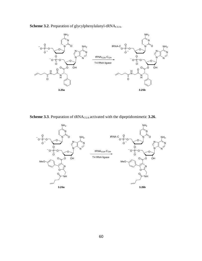
60
Scheme 3.2. Preparation of glycylphenylalanyl-tRNACUA.
tRNA-C
N
NN
N
NH2
O
OHO
OPO
O
O
N
N
NH2
O
O
OPO
O
O
tRNACUA-COH
T4 RNA ligase
3.25a
O
NH
OHN
O
N
NN
N
NH2
O
OHO
OPO
O
O
N
N
NH2
O
O
OPO
O
O
O
NH
OHN
O
3.25b
Scheme 3.3. Preparation of tRNACUA activated with the dipeptidomimetic 3.26.
tRNA-C
N
NN
N
NH2
O
OHO
OPO
O
O
N
N
NH2
O
O
OPO
O
O
tRNACUA-COH
T4 RNA ligaseO
O
N
MeO
NHO
N
NN
N
NH2
O
OHO
OPO
O
O
N
N
NH2
O
O
OPO
O
O
O
O
N
MeO
NHO
3.26a 3.26b

61
1 2 3
Figure 3.7. Preparation of misacylated tRNAs. Lane 1, glycylphenylalany-tRNACUA;
lane 2, tRNACUA activated with the dipeptidomimetic 3.26; lane 3, nonacylated
abbreviated tRNACUA.
3.2.2. In vitro translation using glycylphenylalanine (3.25) with wild-type ribosome
Wild-type ribosomes synthesize polypeptide chains by incorporating one α-L-
amino acid at a time via one monoacylated tRNA for each corresponding codon in the
mRNA transcript.42
Ribosome mediated sequential incorporation of two amino acids for
one codon using bisacylated tRNAs has not been reported to date. Therefore, the ability
of the wild-type ribsome to enable the incorporation of glycylphenylalanine using DHFR
mRNA having a UAG codon at position 10 in the presence of glycylphenylalanyl-
tRNACUA was studied. The suppression efficiency of glycylphenylalanyl-tRNACUA was
compared to phenylalanyl-tRNACUA suppression of the UAG codon using an S-30
preparation having wild-type ribosomes (Figure 3.8). The suppression efficiency was
<2% above the background for glycylphenylalanine. This data further proves that during
translation, the wild-type ribosome cannot transfer a dipeptide from dipeptidyl-tRNA to a
polypeptide chain.90

62
1 2 3
100 0.2 1.4 % suppression
Figure 3.8. Translation of DHFR from wild-type (lane 1) and modified (lanes 2-3) (UAG
codon in position 10) mRNA in the presence of different suppressor tRNACUAs. Lane 2,
nonacylated tRNACUA; lane 3, tRNACUA activated with glycylphenylalanine (3.25). The
suppression efficiency relative to wild type is shown below each lane.
3.2.3. In vitro translation using glycylphenylalanine (3.25) and dipeptidomimetic
3.26 with the modified ribosomes
A modified DHFR contruct, having a TAG codon in position 10 (pETDH10
plasmid) was used for the incorporation of glycylphenylalanine (3.25) and the
dipeptidomimetic 3.26. Six different S-30 preparations having modified ribosomes
produced different levels of full length DHFR from the wild-type gene relative to the S-
30 system having the wild-type ribosome. Therefore, the suppression efficiencies were
expressed relative to the suppression obtained using phenylalanyl-tRNACUA (Table 3.3).
As a negative control, DHFR synthesis in the presence of nonacylated-tRNACUA was
measured for each experiment. The amounts of DHFR produced were quantified with a
phosphoimager, which monitored the incorporation of 35
S-methionine into DHFR.
← full length DHFR
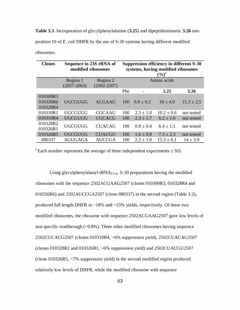
63
Table 3.3. Incorporation of glycylphenylalanine (3.25) and dipeptidomimetic 3.26 into
position 10 of E. coli DHFR by the use of S-30 systems having different modified
ribosomes.
Clones Sequence in 23S rRNA of
modified ribosomes
Suppression efficiency in different S-30
systems, having modified ribosomes
(%)a
Region 1
(2057-2063)
Region 2
(2502-2507)
Amino acids
Phe - 3.25 3.26 010309R3
010326R6
010328R4
UGCGUGG
ACGAAG
100
0.8 ± 0.2
18 ± 4.0
15.3 ± 2.5
010310R1 UGCGUGG CGCAAU 100 2.3 ± 1.0 10.2 ± 0.6 not tested
010310R4 UGCGUGG CGCACG 100 2.3 ± 1.7 6.2 ± 1.6 not tested
010328R2
010326R1 UGCGUGG CUACAG
100 0.8 ± 0.4 6.4 ± 1.1 not tested
010326R5 UGCGUGG CUAUGU 100 1.6 ± 0.8 7.3 ± 2.3 not tested
080337 AGUGAGA AUCCGA 100 2.2 ± 1.0 15.3 ± 6.1 14 ± 3.0
a Each number represents the average of three independent experiments ± SD.
Using glycylphenylalanyl-tRNACUA, S-30 preparations having the modified
ribosomes with the sequence 2502ACGAAG2507 (clones 010309R3, 010328R4 and
010326R6) and 2502AUCCGA2507 (clone 080337) in the second region (Table 3.2),
produced full length DHFR in ~18% and ~15% yields, respectively. Of these two
modified ribosomes, the ribosome with sequence 2502ACGAAG2507 gave low levels of
non specific readthrough (~0.8%). Three other modified ribosomes having sequence
2502CGCACG2507 (clones 010310R4, ~6% suppression yield), 2502CUACAG2507
(clones 010328R2 and 010326R1, ~6% suppression yield) and 2502CUAUGU2507
(clone 010326R5, ~7% suppression yield) in the second modified region produced
relatively low levels of DHFR, while the modified ribosome with sequence

64
2502CGCAAU2507 incorporated glycylphenylalanine into DHFR in a moderate yield of
~10%. Figure 3.9 illustrates the incorporation of glycylphenylalanine into position 10 of
DHFR using S-30 preparations having different modified ribosomes. The S-30 systems,
prepared from the ribosomes having mutations of 2502ACGAAG2507 and
2502AUCCGA2507, were used for the incorporation of the dipeptidomimetic 3.26 into
position 10 of DHFR and gave full length DHFR in ~15% and ~14% yields, respectively.
Figure 3.10 illustrates the formation of full length DHFR using an S-30 system prepared
from ribosomal clone 010328R4 in presence of glycylphenylalanyl-tRNACUA (3.25b) and
tRNACUA (3.26b) activated with dipeptidomimetic 3.26.
1 2 3 4 5 6 7 8 9
100 2.2 12.5 100 0.6 8.2 100 0.1 5.5 % suppressiona
Figure 3.9. Translation of DHFR from a modified DHFR mRNA (UAG codon in
position 10) by the use of S-30 systems prepared from clones 080337 (lanes 1-3),
010326R5 (lanes 4-6) and 010310R4 (lanes 7-9) in the presence of different suppressor
tRNAs. Lanes 1, 4 and 7, phenylalanyl-tRNACUA; lanes 2, 5 and 8, nonacylated
tRNACUA; lanes 3, 6 and 9, glycylphenylalanyl-tRNACUA. a The suppression efficiency
values are one of the data set used for the average suppression efficiency values in Table
3.2.
← full length DHFR

65
1 2 3 4
100 18 16 0.6 % suppressiona
Figure 3.10. Translation of DHFR from a modified DHFR mRNA (UAG codon in
position 10) by the use of an S-30 system prepared from clone 010328R4 in the presence
of different suppressor tRNAs. Lane 1, phenylalanyl-tRNACUA; lane 2,
glycylphenylalanyl-tRNACUA; lane 3, tRNACUA activated with the dipeptide mimic; lane
4, nonacylated-tRNA. a The suppression efficiency values are one of the data set used for
the average suppression efficiency values in Table 3.2.
3.2.4. Characterization of the incorporation of glycylphenylalanine (3.25) and
dipeptidomimetic 3.26 into DHFR by MALDI-MS analysis of the peptides resulting
from “in-gel” trypsin digestion
In order to provide direct evidence for the successful suppression of the UAG
codon at position 10 of DHFR mRNA by glycylphenylalanyl-tRNACUA or tRNACUA
activated with 3.26, two modified DHFRs putatively bearing glycylphenylalanine (3.25)
(modified DHFR 1) or dipeptidomimetic 3.26 (modified DHFR 2) were prepared on a
larger scale for an “in-gel” trypsin digestion followed by MALDI-MS analysis.68
As a
control, DHFR V10F was also prepared in amounts similar to the two modified DHFRs.
The modified DHFRs were purified by the use of Ni-NTA and DEAE-Sephadex
chromatography followed by SDS-polyacrylamide gel electrophoresis.51
Table 3.4
compares the estimated and observed molecular weights in MALDI-MS of the tryptic
peptides from DHFR V10F and the modified DHFRs 1 and 2. The tryptic digest of
← full length DHFR
← internal initiation at Met1

66
DHFR V10F gave an ion peak at m/z 1321.48 (Table 3.4 and Figure 3.11A) and
confirmed the presence of peptide fragment (amino acids 1-12) having phenylalanine at
position 10. For DHFR 1, a tryptic fragment encompassing amino acids 1-12 was
anticipated to have a molecular mass of 1377 Da. As shown in Table 3.4 and Figure
3.11B, there was an ion peak at m/z 1377.33 consistent with the presence of
glycylphenylalanine at position 10 of DHFR. As illustrated in Figure 3.11C (modified
DHFR 2, Table 3.4), an ion peak at m/z 1404.28 generated by the peptide having amino
acids 1-12 confirmed the incorporation of dipeptidomimetic 3.26 into position 10 of
DHFR (estimated value 1404 Da). The other six tryptic peptides encompassing amino
acids 13-106 of DHFR were observed for DHFR V10F and the two modified DHFRs and
gave the expected ion peaks in their respective MALDI-MS spectrum. In replicate
experiments, the (large) C-terminal fragment was never observed for any of the DHFRs.
Table 3.4. MALDI-MS analysis of tryptic digests of the wild-type and modified DHFR
samples.
Position Peptide sequence MALDI-MS analysis, molecular mass, Da
DHFR
(V10F) Modified
DHFR 1 Modified
DHFR 2
Est MS Est MS Est MS
-9 - -1 MVHHHHHHR 1272 – 1272 – 1272 –
1-12 MISLIAALAFDR 1321
1321.48
1377
1377.33
1404 1404.28
13-32 VIGMENAMPWNLPADLAWFK 2304 2304.43 2304 2303.13 2304 2303.19
34-44 NTLNK PVIMGR 1242 1242.46 1242 1242.29 1242 1242.41
45-57 HTWESIGRPLPGR 1506 1506.47 1506 1506.26 1506 1506.33
59-71 NIILSSQPGTDDR 1415 1415.43 1415 1415.16 1415 1415.08
72-76 VTWVK 632 632.28 632 634.21 632 634.28
77-98 SVDEAIAACGDVPEIMVIGGGR 2159 2159.40 2159 2159.41 2159 2159.12
99-106 VYEQFLPK 1023 1023.35 1023 1023.18 1023 1023.22
DHFR V10F, phenylalanine at position 10 of DHFR; modified DHFR 1,
glycylphenylalanine at position 10 of DHFR; modified DHFR 2, dipeptidomimetic 3.26
atposition 10 of DHFR. The values in boldface reflect the presence of phenylalanine (for

67
DHFR V10F), glycylphenylalanine (for modified DHFR 1) and dipeptidomimetic (for
modified DHFR 2). Position 10 is indicated in red.
A.
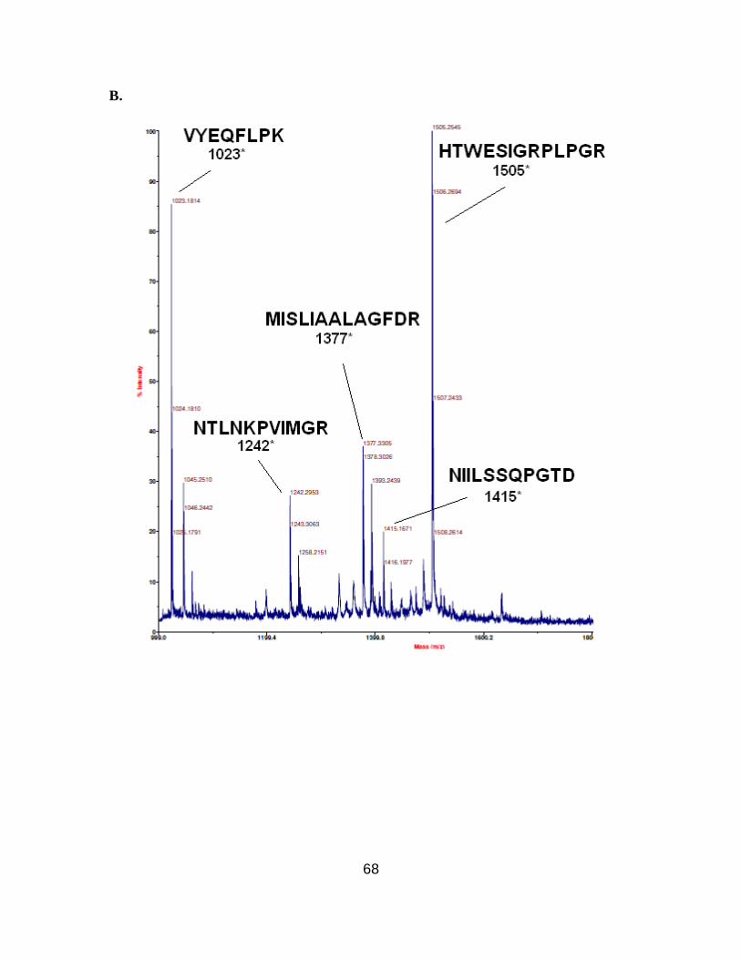
68
B.
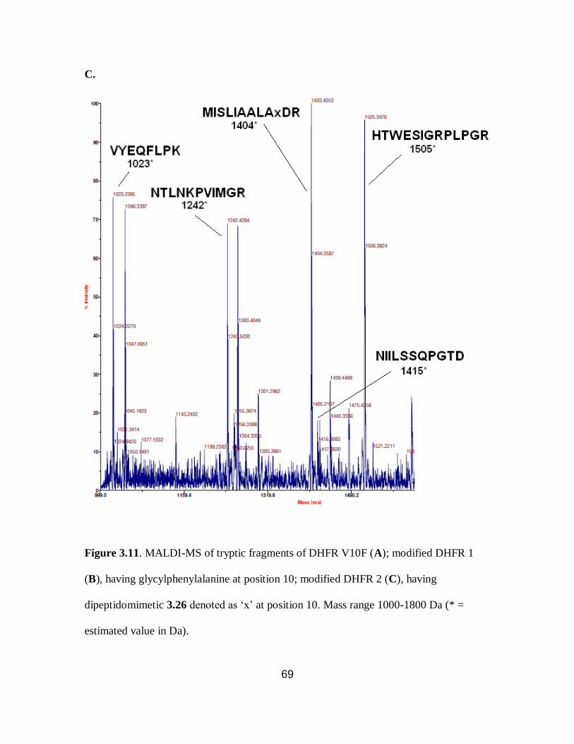
69
C.
Figure 3.11. MALDI-MS of tryptic fragments of DHFR V10F (A); modified DHFR 1
(B), having glycylphenylalanine at position 10; modified DHFR 2 (C), having
dipeptidomimetic 3.26 denoted as ‘x’ at position 10. Mass range 1000-1800 Da (* =
estimated value in Da).

70
3.2.5. Fluorescence emission spectra of wild-type DHFR and modified DHFR having
dipeptidomimetic 3.26 at position 10
Dipeptidomimetic 3.26 is a highly fluorescent structural analogue of the GFP
chromophore 3.23 (Scheme 3.1). Figure 3.12 shows the fluorescence emission spectrum
of 3.26 in water at four different concentrations. The absorption maximum for 3.26 was
at 302 nm, therefore, this wavelength was chosen as excitation wavelength.
Dipeptidomimetic 3.26 gave a fluorescence emission maximum at ~403 nm.
The fluorescence emission spectra of wild-type DHFR and modified DHFR
having 3.26 at position 10 were compared (Figures 3.13) at 302 nm excitation
wavelength. For both the wild-type and modified DHFRs, fluorescence spectra were
recorded at three different protein concentrations. The protein concentrations were
Figure 3.12. Fluorescence emission spectrum of dipeptidomimetic 3.26. The
fluorescence emission was monitored from 340-500 nm following irradiation at 302 nm.
The sample concentration range was 200-800 nM in water.

71
A.
B.
Figure 3.13. Fluorescence emission spectrum of the wild-type (A) and modified DHFR
containing dipeptidomimetic 3.26 at position 10 (B). The fluorescence emission was
monitored from 350-450 nm following irradiation at 302 nm. For wild-type DHFR (A),
the sample concentration range was 50-150 nM and for modified DHFR having 3.26 at
position 10 (B), the sample concentration range was 1-3 nM.
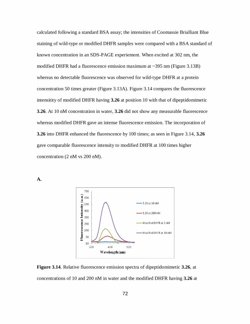
72
calculated following a standard BSA assay; the intensities of Coomassie Brialliant Blue
staining of wild-type or modified DHFR samples were compared with a BSA standard of
known concentration in an SDS-PAGE experiement. When excited at 302 nm, the
modified DHFR had a fluorescence emission maximum at ~395 nm (Figure 3.13B)
whereas no detectable fluorescence was observed for wild-type DHFR at a protein
concentration 50 times greater (Figure 3.13A). Figure 3.14 compares the fluorescence
intensitity of modified DHFR having 3.26 at position 10 with that of dipeptidomimetic
3.26. At 10 nM concentration in water, 3.26 did not show any measurable fluorescence
whereas modified DHFR gave an intense fluorescence emission. The incorporation of
3.26 into DHFR enhanced the fluorescence by 100 times; as seen in Figure 3.14, 3.26
gave comparable fluorescence intensity to modified DHFR at 100 times higher
concentration (2 nM vs 200 nM).
A.
Figure 3.14. Relative fluorescence emission spectra of dipeptidomimetic 3.26, at
concentrations of 10 and 200 nM in water and the modified DHFR having 3.26 at
73
concentrations of 2 and 10 nM in water.The fluorescence emission was monitored from
320-550 nm following irradiation at 302 nm.
3.3. Discussion
In this study, glycylphenylalanyl-tRNACUA and tRNACUA activated with
dipeptidomimetic 3.26 were prepared and the ability of these misacylated tRNAs to
suppress an UAG codon at position 10 of a DHFR mRNA transcript by using the S-30
preparations having different modified ribosomes was studied (Table 3.2). The
glycylphenylalanyl-pdCpA derivative was prepared by Dr. Rakesh Paul, while the
pdCpA derivative activated with dipeptidomimetic 3.26 was synthesized by Dr.
Maninkandadas M. Madathil. These pdCpA derivatives were ligated to abbreviated
tRNACUA using T4 RNA ligase and the full length tRNACUAs were obtained in
quantitative yields as shown by analysis on an 8% polyacrylamide-7 M urea gel at pH 5.0
(Figure 3.7).
Table 3.3 summarizes the suppression yields of the UAG codon at position 10 of
DHFR mRNA by the misacylated tRNACUA activated with glycylphenylalanine (3.25) or
dipeptidomimetic 3.26. S-30 preparations having ribosomes with nucleotide sequences
2502ACGAAG2507 and 2502AUCCGA2507 gave the best results for the incorporation
of glycylphenylalanine (~18% and ~15% suppression yields, respectively) into position
10 of DHFR. The same two S-30 systems produced full length DHFR in presence of
tRNACUA activated with the dipeptidomimetic 3.26 in ~15% and ~14% yields,
respectively. The four other modified ribosomes produced moderate levels (~6% to ~10%
74
suppression yields) of full length DHFR using glycylphenylalanyl-tRNACUA and DHFR
mRNA having a UAG codon at position 10.
Table 3.2 shows the nucleotide sequence of the wild-type and nine modified
ribosomal clones used for S-30 preparations. Clone 080337 had a different sequence in
the first modified region (2057AGUGAGA2063), than the other eight ribosomal clones
(2057UGCGUGG2063). Three clones, 010309R3, 010326R6 and 010328R4 shared the
same nucleotide sequence in the second region (2502ACGAAG2507), while a different
set of clones (010326R1 and 010328R2) also had the same sequence in the second region
(2502CUACAG2507). Therefore, six different modified ribosomes were tested for the
incorporation of glycylphenylalanine (3.25) into DHFR.
In the first modified region, the wild type sequence 2057GAAAGAC2063 was
modified to either 2507UGCGUGG2063 (for five modified ribosomes) or
2507AGUGAGA2063 (for clone 080337). Interestingly, A2058G, A2060G and A2062G
substitutions were present in all six modified ribosomes suggesting the possible
importance of a purine nucleotide at these positions. Nucleotide C2063 partcipates in a
non-conventional base pair with A2450 which is located in the vicinity of A2451.69
A
C2063G substitution was found to be present in five of the modified ribosomes, whereas
a C2063A mutation was present in clone 080337. Nucleotide G2061 is a member of the
hydrogen bonding network that orients A2451 in the peptidyltransferase center.20
Either
G2061U or G2061A substitution was present in the selected clones. The mutations at
G2061 and C2063 might well play a role in the relaxation of the specificity of the PTC to
accommodate a dipeptidyl-tRNA. C2063A and G2061U substitutions were also present
in all the ribosomal clones enabling β-amino acid incorporation (Chapter 2).

75
In the second region (2502-2507), there was lower homology among the modified
ribosomes. As discussed in the previous chapter, this region is responsible for
stabilization of the 3'-ACC end of aminoacyl-tRNA in the A-site. Nucleotide A76 of the
aminoacyl-tRNA interacts with wobble base pair U2506-G2583 in the wild-type
ribosome.20
For five of the modified ribosomes, U2506 was substituted by a purine
nucleotide G or A, whereas only one modified ribosome had a U2506C substitution. Two
modified ribosomes, which gave the best results for the incorporation of both
glycylphenylalanine and dipeptidomimetic 3.26, had G2502A substitutions in the second
region. As shown in Chapter 2, clones 0403x4 and 040217 were efficient in incorporating
β-amino acids and these two clones also had the same G2502A substitution.
Modified DHFRs having glycylphenylalanine (modified DHFR 1) and
dipeptidomimetic 3.26 (modified DHFR 2) at position 10 were prepared on a larger scale
for an “in-gel” trypsin digestion followed by MALDI-MS analysis. As a control, DHFR
V10F was also prepared in a similar amount as the modified DHFRs. For modified
DHFRs having β-amino acids 2.3 and 2.5, the tryptic peptide, encompassing amino acids
-9 to 12, was never observed in MALDI. We speculate that the six-histidine tag attached
at the N-terminal of this peptide causes aggregation of the peptide. Therefore, a different
DHFR construct having an arginine residue at -1 position was used for the preparation of
the modified DHFR having glycylphenylalanine. This enabled hydrolysis at position -1
by trypsin, and removal of the six-histidine tag to generate a peptide with amino acids 1-
12.
The tryptic digest of the DHFR V10F gave an ion peak at m/z 1321.48 (Table 3.4
and Figure 3.11A) which corresponded well with the estimated molecular mass (1321

76
Da) for the peptide fragment (amino acids 1-12) having phenylalanine at position 10. In
the case of the modified DHFR 1 putatively containing glycylphenylalanine at position
10, a tryptic fragment encompassing amino acids 1-12 was anticipated to have a
molecular mass of 1377 Da. As shown in Table 3.4 and Figure 3.11B, there was an ion
peak at m/z 1377.33 consistent with the presence of the glycylphenylalanine at position
10 of DHFR.
The estimated mass of the peptide fragment (amino acids 1-12) having 3.26 at
position 10 was 1404 Da. Figure 3.11C demonstrates the presence of 3.26 in DHFR
where an ion peak at 1404.28 is consistent with the estimated value. The other six tryptic
peptides encompassing amino acids 13-106 of DHFR were all observed in MALDI-MS
spectrum of the wild-type and modified DHFRs. The (large) C-terminal fragment was
never observed as was also true in the case of modified DHFRs having β-amino acids
discussed in Chapter 2.
The structures of the GFP chromophore before (3.23) and after the deprotonation
(3.24) of the phenolic hydroxyl group are shown in Scheme 3.1. Dipeptidomimetic 3.26
is a highly fluorescent structural analogue of protonated GFP chromophore 3.23 as shown
by Figure 3.12. Therefore, the fluorescence emission spectrum of modified DHFR having
3.26 at position 10 was measured and compared with that of wild-type DHFR (Figures
3.13). Figure 3.13 shows the fluorescence emission spectrum following irradiation at 302
nm. Wild-type DHFR did not show any detectable fluorescence at high micro-molar
protein concentration (Figures 3.13A). For modified DHFR having 3.26 at position 10,
strong fluorescence emission was observered at 50 times lower protein concentration
(Figures 3.13B). As evident from Figure 3.14, nearly 100-fold enhancement in
77
fluorescence intensity was achieved when dipeptidomimetic 3.26 was incorporated into
position 10 of DHFR. There was no detectable fluorescence for 3.26 at 10 μM
concentration in water. In contrast, modified DHFR having 3.26 was highly fluorescent
as compared to 3.26 at 10 μM.
This study demonstrated the unprecedented incorporation of a dipeptide and
dipeptidomimetic into a protein in vitro using modified ribosomes and mRNA having a
UAG codon in presence of respective misacylated suppressor tRNAs. Novel or improved
therapeutic proteins can now be prepared by the incorporation of peptidomimetics into
such proteins using the modified ribosomes described in this study. Using a similar
strategy described in this chapter, modified ribosomes can be selected for the
incorporation of structurally varied peptidomimetics into proteins. Reverse turn mimics
described in Section 3.1.1 and peptide isosteres in Section 3.1.2 can possibly be
incorporated into specific positions in proteins using the modified ribosomes discussed in
this study. This can create an important tool in the future to study structure-function
relationships in proteins using peptidomimetics. The GFP chromophore 3.23 described in
Section 3.1.3 can be incorporated into non-GFPs having similar structure to GFP to
prepare artificial GFP-like proteins.
3.4. Experimental
3.4.1. General materials and methods
Tris, acrylamide, bis-acrylamide, urea, ammonium persulfate, N,N,N',N'-
tetramethylenediamine (TEMED), dihydrofolic acid, glycerol, ampicillin, pyruvate
kinase, lysozyme, erythromycin, isopropyl- -D-thiogalactopyranoside (IPTG),
dithiothreitol (DTT) and 2-mercaptoethanol were purchased from Sigma Chemicals (St.
78
Louis, MO). 35
S-Methionine (10 µCi/µL) was obtained from Amersham (Pitscataway,
NJ). BL-21 (DE-3) competent cells and T4 RNA ligase were from Promega (Madison,
WI). Plasmid MaxiKit (Life Science Products, Inc., Frederick, CO) and GenEluteTM
HP
plasmid miniprep kit (Sigma) were used for plasmid purification.
Phosphorimager analysis was performed using a Molecular Dynamics 400E
PhosphoImager equipped with ImageQuant version 3.2 software. Ultraviolet and visible
spectral measurements were made using a Perkin-Elmer lambda 20 spectrophotometer.
MALDI-TOF mass spectra were obtained at the Arizona State University CLAS High
Resolution Mass Spectrometry Facility.
3.4.2. Preparation of aminoacyl-tRNACUAs (3.25b and 3.26b). The activation of
suppressor tRNACUAs was carried out as described previously.50
Briefly, 100-µL reaction
mixtures of 100 mM Na Hepes, pH 7.5, contained 1.0 mM ATP, 15 mM MgCl2, 100 µg
of abbreviated suppressor tRNACUA-COH, 0.5 A260 unit of N-pentenoyl- -
aminoacyl-pdCpA, 15% DMSO, and 100 units of T4 RNA ligase. The reaction mixtures
were incubated at 37 °C for 1 h, then quenched by the addition of 0.1 vol of 3 M NaOAc,
pH 5.2. The N-protected aminoacylated tRNA was precipitated with 3 vol of cold
ethanol. The efficiency of ligation was estimated by 8% polyarylamide–7 M urea gel
electrophoresis (pH 5.0) (Figure 3.6).67
The N-pentenoyl-protected aminoacyl-tRNACUAs were deprotected by treatment
with 5 mM aqueous I2 at 25 °C for 15 min. The solution was centrifuged, and the
supernatant was adjusted to 0.3 M NaOAc and treated with 3 vol of cold ethanol to
precipitate the aminoacylated tRNA. The tRNA pellet was collected by centrifugation,
washed with 70% aq EtOH, air dried and the dissolved in 50 µL of RNase free water.

79
3.4.3. Preparation of S-30 extracts from cells having modified ribosomes. Aliquots
(5-10 μL) from liquid stocks of E. coli BL-21(DE-3) cells, harboring plasmids with a
wild-type or modified rrnB gene, were placed on LB agar supplemented with 100 g/mL
of ampicillin and grown at 37 ºC for 16-18 h. One colony was picked from each agar
plate and transferred into 3 mL of LB medium supplemented with 100 g/mL of
ampicillin and 0.5 mM IPTG. The cultures were grown at 37 ºC for 3-6 h in a
thermostated shaker until OD600 ~ 0.15-0.3 was reached (about 3-5 h), diluted with LB
medium supplemented with 100 g/mL ampicillin, 1 mM IPTG and 3 µg/mL of
erythromycin (for selectively enhancing the modified ribosome fraction) until OD600 0.01
was reached, and then grown at 37 ºC for 12-18 h. The optimal concentration of the final
cultures was OD600 0.5-1.0. Cells were harvested by centrifugation (5000 × g, 4 ºC, 10
min), washed three times with S-30 buffer (1 mM Tris-OAc, pH 8.2, containing 1.4 mM
Mg(OAc)2, 6 mM KOAc and 0.1mM DTT) supplemented with β-mercaptoethanol (0.5
mL/L) and once with S-30 buffer having 0.05 mL/L β-mercaptoethanol. The weight of
the wet pellet was estimated and 1.27 mL of S-30 buffer was added to suspend each 1 g
of cells. The volume of the suspension was measured and used for estimating the amount
of the other components. Pre-incubation mixture (0.3 mL) (0.29 M Tris, pH 8.2,
containing 9 mM Mg(OAc)2, 13 mM ATP, 84 mM phosphoenol pyruvate, 4.4 mM DTT
and 5 µM amino acids mixture), 15 units of pyruvate kinase and 10 μg of lyzozyme were
added per 1 mL of cell suspension and the resulting mixture was incubated at 37 ºC for
30 min. The incubation mixture was then frozen at – 80 ºC (~ 30 min), melted (37 ºC,
30 min), and again frozen and melted at room temperature (~ 30 min). Ethylene glycol
tetraacetic acid (EGTA) was then added to 2.5 mM final concentration and the cells were

80
incubated at 37 ºC for 30 min and again frozen (– 80 ºC, 30 min). The frozen mixture was
centrifuged (15,000 × g, 4 ºC, 1 h) and the supernatant was stored in aliquots at – 80 ºC.
3.4.4. In vitro protein translation. Protein translation reactions were carried out in 12-
2000 L of incubation mixture containing 0.2-0.4 L/ L of S-30 system, 100 ng/ L of
plasmid, 35 mM Tris acetate, pH 7.4, 190 mM potassium glutamate, 30 mM ammonium
acetate, 2 mM DTT, 0.2 mg/mL total E. coli tRNA, 3.5% PEG 6000, 20 g/mL folinic
acid, 20 mM ATP and GTP, 5 mM CTP and UTP, 100 M amino acids mixture, 0.5
Ci/ L of 35
S-methionine and 1 μg/mL rifampicin. In the case of plasmids having a gene
with a TAG codon, a suppressor tRNA was added to a concentration of 0.3-0.5 g/ L
(for α-aminoacyl-tRNAs) and 0.6-1.0 g/ L (for β-aminoacyl-tRNAs). Reactions were
carried out at 37 C for 1 h and terminated by chilling on ice. Aliquots from in vitro
translation mixtures were analyzed by SDS-PAGE followed by quantification of the
radioactive bands by phosphorimager analysis.
3.4.5. Purification of DHFR. Samples of DHFR, prepared during in vitro translation,
were diluted with 50 mM Na phosphate, pH 8.0, containing 0.3 M NaCl and 0.1 mg/ml
BSA and applied to a 50- L Ni-NTA agarose column that had been equilibrated with the
same buffer. The column was washed with three 500- L portions of the same buffer and
DHFR was eluted by washing with 150 L of the same buffer also containing 250 mM
imidazole.
A 50- L column of DEAE-Sepharose was equilibrated with 500 L of 10 mM Na
phosphate, pH 7.4. Samples of DHFR purified by Ni-NTA chromatography were diluted
10-fold in the same buffer and applied to the resin. The column was washed with 500 L

81
of the same buffer. DHFR was eluted with a NaCl step gradient (0.1; 0.2; 0.3; 0.4; 0.5 M)
in 10 mM Na phosphate, pH 7.4. The fractions were analyzed on a 12% polyacrylamide
gel, stained using Coomassie R-250, then destained in 20% ethanol–7% acetic acid. The
fractions containing DHFR were combined, dialyzed against 50 mM Na phosphate, pH
7.4, and concentrated by ultrafiltration using a YM-10 filter (Amicon Ultra, Millipore
Corp, Billerica, MA).
3.4.6. “In gel” trypsin digestion. Samples to be digested in the gel were run in 3-4 lanes
of a 12% SDS-polyacrylamide gel, stained with Coomassie R-250 and destained until the
background was clear. That area of the gel having the DHFR was cut from the gel and
washed with 0.1 M ammonium bicarbonate (1 h, room temperature). The solution was
discarded and 0.1-0.2 mL of 0.1 M ammonium bicarbonate and 10-30 μL of 0.045 mM
DTT were added. Gel pieces were incubated at 60 ºC for 30 min, cooled to room
temperature and incubated at room temperature for 30 min in the dark after the addition
of 10-30 μL of 0.1 M iodoacetamide. Gel pieces were washed in 50% acetonitrile–0.1 M
ammonium bicarbonate until they became colorless. After discarding the solution, the gel
pieces were incubated in 0.1-0.2 mL of acetonitrile (10-20 min at room temperature) and,
after removal of solvent, were re-swelled in 50-100 μL of 25 mM ammonium bicarbonate
containing 0.02µg/µL trypsin. After incubation at 37 ºC for 4 h, the supernatant was
removed to a new tube and the peptides were extracted with 60% acetonitrile–0.1% aq
TFA (20 min at room temperature). The combined fractions were dried and reconstituted
in 10 μL of 60% acetonitrile–0.1% aq TFA for each 1 μg of the protein (measured before
trypsin digestion). Approximately, 5 μL of this solution was mixed thoroughly with 2.5
μL of the matrix solution (saturated solution of dihydroxybenzoic acid in 60%

82
acetonitrile–0.1% aq TFA). A 2 μL aliqout from the resulting solution was deposited on
one of the wells in a 90-well MALDI-MS plate and dried.This was repeated twice and the
plate was subjected to MALDI-MS analysis.

83
Chapter 4
SYNTHESIS AND BIOLOGICAL EVALUATION OF TOPOPYRONES
4.1. Introduction
The diameter of the nucleus in a typical human cell is approximately 10 µm.
Human DNA contains 6 billion base pairs with each base pair about 0.34 nm in length,
making the total length of DNA in a human cell approximately 2 m.1 DNA in the cell
nucleus is wrapped around histone proteins having positively charged side chains and is
packaged as chromosomes in a space with approximately 10 µm diameter.1 Processes
such as DNA replication, DNA repair, DNA recombination and transcription require
unpackaging of the DNA followed by strand separation. The process for generating
single-stranded DNA is performed by motor proteins called DNA helicases.91
During
replication or transcription, strand separation generates DNA supercoiling in the flanking
regions where two strands are separated by the polymerase–helicase complex. As a result,
positive and negative supercoiling in front and behind, respectively, of the replication or
transcription sites are generated.92
Positive supercoiling can stall the replication (or
transcription) complex and negative supercoiling can generate abnormal DNA
structures.93
DNA topoisomerases are the class of enzymes which relax supercoiled DNA
by generating transient single or double stranded DNA breaks.1 The first member of the
topoisomerase class of enzymes was discovered by James C. Wang in 1971 and named
“ω protein”, which is E. coli topoisomerase I.94
DNA topoisomerases are divided into two main categories - type I and type II.
The type I enzymes form a transient covalent bond with one DNA strand at a time. By
contrast, a pair of strands in a DNA double helix is transiently broken in concert by type

84
II enzymes (Figure 4.1).95
Another important difference is that the type II topoisomerases
require ATP for the transport of one intact DNA double helix through another, whereas
type I enzymes do not require ATP to function.96,97
All topoisomerases
Figure 4.1. Stabilization of DNA–topoisomerase I or DNA–topoisomerase II covalent
binary complexes by topoisomerase I or II poisons, respectively.93
have a conserved tyrosine residue which is responsible for the catalytic activity. The
hydroxyl group of the active site tyrosine moiety mediates a nucleophilic attack on the

85
DNA phosphodiester bond, yielding a phosphorotyrosine covalent bond and breaking the
DNA phosphodiester linkage (Figure 4.2).93
Rejoining of the DNA strand occurs by
another transesterfication reaction, where the hydroxyl group in the sugar moiety attacks
the phosphorotyrosine linkage and displaces the tyrosine residue to reform the DNA
backbone (Figure 4.2).93
O
P O
O-
O
O
5
O
'
3'
-1 B
+1 B
O
P O-
O
O
O
5 ' -1 B
HO
O
3'
+1 B
HN
O
tyrosine-topoisomerase IB
HN
O
OH
DNA cleavage
DNA religation
tyrosine-topoisomerase IB
DNA-topoisomerase IBcovalent binary complex
O
P O
O-
O
O
5
O
'
3'
-1 B
+1 B
HN
O
OH
DNA cleavage
DNA religation
tyrosine-topoisomerase IA
HO
O
5 ' -1 B
O
O
3'
+1 B
tyrosine-topoisomerase IA
DNA-topoisomerase IAcovalent binary complex
P
O-
OO
HN
O
5' 3'
BA
Figure 4.2. Mechanism of DNA relaxation by topoisomerases. (A) type IB (B) type IA
(or type II).93
Type I topoisomerases are further divided into two sub-groups, type IA and IB. In
general, type IA enzymes generate a covalently bound tyrosyl–DNA intermediate at the
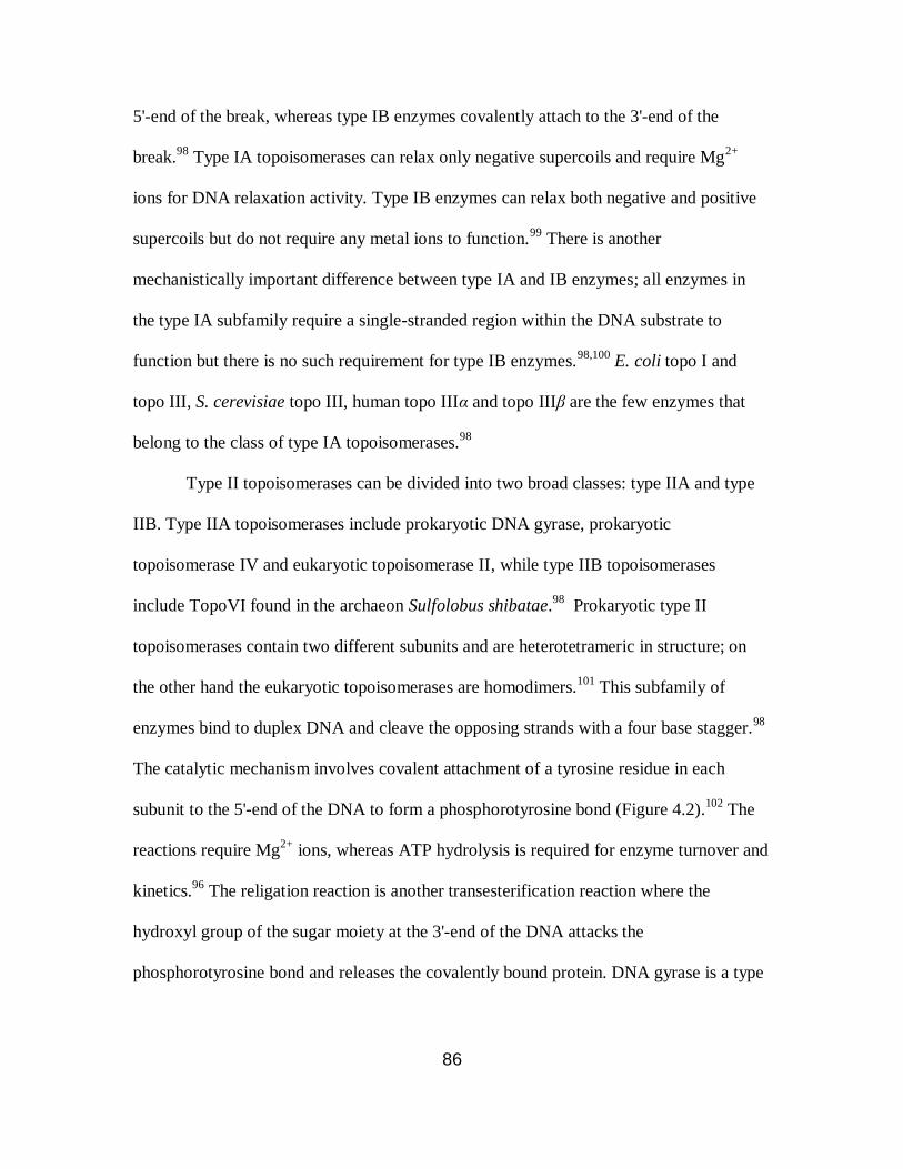
86
5'-end of the break, whereas type IB enzymes covalently attach to the 3'-end of the
break.98
Type IA topoisomerases can relax only negative supercoils and require Mg2+
ions for DNA relaxation activity. Type IB enzymes can relax both negative and positive
supercoils but do not require any metal ions to function.99
There is another
mechanistically important difference between type IA and IB enzymes; all enzymes in
the type IA subfamily require a single-stranded region within the DNA substrate to
function but there is no such requirement for type IB enzymes.98,100
E. coli topo I and
topo III, S. cerevisiae topo III, human topo IIIα and topo IIIβ are the few enzymes that
belong to the class of type IA topoisomerases.98
Type II topoisomerases can be divided into two broad classes: type IIA and type
IIB. Type IIA topoisomerases include prokaryotic DNA gyrase, prokaryotic
topoisomerase IV and eukaryotic topoisomerase II, while type IIB topoisomerases
include TopoVI found in the archaeon Sulfolobus shibatae.98
Prokaryotic type II
topoisomerases contain two different subunits and are heterotetrameric in structure; on
the other hand the eukaryotic topoisomerases are homodimers.101
This subfamily of
enzymes bind to duplex DNA and cleave the opposing strands with a four base stagger.98
The catalytic mechanism involves covalent attachment of a tyrosine residue in each
subunit to the 5'-end of the DNA to form a phosphorotyrosine bond (Figure 4.2).102
The
reactions require Mg2+
ions, whereas ATP hydrolysis is required for enzyme turnover and
kinetics.96
The religation reaction is another transesterification reaction where the
hydroxyl group of the sugar moiety at the 3'-end of the DNA attacks the
phosphorotyrosine bond and releases the covalently bound protein. DNA gyrase is a type
87
II enzyme found in all bacteria and is the only topoisomerase II enzyme capable of
generating negative supercoils in plasmid DNA.103
Type I and II DNA topoisomerases have been firmly established as effective
molecular targets for many antitumor drugs.104
There are three type I topoisomerases that
have been identified in human cells; topoisomerase I (topo I), topoisomerase IIIα (topo
IIIα) and topoisomerase IIIβ (topo IIIβ). Topo I functions as a pivot in DNA replication,
RNA transcription and chromosome condensation and segregation.104
It has been
demonstrated that topo I is essential in Drosophila and vertebrates but not in yeast.105,106
Murine or human cells having no or low levels of topo I were found to have replication
defects and exhibited altered gene expression.107,108
Topo I has transcriptional activities
independent of its DNA nicking-ligation activity such as promoter regulation109
and
phosphorylation/activation of splicing factors.110
Human cells have two topoisomerase II
isoenzymes; topoisomerase IIα (topo IIα) and topoisomerase IIβ (topo IIβ).99
Topo IIα is
crucial for cell growth and its levels are maximal in the late S/G2 phase of the cell cycle,
where it plays an important role in chromosome condensation and segregation.111
Despite
having similar enzymatic functions in vitro, topo IIβ expressions levels are always
constant throughout the cell cycle. Human topo IIα is mainly expressed in proliferating
cells, and is required for the completion of mitosis.112
By contrast, topo IIβ is required for
normal cell development but is not required for proliferation.113
A “classical” topoisomerase I or II poison acts by misaligning the free hydroxyl
group of the sugar moiety of DNA in the enyme–DNA covalent binary complex and
prevents the reverse transesterfication reaction to religate the DNA (Figure 4.1). In this
way topoisomerase poisons stabilize the normally transient enzyme–DNA covalent

88
intermediate and convert the binary covalent complex into a potential “suicide complex”
which leads to preferential cell death in cancer cells.93
A widely studied group of DNA
topoisomerase I poisons are camptothecin (CPT) and its derivatives.93
CPT was first
isolated from Camptotheca acuminate, a Chinese tree, in 1966 and is shown to have
exclusive topoisomerase I poison activity.114
In animal studies, CPT showed potent
antitumor activity against a broad spectrum of tumors which was accompanied by
excessive toxicity during phase I/II trials.115
CPT is inactive in its ring opened form
shown in Figure 4.3. Human topo I was identified as a molecular target of CPT and
subsequently, it was established as the sole antitumor target of CPT.114,116
Two families
of non-camptothecin topo I inhibitors have been to the clinical development stage,
namely indenoisoquinolines (NSC706744, NSC725766 and NSC724998) and
dibenzonaphthyridinones (Topovale, a Genzyme derivative).93
N
N
O
O
OH O
N
NO
OH O
OH
O-
Camptothecin (active) Camptothecin carboxylate (inactive)
OH-
H+
Figure 4.3. Hydrolysis of camptothecin to its inactive carboxylate form.93
Human topo II is a molecular target of many anti-cancer drugs including
doxorubicin, mitoxantrone, m-AMSA and etoposide. These topo II poisons stabilize the
covalent complex between the enzyme and DNA. Clinically useful topo II poisons are
highly sensitive to ATP and the ATP/ADP ratio for their poisoning activity.115
It has been

89
shown that the activity of etoposide increased by 50- to 100-fold in presence of ATP.115
This reflects the need for ATP for type II topoisomerase enzymes to function. The anti-
cancer activity of topo II poisons cannot be fully accounted for by drug-induced DNA
cleavage. Both DNA replication and RNA transcription have been suggested to be
involved in processing the covalent binary complex stabilized by topo II poisons.117
Selective inhibition of one type of topoisomerase can result in some negative
consequences. Topoisomerase I inhibition can cause a compensatory increase in
topoisomerase II levels, which may be associated with drug resistance against
topoisomerase I inhibitors/poisons.118
Simultaneous treatment with topo I poisons like
CPT and topo II poisons such etoposide has been shown to be antagonistic.117,119,120
Etoposide can potentially antagonize CPT by DNA replication inhibition, whereas CPT
may decrease the effects of etoposide by inhibiting transcription. Also, combination
therapies to target both topo I and topo II have shown significant toxicities, including
severe to life threating neutopenia and anemia.121
Recently, anticancer agents with dual
topoisomerase I and II inhibitory activities have generated significant interest. A number
of dual topoisomerase I and II inhibitors have been reported118,121
whereas only
saintopin122
and topopyrones A to D,123
have been reported as dual topoisomerase I and II
poisons to date.
Topopyrones A, B, C and D were first isolated from culture broths of two fungi,
Phoma sp. BAUA2861 and Pencillium sp. BAUA4206 by Kanai et al.124
in 2000 and
were reported to be human topoisomerase I poisons. Topopyrones, structurally similar to
both doxorubicin and some pluramycins, are anthraquinone polyphenols having a fused
1,4-pyrone ring in an angular or linear orientation and may or may not have a chlorine

90
substitution at position 7 (Figure 4.4).124
The total synthesis of all four topopyrones A to
D was reported by Elban and Hecht,125
followed by the report of topopyrones as dual
topoisormerase I and II poisons by Khan et al. in 2008.123
In the report by Khan et al.,
each of the topopyrones was able to stabilize enzyme–DNA covalent binary complexes
for both topoisomerases I and II, thus showing dual poison activity. A covalent binary
complex between 23 bp double stranded DNA, having a strong topoisomerase I cleavage
site, and topoisomerase I was shown to stabilize at the same DNA sites as camptothecin.
Alternatively, topopyrones A to D were compared with the known topoisomerase II
poison, etoposide, for their ability to stabilize topoisomerase IIα–DNA binary complex.
At the concentrations tested, all four topopyrones were shown to have potencies similar
to etoposide.
With the encouraging biological results for topopyrones A to D and the synthetic
route to access all four topopyrones in hand, an SAR study was the next logical step. We
initially chose to replace the chlorine substituent with a bromine substituent at position 7
for topopyrones A and B to obtain derivatives 4.8 and 4.11 (Figure 4.4), respectively.
The synthetic route followed to obtain the bromo-derivatives was similar as reported
earlier for topopyrones A and B.125
Along with the synthesis of topopyrones A to D, the
synthesis of two novel topopyrone derivatives 4.9 and 4.10 was also reported.125
For this
study, these two derivatives were synthesized following a similar strategy as reported
earlier.125
X-ray crystallographic analysis of the binding of other topoisomerase I poisons
to the DNA–topoisomerase I covalent binary complex indicates the H-bonding between
the hydroxyl groups of the poisons and the binary complex.127,128
Also, in a docking study
done between topopyrones and topopisomerase I, a potential hydrogen bond between

91
O
O
O
HO
OOH
OH
O
O
O
MeO
OOH
OMe
O
O
O
MeO OMe
Br
OMe
O
O
O
O
HO OH
Br
OH
O
O
O
OH
O OH
Br
OHO
O
O
O
HO OH
Cl
OH
O
O
O
OH
O OH
Cl
OHO
O
O
O
HO OH
OH
O
O
O
OH
O OH
OHO
topopyrone A (4.1) topopyrone B (4.2)
topopyrone C (4.3) topopyrone D (4.4)
O
O
O
HO OH
OH
O
O
O
OH
O OH
OHO
Cl Cl
4.5 4.6 4.7
4.8 4.9 4.10
4.11
N
N
O
O
OOH
O O
O
O
OO
O
OH
O
H
HHO
O
H
HOH
camptothecin
etoposide
Figure 4.4. Structures of topopyrones A to D, topopyrone derivatives, etoposide and
camptothecin.

92
the C6 OH group and Asn352 was observed for topopyrone C.129
Therefore, to
understand the role of the OH groups, compounds 4.5, 4.6 and 4.7 were also synthesized.
4.2. Results
The total synthesis of topopyrones A, B, C, and D was reported by Elban and
Hecht125
in 2008 along with the synthesis of two novel topopyrone derivatives (4.9 and
4.10). In the present study, a similar synthetic approach was adopted to gain access to two
new topopyrone C derivatives (4.5 and 4.6), two new topopyrone A derivatives (4.7 and
4.8) and one new topopyrone B derivative (4.11). Topopyrones 4.9 and 4.10 were
obtained in analogy with the procedure reported previously.125
Highlights of the synthetic
route shown here are the Diels–Alder addition reaction (4.20 and 4.21, Scheme 4.1),
Friedel-Crafts acylation reaction at the ortho position of phenol (4.16, Scheme 4.1), the
pyrone ring formation using a strong base (4.18, Schemes 4.1 and 4.7, Scheme 4.2), and
regioselective mono-bromination (4.23, Scheme 4.2).
4.2.1. Synthesis of topopyrones 4.5 and 4.6
Diels–Alder addition of the previously reported 1,3-dimethoxy-1-trimethylsiloxy-
1,3-butadiene125,130
and commercially available 2,5-dichloro-1,4-benzoquinone in the
presence of pyridine gave quinone 4.12. Protection of the phenol using p-toluenesulfonyl
chloride (p-TsCl) and K2CO3 in acetone at reflux afforded 4.13 in 90% yield. Pd/C
catalyzed hydrogenation effected reduction of the quinone, which was immediately
methylated using dimethyl sulfate and KOH to give 4.14 in 95% overall yield.
Compound 4.15 was obtained by removal of the tosylate group from 4.14 (aq KOH at
reflux). Although this reaction appeared to proceed cleanly as judged by silica gel TLC
analysis, the yield of isolated product was only moderate (56%). An acetyl group was

93
introduced ortho to the free hydroxyl group using TiCl4 and acetyl chloride in 1,2-
dichloroethane at reflux, affording compound 4.16 in 96% yield.131,132
The pyrone ring
was introduced at this stage via base-catalyzed Claisen condensation; compound 4.16 was
treated with NaH (60% dispersion in mineral oil) in freshly distilled ethyl acetate at
reflux to afford the β-diketone. This intermediate was subsequently treated with TFA to
obtain 4.17 (56%, two steps). Finally, compound 4.17 was oxidized to quinone 4.18 (72%
yield) using cerium(IV) ammonium nitrate in CH3CN. A second Diels−Alder addition
reaction was used to obtain the precursors for topopyrones 4.5 and 4.6. Thus quinone
4.18 was treated with 1-methoxy-1-trimethylsiloxy-3-tert-butyldimethylsiloxy-1,3-
butadiene (4.19)125
in the presence of pyridine to facilitate the aromatization of the
intermediate Diels−Alder addition product. Compounds 4.20 and 4.21 were thereby
obtained in 40% and 25% yields, respectively. Finally, compound 4.20 was treated with
tetra-n-butylammonium fluoride hydrate (TBAF·H2O) to remove the
tertbutyldimethylsilyl protecting group, followed by demethylation using 1 M BBr3 in
CH2Cl2. This afforded topopyrone C derivative 4.5 in 23% overall yield. Similarly,
topopyrone C derivative 4.6 was obtained in 50% yield by the treatment of 4.21 with
TBAF·H2O in THF.
4.2.2. Synthesis of topopyrones 4.7, 4.8 and 4.11
Monobrominated intermediate 4.23 was obtained in 20% overall yield by treating 4.2237
with N-bromosuccinimide (Scheme 4.2)133,134
in the presence of (iPr)2NH at −78 °C in
freshly distilled CH2Cl2, followed by methylation of the phenolic hydroxyl groups with
dimethyl sulfate and K2CO3 in acetone. Removal of the tosylate group, followed by O-
acetylation of the intermediate phenol afforded 4.24 (52%, two steps). This was achieved

94
Scheme 4.1. Synthesis of topopyrones 4.5 and 4.6.126
OH
MeO
O
O TsCl, K2CO3
acetone, reflux
90%
O
O
OTs
MeO
1) Pd/C, H2 (g), 1 atm
2) (MeO)2SO2, KOH OMe
OMe
OTs
MeO
THF-MeOH
THF-H2O
95%
4.12 4.13 4.14
Cl Cl Cl
KOH OMe
OMe
OH
MeO
EtOH-H2O, reflux
TiCl4, AcCl
OMe
OMe
OH
MeO
1,2-dichloroethane O
56% 96% ClCl
4.15 4.16
-10 oC rt
two steps
1) NaH, EtOAc, refluxOMe
OMe
O
MeO
O
2) TFA
cerium(IV) ammonium
O
O
O
MeO
OCH3CN-H2O
Cl
56% 72%
4.17 4.18
TBSO
OMe
OTMS
pyridine,benzene, reflux
4.19
Cl
4.16
two steps40% (4.20)
25% (4.21)
nitrate
1) TBAF.H2O, THF
2) BBr3
O
O
O
HO
OOH
OH
23%
O
O
O
MeO
OOH
OMe
TBAF.H2O, THF
4.6
50%
4.5
two steps
O
O
O
MeO
OOTBS
OH
4.20
O
O
O
MeO
OOTBS
OMe
4.21
+
using KOH in EtOH−H2O at reflux followed by treatment with acetic anhydride in the
presence of triethylamine and 4-N,N-dimethylaminopyridine.125
Treatment of acetylated
intermediate 4.24 with LiH in freshly distilled THF at reflux, followed by treatment with

95
Scheme 4.2. Synthesis of topopyrones 4.7, 4.8 and 4.11.126
O
O
OTs
MeO
O OH
OH
1) NBS, (iPr)2NH
CH2Cl2, -78 °CO
O
OTs
MeO
O OMe
OMe
Br2) (MeO)2SO2,K2CO3
acetone, reflux
1) KOH, 1:1 EtOH-H2O, reflux
2) Ac2O, TEA, DMAP, CH2Cl2
O
O
OAc
MeO
O
OMe
Br
OMe
4.22 4.23
4.24
52%
20%
two steps
O
O
O
MeO OMe
Br
OMe1) LiH, THF, reflux
2) TFA, CH2Cl2, 0 °CO
45%
4.7
two steps
two steps
O
O
O
HO OH
Br
OH
O
1M AlCl3 in NO2Ph
90 oC
50%
4.8
O
O
OH
O OH
Br
OHO
1% NaOH in MeOH, rt
50%
4.11
CF3COOH at 0 °C (Baker−Venkataraman rearrangement)135,136
led to pyrone ring
formation, affording topopyrone 4.7 in 45% yield for the two steps. Removal of the
methyl groups using 1 M AlCl3 in nitrobenzene at 90 °C gave brominated topopyrone A
derivative 4.8 in 50% yield.125
Brominated topopyrone B derivative 4.11 was obtained by
the rearrangement of the pyrone ring in 4.8 using 1% NaOH in MeOH (50% yield).124,125
4.2.3. Biological evaluation of topopyrone derivatives
Biological evaluation of the topopyrones shown in Figure 4.4 was performed by
Dr. Paul A. Zaleski.126
Compounds 4.5, 4.6, 4.7 and 4.8 were analyzed for their ability to
stabilize the DNA–topoisomerase I covalent binary complex by polyacrylamide gel
electrophoresis (Table 4.1). In this experiment, the amount of DNA cleaved by 1 µM
96
CPT was arbitrarily assigned a value of 100% and the amount of DNA cleaved by each
topopyrone was measured relative to that cleaved by CPT. Topopyrone 4.8 showed the
best results of the four topopyrones studied, producing 25% DNA cleavage at 10 µM
concentration whereas topopyrone 4.7, a trimethoxy derivative of topopyrone 4.8, failed
to stabilize the DNA-topoisomerase I covalent binary complex at 1 or 10 µM
concentration. Topopyronce C derivatives 4.5 and 4.6, on the other hand, did not show
much difference at 10 µM concentration (Table 4.1) and produced 11% and 10% DNA
cleavage, respectively.
In a separate experiment, a nitrocellulose filter binding assay was performed to
measure the amounts of DNA–topoisomerase I covalent binary complex stabilized by
topopyrones 4.3, 4.9 and 4.10 at 10 or 20 µM concentrations (Table 4.2). In the absence
of any poison, topoisomerase I was covalently bound to 1.4 ± 0.5% of the linearized
plasmid DNA substrate; in the presence of 0.5 µM CPT this increased to 39 ± 4.6%. Of
the three topopyrones, compound 4.10 employed at 20 µM was as efficient as 0.5 μM
CPT in stabilizing the covalent binary complex. Compound 4.9, having pyrone ring in an
angular position, did poorly in stabilizing the covalent binary complex. Only 5-6% DNA
covalently bound to topoisomerase I was observed.
Stabilization of the DNA–topoisomerase II covalent binary complex by either
etoposide or topopyrone 4.10 was also analyzed by a nitrocellulose filter binding assay.
Of the two human type II topoisomerases, isoform IIα was used for this study as it was
shown to be more relevant in proliferating cells.137
In the nitrocellulose filter binding
assay, the amount of covalent DNA–topoisomerase II binary complex was relatively high

97
Table 4.1. Topoisomerase I-mediated DNA cleavage by CPT and topopyrones on a 3′-32
P
end labeled 23-base pair oligonucleotide substrate.126
topoisomerase I poison % DNA cleavagea
DNA alone 1
Topoisomerase I alone 0
1 μM CPT 100
1 μM topopyrone 4.5 6
10 μM topopyrone 4.5 11
1 μM topopyrone 4.6 3
10 μM topopyrone 4.6 10
1 μM topopyrone 4.7 1
10 μM topopyrone 4.7 1
1 μM topopyrone 4.8 2
10 μM topopyrone 4.8 25
a Relative to CPT; determined by densitometric analysis.
This experiment was carried out by Dr. Paul A. Zaleski.
even in the absence of any poison. Etoposide at 0.5 µM concentration produced 34 ±
4.6% of the covalent binary complex. At 1 and 5 µM concentrations of 4.10, a similar
amount of the DNA was covalently bound to the enzyme.
Topopyrones 4.2, 4.9 and 4.10 were also analyzed for cleavage site specificity in
topopyrone–DNA–topoisomerase I ternary complex. These compounds were shown to
stabilize topoisomerase I cleavage at sites similar to CPT along with a new 5'-TG72-3'
cleavage site in a 158-bp DNA duplex substrate.126
Alternatively, topopyrones 4.2 and
4.11 stabilized topoisomerase II cleavage sites in 5'-32
P labeled 158 base pair DNA
duplex similar to etoposide (5'-CA23-3' and 5'-CG74-3') but with varying efficiencies.

98
Table 4.2. Nitrocellulose filter binding of CPT or topopyrone stabilized DNA–
topoisomerase covalent binary complexes.126
topoisomerase poisons DNA covalently bound to topoisomerase (%)a
topoisomerase I
none 1.4 ± 0.5
0.5 μM CPT 39 ± 4.6
10 μM topopyrone C (4.3) 16 ± 4.3
20 μM topopyrone C (4.3) 21 ± 7.8
10 μM topopyrone 4.9 6.1 ± 2.4
20 μM topopyrone 4.9 5.1 ± 4.4
10 μM topopyrone 4.10 11 ± 3.6
20 μM topopyrone 4.10 30 ± 5.2
topoisomerase IIα
none 23 ± 2.7
0.5 μM etoposide 34 ± 4.6
1 μM topopyrone 4.10 40 ± 13
5 μM topopyrone 4.10 34 ± 2.5 a Topoisomerase mediated cleavage conditions described in experimental procedures;
reaction mixtures contained 6 units of topoisomerase I or 5 units of topoisomerase IIα.
Values represent the mean ± SD (n = 3).
This experiment was carried out by Dr. Paul A. Zaleski.
4.3. Discussion
Elban and Hecht125
reported the total synthesis of topopyrones A to D (Figure 4.4)
and two novel topopyrone derivatives (4.9 and 4.10). At the time the topoyrones were
discovered, they were reported to be only topoisomerase I poisons.12436
In contrast, Khan
et al.123
found that all four topopyrones were dual topopisomerase I and II poisons. In this
report each of the topopyrones was able to stabilize enzyme–DNA covalent binary
complexes for both topoisomerases I and II, thus showing the dual poison activity.
Covalent binary complex formation between a 23 bp double-stranded DNA, having a

99
strong topoisomerase I cleavage site, and topoisomerase I was shown to stabilize the
same cleavage sites as camptothecin. On the other hand, topopyrones A to D were
compared with known topoisomerase II poison, etoposide, for their ability to stabilize
topoisomerase IIα–DNA binary complex. At the concentrations tested, all four
topopyrones were shown to have potency similar to etoposide.
In this thesis, the synthesis and biological activities of seven topopyrone
derivatives are reported. Initially compounds 4.8 and 4.11 were envisioned where the
chlorine substitution at position 7 was changed to a bromine substitution (Figure 4.4).
The synthetic route to obtain the bromo-derivatives was similar to that reported earlier for
topopyrones A and B.125
Compounds 4.9 and 4.10 were synthesized following a similar
strategy as reported earlier by Elban and Hecht.125
To understand the role of the hydroxyl
groups in stabilization of DNA–topoisomerase I or II covalent binary complexes,
compounds 4.5, 4.6 and 4.7 were also synthesized.
Scheme 4.1 illustrates the synthesis of compounds 4.5 and 4.6, which began with
the protection of the phenol in a previously reported compound, 4.12. The phenol was
protected as a tosylate, where treatment of 4.12 with p-toluenesulfonyl chloride and
K2CO3 in acetone at reflux gave product 4.13 in 90% yield. The next step was to protect
the quinone via catalytic reduction followed by methylation. Pd/C catalyzed
hydrogenation in 1:1 methanol–THF afforded the reduced quinone, which was
immediately derivatized by methylation using dimethyl sulfate and KOH in 1:1 THF–
water to give 4.14 in 95% overall yield. The next reaction was the deprotection of the
tosylate group in aq KOH at reflux for 4 h. Although this reaction appeared to proceed
cleanly as judged by silica gel TLC analysis, the yield of the isolated product was only
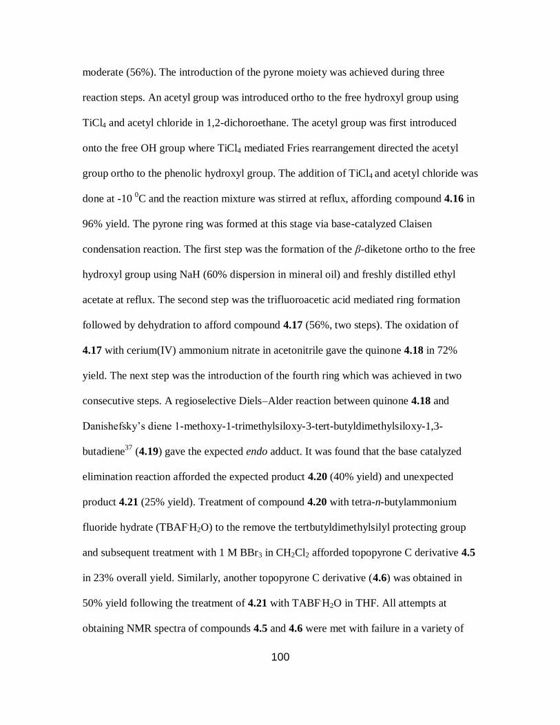
100
moderate (56%). The introduction of the pyrone moiety was achieved during three
reaction steps. An acetyl group was introduced ortho to the free hydroxyl group using
TiCl4 and acetyl chloride in 1,2-dichoroethane. The acetyl group was first introduced
onto the free OH group where TiCl4 mediated Fries rearrangement directed the acetyl
group ortho to the phenolic hydroxyl group. The addition of TiCl4 and acetyl chloride was
done at -10 0C and the reaction mixture was stirred at reflux, affording compound 4.16 in
96% yield. The pyrone ring was formed at this stage via base-catalyzed Claisen
condensation reaction. The first step was the formation of the β-diketone ortho to the free
hydroxyl group using NaH (60% dispersion in mineral oil) and freshly distilled ethyl
acetate at reflux. The second step was the trifluoroacetic acid mediated ring formation
followed by dehydration to afford compound 4.17 (56%, two steps). The oxidation of
4.17 with cerium(IV) ammonium nitrate in acetonitrile gave the quinone 4.18 in 72%
yield. The next step was the introduction of the fourth ring which was achieved in two
consecutive steps. A regioselective Diels–Alder reaction between quinone 4.18 and
Danishefsky’s diene 1-methoxy-1-trimethylsiloxy-3-tert-butyldimethylsiloxy-1,3-
butadiene37
(4.19) gave the expected endo adduct. It was found that the base catalyzed
elimination reaction afforded the expected product 4.20 (40% yield) and unexpected
product 4.21 (25% yield). Treatment of compound 4.20 with tetra-n-butylammonium
fluoride hydrate (TBAF.H2O) to the remove the tertbutyldimethylsilyl protecting group
and subsequent treatment with 1 M BBr3 in CH2Cl2 afforded topopyrone C derivative 4.5
in 23% overall yield. Similarly, another topopyrone C derivative (4.6) was obtained in
50% yield following the treatment of 4.21 with TABF.H2O in THF. All attempts at
obtaining NMR spectra of compounds 4.5 and 4.6 were met with failure in a variety of

101
solvent systems. Therefore, 4.5 and 4.6 were converted to 3,5,7-trimethoxy and 7-acetoxy
derivatives, respectively. Treatment of 4.5 with dimethyl sulfate and K2CO3 in acetone at
reflux gave the derivative as an orange solid in 45% yield. Compound 4.6 was treated
with acetic anhydride in pyridine to obtain the corresponding acetoxy derivative (68%
yield, yellow solid). The NMR data obtained for the trimethoxy or acetoxy derivative was
in accordance with the expected structures of compounds 4.5 and 4.6.
Scheme 4.2 outlines the synthesis of the brominated derivatives 4.7, 4.8 and 4.11.
Previously reported compound 4.22125
was treated with N-bromosuccinimide in the
presence of diisopropylamine to obtain the monobrominated crude product. The
bromination was done at -78 oC and the reaction was complete within 5 minutes as
judged by silica gel TLC analysis. The crude product was then treated with dimethyl
sulfate in presence of K2CO3 to obtain the methylated product 4.23 in 20% overall yield.
The tosylate protecting group was removed by treatment with KOH in 1:1 ethanol–water
at reflux to afford the phenolic intermediate which was subsequently acetylated by using
acetic anhydride in presence of triethylamine and 4-N,N-dimethylaminopyridine to obtain
4.24 (52%, two steps). At this stage, the pyrone ring was introduced following Baker-
Venkataraman rearrangement. Treatment of 4.24 with LiH in freshly distilled THF at
reflux gave the β-diketone intermediate which lead to the formation of pyrone ring by the
treatment of trifluoroacetic anhydride at 0 oC, affording topopyrone 4.7 in 45% yield for
the two steps. This reaction was highly sensitive to moisture; deacylated product was
often recovered with no formation of the desired product. A harsh demethylation
procedure was applied to obtain topopyrone 4.8. Treatment of 4.7 with 1 M AlCl3 in
nitrobenzene over two days at 90 oC gave the brominated topopyrone in 50% yield. Kanai

102
et al.124
reported that the angular pyrone ring (topopyrone A) underwent a base catalyzed
rearrangement to give thermodynamically stable linear pyrone ring (topopyrone B). A
similar method was applied to obtain compound 4.11 in 50% yield, where 4.8 was treated
with 1% NaOH in methanol. Again, compounds 4.8 and 4.11 could not be characterized
by NMR due to poor solubility in a variety of solvents. Only 4.11 was successfully
converted to its trimethoxy derivative by using dimethyl sulfate and K2CO3. The NMR
characterization of the trimethoxy derivative of 4.11 confirmed the structure of
compound 4.11. All attempts to convert compound 4.8 into its methoxy or acetoxy
derivative met with failure; therefore, it was only characterized by high resolution mass
spectrometry.
The biological evaluation of the topopyrones was done by Dr. Paul A. Zaleski.126
The ability of the novel topopyrones 4.5 and 4.6 to stabilize the DNA−topoisomerase I
covalent binary complex was investigated using a 23-base pair oligonucleotide substrate
that contained a strong topoisomerase I cleavage site having guanosine at the +1 position
and thymidine at the −1 position.138
Both compounds stabilized the DNA–topoisomerase
I covalent binary complex with similar efficiencies as analyzed by the polyacrylamide gel
electrophoresis. In the densitometric analysis, the percentage of DNA cleavage obtained
by 1 μM CPT was arbitrarily assigned a value of 100% (Table 4.1). Topopyrone 4.5 gave
6% and 11% DNA cleavage at 1 and 10 μM concentrations, respectively, which is similar
to that obtained with topopyrone 4.6. On the other hand, considerable variations were
seen in DNA cleavage stabilized by topopyrones 4.7 and 4.8 (Table 4.1). Topopyrone 4.8
produced 25% DNA cleavage at 10 μM concentration, whereas topopyrone 4.7 failed to
stabilize the DNA–topoisomerase I covalent binary complex at either 1 or 10 μM
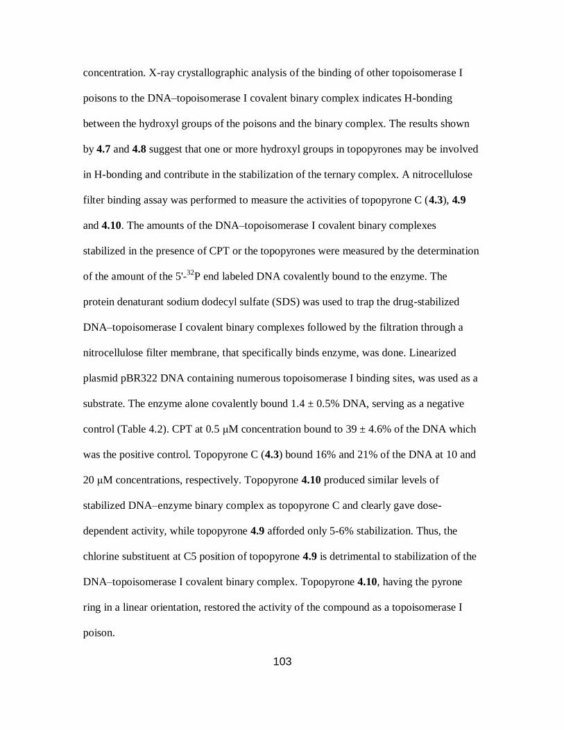
103
concentration. X-ray crystallographic analysis of the binding of other topoisomerase I
poisons to the DNA–topoisomerase I covalent binary complex indicates H-bonding
between the hydroxyl groups of the poisons and the binary complex. The results shown
by 4.7 and 4.8 suggest that one or more hydroxyl groups in topopyrones may be involved
in H-bonding and contribute in the stabilization of the ternary complex. A nitrocellulose
filter binding assay was performed to measure the activities of topopyrone C (4.3), 4.9
and 4.10. The amounts of the DNA–topoisomerase I covalent binary complexes
stabilized in the presence of CPT or the topopyrones were measured by the determination
of the amount of the 5'-32
P end labeled DNA covalently bound to the enzyme. The
protein denaturant sodium dodecyl sulfate (SDS) was used to trap the drug-stabilized
DNA–topoisomerase I covalent binary complexes followed by the filtration through a
nitrocellulose filter membrane, that specifically binds enzyme, was done. Linearized
plasmid pBR322 DNA containing numerous topoisomerase I binding sites, was used as a
substrate. The enzyme alone covalently bound 1.4 ± 0.5% DNA, serving as a negative
control (Table 4.2). CPT at 0.5 μM concentration bound to 39 ± 4.6% of the DNA which
was the positive control. Topopyrone C (4.3) bound 16% and 21% of the DNA at 10 and
20 μM concentrations, respectively. Topopyrone 4.10 produced similar levels of
stabilized DNA–enzyme binary complex as topopyrone C and clearly gave dose-
dependent activity, while topopyrone 4.9 afforded only 5-6% stabilization. Thus, the
chlorine substituent at C5 position of topopyrone 4.9 is detrimental to stabilization of the
DNA–topoisomerase I covalent binary complex. Topopyrone 4.10, having the pyrone
ring in a linear orientation, restored the activity of the compound as a topoisomerase I
poison.

104
Compound 4.10 produced about the same amount of bound topo I as CPT, albeit
at a higher concentration. Therefore, it was an ideal topopyrone derivative for testing
activity as a topoisomerase II poison. As already discussed in section 4.1, topoisomerase
IIα is relevant in proliferating cells. Therefore, this isoform of the human topoisomerase
II enzyme was used for the study. In contrast to topoisomerase I, high levels of ostensibly
covalently bound DNA (23 ± 2.7%) were observed without any topoisomerase II poison.
In the presence of 0.5 μM etoposide, 34 ± 4.6% of the DNA was covalently bound to
topoisomerase II enzyme. Topopyorone 4.10 produced similar levels of stabilized DNA–
enzyme binary complex at both 1 and 5 μM concentrations.
The analysis of the cleavage site specificity of topopyrone-stabilized DNA–
topoisomerase I or II covalent binary complexed was also done using either 3'-32
P or 5'-
32P end labeled 158-base pair DNA duplex substrate. At 1 μM CPT, several DNA
cleavage sites at 5'-TG-3' and 5'-TA-3' were observed. The topopyrones were found to
stabilize topoisomerase I cleavage at sites similar to CPT but with varying efficiencies.
Interestingly, three topopyrones (4.3, 4.9 and 4.10) stabilized a topoisomerase I cleavage
site at 5'-GT72-3' which was not observed in case of CPT. Topopyrones 4.2 and 4.11 were
used to study the cleavage site specificity of stabilized DNA–topoisomerase II covalent
binary complexes and were compared with etoposide. Both the topopyrones were found
to produce similar DNA cleavage sites as etoposide (5'-CA23-3' and 5'-CG74-3') with
varying efficiencies.

105
4.4. Experimental
4.4.1. General experimental procedures
Chemicals and solvents were purchased as reagent grade from Sigma-Aldrich
Chemicals and were used without further purification. All the reactions were performed
under an argon atmosphere, unless otherwise specified. Thin-layer chromatography
(TLC) plates (precoated glass plates with silica gel 60 F254, 0.25 mm thickness) were
used for analytical TLC and were visualized by UV irradiation (254 nm). Flash
chromatography was carried out using Silicycle 200−400 mesh silica gel. 1H and
13C
NMR spectra were obtained using a Varian 400 MHz NMR spectrometer. Chemical
shifts (δ) are reported in parts per million (ppm) and are referenced to residual CHCl3 (δ
7.26 ppm for 1H NMR and δ 77.16 for 13
C NMR) as the internal strandard. Splitting
patterns are designated as s, singlet; d, doublet; m, multiplet. High-resolution mass
spectra were obtained at the Michigan State Mass Spectrometry Facility or the Arizona
State University CLAS High Resolution Mass Spectrometry Facility.
4.4.2. Synthesis of topopyrone derivatives
O
O
OTs
MeO Cl
Toluene-4-sulfonic Acid 6-Chloro-3-methoxy-5,8-dioxo-5,8-dihydronaphthalen-1-yl
Ester (4.13). To a solution containing 0.41g (1.7 mmol) of 4.1243
in 100 mL of acetone at
room temperature was added 0.65 g (3.4 mmol) of p-toluenesulfonyl chloride (p-TsCl)
and 0.47 g (3.4 mmol) of K2CO3. The reaction mixture was heated at reflux for 6 h, and
the cooled solution was filtered through a silica gel plug. The silica gel was washed with
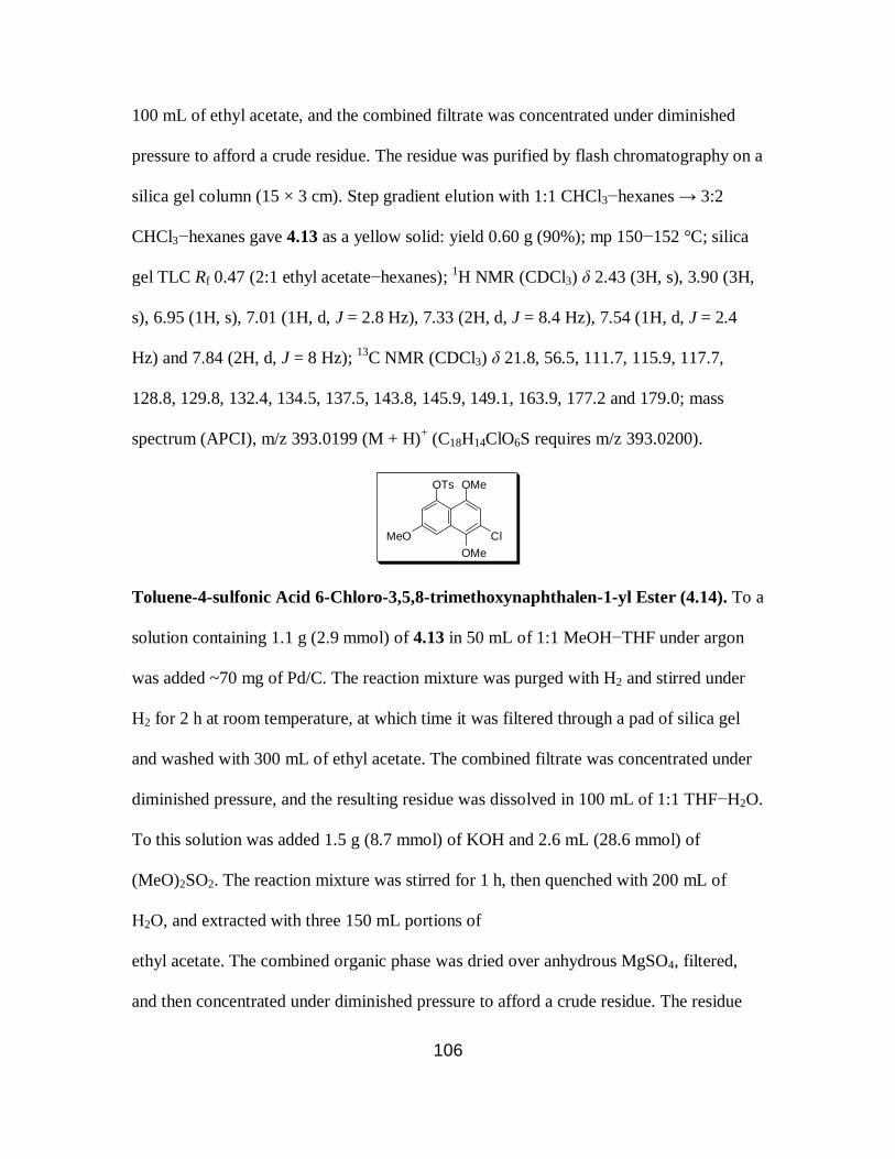
106
100 mL of ethyl acetate, and the combined filtrate was concentrated under diminished
pressure to afford a crude residue. The residue was purified by flash chromatography on a
silica gel column (15 × 3 cm). Step gradient elution with 1:1 CHCl3−hexanes → 3:2
CHCl3−hexanes gave 4.13 as a yellow solid: yield 0.60 g (90%); mp 150−152 °C; silica
gel TLC Rf 0.47 (2:1 ethyl acetate−hexanes); 1H NMR (CDCl3) δ 2.43 (3H, s), 3.90 (3H,
s), 6.95 (1H, s), 7.01 (1H, d, J = 2.8 Hz), 7.33 (2H, d, J = 8.4 Hz), 7.54 (1H, d, J = 2.4
Hz) and 7.84 (2H, d, J = 8 Hz); 13
C NMR (CDCl3) δ 21.8, 56.5, 111.7, 115.9, 117.7,
128.8, 129.8, 132.4, 134.5, 137.5, 143.8, 145.9, 149.1, 163.9, 177.2 and 179.0; mass
spectrum (APCI), m/z 393.0199 (M + H)+ (C18H14ClO6S requires m/z 393.0200).
OMe
OMe
OTs
MeO Cl
Toluene-4-sulfonic Acid 6-Chloro-3,5,8-trimethoxynaphthalen-1-yl Ester (4.14). To a
solution containing 1.1 g (2.9 mmol) of 4.13 in 50 mL of 1:1 MeOH−THF under argon
was added ~70 mg of Pd/C. The reaction mixture was purged with H2 and stirred under
H2 for 2 h at room temperature, at which time it was filtered through a pad of silica gel
and washed with 300 mL of ethyl acetate. The combined filtrate was concentrated under
diminished pressure, and the resulting residue was dissolved in 100 mL of 1:1 THF−H2O.
To this solution was added 1.5 g (8.7 mmol) of KOH and 2.6 mL (28.6 mmol) of
(MeO)2SO2. The reaction mixture was stirred for 1 h, then quenched with 200 mL of
H2O, and extracted with three 150 mL portions of
ethyl acetate. The combined organic phase was dried over anhydrous MgSO4, filtered,
and then concentrated under diminished pressure to afford a crude residue. The residue
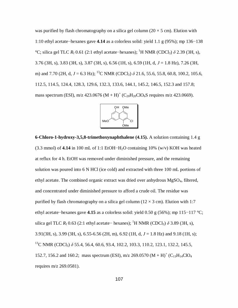
107
was purified by flash chromatography on a silica gel column (20 × 5 cm). Elution with
1:10 ethyl acetate−hexanes gave 4.14 as a colorless solid: yield 1.1 g (95%); mp 136−138
°C; silica gel TLC Rf 0.61 (2:1 ethyl acetate−hexanes); 1H NMR (CDCl3) δ 2.39 (3H, s),
3.76 (3H, s), 3.83 (3H, s), 3.87 (3H, s), 6.56 (1H, s), 6.59 (1H, d, J = 1.8 Hz), 7.26 (3H,
m) and 7.70 (2H, d, J = 6.3 Hz); 13
C NMR (CDCl3) δ 21.6, 55.6, 55.8, 60.8, 100.2, 105.6,
112.5, 114.5, 124.4, 128.3, 129.6, 132.3, 133.6, 144.1, 145.2, 146.5, 152.3 and 157.8;
mass spectrum (ESI), m/z 423.0676 (M + H)+ (C20H20ClO6S requires m/z 423.0669).
OMe
OMe
OH
MeO Cl
6-Chloro-1-hydroxy-3,5,8-trimethoxynaphthalene (4.15). A solution containing 1.4 g
(3.3 mmol) of 4.14 in 100 mL of 1:1 EtOH−H2O containing 10% (w/v) KOH was heated
at reflux for 4 h. EtOH was removed under diminished pressure, and the remaining
solution was poured into 6 N HCl (ice cold) and extracted with three 100 mL portions of
ethyl acetate. The combined organic extract was dried over anhydrous MgSO4, filtered,
and concentrated under diminished pressure to afford a crude oil. The residue was
purified by flash chromatography on a silica gel column (12 × 3 cm). Elution with 1:7
ethyl acetate−hexanes gave 4.15 as a colorless solid: yield 0.50 g (56%); mp 115−117 °C;
silica gel TLC Rf 0.63 (2:1 ethyl acetate− hexanes); 1H NMR (CDCl3) δ 3.89 (3H, s),
3.91(3H, s), 3.99 (3H, s), 6.55-6.56 (2H, m), 6.92 (1H, d, J = 1.8 Hz) and 9.18 (1H, s);
13C NMR (CDCl3) δ 55.4, 56.4, 60.6, 93.4, 102.2, 103.3, 110.2, 123.1, 132.2, 145.5,
152.7, 156.2 and 160.2; mass spectrum (ESI), m/z 269.0570 (M + H)+ (C13H14ClO4
requires m/z 269.0581).
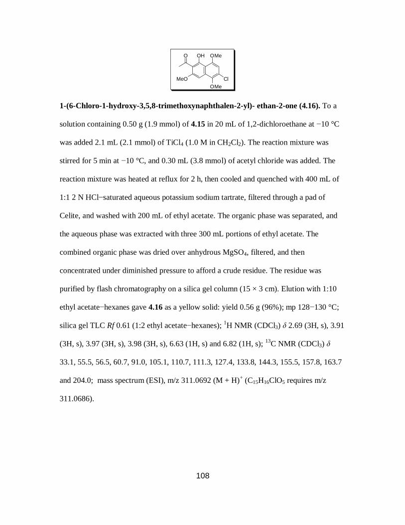
108
OMe
OMe
OH
MeO
O
Cl
1-(6-Chloro-1-hydroxy-3,5,8-trimethoxynaphthalen-2-yl)- ethan-2-one (4.16). To a
solution containing 0.50 g (1.9 mmol) of 4.15 in 20 mL of 1,2-dichloroethane at −10 °C
was added 2.1 mL (2.1 mmol) of TiCl4 (1.0 M in CH2Cl2). The reaction mixture was
stirred for 5 min at −10 °C, and 0.30 mL (3.8 mmol) of acetyl chloride was added. The
reaction mixture was heated at reflux for 2 h, then cooled and quenched with 400 mL of
1:1 2 N HCl−saturated aqueous potassium sodium tartrate, filtered through a pad of
Celite, and washed with 200 mL of ethyl acetate. The organic phase was separated, and
the aqueous phase was extracted with three 300 mL portions of ethyl acetate. The
combined organic phase was dried over anhydrous MgSO4, filtered, and then
concentrated under diminished pressure to afford a crude residue. The residue was
purified by flash chromatography on a silica gel column (15 × 3 cm). Elution with 1:10
ethyl acetate−hexanes gave 4.16 as a yellow solid: yield 0.56 g (96%); mp 128−130 °C;
silica gel TLC Rf 0.61 (1:2 ethyl acetate−hexanes); 1H NMR (CDCl3) δ 2.69 (3H, s), 3.91
(3H, s), 3.97 (3H, s), 3.98 (3H, s), 6.63 (1H, s) and 6.82 (1H, s); 13
C NMR (CDCl3) δ
33.1, 55.5, 56.5, 60.7, 91.0, 105.1, 110.7, 111.3, 127.4, 133.8, 144.3, 155.5, 157.8, 163.7
and 204.0; mass spectrum (ESI), m/z 311.0692 (M + H)+ (C15H16ClO5 requires m/z
311.0686).

109
OMe
OMe
O
MeO
O
Cl
8-Chloro-2-methyl-5,7,10-trimethoxybenzo[h]chromen-4-one (4.17). To a solution
containing 0.40 g (1.29 mmol) of 4.16 in 20 mL of freshly distilled ethyl acetate was
added 0.57 g (14.1 mmol) of NaH (60% dispersion in mineral oil). The reaction mixture
was heated at reflux for 1 h, cooled, and quenched with 100 mL of 2 N HCl (ice cold).
The solution was extracted with three 200 mL portions of CHCl3, dried over anhydrous
MgSO4, filtered, and concentrated under diminished pressure to afford a crude residue.
The residue was dissolved in 15 mL of TFA at 0 °C and stirred for 15 min at 0 °C and
then for 30 min at room temperature. The solvent was concentrated under diminished
pressure, and the resulting residue was coevaporated with two 50 mL portions of toluene.
The resulting residue was purified by flash chromatography on a silica gel column (15 ×
2 cm). Step gradient elution with 1:1 ethyl acetate−hexanes → 20% MeOH in CHCl3
gave 4.17 as a colorless solid: yield 0.24 g (56%); mp 182−184 °C; silica gel TLC Rf 0.43
(15% MeOH in 1:1 ethyl acetate− hexanes); 1H NMR (CDCl3) δ 2.42 (s, 3H), 3.95 (s,
3H), 3.97 (s, 3H), 4.06 (s, 3H), 6.26 (s, 1H), 6.76 (s, 1H) and 7.23 (s, 1H); 13
C NMR
(CDCl3) δ 19.8, 56.3, 56.4, 60.9, 96.4, 106.5, 110.4, 113.5, 113.9, 127.3, 132.7, 143.9,
154.2, 157.2, 157.4, 163.2 and 177.6; mass spectrum (ESI), m/z 335.0675 (M + H)+
(C17H16ClO5 requires m/z 335.0686).

110
O
O
O
MeO
O
Cl
8-Chloro-5-methoxy-2-methylbenzo[h]chromene-4,7,10-trione (4.18). To a solution
containing 0.24 g (0.71 mmol) of 4.17 in 50 mL of CH3CN at 0 °C was added 0.77 g (1.4
mmol) of ceric ammonium nitrate in 8 mL of H2O. The reaction mixture was warmed to
room temperature and stirred for 0.5 h, at which time 150 mL of H2O was added. The
reaction mixture was extracted with four 150 mL portions of CHCl3, and the combined
organic phase was dried over anhydrous MgSO4, filtered, and concentrated under
diminished pressure to afford a crude residue. The residue was purified by flash
chromatography on a silica gel column (15 × 2 cm). Elution with CHCl3 as eluant gave
4.18 as a yellow solid: yield 0.16 g (72%); mp 257−259 °C; silica gel TLC Rf 0.50 (15%
MeOH in 1:1 ethyl acetate−hexanes); 1H NMR (CDCl3) δ 2.43 (3H, s), 4.13 (3H, s), 6.18
(1H, s), 7.12 (1H, s) and 7.52 (1H, s); 13
C NMR (CDCl3) δ 20.0, 57.4, 104.9, 113.1,
113.3, 117.6, 135.9, 138.2, 143.1, 157.3, 164.2, 165.4, 176.4, 177.6 and 179.3; mass
spectrum (ESI), m/z 305.0213 (M + H)+ (C15H10O5Cl requires m/z 305.0217).
O
O
O
MeO
OOTBS
OH
O
O
O
MeO
OOTBS
OMe
10-(tert-Butyldimethylsilyloxy)-8-hydroxy-5-methoxy-2- methyl-1-
oxabenzo[a]anthracene-4,7,12-trione (4.20) and 10-(tert-Butyldimethylsilyloxy)-5,8-
dimethoxy-2-methyl-1-oxabenzo[a]anthracene-4,7,12-trione (4.21). To a solution
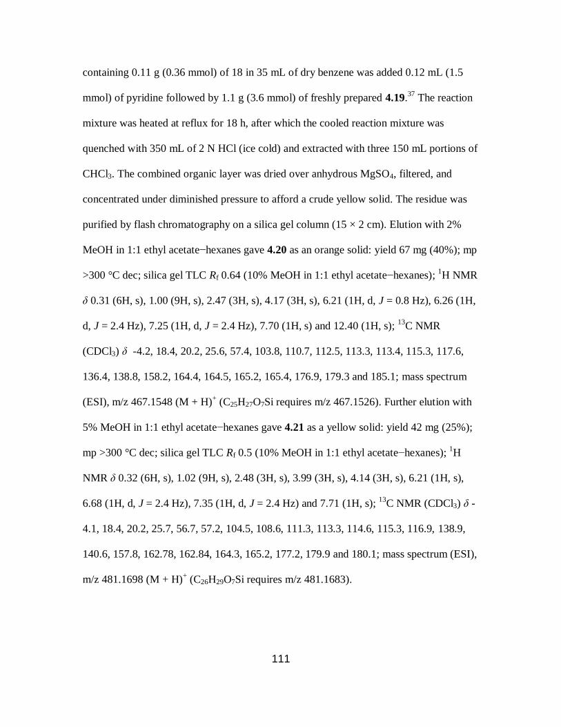
111
containing 0.11 g (0.36 mmol) of 18 in 35 mL of dry benzene was added 0.12 mL (1.5
mmol) of pyridine followed by 1.1 g (3.6 mmol) of freshly prepared 4.19.37
The reaction
mixture was heated at reflux for 18 h, after which the cooled reaction mixture was
quenched with 350 mL of 2 N HCl (ice cold) and extracted with three 150 mL portions of
CHCl3. The combined organic layer was dried over anhydrous MgSO4, filtered, and
concentrated under diminished pressure to afford a crude yellow solid. The residue was
purified by flash chromatography on a silica gel column (15 × 2 cm). Elution with 2%
MeOH in 1:1 ethyl acetate−hexanes gave 4.20 as an orange solid: yield 67 mg (40%); mp
>300 °C dec; silica gel TLC Rf 0.64 (10% MeOH in 1:1 ethyl acetate−hexanes); 1H NMR
δ 0.31 (6H, s), 1.00 (9H, s), 2.47 (3H, s), 4.17 (3H, s), 6.21 (1H, d, J = 0.8 Hz), 6.26 (1H,
d, J = 2.4 Hz), 7.25 (1H, d, J = 2.4 Hz), 7.70 (1H, s) and 12.40 (1H, s); 13
C NMR
(CDCl3) δ -4.2, 18.4, 20.2, 25.6, 57.4, 103.8, 110.7, 112.5, 113.3, 113.4, 115.3, 117.6,
136.4, 138.8, 158.2, 164.4, 164.5, 165.2, 165.4, 176.9, 179.3 and 185.1; mass spectrum
(ESI), m/z 467.1548 (M + H)+ (C25H27O7Si requires m/z 467.1526). Further elution with
5% MeOH in 1:1 ethyl acetate−hexanes gave 4.21 as a yellow solid: yield 42 mg (25%);
mp >300 °C dec; silica gel TLC Rf 0.5 (10% MeOH in 1:1 ethyl acetate−hexanes); 1H
NMR δ 0.32 (6H, s), 1.02 (9H, s), 2.48 (3H, s), 3.99 (3H, s), 4.14 (3H, s), 6.21 (1H, s),
6.68 (1H, d, J = 2.4 Hz), 7.35 (1H, d, J = 2.4 Hz) and 7.71 (1H, s); 13
C NMR (CDCl3) δ -
4.1, 18.4, 20.2, 25.7, 56.7, 57.2, 104.5, 108.6, 111.3, 113.3, 114.6, 115.3, 116.9, 138.9,
140.6, 157.8, 162.78, 162.84, 164.3, 165.2, 177.2, 179.9 and 180.1; mass spectrum (ESI),
m/z 481.1698 (M + H)+ (C26H29O7Si requires m/z 481.1683).
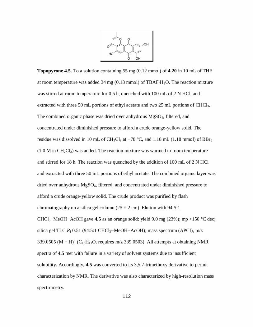
112
O
O
O
HO
OOH
OH
Topopyrone 4.5. To a solution containing 55 mg (0.12 mmol) of 4.20 in 10 mL of THF
at room temperature was added 34 mg (0.13 mmol) of TBAF·H2O. The reaction mixture
was stirred at room temperature for 0.5 h, quenched with 100 mL of 2 N HCl, and
extracted with three 50 mL portions of ethyl acetate and two 25 mL portions of CHCl3.
The combined organic phase was dried over anhydrous MgSO4, filtered, and
concentrated under diminished pressure to afford a crude orange-yellow solid. The
residue was dissolved in 10 mL of CH2Cl2 at −78 °C, and 1.18 mL (1.18 mmol) of BBr3
(1.0 M in CH2Cl2) was added. The reaction mixture was warmed to room temperature
and stirred for 18 h. The reaction was quenched by the addition of 100 mL of 2 N HCl
and extracted with three 50 mL portions of ethyl acetate. The combined organic layer was
dried over anhydrous MgSO4, filtered, and concentrated under diminished pressure to
afford a crude orange-yellow solid. The crude product was purified by flash
chromatography on a silica gel column (25 × 2 cm). Elution with 94:5:1
CHCl3−MeOH−AcOH gave 4.5 as an orange solid: yield 9.0 mg (23%); mp >150 °C dec;
silica gel TLC Rf 0.51 (94:5:1 CHCl3−MeOH−AcOH); mass spectrum (APCI), m/z
339.0505 (M + H)+ (C18H11O7 requires m/z 339.0503). All attempts at obtaining NMR
spectra of 4.5 met with failure in a variety of solvent systems due to insufficient
solubility. Accordingly, 4.5 was converted to its 3,5,7-trimethoxy derivative to permit
characterization by NMR. The derivative was also characterized by high-resolution mass
spectrometry.

113
To a solution containing 10.0 mg (0.03 mmol) of 4.5 in 5 mL of acetone was
added 0.04 mL (0.30 mmol) of (MeO)2SO2 and 42 mg (0.30 mmol) of K2CO3. The
reaction mixture was heated at reflux for 18 h, quenched with 50 mL of 1 N HCl, and
extracted with two 20 mL portions of ethyl acetate. The combined organic extract was
dried over anhydrous MgSO4, then filtered and concentrated under diminished pressure to
afford a crude residue. The crude product was purified by flash chromatography on a
silica gel column (15 × 2 cm). Elution with 2% MeOH in CHCl3 gave the 3,5,7-
trimethoxy derivative of topopyrone 4.5 as an orange solid: yield 5.0 mg (45%); silica gel
TLC Rf 0.48 (2% MeOH in CHCl3); 1H NMR (CDCl3) δ 2.48 (3H, d, J = 0.4 Hz), 3.98
(3H, s), 4.01 (3H, s), 4.14 (3H, s), 6.20 (1H, d, J = 0.4 Hz), 6.82 (1H, d, J = 2.8 Hz), 7.35
(1H, d, J = 2.4 Hz) and 7.58 (1H, s); 13
C NMR (CDCl3) δ 20.7, 56.5, 57.2, 57.5, 103.0,
103.5,106.2, 113.5, 117.6, 118.1, 118.5, 136.3, 137.4, 158.1, 162.4, 163.3, 164.5, 165.9,
177.6, 179.6, and 183.5; mass spectrum (ESI), m/z 381.0983 (M + H)+ (C21H17O7
requires m/z 381.0974).
O
O
O
MeO
OOH
OMe
Topopyrone 4.6. To a solution containing 52 mg (0.11 mmol) of 4.21 in 10 mL of THF
at room temperature was added 30 mg (0.11 mmol) of TBAF·H2O. The reaction mixture
was stirred for 1 h at room temperature, quenched with 100 mL of 2 N HCl, and extracted
with three 50 mL portions of ethyl acetate and two 25 mL portions of CHCl3. The
combined organic phase was dried over anhydrous MgSO4, filtered, and then
concentrated under diminished pressure to afford a crude orange-yellow solid. The crude

114
product was purified by flash chromatography on a silica gel column (20 × 2 cm). Elution
with 94:5:1 CHCl3−MeOH−AcOH gave 4.6 as a yellow solid: yield 20 mg (50%); mp
>180 °C dec; silica gel TLC Rf 0.42 (94:5:1 CHCl3− MeOH−AcOH); mass spectrum
(ESI), m/z 367.0807 (M + H)+ (C20H15O7 requires m/z 367.0818). All attempts at
obtaining NMR spectra of 4.6 met with failure in a variety of solvent systems due to
insufficient solubility. Accordingly, 4.6 was converted to its 7-acetoxy derivative to
permit characterization by NMR. The derivative was also characterized by high-
resolution mass spectrometry.
A mixture of 12 mg (33 μmol) of 4.6, 3 mL of acetic anhydride, and 1.5 mL of
pyridine was stirred at room temperature for 3 h. The reaction mixture was quenched
with 50 mL of 2 N HCl and extracted with two 25 mL portions of ethyl acetate. The
combined organic extract was dried over anhydrous MgSO4, filtered, and concentrated
under diminished pressure to afford a crude residue. The crude product was purified by
flash chromatography on a silica gel column (15 × 2 cm). Elution with 3% MeOH in
CHCl3 gave the 7-acetoxy derivative of topopyrone 4.6 as a yellow solid: yield 9.0 mg
(68%); silica gel TLC Rf 0.50 (2% MeOH in CHCl3); 1H NMR (CDCl3) δ 2.37 (3H, s),
2.48 (3H, s), 4.04 (3H, s), 4.15 (3H, s), 6.22 (1H, s), 7.07 (1H, d, J = 2.0 Hz), 7.66 (1H, d,
J = 2.4 Hz), and 7.69 (1H, s); 13
CNMR (CDCl3) δ 19.9, 21.1, 56.8, 57.1, 104.3, 110.4,
113.1, 113.2, 114.4, 116.9, 118.3, 138.4, 139.9, 156.3, 157.7, 161.7, 164.3, 165.1, 168.3,
176.8, 179.0, and 180.2; mass spectrum (ESI), m/z 409.0931 (M + H)+ (C22H17O8
requires m/z 409.0923).

115
O
O
OTs
MeO
O OMe
OMe
Br
2-Acetyl-7-bromo-3,6,8-trimethoxy-1-tosyloxyanthraquinone (4.23). To a solution
containing 0.05 g (0.10 mmol) of 4.2211
in 10 mL of CH2Cl2 at −78 °C was added 0.02
mL (0.15 mmol) of (iPr)2NH followed by 0.02 g (0.11 mmol) of N-bromosuccinimide in
10 mL of CH2Cl2. After the additions, the reaction was quenched by the addition of 20
mL of 1:1 H2O−CH2Cl2. The phases were separated, and the aqueous phase was extracted
with three 10 mL portions of CH2Cl2. The combined organic extract was dried over
anhydrous MgSO4, filtered, and then concentrated under diminished pressure to afford a
crude red solid. The residue was purified by flash chromatography on a silica gel column
(15 × 2 cm). Elution with 2% MeOH in 1:1 ethyl acetate−hexanes gave the crude
monobrominated product 4.23 as a yellow solid. The product obtained was dissolved in 4
mL of acetone, and 0.05 g (0.36 mmol) of K2CO3 was added, followed by 0.03 mL (0.40
mmol) of (MeO)2SO2. The reaction mixture was stirred at reflux for 3 h, and the cooled
reaction mixture was quenched with 50 mL of 2 N HCl and then extracted with three 50
mL portions of ethyl acetate. The combined organic extract was dried over
anhydrous MgSO4, filtered, and then concentrated under diminished pressure to afford a
crude yellow solid. The residue was purified by flash chromatography on a silica gel
column (15 × 2 cm). Elution with 1:2 ethyl acetate−hexanes gave 4.23 as a yellow solid:
yield 12 mg (20%); silica gel TLC Rf 0.35 (1:1 ethyl acetate−hexanes); 1H NMR (CDCl3)
δ 2.46 (3H, s), 2.53 (3H, s), 4.01 (3H, s), 4.02 (3H, s), 4.09 (3H, s), 7.36 (2H, d, J = 8.4
Hz), 7.54 (1H, s), 7.72 (1H, s) and 7.93 (2H, d, J = 8.4 Hz), 13
C NMR (CDCl3) 21.8,

116
31.9, 55.9, 56.66, 56.72, 102.6, 105.6, 107.2, 117.9, 124.5, 129.0, 129.6, 132.3, 132.7,
135.4, 136.0, 143.2, 145.6, 159.3, 161.7, 164.1, 179.2, 182.4 and 198.1; mass spectrum
(ESI), m/z 589.0182 (M + H)+ (C26H22BrO9S requires m/z 589.0168).
O
O
OAc
MeO
O
OMe
Br
OMe
1-Acetoxy-2-acetyl-7-bromo-3,6,8-trimethoxyanthraquinone (4.24). To a solution
containing 0.07 g (0.12 mmol) of 4.23 in 15 mL of 1:1 EtOH−H2O was added 0.06 g (1.0
mmol) of KOH. The reaction mixture was heated at reflux for 4 h. The cooled solution
was poured into 2 N HCl and extracted with three 50 mL portions of ethyl acetate. The
combined organic extract was dried over anhydrous MgSO4, filtered, and then
concentrated under diminished pressure to afford a crude yellow solid. The residue was
purified by flash chromatography on a silica gel column (15 × 2 cm). Elution with 1:2
ethyl acetate−hexanes afforded the product having a free hydroxyl group. This material
was dissolved in 5 mL of CH2Cl2, and 0.03 mL (0.30 mmol) of acetic anhydride was
added followed by 0.04 mL (0.30 mmol) of Et3N and a catalytic amount of 4-N,N-
dimethylaminopyridine. The reaction mixture was stirred at room temperature for 18 h,
quenched with 50 mL of 2 N HCl, and extracted with three 50 mL portions of ethyl
acetate. The combined organic extract was dried over anhydrous MgSO4, filtered, and
then concentrated under diminished pressure to afford a crude yellow solid. The residue
was purified by flash chromatography on a silica gel column (15 × 2 cm). Elution with
1:2 ethyl acetate−hexanes gave 4.24 as a yellow solid: yield 30 mg (52%); silica gel TLC
Rf 0.29 (1:1 ethyl acetate−hexanes); 1H NMR (CDCl3) δ 2.42 (3H, s), 2.50 (3H, s), 3.91

117
(3H, s), 4.03 (3H, s), 4.08 (3H, s), 7.55 (1H, s) and 7.66 (1H, s); 13
C NMR (CDCl3) δ
21.0, 31.7, 56.6, 57.0, 61.8, 105.2, 106.5, 117.4, 120.2, 121.4, 131.4, 134.1, 135.4, 147.5,
159.4, 159.8, 160.7, 169.1, 178.8, 181.8 and 198.9; mass spectrum (ESI), m/z 477.0162
(M + H)+ (C21H18BrO8 requires m/z 477.0185).
O
O
O
MeO OMe
Br
OMe
O
Topopyrone 4.7. To a solution containing 0.04 g (0.08 mmol) of 4.24 in 10 mL of
freshly distilled THF was added 0.02 g (2.5 mmol) of LiH. The reaction mixture was
heated at reflux for 18 h, and the cooled reaction mixture was quenched carefully with 50
mL of 2 N HCl. The resulting solution was extracted with three 75 mL portions of ethyl
acetate, dried over anhydrous MgSO4, filtered, and then concentrated under diminished
pressure to afford a crude residue. The residue was dissolved in 5 mL of CF3COOH. The
reaction mixture was stirred at room temperature for 1 h and concentrated under
diminished pressure, and the residue so obtained was co-evaporated with two 5 mL
portions of toluene. The resulting crude residue was purified by flash chromatography on
a silica gel column (7 × 2 cm). Gradient elution with 1:1 ethyl acetate−hexanes → 2%
MeOH in 1:1 ethyl acetate−hexanes gave 4.7 as a yellow solid: yield 17 mg (45%); mp
>280 °C dec; silica gel TLC Rf 0.21 (1:1 ethyl acetate−hexanes); 1H NMR (CDCl3) δ
2.48 (3H, d, J = 0.4 Hz), 4.04 (3H, s), 4.09 (3H, s), 4.16 (3H, s), 6.21 (1H, d, J = 0.8 Hz),
7.55 (1H, s) and 7.59 (1H, s); 13
C NMR (CDCl3) δ 19.9, 56.9, 57.0, 61.8, 103.1, 104.9,
112.9, 116.7, 117.7, 117.9, 122.3, 133.5, 136.8, 157.4, 158.9, 160.3, 163.1, 165.2, 176.8,
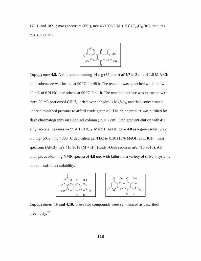
118
178.1, and 182.1; mass spectrum (ESI), m/z 459.0060 (M + H)+ (C21H16BrO7 requires
m/z 459.0079).
O
O
O
HO OH
Br
OH
O
Topopyrone 4.8. A solution containing 14 mg (35 μmol) of 4.7 in 2 mL of 1.0 M AlCl3
in nitrobenzene was heated at 90 °C for 48 h. The reaction was quenched while hot with
20 mL of 6 N HCl and stirred at 90 °C for 1 h. The reaction mixture was extracted with
three 50 mL portionsof CHCl3, dried over anhydrous MgSO4, and then concentrated
under diminished pressure to afford crude green oil. The crude product was purified by
flash chromatography on silica gel column (15 × 2 cm). Step gradient elution with 4:1
ethyl acetate−hexanes → 95:4:1 CHCl3−MeOH−AcOH gave 4.8 as a green solid: yield
6.5 mg (50%); mp >300 °C dec; silica gel TLC Rf 0.30 (10% MeOH in CHCl3); mass
spectrum (APCI), m/z 416.9618 (M + H)+ (C18H10O7Br requires m/z 416.9610). All
attempts at obtaining NMR spectra of 4.8 met with failure in a variety of solvent systems
due to insufficient solubility.
O
O
O
HO OH
OH
O
Cl
O
O
OH
O OH
OHO
Cl
Topopyrones 4.9 and 4.10. These two compounds were synthesized as described
previously.37

119
O
O
OH
O OH
Br
OHO
Topopyrone 4.11. A solution containing 8.0 mg (19 μmol) of 4.8 in 4 mL of 1% NaOH
in MeOH was heated at 60 °C for 48 h. The reaction mixture was neutralized carefully
with 1 N HCl and concentrated under diminished pressure. The resulting residue was
dissolved in 10 mL of H2O and extracted successively with three 20 mL portions of
CHCl3 and two 10 mL portions of ethyl acetate. The combined organic phase was dried
over anhydrous MgSO4, filtered, and then concentrated under diminished pressure to
afford a crude residue. The residue was purified by flash chromatography on a silica gel
column (10 × 2 cm). Elution with 94:5:1 CHCl3−MeOH−AcOH gave 4.11 as a green
solid: yield 4.0 mg (50%); mp >300 °C dec; silica gel TLC Rf 0.23 (10% MeOH in
CHCl3); mass spectrum (ESI), m/z 416.9613 (M + H)+ (C18H10BrO7 requires m/z
416.9610). All attempts at obtaining NMR spectra of 4.11 met with failure in a variety of
solvent systems due to insufficient solubility. Accordingly, 4.11 was converted to its
1,6,8-trimethoxy derivative to permit characterization by NMR. The derivative was also
characterized by high-resolution mass spectrometry.
To a solution containing 6.0 mg (14 μmol) of 4.11 in 4 mL of acetone at room
temperature was added 0.03 mL (0.23 mmol) of (MeO)2SO2 and 32 mg (0.23 mmol) of
K2CO3. The reaction mixture was heated at reflux for 18 h, quenched with 50 mL of 1 N
HCl, and extracted with two 20 mL portions of ethyl acetate. The combined organic
extract was dried over anhydrous MgSO4, filtered, and concentrated under diminished
pressure to afford a crude residue. The crude product was purified by flash

120
chromatography on a silica gel column (10 × 1 cm). Elution with 2% MeOH in CHCl3
gave the 1,6,8-trimethoxy derivative of topopyrone 4.11 as an orange solid: yield 4.0 mg
(66%); silica gel TLC Rf 0.55 (2% MeOH in CHCl3); 1H NMR (CDCl3) δ 2.49 (3H, s),
4.02 (3H, s), 4.10 (3H, s), 4.17 (3H, s), 6.24 (1H, s), 7.66 (1H, s), and 7.71 (1H, s); 13
C
NMR (CDCl3) δ 20.0, 57.1, 57.3, 61.5, 104.4, 105.9, 113.3, 114.3, 115.9, 119.7, 136.7,
139.6, 157.8, 159.3, 161.7, 164.4, 165.2, 179.19, and 179.21; mass spectrum (ESI), m/z
459.0077 (M + H)+ (C21H16O7Br requires m/z 459.0079).
.

121
REFERENCES
1. Karp, G. Cell and Molecular Biology: Concepts and Experiments; John Wiley
& Sons, Inc.: 2002; .
2. Green, R.; Noller, H. F. Annu. Rev. Biochem. 1997, 66, 679-716.
3. Schmeing, T. M.; Ramakrishnan, V. Nature 2009, 461, 1234-1242.
4. Lafontaine, D. L.; Tollervey, D. Nat. Rev. Mol. Cell Biol. 2001, 2, 514-520.
5. Cech, T. R. Science 2000, 289, 878-879.
6. Kurland, C. G.; Ehrenberg, M. Annu. Rev. Biophys. Biophys. Chem. 1987, 16,
291-317.
7. Ling, J.; Reynolds, N.; Ibba, M. Annu. Rev. Microbiol. 2009, 63, 61-78.
8. Baldwin, A. N.; Berg, P. J. Biol. Chem. 1966, 241, 839-845.
9. Eldred, E. W.; Schimmel, P. R. J. Biol. Chem. 1972, 247, 2961-2964.
10. Yarus, M. Proc. Natl. Acad. Sci. U.S.A. 1972, 69, 1915-1919.
11. Rodnina, M. V.; Wintermeyer, W. Trends Biochem. Sci. 2001, 26, 124-130.
12. Sievers, A.; Beringer, M.; Rodnina, M. V.; Wolfenden, R. Proc. Natl. Acad.
Sci. U.S.A. 2004, 101, 7897-7901.
13. Crick, F. J. Mol. Biol. 1968, 38, 367-379.
14. Doherty, E. A.; Doudna, J. A. Annu. Rev. Biophys. Biomol. Struct. 2001, 30,
457-475.
15. Guerrier-Takada, C.; Gardiner, K.; Marsh, T.; Pace, N.; Altman, S. Cell 1983,
35, 849-857.
16. Kruger, K.; Grabowski, P. J.; Zaug, A. J.; Sands, J.; Gottschling, D. E.; Cech,
T. R. Cell 1982, 31, 147-157.
17. Noller, H. F.; Hoffarth, V.; Zimniak, L. Science 1992, 256, 1416-1419.
18. Nitta, I.; Kamada, Y.; Noda, H.; Ueda, T.; Watanabe, K. Science 1998, 281,
666-669.
122
19. Ban, N.; Nissen, P.; Hansen, J.; Moore, P. B.; Steitz, T. A. Science 2000, 289,
905-920.
20. Nissen, P.; Hansen, J.; Ban, N.; Moore, P. B.; Steitz, T. A. Science 2000, 289,
920-930.
21. Hansen, J. L.; Schmeing, T. M.; Moore, P. B.; Steitz, T. A. Proc. Natl. Acad.
Sci. U.S.A. 2002, 99, 11670-11675.
22. Bieling, P.; Beringer, M.; Adio, S.; Rodnina, M. V. Nat. Struct. Mol. Biol.
2006, 13, 423-428.
23. Weinger, J. S.; Parnell, K. M.; Dorner, S.; Green, R.; Strobel, S. A. Nat.
Struct. Mol. Biol. 2004, 11, 1101-1106.
24. Koch, M.; Huang, Y.; Sprinzl, M. Angew. Chem. Int. Ed Engl. 2008, 47,
7242-7245.
25. Marshall, S. A.; Lazar, G. A.; Chirino, A. J.; Desjarlais, J. R. Drug Discov.
Today 2003, 8, 212-221.
26. Leblond, F.; Davis, S. C.; Valdes, P. A.; Pogue, B. W. J. Photochem.
Photobiol. B. 2010, 98, 77-94.
27. Lai, Y. T.; Tsai, K. L.; Sawaya, M. R.; Asturias, F. J.; Yeates, T. O. J. Am.
Chem. Soc. 2013, 135, 7738-7743.
28. Sheldon, R. A.; van Pelt, S. Chem. Soc. Rev. 2013, 42, 6223-6235.
29. Hendrickson, T. L.; de Crecy-Lagard, V.; Schimmel, P. Annu. Rev. Biochem.
2004, 73, 147-176.
30. Cohen, G. N.; Cowie, D. B. C. R. Hebd. Seances. Acad. Sci. 1957, 244, 680-
683.
31. Hendrickson, W. A.; Horton, J. R.; LeMaster, D. M. EMBO J. 1990, 9, 1665-
1672.
32. Hecht, S. M.; Alford, B. L.; Kuroda, Y.; Kitano, S. J. Biol. Chem. 1978, 253,
4517-4520.
33. Heckler, T. G.; Chang, L. H.; Zama, Y.; Naka, T.; Chorghade, M. S.; Hecht,
S. M.. Biochemistry 1984, 23, 1468-1473.
34. Noren, C. J.; Anthony-Cahill, S. J.; Griffith, M. C.; Schultz, P. G. Science
1989, 244, 182-188.

123
35. Wang, L.; Brock, A.; Herberich, B.; Schultz, P. G. Science 2001, 292, 498-
500.
36. Farnum, M. F.; Magde, D.; Howell, E. E.; Hirai, J. T.; Warren, M. S.;
Grimsley, J. K.; Kraut, J. Biochemistry 1991, 30, 11567-11579.
37. Miller, G. P.; Benkovic, S. J. Chem. Biol. 1998, 5, R105-R113.
38. Huennekens, F. M. Protein Sci. 1996, 5, 1201-1208.
39. Hitchings, G. H.; Burchall, J. J. Adv. Enzymol. Relat. Areas Mol. Biol. 1965,
27, 417-468.
40. Matthews, D. A.; Alden, R. A.; Bolin, J. T.; Filman, D. J.; Freer, S. T.;
Hamlin, R.; Hol, W. G.; Kisliuk, R. L.; Pastore, E. J.; Plante, L. T.; Xuong, N.;
Kraut, J. J. Biol. Chem. 1978, 253, 6946-6954.
41. Fierke, C. A.; Johnson, K. A.; Benkovic, S. J. Biochemistry 1987, 26, 4085-
4092.
42. Polacek, N.; Mankin, A. S. Crit. Rev. Biochem. Mol. Biol. 2005, 40, 285-311.
43. Pape, T.; Wintermeyer, W.; Rodnina, M. V. EMBO J. 1998, 17, 7490-7497.
44. Nathans, D.; Neidle, A. Nature 1963, 197, 1076-1077.
45. Pestka, S. Annu. Rev. Microbiol. 1971, 25, 487-562.
46. Hohsaka, T.; Sato, K.; Sisido, M.; Takai, K.; Yokoyama, S. FEBS Lett. 1993,
335, 47-50.
47. Starck, S. R.; Qi, X.; Olsen, B. N.; Roberts, R. W. J. Am. Chem. Soc. 2003,
125, 8090-8091.
48. Dedkova, L. M.; Fahmi, N. E.; Golovine, S. Y.; Hecht, S. M. J. Am. Chem.
Soc. 2003, 125, 6616-6617.
49. Dedkova, L. M.; Fahmi, N. E.; Golovine, S. Y.; Hecht, S. M. Biochemistry
2006, 45, 15541-15551.
50. Dedkova, L. M.; Fahmi, N. E.; Paul, R.; del Rosario, M.; Zhang, L.; Chen, S.;
Feder, G.; Hecht, S. M. Biochemistry 2012, 51, 401-415.
51. Maini, R.; Nguyen, D. T.; Chen, S.; Dedkova, L. M.; Chowdhury, S. R.;
Alcala-Torano, R.; Hecht, S. M. Bioorg. Med. Chem. 2013, 21, 1088-1096.
124
52. Porter, E. A.; Weisblum, B.; Gellman, S. H. J. Am. Chem. Soc. 2002, 124,
7324-7330.
53. Hart, S. A.; Bahadoor, A. B.; Matthews, E. E.; Qiu, X. J.; Schepartz, A. J. Am.
Chem. Soc. 2003, 125, 4022-4023.
54. Cheng, R. P.; DeGrado, W. F. J. Am. Chem. Soc. 2001, 123, 5162-5163.
55. Frackenpohl, J.; Arvidsson, P. I.; Schreiber, J. V.; Seebach, D. Chembiochem
2001, 2, 445-455.
56. Appella, D. H.; Christianson, L. A.; Klein, D. A.; Powell, D. R.; Huang, X.;
Barchi, J. J.,Jr; Gellman, S. H. Nature 1997, 387, 381-384.
57. Seebach, D.; Gardiner, J. Acc. Chem. Res. 2008, 41, 1366-1375.
58. Kritzer, J. A.; Stephens, O. M.; Guarracino, D. A.; Reznik, S. K.; Schepartz,
A. Bioorg. Med. Chem. 2005, 13, 11-16.
59. Porter, E. A.; Wang, X.; Lee, H. S.; Weisblum, B.; Gellman, S. H. Nature
2000, 404, 565.
60. Cristau, M.; Devin, C.; Oiry, C.; Chaloin, O.; Amblard, M.; Bernad, N.; Heitz,
A.; Fehrentz, J. A.; Martinez, J. J. Med. Chem. 2000, 43, 2356-2361.
61. Heckler, T. G.; Roesser, J. R.; Xu, C.; Chang, P. I.; Hecht, S. M. Biochemistry
1988, 27, 7254-7262.
62. Roesser, J. R.; Xu, C.; Payne, R. C.; Surratt, C. K.; Hecht, S. M. Biochemistry
1989, 28, 5185-5195.
63. Lodder, M.; Golovine, S.; Hecht, S. M. J. Org. Chem. 1997, 62, 778.
64. Lodder, M.; Golovine, S.; Laikhter, A. L.; Karginov, V. A.; Hecht, S. M. J.
Org. Chem. 1998, 63, 794-803.
65. Robertson, S. A.; Ellman, J. A.; Schultz, P. G. J.Am. Chem. Soc. 1991, 113,
2722.
66. Robertson, S. A.; Noren, C. J.; Anthony-Cahill, S. J.; Griffith, M. C.; Schultz,
P. G. Nucleic Acids Res. 1989, 17, 9649-9660.
67. Varshney, U.; Lee, C. P.; RajBhandary, U. L. J. Biol. Chem. 1991, 266,
24712-24718.
68. Huynh, M. L.; Russell, P.; Walsh, B. Methods Mol. Biol. 2009, 519, 507-513.

125
69. Muth, G. W.; Chen, L.; Kosek, A. B.; Strobel, S. A. RNA 2001, 7, 1403-1415.
70. Renukuntla, J.; Vadlapudi, A. D.; Patel, A.; Boddu, S. H.; Mitra, A. K. Int. J.
Pharm. 2013, 447, 75-93.
71. Lohan, S.; Bisht, G. S. Mini Rev. Med. Chem. 2013, 13, 1073-1088.
72. Richardson, J. S. Adv. Protein Chem. 1981, 34, 167-339.
73. Marcelino, A. M.; Gierasch, L. M. Biopolymers 2008, 89, 380-391.
74. Che, Y.; Marshall, G. R. Biopolymers 2006, 81, 392-406.
75. Nagai, U.; Sato, K.; Nakamura, R.; Kato, R. Tetrahedron 1993, 49, 3577-
3592.
76. Nachman, R. J.; Kaczmarek, K.; Williams, H. J.; Coast, G. M.; Zabrocki, J.
Biopolymers 2004, 75, 412-419.
77. Hartley, O.; Gaertner, H.; Wilken, J.; Thompson, D.; Fish, R.; Ramos, A.;
Pastore, C.; Dufour, B.; Cerini, F.; Melotti, A.; Heveker, N.; Picard, L.; Alizon,
M.; Mosier, D.; Kent, S.; Offord, R. Proc. Natl. Acad. Sci. U.S.A. 2004, 101,
16460-16465.
78. Berger, E. A.; Murphy, P. M.; Farber, J. M. Annu. Rev. Immunol. 1999, 17,
657-700.
79. Grauer, A.; Konig, B. Eur. J. Org. Chem. 2009, 5099-5111.
80. Wissner, R. F.; Batjargal, S.; Fadzen, C. M.; Petersson, E. J. J. Am. Chem.
Soc. 2013, 135, 6529-6540.
81. Chatterjee, J.; Gilon, C.; Hoffman, A.; Kessler, H. Acc. Chem. Res. 2008, 41,
1331-1342.
82. Oh, K.; Guan, Z. Chem. Commun. (Camb) 2006, 29, 3069-3071.
83. Shimomura, O.; Johnson, F. H.; Saiga, Y. J. Cell. Comp. Physiol. 1962, 59,
223-239.
84. Prasher, D. C.; Eckenrode, V. K.; Ward, W. W.; Prendergast, F. G.; Cormier,
M. J. Gene 1992, 111, 229-233.
85. Chalfie, M.; Tu, Y.; Euskirchen, G.; Ward, W. W.; Prasher, D. C. Science
1994, 263, 802-805.

126
86. Chudakov, D. M.; Matz, M. V.; Lukyanov, S.; Lukyanov, K. A. Physiol. Rev.
2010, 90, 1103-1163.
87. Ormo, M.; Cubitt, A. B.; Kallio, K.; Gross, L. A.; Tsien, R. Y.; Remington, S.
J. Science 1996, 273, 1392-1395.
88. Zimmer, M. Chem. Rev. 2002, 102, 759-781.
89. Ivashkin, P. E.; Iampol'skii, I. V.; Luk'ianov, K. A. Bioorg. Khim. 2009, 35,
726-743.
90. Wang, B.; Zhou, J.; Lodder, M.; Anderson, R. D.,3rd; Hecht, S. M. J. Biol.
Chem. 2006, 281, 13865-13868.
91. Thommes, P.; Hubscher, U. Chromosoma 1992, 101, 467-473.
92. Liu, L. F.; Wang, J. C. Proc. Natl. Acad. Sci. U.S.A. 1987, 84, 7024-7027.
93. Pommier, Y.; Leo, E.; Zhang, H.; Marchand, C. Chem. Biol. 2010, 17, 421-
433.
94. Wang, J. C. J. Mol. Biol. 1971, 55, 523-533.
95. Wang, J. C. Nat. Rev. Mol. Cell Biol. 2002, 3, 430-440.
96. Lindsley, J. E.; Wang, J. C. Proc. Natl. Acad. Sci. U.S.A. 1991, 88, 10485-
10489.
97. Wang, J. C. Q. Rev. Biophys. 1998, 31, 107-144.
98. Champoux, J. J. Annu. Rev. Biochem. 2001, 70, 369-413.
99. Wang, J. C. Annu. Rev. Biochem. 1996, 65, 635-692.
100. Kirkegaard, K.; Wang, J. C. J. Mol. Biol. 1985, 185, 625-637.
101. Watt, P. M.; Hickson, I. D. Biochem. J. 1994, 303 ( Pt 3), 681-695.
102. Liu, L. F.; Liu, C. C.; Alberts, B. M. Cell 1980, 19, 697-707.
103. Gellert, M.; Mizuuchi, K.; O'Dea, M. H.; Nash, H. A. Proc. Natl. Acad. Sci.
U.S.A. 1976, 73, 3872-3876.
104. Liu, L. F. Annu. Rev. Biochem. 1989, 58, 351-375.
105. Lee, M. P.; Brown, S. D.; Chen, A.; Hsieh, T. S. Proc. Natl. Acad. Sci.
U.S.A. 1993, 90, 6656-6660.

127
106. Goto, T.; Wang, J. C. Proc. Natl. Acad. Sci. U.S.A. 1985, 82, 7178-7182.
107. Miao, Z. H.; Player, A.; Shankavaram, U.; Wang, Y. H.; Zimonjic, D. B.;
Lorenzi, P. L.; Liao, Z. Y.; Liu, H.; Shimura, T.; Zhang, H. L.; Meng, L. H.;
Zhang, Y. W.; Kawasaki, E. S.; Popescu, N. C.; Aladjem, M. I.; Goldstein, D. J.;
Weinstein, J. N.; Pommier, Y. Cancer Res. 2007, 67, 8752-8761.
108. Tuduri, S.; Crabbe, L.; Conti, C.; Tourriere, H.; Holtgreve-Grez, H.; Jauch,
A.; Pantesco, V.; De Vos, J.; Thomas, A.; Theillet, C.; Pommier, Y.; Tazi, J.;
Coquelle, A.; Pasero, P. Nat. Cell Biol. 2009, 11, 1315-1324.
109. Merino, A.; Madden, K. R.; Lane, W. S.; Champoux, J. J.; Reinberg, D.
Nature 1993, 365, 227-232.
110. Soret, J.; Gabut, M.; Dupon, C.; Kohlhagen, G.; Stevenin, J.; Pommier, Y.;
Tazi, J. Cancer Res. 2003, 63, 8203-8211.
111. Hsiang, Y. H.; Wu, H. Y.; Liu, L. F. Cancer Res. 1988, 48, 3230-3235.
112. Nitiss, J. L. Nat. Rev. Cancer. 2009, 9, 327-337.
113. Austin, C. A.; Marsh, K. L. Bioessays 1998, 20, 215-226.
114. Hsiang, Y. H.; Hertzberg, R.; Hecht, S.; Liu, L. F. J. Biol. Chem. 1985, 260,
14873-14878.
115. Kupfer, G.; Bodley, A. L.; Liu, L. F. NCI Monogr. 1987, 4, 37-40.
116. Hsiang, Y. H.; Liu, L. F. Cancer Res. 1988, 48, 1722-1726.
117. D'Arpa, P.; Beardmore, C.; Liu, L. F. Cancer Res. 1990, 50, 6919-6924.
118. Van, G. R.; Lendfers, R. R.; Schellens, J.; Bult, A.; Beijnen, J. J. Oncol.
Pharm. Pract. 2000, 6, 92-108.
119. Bertrand, R.; O'Connor, P. M.; Kerrigan, D.; Pommier, Y. Eur. J. Cancer
1992, 28A, 743-748.
120. Kaufmann, S. H. Cancer Res. 1991, 51, 1129-1136.
121. Salerno, S.; Da Settimo, F.; Taliani, S.; Simorini, F.; La Motta, C.;
Fornaciari, G.; Marini, A. M. Curr. Med. Chem. 2010, 17, 4270-4290.
122. Yamashita, Y.; Kawada, S.; Fujii, N.; Nakano, H. Biochemistry 1991, 30,
5838-5845.

128
123. Khan, Q. A.; Elban, M. A.; Hecht, S. M. J. Am. Chem. Soc. 2008, 130,
12888-12889.
124. Kanai, Y.; Ishiyama, D.; Senda, H.; Iwatani, W.; Takahashi, H.; Konno, H.;
Tokumasu, S.; Kanazawa, S. J. Antibiot. (Tokyo) 2000, 53, 863-872.
125. Elban, M. A.; Hecht, S. M. J. Org. Chem. 2008, 73, 785-793.
126. Zaleski, P. A.; Maini, R.; Leiris, S. J.; Elban, M. A.; Hecht, S. M. J. Nat.
Prod. 2012, 75, 577-585.
127. Staker, B. L.; Hjerrild, K.; Feese, M. D.; Behnke, C. A.; Burgin, A. B.,Jr;
Stewart, L. Proc. Natl. Acad. Sci. U.S.A. 2002, 99, 15387-15392.
128. Staker, B. L.; Feese, M. D.; Cushman, M.; Pommier, Y.; Zembower, D.;
Stewart, L.; Burgin, A. B. J. Med. Chem. 2005, 48, 2336-2345.
129. Elban, M. A. Synthesis of DNA Damaging Agents, University of Virginia,
Charlottesville, VA, 2002.
130. Savard, J.; Brassard, P. Tetrahedron 1984, 40, 3455-3464.
131. Rozenberg, V.; Danilova, T.; Sergeeva, E.; Vorontsov, Z. S.; Lysenko, K.;
Belokon, Y. Eur. J. Org. Chem. 2000, 2000, 3295-3303.
132. Bensari, A.; Zaveri, N. T. Synthesis 2003, 2, 267-271.
133. Krohn, K.; Bernhard, S.; Florke, U.; Hayat, N. J. Org. Chem. 2000, 65,
3218-3222.
134. Tietze, L. F.; Gericke, K. M.; Schuberth, I. Eur. J. Org. Chem. 2007, 2007,
4563-4577.
135. Mahal, H. S.; Venkataraman, K. J. Chem. Soc. 1934, 56, 1767-1769.
136. Baker, W. J Chem Soc 1933, 55, 1381-1391.
137. Drake, F. H.; Hofmann, G. A.; Bartus, H. F.; Mattern, M. R.; Crooke, S. T.;
Mirabelli, C. K. 1989, 28, 8154-8160.
138. Bonven, B. J.; Gocke, E.; Westergaard, O. Cell 1985, 41, 541-551.