t lymphocyte function in the delayed phase of ischemic brain … · 102 t lymphocytes mediate...
TRANSCRIPT

Mini Review
102 T lymphocytes mediate delayed phase of ischemic brain injury
Mini Review
T lymphocyte function in the delayed phase of ischemic brain injury
Takashi Shichita, Go Muto and Akihiko Yoshimura
Department of Microbiology and Immunology, School of Medicine, Keio University, Tokyo, Japan and Japan Science and Technology Agency (JST), Core Research for Evolutional Science and Technology (CREST), Tokyo, Japan
Lymphocyte recruitment and activation have been implicated in the progression of cerebral ischemia-reperfusion (I/R) injury, yet the roles of specific lymphocyte subpo-pulations and cytokines in stroke remain to be clarified. We demonstrated that IL-23 and IL-17, rather than IFN-γ, play pivotal roles in the evolution of brain infarction. IL-23 was produced from infiltrated macrophages in the immediate phase of brain ischemia; thereafter, IL-17-producing T lymphocytes were infiltrated into the ischemic brain tissue in the delayed phase. IL-17 was mostly produced in γδT lymphocytes, and was de-pendent on IL-23. We discovered that not only IL-23 but also IL-17 deficiency prevented neural cell death in the delayed phase of ischemic brain injury. We also demonstrated that FTY720 administration, which blocked T cell infiltration into the brain, suppressed ischemic brain injury. Furthermore, the depletion of γδT cells also attenuated ischemic brain damages. Therefore, we propose that cerebral T lymphocytes, including γδT lymphocytes, be considered as new therapeutic targets in a novel neuroprotective strategy for ischemic brain injury during the delayed phase.
Rec.11/24/2009,Acc.6/8/2010
Corresponding author: Akihiko Yoshimura, Ph.D. Department of Microbiology and Immunology, Keio University School of Medicine, 35 Shina-nomachi, Shinjuku-ku, Tokyo, 160-8582, Japan Ph: +81 3 5363 3483, Fax: +81 3 5360 1508, Email address: [email protected]
Key words: Inflammasome, autoinflammation, inflammasome, ischemic injury, transplant immunology

Inflammation and Regeneration Vol.31 No.1 January 2011 103
Introduction Stroke is a leading cause of death and dis-
ability worldwide and approximately 80% of strokes in Japan are caused by brain infarc-tion. So far, no successful therapy has been established that can benefit patients beyond the narrow time window of thrombolysis1,2). Brain infarction is the death of brain tissue caused by brain ischemia, which is the loss of cerebral blood flow mainly caused by the stenosis or occlusion of cerebral arteries. In the immediate phase of brain infarction, ischemia causes the necrotic cell death of brain tissue, and moreover, macrophages be-gin to infiltrate into the ischemic brain tissue and promote rapid inflammatory responses3)
(Fig. 1). These inflammatory responses continue in
the delayed phase of brain infarction and cause the evolution of infarct volume and neurological worsening. In this phase, vari-ous types of inflammatory cells including macrophages, neutrophils and lymphocytes infiltrate into the ischemic brain tissue and exacerbate ischemic injury, now recognized as a secondary ischemic injury. Brain edema and apoptotic neuronal death have been suggested to contribute to the evolution of infarct volume in the delayed phase4-6), how-ever, both the mechanism and the regulation of such events have not been fully unders-tood.
Fig. 1 The inflammation and brain infarction.
Many inflammatory factors play a major role in the inflammation during the immediate phase. In this phase, macrophages begin to infiltrate the ischemic brain tissue and mediate ischemic injury. It has not, however, been clarified which cells exacerbate the ischemic damage in the delayed phase of brain ischemia. Because lymphocytes infiltrate the ischemic brain tissue in the delayed phase, we hypothesized that these infiltrating lymphocytes caused secondary inflammatory responses. If this is true, we can develop a new neuroprotective strategy which can be initiated even at the delayed phase of brain infarction.
T lymphocytes in ischemic brain injury
Recently, T lymphocytes have been sug-gested to play a role as mediators in the de-layed phase of brain ischemia. In fact, as early as 24 hours after reperfusion, T lym-phocytes are present in the ischemic brain tissue and appear to be localized to the in-farction boundary zones, typically close to blood vessels7,8). The number of infiltrated T lymphocytes was reported to reach its peak
in the delayed phase (day 3 from the onset) of brain I/R injury9). Furthermore, recent stu-dies revealed that the depletion of these T lymphocytes attenuated cerebral ischemic damage. A significant reduction of infarct volume was observed in severe combined im-munodeficient (SCID) mice and recombina-tion activating gene (RAG)-deficient mice both of which are lacking T and B lympho-cytes10,11). A previous study revealed that CD4-positive and CD8-positive T lympho-cytes, but not B lymphocytes, exacerbate in-

Mini Review
104 T lymphocytes mediate delayed phase of ischemic brain injury flammatory responses and neurological defi-cit with experimental stroke11).
It has not been clarified whether a specific antigen of the brain is involved in the activa-tion of these infiltrated T cells. Because the inflammatory responses induced by ischemic injury have been shown to be driven by the innate immune system, it is highly possible that these T lymphocytes mediate anti-gen-independent, innate inflammatory res-
ponses11-14). However, some reports suggested the role of antigen recognition by the T lym-phocytes in ischemic brain injury. Treatment with T cell receptor ligands, major histo-compatibility complex class II molecules bound to myelin peptides, attenuated ischemic brain injury15). In addition, myelin basic protein-tolerized animals have been shown to develop reduced infarction com-pared to control animals16,17).
Fig. 2 FTY720 administration attenuated ischemic brain injury. The infarct volume of FTY720- or water-dosed mice measured by anti-MAP2 staining at day 4 (“H2O” or “FTY720 day 0”: H2O or FTY720 was administered five minutes before reperfusion; “FTY720 day 0,1,2,3”: FTY720 was administered be-fore reperfusion and every 24 hours ). The number of mice is shown on each bar. **P < 0.01 vs. control mice (one-way ANOVA with Dunnett’s correction).
Regardless of antigen specificity, these data indicate that T lymphocytes play a pro-gressive role in the delayed phase of ischemic brain injury by enhancing secondary ischemic inflammatory responses. However, it remains unclear whether these T lympho-cytes act centrally or peripherally. To at-tempt to answer this question, we evaluated the therapeutic effect of FTY720, an immu-nomodulatory prodrug which is well known to prevent T lymphocyte infiltration into in-flammatory tissues18,19). We discovered that FTY720 suppressed T lymphocyte infiltration into the ischemic brain tissue without in-fluencing macrophage infiltration. Addition-ally, FTY720 administration significantly reduced the infarct volume, compared to control mice (Fig. 2). These data support the contention that T lymphocytes act centrally and mediate ischemic injury. However, T lymphocytes infiltration begins 24 hours af-ter the onset of brain ischemia. In order to develop a new neuroprotective therapy, the dose and therapeutic time window of FTY720 administration should be established in fu-ture study.
T lymphocyte cytokines in ischemic brain injury
Although recent studies clearly demon-strate the relationship between ischemic stroke and T lymphocytes, the specific func-tion of these T lymphocytes has not been suf-ficiently clarified. IFN-γ from activated T cells has been proposed to promote infarction. However, the role of IFN-γ in ischemic brain injury is the subject of controversy11,22). IL-10 is well-known to function as a neuroprotec-tive mediator derived from T lymphocytes, especially from regulatory T cells20,21).
We have been interested in the newly identified cytokines, IL-23 and IL-17, be-cause they play a pivotal role in the onset and the progression of experimental autoimmune encephalomyelitis (EAE). EAE is a well-es-tablished T cell-mediated brain and spinal cord inflammation model. It has been demon-strated that IL-23 is a critical cytokine for the onset of experimental autoimmune en-cephalomyelitis (EAE)23,24). IL-23 is a hetero-dimer of IL-23p19 and IL-12p40 and has been shown to be essential for the induction
Inflammation and Regeneration Vol.31 No.1 January 2011 105 of IL-17 production from helper T cells (Th17)25) and γδT lymphocytes26,27) IL-23 is mostly produced from antigen-presenting cells, such as dendritic cells.
On the other hand, IL-17 is produced not only from CD4-positive helper T cells (Th17) but also from γδT lymphocytes. In EAE, IL-17 and other inflammatory mediators from Th17 cells have been implicated in the onset as well as the progression of the disease28-30). IL-17 has been reported to modify many in-flammatory responses in the central nervous system by promoting the production of neu-rotoxic cytokine33) and blood brain barrier
disruption34,35). Therefore, we hypothesized that IL-23 and
IL-17 were implicated in cerebral ischemic injury. Actually, elevated levels of IL-17 were found in the ischemic hemispheres of human brains, and the levels peaked 3-5 days after brain ischemia36). It remains to be deter-mined which T lymphocyte subsets and in-flammatory mediators participate in the evolution of brain infarction. If we can mod-ulate these inflammatory T lymphocyte functions, a new neuroprotective therapy could be developed (Fig. 1).
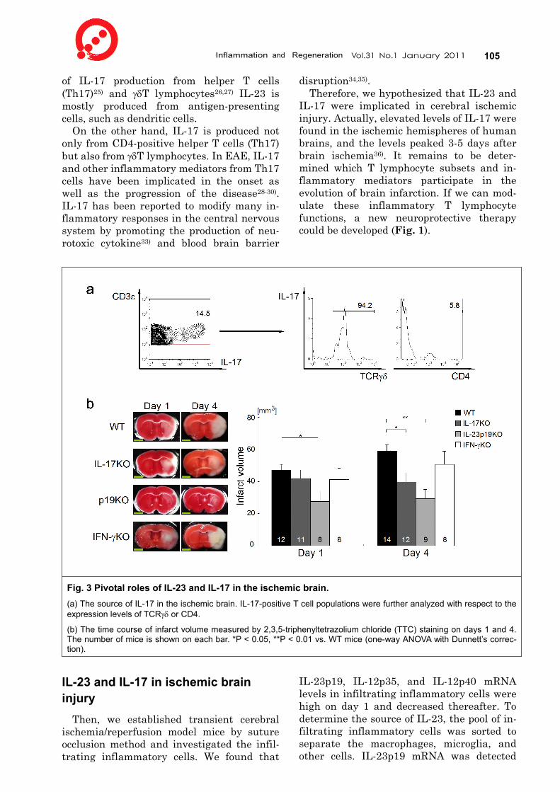
Fig. 3 Pivotal roles of IL-23 and IL-17 in the ischemic brain. (a) The source of IL-17 in the ischemic brain. IL-17-positive T cell populations were further analyzed with respect to the expression levels of TCRγδ or CD4.
(b) The time course of infarct volume measured by 2,3,5-triphenyltetrazolium chloride (TTC) staining on days 1 and 4. The number of mice is shown on each bar. *P < 0.05, **P < 0.01 vs. WT mice (one-way ANOVA with Dunnett’s correc-tion).
IL-23 and IL-17 in ischemic brain injury
Then, we established transient cerebral ischemia/reperfusion model mice by suture occlusion method and investigated the infil-trating inflammatory cells. We found that
IL-23p19, IL-12p35, and IL-12p40 mRNA levels in infiltrating inflammatory cells were high on day 1 and decreased thereafter. To determine the source of IL-23, the pool of in-filtrating inflammatory cells was sorted to separate the macrophages, microglia, and other cells. IL-23p19 mRNA was detected

Mini Review
106 T lymphocytes mediate delayed phase of ischemic brain injury only in the macrophage fraction (CD45 high, CD11b high population). By using bone marrow-transferred chimeric mice, we also showed that infiltrated macrophages, but not brain cells, were the major source of IL-23, which was essential for the induction of IL-17 producing cells in the ischemic brain.
To identify the IL-17-producing cells in ischemic brain tissue, we performed intra-cellular FACS analysis on the infiltrating inflammatory cells. The number of IL-17-pos-itive cells as well as IFN-γ-positive cells in ischemic brain tissue was first measurable on day 3 and decreased on day 6. Importantly, we barely detected IL-17-producing cells in infiltrating mononuclear cells from IL-23 knockout (KO) mice. Therefore, IL-23 is a key factor for the induction of IL-17 producing cells in the ischemic brain.
All of the IL-17-positive and IFN-γ-positive cells were also CD45-positive. Approximately 90% of the IL-17-positive cells and IFN--γ-positive cells were CD3-positive T lym-phocytes. Surprisingly, most of the IL-17--producing T lymphocytes were CD4-negative but γδTCR-positive (Fig. 3a). These data in-dicate that γδT lymphocytes, rather than Th17 cells, are the source of IL-17.
Furthermore, to investigate the functional significance of IL-23 and IL-17 in brain in-farction, we used an experimental brain ischemia model in gene-disrupted mice. IL-23p19 KO mice exhibited a significant reduction in infarct volume at day 1 and 4, compared to wild-type (WT) mice (Fig. 3b). IL-17 KO mice also exhibited a significant reduction of infarct volume on day 4, but no drastic difference was observed on day 1 (Fig. 3b). However, there was no statistical differ-ence between IFN-γ KO and WT mice at day 1 and 4. We also revealed the importance of IL-17-producing T lymphocytes rather than brain cells for the progression of ischemic damages by an adoptive transfer experiment. These data indicate that IL-23p19 functions in the immediate phase of ischemic brain in-jury, while IL-17 plays a role in the delayed phase (after day 3). Additionally, our results suggest that IL-23 promoted ischemic brain injury at the immediate phase by IL-17--independent mechanism.
γδT lymphocytes in ischemic brain injury
Infiltrating γδT lymphocytes were found in
the ischemic brain tissue and their presence peaked at day 3. These γδT lymphocytes ap-peared to be localized to the infarct boundary zones (Fig. 4a), suggesting a role in neural cell death in the penumbral region. We con-firmed that the administration of anti-TCRγδ antibody almost completely depleted cerebral γδT lymphocytes and IL-17-positive T lym-phocytes in ischemic brain tissue. Further-more, anti-TCRγδ antibody treatment exhi-bited a neuroprotective effect even 24 hours after the induction of brain ischemia (Fig. 4b). These data suggest that IL-17 is the most potent effector of γδT lymphocytes.
Discussion
In the immediate phase of brain ischemia, infiltrated macrophages produce IL-23. The-reafter, in the delayed phase of brain ische-mia, T lymphocytes, including γδT lympho-cytes, infiltrate the ischemic brain tissue, and IL-23 induces IL-17 production by γδT lymphocytes. IL-17 exacerbates ischemic brain injury by promoting blood brain barrier disruption and the production of neurotoxic factors.
Recent studies have revealed that γδT lymphocytes can more rapidly produce IL-17 than can Th17, and they exacerbate au-toimmune disease such as EAE31). IL-17-producing γδT lymphocytes have also been shown to be involved in the develop-ment of EAE31,32). These γδT lymphocytes, which produce IL-17 but not IFN-γ, were re-ported to be antigen-naïve and a natural-ly-occurring type of γδT lymphocytes31,37). Therefore, it is reasonable to conclude that IL-23-induced IL-17-producing γδT lympho-cytes play a role in ischemic brain injury.
Our findings raise the intriguing possibil-ity that T lymphocytes, including γδT lym-phocytes which produce IL-17, could be a new therapeutic target for stroke, widening the therapeutic time window for neuroprotection against brain ischemia. A recent study iden-tified the IL-17-producing cells in the ischemic brain of stroke patients, and that their presence peaked 3-5 days after injury36). In addition, it has been reported that the in-farct volume evolved until 3-5 days after the onset38,39). Thus, it is highly likely that infil-tration of IL-17-producing T lymphocytes plays a pivotal role not only in mouse but also in human brains. We propose that suppres-

Inflammation and Regeneration Vol.31 No.1 January 2011 107 sion of a specific pathogenic T lymphocyte subset, such as IL-17 producing γδT lympho-cytes, is useful for neuroprotection against
brain ischemia.
Fig. 4 The effects of γδT lymphocyte infiltration in ischemic brain tissue. (a) Anti-mouse TCRγδ immunostaining of ischemic brain tissue at day 4.
(b) The infarct volume of mice which were PBS- or antibody-treated 24 hours after the induction of brain ischemia. Mice were sacrificed at day 7 and the infarct volumes were evaluated by anti-MAP2 immunostaining. ***P = 0.028, vs. control mice [two-sided Student’s t-test]. The number of mice is shown on each bar.
References 1) Sacco RL, Chong JY, Prabhakaran S, Elkind
MS: Experimental treatments for acute ischaemic stroke. Lancet. 2007; 369: 331- 341.
2) Donnan GA, Fisher M, Macleod, M, Davis SM: Stroke. Lancet. 2008; 371: 1612- 1623.
3) Barone FC, Feuerstein GZ: Inflammatory mediators and stroke: new opportunities for novel therapeutics. J Cereb Blood Flow Me-tab. 1999; 19: 819-834.
4) Matsui T, et al. Astrocytic activation and delayed infarct expansion after permanent focal ischemia in rats. Part I: enhanced as-trocytic synthesis of s-100beta in the periin-farct area precedes delayed infarct expan-sion. J Cereb Blood Flow Metab. 2002; 22: 711-722.
5) Charriaut-Marlangue C, et al.: Apoptosis and necrosis after reversible focal ischemia:
an in situ DNA fragmentation analysis. J Cereb Blood Flow Metab. 1996; 16: 186-194.
6) Fink K, et al.: Prolonged therapeutic window for ischemic brain damage caused by delayed caspase activation. J Cereb Blood Flow Me-tab. 1999; 18: 1071-1076.
7) Schroeter M, Jander S, Witte OW, Stoll G: Local immune responses in the rat middle cerebral artery occlusion. J Neuroimmunol. 1994; 55: 195-203.
8) Jander S, Karemer M, Schroeter M, Witte OW, Stoll G: Lymphocytic infiltration and expression of intercellular adhesion mole-cule-1 in photochemically induced ischemia of the rat cortex. J Cereb Blood Flow Metab. 1995; 15: 42-51.
9) Gelderblom M, et al.: Temporal and spatial dynamics of cerebral immune cell accumula-tion in stroke. Stroke. 2009; 40: 1849-1857.
10) Hurn PD, et al.: T- and B-cell-deficient mice with experimental stroke have reduced le-
Mini Review
108 T lymphocytes mediate delayed phase of ischemic brain injury
sion size and inflammation. J Cereb Blood Flow Metab. 2007; 27: 1798-1805.
11) Yilmaz G, Arumugam TV, Stokes KY, Granger DN: Role of T lymphocytes and in-terferon-gamma in ischemic stroke. Circula-tion. 2006; 113: 2105-2112.
12) Marsh BJ, Williams-Karnesky RL, Sten-zel-Poore MP: Toll-like receptor signaling in endogenous neuroprotection and stroke. Neuroscience. 2009; 158: 1007-1020.
13) Tang SC, Arumugam TV, Xu X, Cheng A, Mughal MR, et al.: Pivotal role for neuronal Toll-like receptors in ischemic brain injury and functional deficits. Proc Natl Acad Sci U S A. 2007; 104: 13798-13803.
14) Gee JM, Kalil A, Shea C, Becker KJ: Lym-phocytes: potential mediators of posti-schemic injury and neuroprotection. Stroke. 2007; 38: 783-788.
15) Subramanian S, Zhang B, Kosaka Y, Bur-rows GG, Grafe MR, et al.: Recombinant T cell receptor ligand treats experimental stroke. Stroke. 2009; 40: 2539-2545.
16) Becker KJ: Sensitization and tolerization to brain antigens in stroke. Neuroscience. 2009; 158: 1090-1097.
17) Becker K, Kindrick D, McCarron R, Hallen-beck J, Winn R: Adoptive transfer of myelin basic protein-tolerized splenocytes to naive animals reduces infarct size: a role for lym-phocytes in ischemic brain injury? Stroke. 2003; 34: 1809-1815.
18) Dev KK, Mullershausen F, Mattes H, Kuhn RR, Bilbe G, et al.: Brain sphingo-sine-1-phosphate receptors: implication for FTY720 in the treatment of multiple sclero-sis. Pharmacol Ther. 2008; 117: 77-93.
19) Premenko-Lanier M, Moseley NB, Pruett ST, Romagnoli PA, Altman JD: Transient FTY720 treatment promotes immune-me-diated clearance of a chronic viral infection. Nature. 2008; 454: 894-898.
20) Ooboshi, et al.: Postischemic gene transfer of interleukin-10 protects against both focal and global brain ischemia. Circulation. 2006; 111: 913-919.
21) Liesz A, et al.: Regulatory T cells are key cerebroprotective immunomodulators in acute experimental stroke. Nat Med. 2009; 15: 192-199.
22) Lambertsen KL, et al.: A role for inter-feron-gamma in focal cerebral ischemia in mice. J Neuropathol Exp Neurol. 2004; 63: 942-955.
23) Cua DJ, et al.: Interleukin-23 rather than interleukin-12 is the critical cytokine for autoimmune inflammation of the brain. Nature. 2003; 421: 744-748.
24) Chen Y, et al.: Anti-IL-23 therapy inhibits multiple inflammatory pathways and ame-liorates autoimmune encephalomyelitis. J Clin Invest. 2006; 116: 1317-1326.
25) Langrish CL, et al.: IL-23 drives a patho-genic T cell population that induces au-toimmune inflammation. J Exp Med. 2005; 201: 233-240.
26) Nakamura R, et al.: Tyk2-signaling plays an important role in host defense against Escherichia coli through IL-23-induced IL-17 production by gammadelta T cells. J Immu-nol. 2008; 181: 2071-2075.
27) Ribot JC, et al.: CD27 is a thymic determi-nant of the balance between interfe-ron-gamma- and interleukin 17-producing gammadelta T cell subsets. Nat Immunol. 2009; 10: 427-436.
28) Lock C, et al.: Gene-microarray analysis of multiple sclerosis lesions yields new targets validated in autoimmune encephalomyelitis. Nat Med. 2002; 8: 500-508.
29) Sutton C, Brereton C, Keogh B, Mills KH, Lavelle EC: A crucial role for interleukin (IL)-1 in the induction of IL-17-producing T cells that mediate autoimmune encephalo-myelitis. J Exp Med. 2006; 203: 1685-1691.
30) Park H, et al.: A distinct lineage of CD4 T cells regulates tissue inflammation by pro-ducing interleukin 17. Nat Immunol. 2005; 6, 1133-1141.
31) Jensen KD, et al.: Thymic selection deter-mines gammadelta T cell effector fate: anti-gen-naïve cells make interleukin-17 and an-tigen-experienced cells make interferon gamma. Immunity. 2008; 29: 90-100.
32) Sutton CE, et al.: Interleukin-1 and IL-23 induce innate IL-17 production from gam-madelta T cells, amplifying Th17 responses and autoimmunity. Immunity. 2009; 31: 331-341.
33) Fossiez F, et al.: T cell interleukin-17 in-duces stromal cells to produce proinflam-matory and hematopoietic cytokines. J Exp Med. 1996; 183: 2593-2603.
34) Kebir H, et al.: Human TH17 lymphocytes promote blood-brain barrier disruption and central nervous system inflammation. Nat Med. 2007; 13: 1173-1175.
35) Ifergan I, et al.: The blood-brain barrier in-duces differentiation of migrating monocytes
Inflammation and Regeneration Vol.31 No.1 January 2011 109
into Th17-polarizing dendritic cells. Brain. 2008; 131: 785-799.
36) Li GZ, et al.: Expression of interleukin-17 in ischemic brain tissue. Scand J Immunol. 2005; 62: 481-486.
37) Shibata K, Yamada H, Hara H, Kishihara K, Yoshikai Y: Resident Vδ1+γδ T cells control early infiltration of neutrophils after Esche-richia coli infection via IL-17 production. J Immunol. 2007; 178: 4466-4472.
38) Gaudinski MR, et al.: Establishing final in-farct volume: stroke lesion evolution past 30 days is insignificant. Stroke. 2008; 39: 2765-2768.
39) Shimosegawa E, et al.: Metabolic penumbra of acute brain infarction: a correlation with infarct growth. Ann Neurol. 2005; 57: 495-504.