the lungs · the mechanics of breathing for the efficient exchange of gases between the blood and...
TRANSCRIPT

08CHAPTER
THE RESPIRATORY SYSTEM
UNIT 1 CONTENT
SCIENCE AS A HUMAN ENDEAVOUR❯❯ lifestyle choices, including being active
or sedentary, the use of drugs and type of diet, can compromise body functioning in the short term and may have long-term consequences
SCIENCE UNDERSTANDING
Respiratory system❯❯ the exchange of gases between the
internal and external environments of the body is facilitated by the structure and function of the respiratory system at the cell, tissue and organ levels
❯❯ the efficient exchange of gases in the lungs is maintained by the actions of breathing, blood flow and the structure of the alveoli
Shut
ters
tock
/Ale
ksan
dr M
arki
n
Year
11
Sylla
bus,
Hum
an B
iolo
gy, ©
Sch
ool C
urri
culu
m a
nd S
tand
ards
Aut
hori
ty, 2
014,
Gov
ernm
ent o
f Wes
tern
Aus
tral
ia
UNIT 1 | HUMAN PERSPECTIVES UNITS 1 & 2 | ISBN 978017035112698
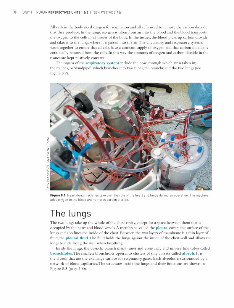
Figure 8.1 Heart–lung machines take over the role of the heart and lungs during an operation. The machine adds oxygen to the blood and removes carbon dioxide.
All cells in the body need oxygen for respiration and all cells need to remove the carbon dioxide that they produce. In the lungs, oxygen is taken from air into the blood and the blood transports the oxygen to the cells in all tissues of the body. In the tissues, the blood picks up carbon dioxide and takes it to the lungs where it is passed into the air. The circulatory and respiratory systems work together to ensure that all cells have a constant supply of oxygen and that carbon dioxide is continually removed from the cells. In this way the amounts of oxygen and carbon dioxide in the tissues are kept relatively constant.
The organs of the respiratory system include the nose, through which air is taken in; the trachea, or ‘windpipe’, which branches into two tubes; the bronchi; and the two lungs (see Figure 8.2).
The lungsThe two lungs take up the whole of the chest cavity, except for a space between them that is occupied by the heart and blood vessels. A membrane, called the pleura, covers the surface of the lungs and also lines the inside of the chest. Between the two layers of membrane is a thin layer of fluid, the pleural fluid. The fluid holds the lungs against the inside of the chest wall and allows the lungs to slide along the wall when breathing.
Inside the lungs, the bronchi branch many times and eventually end in very fine tubes called bronchioles. The smallest bronchioles open into clusters of tiny air sacs called alveoli. It is the alveoli that are the exchange surface for respiratory gases. Each alveolus is surrounded by a network of blood capillaries. The structures inside the lungs and their functions are shown in Figure 8.3 (page 100).
Get
ty Im
ages
/SP
L C
reat
ive/
AJ
Pho
to

Pharynx, or throat – air from nasal cavity passes through here.
Larynx – the organ of voice. Air passes through the larynx,going to and from the lungs.Contains the vocal cords, whichcan vibrate to make sound.
Trachea, or windpipe – carries air toand from the lungs. Is lined with a mucousmembrane and cells with cilia. The cilia beatto move mucus and trapped particlesupwards.
Intercostal muscles – muscles betweenthe ribs. They move the rib cage upwardsand outwards to increase the volume ofthe chest cavity and thus the lungswhen breathing in.
Lungs – occupy all the chest cavity,except the space taken up by the heart.They are covered by a pleural membranethat also lines the inside of the chest.Pleural fluid between the two layers holdsthe lungs against the inside of the chest.
Alveoli – tiny air sacs that makeup most of the lung. They occur inclusters and have very thin wallsthat are well supplied with bloodcapillaries for the exchange ofoxygen and carbon dioxide.
Diaphragm – a muscle that separatesthe chest from the abdomen. It contractsand flattens downwards, thereby increasingthe volume of chest cavity, and lungs,during breathing in.
Nasal cavity – contains projectionsthat increase the internal surface area.Filters, warms and moistens air beforeit enters the lungs. Contains smellreceptors. Acts as a resonatingchamber for speech sounds. Hairs andmucus trap dust.
Epiglottis – a flap of tissue that,during swallowing, closes off thetrachea so food and liquidcannot enter the lungs.
Bronchioles – very fine tubes with wallsof smooth muscle. The finest of them endin groups of air sacs, the alveoli.
Bronchi – two primary bronchibranch from the trachea; theythen divide into secondary and
tertiary bronchi.Ribs – form the framework for thechest.
ISBN 9780170351126 | CHAPTER 8 | THE RESPIRATORY SYSTEM 99
Figure 8.2 The structure and functions of the parts of the respiratory system

Bronchiole
bronchiolesBronchioles
Alveoli(cut open)
Alveoli
From
pul
mon
ary a
rtery
To pulmonary
vein
Alveolus
Capillary
Bronchiole
Primary bronchi
Lung
Trachea
Secondary bronchi
BronchiolesTertiary bronchi
UNIT 1 | HUMAN PERSPECTIVES UNITS 1 & 2 | ISBN 9780170351126100
Respiratory surfaces and the exchange of gasesThe lungs are well suited to their gas exchange function, for the following reasons:❯❯ The alveoli give the lungs a huge internal surface area, so that large amounts of gases can
be exchanged in a relatively short time. Estimates of the number of alveoli in the lungs vary considerably but there are hundreds of millions of them; they probably have a total surface area of 50–80 m2 – about one-third the area of a tennis court.
❯❯ Each alveolus is well supplied with blood vessels, so that as much blood as possible is close to the air in the alveolus (Figure 8.3). The continuous flow of blood helps to maintain a difference in concentrations of oxygen and carbon dioxide in the blood and in the air in the lungs.
❯❯ The membrane that forms the wall of the alveolus is very thin, so that gas molecules do not have far to travel when moving into or out of the blood. The wall has only one layer of cells and is only 1 micrometre (1 µm or 1/1000 of a millimetre) thick (Figure 8.4).
❯❯ The lungs are positioned deep inside the body to prevent excessive evaporation of the fluid that covers the respiratory surfaces. It is important that the membrane of the alveolus be covered by a thin layer of moisture because gases can diffuse into and out of the blood only when they are dissolved in fluid.
❯❯ The lung volume can be changed by movements of the respiratory muscles, so that air is made to flow into and out of the lungs. Constant changing of the air in the alveoli helps to ensure that there is always a difference in the concentrations of oxygen and carbon dioxide in the air and in the blood.
Figure 8.3 The structure of the lungs
Figure 8.4 Photomicrograph of a section of lung tissue. The areas arrowed are the air spaces inside the alveoli.
Pho
tota
ke/©
Car
olin
a B
iolo
gica
l Sup
ply
Com
pany
1 Diaphragm relaxes,pushing up intochest cavity
2 Lung volume decreases
3 Air flows from higherpressure in lungs tolower pressure outside1 Rib cage
moves downand inwards
Trachea
Pleural membranes
Pleural fluid
1 Diaphragm contracts,extending chest cavitydownwards
1 Intercostal musclescontract, extending rib cageupwards and outwards
2 Lung volume increases
3 Air flows from higherpressure to lowerpressure in lungs
ISBN 9780170351126 | CHAPTER 8 | THE RESPIRATORY SYSTEM 101
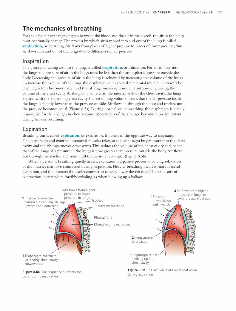
Figure 8.5b The sequence of events that occur during expiration
The mechanics of breathingFor the efficient exchange of gases between the blood and the air in the alveoli, the air in the lungs must continually change. The process by which air is moved into and out of the lungs is called ventilation, or breathing. Air flows from places of higher pressure to places of lower pressure; thus air flows into and out of the lungs due to differences in air pressure.
InspirationThe process of taking air into the lungs is called inspiration, or inhalation. For air to flow into the lungs, the pressure of air in the lungs must be less than the atmospheric pressure outside the body. Decreasing the pressure of air in the lungs is achieved by increasing the volume of the lungs. To increase the volume of the lungs, the diaphragm and external intercostal muscles contract. The diaphragm thus becomes flatter and the rib cage moves upwards and outwards, increasing the volume of the chest cavity. As the pleura adheres to the internal wall of the chest cavity, the lungs expand with the expanding chest cavity. Increased lung volume means that the air pressure inside the lungs is slightly lower than the pressure outside. Air flows in through the nose and trachea until the pressure becomes equal (Figure 8.5a). During normal, quiet breathing, the diaphragm is mainly responsible for the changes in chest volume. Movements of the rib cage become more important during heavier breathing.
ExpirationBreathing out is called expiration, or exhalation. It occurs in the opposite way to inspiration. The diaphragm and external intercostal muscles relax, so the diaphragm bulges more into the chest cavity and the rib cage moves downwards. This reduces the volume of the chest cavity and, hence, that of the lungs. Air pressure in the lungs is now greater than pressure outside the body. Air flows out through the trachea and nose until the pressures are equal (Figure 8.5b).
When a person is breathing quietly at rest, expiration is a passive process, involving relaxation of the muscles that have contracted during inspiration. Heavier breathing involves more forceful expiration, and the intercostal muscles contract to actively lower the rib cage. The same sort of contraction occurs when forcibly exhaling, as when blowing up a balloon.
Figure 8.5a The sequence of events that occur during inspiration

Carbon dioxide diffusesfrom the higherconcentration in the bloodto the lower concentrationin the air in the alveolus.
O2O2
O2
O2
CO2
CO2
CO2
CO2
The artery to thelungs bringsdeoxygenatedblood to the capillaries.
Air flows into and outof the alveoli as webreathe.
Alveolus
Oxygenated blood leavesthe capillaries of thealveoli and is taken to theheart in the veins from thelungs.
Oxygen diffuses from thehigher concentration in theair in the alveolus to thelower concentration in theblood.
The wall of the alveolus is justone cell thick; the wall of thecapillary is one cell thick;the inside of the alveolus islined with a film of moisture.These characteristics make iteasy for the gases to diffuseto and from the body.
UNIT 1 | HUMAN PERSPECTIVES UNITS 1 & 2 | ISBN 9780170351126102
Gas exchangeThe blood in capillaries surrounding the alveoli is brought to the lungs by the pulmonary arteries. This blood has been through the capillaries of the body, where much of the oxygen has been taken up by the body cells. Thus, the blood that comes into the capillaries around the alveoli has a low concentration of oxygen – lower than the concentration in the air in the alveolus. Oxygen therefore dissolves in the moisture on the inside of the alveolus and diffuses through the membrane, through the walls of the capillaries and into the blood (Figure 8.6).
The blood arriving at the capillaries of the alveoli has a higher concentration of carbon dioxide. It has come from the body circulation where it has picked up carbon dioxide produced by respiration in the cells. The concentration of carbon dioxide in the alveolar capillaries is therefore higher than the concentration in the air in the alveolus. Carbon dioxide diffuses out of the blood, into the air in the alveolus. Thus, expired air contains less oxygen, and more carbon dioxide, than inspired air (Table 8.1).
Table 8.1 Oxygen and carbon dioxide concentrations in inspired and expired air
Inspired air (%) Expired air (%)
Oxygen 20.95 15.80
Carbon dioxide 0.04 4.30
Note: The other 79% of the inspired air is made up mainly of nitrogen, with varying amounts of water vapour.
Figure 8.6 Gas exchange between alveolar air and blood

Joinedalveoli
ISBN 9780170351126 | CHAPTER 8 | THE RESPIRATORY SYSTEM 103
For diffusion of gases into and out of the blood, there must be a concentration gradient – that is, a difference in gas concentration between the air in the alveoli and the blood in the capillaries. The concentration gradient for oxygen and carbon dioxide is maintained by:❯❯ the constant flow of blood through the capillaries. As the blood flowing through the capillaries
around each alveolus picks up oxygen and loses carbon dioxide, it is replaced by more blood pumped into the capillaries. This ‘new’ blood is low in oxygen and high in carbon dioxide, so the concentration gradient is maintained
❯❯ the movement of air into and out of the alveoli as we breathe in and out. The air that has picked up carbon dioxide from, and lost oxygen to, the blood is replaced by ‘new’ air with each breath. The ‘new’ air is low in carbon dioxide and high in oxygen.
EXTENSIONThe activities of the circulatory system and respiratory system are closely related. Find out:
❯❯ why rate and depth of breathing change when we exercise❯❯ why heart rate also changes❯❯ how the activities of the circulatory and respiratory systems are coordinated.
Some effects of lifestyle and environment on gas exchangeHow we live and the environments in which we work and spend our leisure time profoundly affect the efficiency of the respiratory surfaces.
EmphysemaEmphysema is a disease usually caused by long-term exposure to irritating particles in the air taken into the lungs. No one can avoid inhaling particles, as there are particles of matter in the air at all times. Some people, however, are exposed to excessively high levels. Smokers constantly inhale irritants in tobacco smoke; those who work in situations where a lot of dust is produced are also at risk. In cities with continually high air pollution, there is a greater risk of emphysema.
The irritating particles cause damage to the alveoli. They lose their elasticity, are often replaced with fibrous tissue, and may break down, reducing the internal surface area of the lung (Figure 8.7). Because of the loss of elasticity of the lung tissue, the lungs are constantly inflated, and breathing out no longer occurs passively but requires voluntary effort. Thus, the emphysema sufferer has two problems – inadequate surface area for gas exchange, and difficulty in ventilating the lungs. Emphysema cannot be cured, and once lung damage begins, the progression of the disease cannot be stopped.
Figure 8.7a Photomicrograph of a section of lung tissue showing normal alveoli
Figure 8.7b Photomicrograph of a section of lung tissue from a patient with emphysema showing how alveolar walls have broken down forming larger, fewer alveoli with reduced total surface area
The respiratory system
Gas exchangeThis website includes an animation of gas exchange.

UNIT 1 | HUMAN PERSPECTIVES UNITS 1 & 2 | ISBN 9780170351126104
Lung cancerLung cancer is similar to most other cancers in that it involves the development of a tumour, a mass of cells that divides in an uncontrolled way. Evidence shows clear links between lung cancer and tobacco smoking, and exposure to asbestos fibres and other pollutants. Smoking poses by far the greatest risk for lung cancer and if smoking is combined with other risk factors, the chances of contracting lung cancer are even greater. For example, people who have worked with asbestos and who also smoke have a 20–90 times greater risk of contracting lung cancer than similar workers who do not smoke. Some chemical substances seem to initiate cancerous growths; others seem to promote the growth of the tumour. Tobacco smoke contains both initiators and promoters of lung cancer.
The most common form of lung cancer begins in the walls of the air passages, usually the bronchi. Inhaled smoke particles constantly irritate the mucous membrane that lines the air passages. This results in excessive production of mucus. Cells at the base of the membrane begin to divide more rapidly and the accumulating mucus cannot be removed. This results in ‘smoker’s cough’. The trapped mucus causes rupture of the alveoli. Emphysema has then developed. Ultimately a cancerous growth develops in an air passage and may spread to other parts of the body.
Lung infectionsPneumonia is an infection of the lungs caused by bacteria, viruses, fungi or other organisms. The inflammation resulting from the infection causes secretion of fluid and mucus into the alveoli, thus reducing the amount of air that they can contain. The surface area available for exchange of gases is also reduced, and breathing difficulty is a symptom of many types of pneumonia.
Tuberculosis (TB) is an infection, usually of the lung, by the bacterium Mycobacterium tuberculosis. As a cause of death, tuberculosis, along with HIV/AIDS and malaria, is one of the top three infectious diseases in the world. Fortunately, in Australia there is a very low incidence of tuberculosis – about 1000 cases per year, most of them in people born overseas.
Most lung infections, such as tuberculosis and pneumonia, are spread by droplets. When infected people cough, sneeze or spit, tiny droplets of moisture containing the bacteria, viruses or fungi may be inhaled by others, so spreading the infection. Good hygiene practices, such as coughing and sneezing into a handkerchief and not spitting, help to reduce the spread of lung infections.
AsthmaAsthma is an allergic response to foreign substances that enter the body. Some of the more common sources of such foreign particles are animal skins, feathers, bird excreta, house dust mites and pollen grains. Substances in food may also trigger an asthma attack. During such an attack, the muscles that surround the bronchioles go into spasm – sudden involuntary contractions. This causes narrowing of the air passages and difficulty in breathing.
Usually the irritation of the membranes lining the air passages causes secretion of excessive mucus and this also restricts movement of air. The reduced volume of air going into and out of the lungs means that the exchange of gases is impaired and the blood does not carry the usual amount of oxygen.

ISBN 9780170351126 | CHAPTER 8 | THE RESPIRATORY SYSTEM 105
Science inquiry
ACTIVITY 8.1 Structure of the lungsThis activity may be done as a demonstration by your teacher.
Examine a set of sheep or pig lungs.1 Identify the structures that can be seen:
a the lungs themselves divided into a number of lobesb the trachea with its rings of cartilage; examine the rings to see whether they form
a complete circlec the two bronchi that branch from the trachead the thin transparent membrane that covers the lungs.
2 Squeeze the lungs between your thumb and a finger. Describe what you feel.3 Cut off a piece of lung and place it in a beaker of water. Does it float? What does this tell you
about the lung?4 Cut open the trachea and observe the interior. Record your observations.5 Continue the cut in the trachea down through one of the bronchi, then through a secondary
bronchus. Keep cutting until the air tubes become too small to see. Do the secondary bronchi have rings of cartilage? As you go along the air tubes from large to small, where do the cartilage rings stop?
ACTIVITY 8.2 Investigating breathingWorking on your own, or with a partner, think of a factor that could influence breathing. ❯ Propose a hypothesis that links the two variables – some aspect of breathing and the factor that
you have selected. ❯ Design an experiment to test your hypothesis. ❯ Your teacher may want you to carry out your experiment and present a report in some
appropriate format.

UNIT 1 | HUMAN PERSPECTIVES UNITS 1 & 2 | ISBN 9780170351126106
Review questions1 List the characteristics of the lungs that make them well suited for gas exchange.2 Why is it that, in the lungs, oxygen diffuses into and carbon dioxide out of the blood, whereas in
other body tissues oxygen diffuses out of and carbon dioxide into the blood?3 a Draw a diagram showing inspiration. As labels for your diagram, list the sequence of
events that occur in inspiration.b Draw a diagram showing expiration. As labels for your diagram, list the sequence of events
that occur in expiration.4 a Why is a concentration gradient important for the exchange of gases?
b Explain how a concentration gradient for oxygen and carbon dioxide is maintained between the blood and the air in the alveoli.
5 a Describe how oxygen is carried in the blood.b Describe how carbon dioxide is carried in the blood.
6 Describe precautions that you can take to reduce your risk of developing emphysema or lung cancer.
7 Describe the types of lung damage that can be caused by smoking.8 Why does pneumonia often cause difficulty in breathing?
Apply your knowledge1 To be effective, any surface where materials are taken into the body, or passed out of the body,
must have a very large surface area. For the lungs, explain how a large surface area is achieved.2 The exchange surfaces of the lungs rely on concentration differences so that substances diffuse
across the surface. Explain how the concentration difference is maintained.3 If air enters the chest cavity through a puncture wound to the chest wall, the lung may collapse.
As the collapsed lung is no longer attached to the chest wall, air cannot be made to move into and out of the lung. However, a person with a collapsed lung can function fairly normally.a Explain how it would be possible for such a person to function in a fairly normal way.b Would there be any activities that such a person would not be able to perform?
4 Students measured the breathing rate and depth of breathing of a girl before and after exercise. Their results are shown in Table 8.2.
Table 8.2 Breathing rate and depth before and after exercise
Breaths per minute Volume of air per breath (cm3)
At rest 19 460
After running 38 1075
a Calculate the total volume of air that the girl breathed in 1 minute before and after exercise.
b What is the reason for the increase in rate and depth of breathing after exercise?c Describe the changes that would occur in the body to bring breathing back to the normal
resting level after exercise.5 The ability to voluntarily control breathing is important when speaking, but it is also important
when eating or drinking. Explain why this is so.6 List five occupations in which people could be at risk of contracting emphysema. What
precautions could be taken to reduce the risk of workers contracting the disease?7 In expired air resuscitation (mouth-to-mouth resuscitation), air from the rescuer’s lungs
is blown into the patient’s lungs. How is expired air able to keep the patient alive? (Refer to Table 8.1 on page 102.)