third-party central memory cd8 t cells − nonreactive anti a new
TRANSCRIPT
doi:10.1182/blood-2012-06-432443Prepublished online February 27, 2013;2013 121: 3033-3040
Reich-Zeliger, Bar Nathansohn, Matthias Edinger, Robert S. Negrin, David Hagin and Yair ReisnerAssaf Lask, Eran Ophir, Noga Or-Geva, Adva Cohen-Fredarow, Ran Afik, Yaki Eidelstein, Shlomit
third-party central memory CD8 T cells−nonreactive antiA new approach for eradication of residual lymphoma cells by host
http://bloodjournal.hematologylibrary.org/content/121/15/3033.full.htmlUpdated information and services can be found at:
(1903 articles)Transplantation � (1491 articles)Lymphoid Neoplasia �
Articles on similar topics can be found in the following Blood collections
http://bloodjournal.hematologylibrary.org/site/misc/rights.xhtml#repub_requestsInformation about reproducing this article in parts or in its entirety may be found online at:
http://bloodjournal.hematologylibrary.org/site/misc/rights.xhtml#reprintsInformation about ordering reprints may be found online at:
http://bloodjournal.hematologylibrary.org/site/subscriptions/index.xhtmlInformation about subscriptions and ASH membership may be found online at:
Copyright 2011 by The American Society of Hematology; all rights reserved.Washington DC 20036.by the American Society of Hematology, 2021 L St, NW, Suite 900, Blood (print ISSN 0006-4971, online ISSN 1528-0020), is published weekly
For personal use only. at Weizmann Institute of Science on August 28, 2013. bloodjournal.hematologylibrary.orgFrom
Regular Article
TRANSPLANTATION
A new approach for eradication of residual lymphoma cells by hostnonreactive anti–third-party central memory CD8 T cellsAssaf Lask,1 Eran Ophir,1 Noga Or-Geva,1 Adva Cohen-Fredarow,1 Ran Afik,1 Yaki Eidelstein,1 Shlomit Reich-Zeliger,1
Bar Nathansohn,1 Matthias Edinger,2 Robert S. Negrin,3 David Hagin,1 and Yair Reisner1
1Immunology Department, Weizmann Institute of Science, Rehovot, Israel; 2Department of Hematology and Oncology, University Hospital Regensburg,
Regensburg, Germany; and 3Bone Marrow Transplantation, Stanford University, Stanford, CA
Key Points
• Anti–third-party Tcm killmalignant B cells in a T-cellreceptor–independentmechanism while sparingnaive B cells.
Generation of T cells endowed with graft-versus-leukemia (GVL) and depleted of
graft-versus-host (GVH) activity represents a highly desirable goal in bone marrow
transplantation (BMT). Here, we demonstrate that donor anti–third-party CD8 T cells
with central memory phenotype (Tcm) exhibit marked GVL reactivity through a unique
T-cell receptor–independent mechanism. Thus, in a residual disease mouse model, Tcm
therapy following autologous BMT led to significant survival prolongation, with 30% to
40% of the treated mice displaying long-term tumor-free survival. A more impressive
finding was that infusion of donor Tcm in an allogeneic model rapidly eliminated
residual lymphoma cells and led to long-term survival of 100% in the absence of GVH disease. Collectively, the strong GVL
reactivity of anti–third-party Tcm, coupled with their demonstrated enhancement of bone marrow allografting, suggests that the
use of Tcm therapy in conjunction with allogeneic T-cell–depleted BMT could be of particular benefit in patients with B-cell
malignancies who cannot tolerate intensive myeloablative conditioning. (Blood. 2013;121(15):3033-3040)
Introduction
The vital role of donor T cells in promoting engraftment andmediating graft-versus-leukemia (GVL) reactivity of allogeneicbone marrow (BM) transplants was established more than 2decades ago upon the introduction of T-cell depletion for theprevention of graft-versus-host disease (GVHD).1,2 We haverecently shown that host T-cell–mediated rejection of T-cell–depleted BM transplants (TDBMT) can be overcome in a mousemodel by adding to the transplant inoculum activated anti–third-party donor CD81 T cells with central memory phenotype (Tcm);these cells can home to the recipient’s lymph nodes and specif-ically delete host anti–donor T-cell clones.3,4 Importantly, theseTcm were shown to be depleted of graft-versus-host reactivity byvirtue of their initial stimulation against third-party cells undercytokine deprivation.
In the present study, we addressed a second attribute of anti–third-party Tcm, namely their potential GVL reactivity, whichcould be very valuable for patients undergoing bone marrow trans-plantation (BMT) following reduced intensity conditioning (RIC).
The possibility that Tcm might exhibit GVL has been indicatedinitially by our previous unexpected observation in the humansetting that both allogeneic and autologous anti–third-party CD81
cytotoxic T lymphocytes (CTLs) exhibit in vitro significant killingof B-cell chronic lymphocytic leukemia (B-CLL)5 and B-cell non-Hodgkin lymphoma (B-NHL) cells6 while sparing acute myeloidleukemia blasts.5 The killing of B-cell tumors by anti–third-party
CTLs was shown to involve a unique T-cell receptor (TCR)-independent 2-step mechanism. First, long-lasting conjugates areformed between the CTL and the tumor cell. These conjugates arerapidly formed through binding of intercellular adhesion molecule1 (ICAM-1) on tumor cells by leukocyte function-associatedantigen 1 (LFA-1) expressed on effector T cells. Second, a slowerprocess of major histocompatibility complex I (MHC-I)–dependentapoptosis is mediated by binding of theMHC-I a2/3 constant regionon the tumor cells to the CD8 molecule on the CTL membrane.
However, considering the nonconventional characteristics ofthis mechanism, it could be argued that this type of killingrepresents an artificial phenomenon with very little relevance ifany to clinical settings. Thus, it was critical to evaluate in anappropriate mouse model whether murine anti–third-party Tcm canmediate significant GVL reactivity in vivo, in addition to theirpotent tolerizing activity.
Initially, we verified in vitro that mouse anti–third-party Tcmare endowed with antilymphoma reactivity through a TCR-independent mechanism, as was previously shown for their humancounterparts. Subsequently, we tested their antilymphoma reac-tivity in a model simulating minimal residual disease followingBMT using bioluminescence imaging (BLI). Strikingly, we dis-covered that both syngeneic and allogeneic Tcm were able toefficiently eliminate lymphoma cells. This effect was achievedwithout any GVHD and while sparing naive B cells.
Submitted June 7, 2012; accepted February 5, 2013. Prepublished online as
Blood First Edition paper, February 27, 2013; DOI 10.1182/blood-2012-06-
432443.
A.L. and E.O. contributed equally to this study.
The online version of this article contains a data supplement.
The publication costs of this article were defrayed in part by page charge
payment. Therefore, and solely to indicate this fact, this article is hereby
marked “advertisement” in accordance with 18 USC section 1734.
© 2013 by The American Society of Hematology
BLOOD, 11 APRIL 2013 x VOLUME 121, NUMBER 15 3033
For personal use only. at Weizmann Institute of Science on August 28, 2013. bloodjournal.hematologylibrary.orgFrom

Thus, together with their ability to markedly enhance BMallografting, anti–third-party Tcm can uniquely address both thechallenge of engraftment following RIC and the problem of relapsecommonly associated with RIC protocols. This novel cell therapycould be highly attractive, particularly for elderly patients withB-CLL and other B-cell malignancies who might not tolerateaggressive conditioning.
Methods
Animals
For detailed information on mouse strains used, see “supplementalMethods.” Institutional review board approvals were as follows: Institu-tional Animal Care and Use Committee (IACUC) application number00520111-4 “TCM GVL in-vitro”; IACUC application number 00510111-3 “In-vivo GVL effect of anti third-party TCMs”; IACUC applicationnumber 02850711-1 “Humoral response after treatment with anti third partyTcm.”
Flow cytometric analysis
For detailed information on antibodies (Abs) used and fluorescence-activated cell sorting (FACS) analysis, see “supplemental Methods.”
Lymphoma cell lines
A20 lymphoma cells and A20 cells transduced with a luciferase reportergene (A20 luc)7 were cultured in RPMI supplemented with 10% fetal calfserum (FCS) and antibiotics.
BCL1-luc cells7 were thawed and washed twice with RPMI sup-plemented with 10% FCS before injection.
Preparation of host nonreactive anti–third-party cells
Anti–third-party Tcm were grown as previously described.3 Briefly,splenocytes from the donor mouse strain (4 3 106 cells/mL) were culturedat a 1:1 ratio against irradiated (20 Gy) third-party splenocytes (donor,third-party, and host mice were MHC-I disparate) for 60 hours underexogenous cytokine deprivation at 37°C in a 5% CO2 incubator.Subsequently, the cells were fractionated on Ficoll-Paque Plus (AmershamPharmacia Biotech, AB) and CD81 cells were positively selected usingMagnetic Particles (BD Pharmingen) and cultured (1 3 106 cells/mL) withrhIL-15 (20 ng/mL; R&D systems) in an Ag-free environment (in theabsence of stimulators) at 37°C in a 5% CO2 incubator. To attain a purifiedpopulation at the end of the culture (day 16), the Tcm were positivelyselected for L-selectin (CD62L, MACS Cell Separation; Miltenyi Biotec,Bergisch Gladbach, Germany). Cell phenotype was then analyzed by flowcytometry (FACScan; Becton Dickinson) for cell size and expressionof the activation marker (CD44) and L-selectin (CD62L) within theCD8 compartment.
Mixed lymphocyte reaction killing assay
Lymphoma cells were obtained by Ficoll density gradient centrifugation,after which they were labeled according to the manufacturer’s instructionswith 0.15 mg/mL CalceinAM (Invitrogen, Carlsbad, CA), a vital dye that isreleased upon cell death. Cells were brought to a concentration of 1 3 106
cells/mL in RPMI supplemented with 10% FCS and antibiotics. Next,2.5 3 105 Calcein-labeled lymphoma cells were incubated with or withoutanti–third-party Tcm at the indicated ratio for 16 hours in 24-well plates.Cells were recovered and analyzed for survival by measuring the number ofsurviving Calcein-stained lymphoma cells by FACS. To obtain absolutevalues of cells, samples were resuspended in a constant volume and flowcytometric counts for each sample were obtained during a predeterminedperiod of time and compared with flow cytometric counts obtained with
a fixed volume and fixed numbers of input cells.8 Survival rates arepresented relative to the survival of lymphoma cells alone. The percentageof lymphoma cell killing was calculated by the formula:�12
The number of live lymphoma cells in the assessed wellThe number of live lymphoma cells in the control well
�3 100
Inhibition of B-cell lymphoma killing by blocking Abs
Anti–third-party Tcm or lymphoma cells were preincubated for 30 minutesin a minimal volume with the indicated neutralizing Ab at the indicatedconcentrations. Blocked cells were then incubated with the other non-blocked component of the mixed lymphocyte reaction for 16 hours at a1:5 ratio in favor of the anti–third-party cells. Lymphoma cell survival wasanalyzed by FACS. The following neutralizing Abs were used: LFA-1(CD11a) blocking Ab (clone M17/4; Biolegend), ICAM-1 (CD54) blockingAb (clone YN1/1.7.4; Biolegend), anti–CD8a blocking Ab (clone 53-6.7;Biolegend).
GVL mouse models
The A20-luc model. Host mice (BALB/c; 12-13 weeks of age) wereexposed to a single dose of lethal total body irradiation (TBI; 8 Gy)on day 21. The following day (day 0), the mice were intravenouslyadministered a transplant of 3 3 106 syngeneic (BALB/c) or allogeneic(B6) nude BM cells with or without 5 3 103 A20-luc cells. On day 11,selected mice received anti–third-party Tcm cells in the indicated amounts.Tumor localization, migratory patterns of A20 cells, and tumor load weremonitored using an in vivo imaging system.
The BCL1-luc model. Host mice (BALB/c; 12 weeks of age)were exposed to a single dose of lethal TBI (8Gy) on day 21. Thefollowing day (day 0), the mice were intravenously administered atransplant of 3 3 106 syngeneic (BALB/c) nude BM cells with or without1 3 103 BCL1-luc cells. On day 11, selected mice received anti–third-partyTcm cells in the indicated amounts. Tumor localization, migratory patterns ofBCL1 cells, and tumor load were monitored using an in vivo imagingsystem.
In vivo imaging
The in vivo GVL model was established as described in the previoussection. At the indicated times posttransplant, the mice were anesthetizedwith ketamine (100 mg/kg; Kepro, Holland, Netherlands) and xylazine(20 mg/kg; Kepro) injected intraperitoneally. Mice were then injectedintraperitoneally with an aqueous solution of D-luciferin (150 mg/kgCat#XR-1001, Xenogen; 30 mg/mL in phosphate-buffered saline) 10minutes prior to imaging. The mice were then monitored using the opticalwhole-body imaging system (IVIS 100, Xenogen) coupled with a PixelflyQE (PCO, Kelheim, Germany) charge-coupled device camera. Imageprocessing and data analysis were performed using Living Image 3.2software.
GVHD evaluation
Mice receiving allogeneic transplants were evaluated for symptoms ofGVHD; the mice were monitored for survival and for external signs ofGVHD including ruffled fur, hunched back, and weight loss.
Purification of naive CD8 T and B cells and lipopolysaccharide
activation
For detailed information, see “supplemental Methods.”
Measurement of antigen-specific immunoglobulins by ELISA
Anti-TNP antibodies in the serum of immunized mice were measured byenzyme-linked immunosorbent assay (ELISA). ELISA plates (Nunc;Maxisorp, Rochester, NY) were coated with 10 mg/mL TNP bovine serumalbumin prepared as previously described.9 Doubling dilutions of serum
3034 LASK et al BLOOD, 11 APRIL 2013 x VOLUME 121, NUMBER 15
For personal use only. at Weizmann Institute of Science on August 28, 2013. bloodjournal.hematologylibrary.orgFrom
were made and incubated for 30 min at 37°C. Following washing inphosphate-buffered saline/0.05% Tween-20, plates were incubated withhorseradish-peroxidase–conjugated polyclonal goat anti-mouse Fab(Jackson ImmunoResearch Laboratories, West Grove, PA). Antibodybinding was detected by addition of TMB substrate (Sigma, St. Louis,MO) to the washed plates and the optical density at 630 nm filter wasmeasured 10 minutes after substrate addition using an ELISA reader (Bio-Tek, Winooski, VT).
Statistical analysis
The analysis of survival data was performed using Kaplan-Meier curves(log-rank test). Comparison of means was conducted using the Studentt test.
Results
Anti–third-party CD8 Tcm induce apoptosis of lymphoma cells
ex vivo through a TCR-independent mechanism mediated via
LFA-1/ICAM-1 interactions
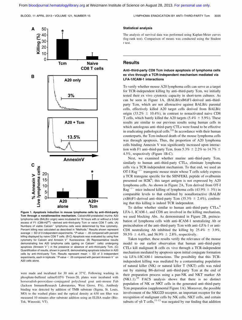
To verify whether mouse A20 lymphoma cells can serve as a targetfor TCR-independent killing by anti–third-party Tcm, we initiallytested their ex vivo cytotoxic capacity in short-term cultures. Ascan be seen in Figure 1A, (BALB/cxB6)F1-derived anti–third-party Tcm, which are not alloreactive against BALB/c parentalcells, effectively killed A20 target cells derived from BALB/corigin (33.2% 6 10.4%), in contrast to nonactivated naive CD8T cells, which barely killed the A20 targets (5.4% 6 5.9%). Theseresults are similar to our previous results using human cells inwhich autologous anti–third-party CTLs were found to be effectivein eradicating pathological cells.5,6 In accordance with their humancounterparts, the Tcm-induced death of the mouse lymphoma cellswas through apoptosis. Thus, the proportion of A20 lymphomacells binding Annexin V was significantly increased upon interac-tion with F1 anti–third-party Tcm, from 5.3% 6 2.2% to 14.7% 64.5%, respectively (Figure 1B-C).
Next, we examined whether murine anti–third-party Tcm,similarly to human anti–third-party CTLs, eliminate lymphomacells via a TCR-independent mechanism. To that end, we used anOT-I Rag2/2 transgenic mouse strain whose T cells solely expressa TCR transgene specific for the SIINFEKL peptide of ovalbuminpresented on H2Kb; this target antigen is not expressed by A20lymphoma cells. As shown in Figure 2A, Tcm derived from OT-IRag2/2 mice induced killing of lymphoma cells (43.9% 6 3%) incomparable levels to that exhibited by nonalloreactive (BALB/cxB6)F1-derived anti–third-party Tcm (35.3% 6 2.4%), confirm-ing that this killing is indeed TCR independent.
To define whether similar to human anti–third-party CTLs,6
LFA-1, ICAM-1, and CD8 are involved in the killing mechanism,we used blocking Abs. As demonstrated in Figure 2B, preincu-bation of lymphoma cells with anti–ICAM-1 neutralizing Ab orpreincubation of the anti–third-party Tcm with anti–LFA-1 or anti-CD8 neutralizing Ab inhibited the killing by 25.4% 6 3.9%,59.5% 6 4.4%, and 56.9% 6 2.8%, respectively.
Taken together, these results verify the relevance of the mousemodel to our earlier observation that human anti–third-partyCTLs kill malignant B cells ex vivo through a TCR-independentmechanism mediated by apoptosis upon initial conjugate formationvia LFA-1/ICAM-1 interactions. The possibility that this TCR-independent killing was mediated by a contaminating populationof natural killer (NK) or natural killer T (NKT) cells was ruledout by staining B6-derived anti–third-party Tcm at the end oftheir preparation process using a pan-NK and NKT marker Ab(NK1.1).10 FACS analysis shows that there is no distinctpopulation of NK or NΚT cells in the generated anti–third-partyTcm preparation (supplemental Figure 1A). Moreover, the possibleinvolvement of the NKG2D receptor, an important receptor for therecognition of malignant cells by NK cells, NKT cells, and certainsubsets of gd T cells,11-13 was negated by our finding that addition
Figure 1. Apoptosis induction in mouse lymphoma cells by anti–third-party
Tcm through a nonalloreactive mechanism. CalceinAM-prelabeled murine A20
lymphoma cells (BALB/c origin) were incubated for 16 hours with or without a 5-fold
excess of F1 (CB6-H2bd) –derived anti–third-party Tcm or naive CD8 T cells. (A)
Numbers of viable Calcein1 lymphoma cells were determined by flow cytometry.
Percent killing was calculated as described in “Methods.” Results shown represent
average 6 SD of 3 independent experiments. *P value , .05 compared with percent
killing displayed by naive CD8 T cells. (B-C) Apoptosis was evaluated by using flow
cytometry for Calcein and Annexin V1 fluorescence. (B) Representative results
demonstrating live A20 lymphoma cells (gating on Calcein1 cells) undergoing
apoptosis (Annexin V1) in the presence or absence of anti–third-party Tcm. (C)
Quantification of results, shown in panel B, demonstrating apoptosis induction in A20
cells by anti–third-party Tcm. Results represent mean 6 SD of 3 independent
experiments, each in triplicate. *P value, .05 compared with percent Annexin V1 on
A20 cells alone.
BLOOD, 11 APRIL 2013 x VOLUME 121, NUMBER 15 LYMPHOMA ERADICATION BY ANTI–THIRD-PARTY Tcm 3035
For personal use only. at Weizmann Institute of Science on August 28, 2013. bloodjournal.hematologylibrary.orgFrom
of NKG2D blocking Ab did not inhibit the killing of lymphomacells by anti–third-party Tcm (supplemental Figure 1B).
Syngeneic anti–third-party Tcm mediate effective lymphoma
regression in vivo and contribute to prolongation of survival in
mice with minimal residual disease
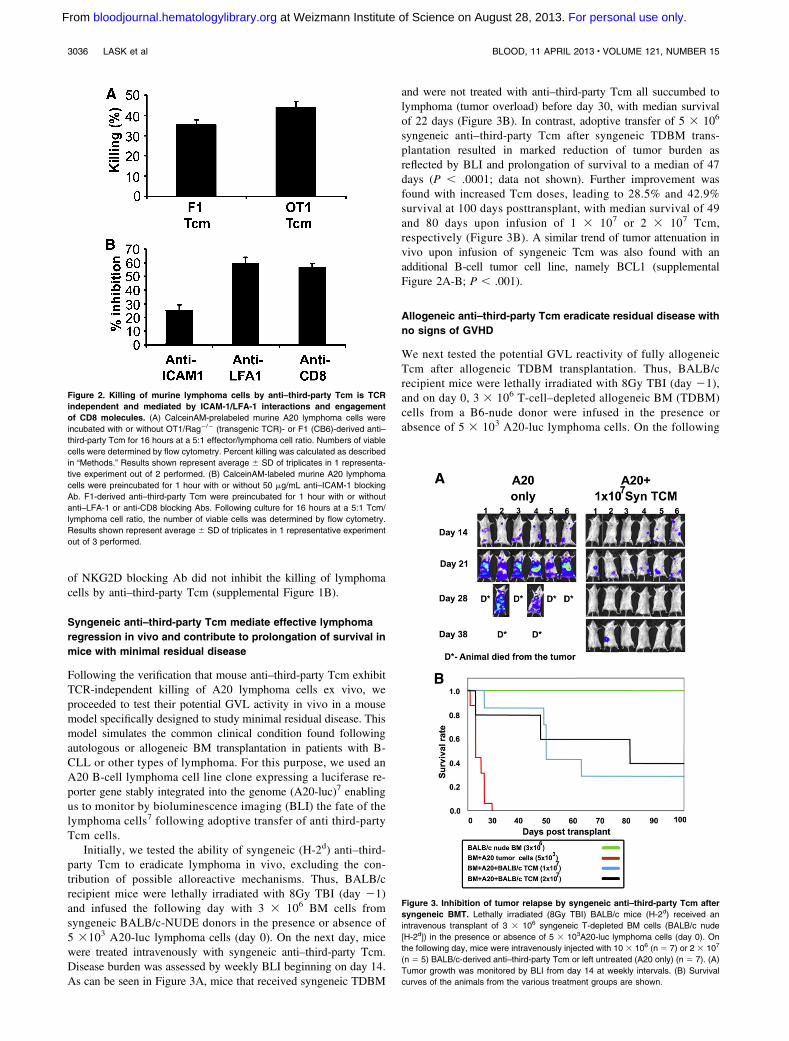
Following the verification that mouse anti–third-party Tcm exhibitTCR-independent killing of A20 lymphoma cells ex vivo, weproceeded to test their potential GVL activity in vivo in a mousemodel specifically designed to study minimal residual disease. Thismodel simulates the common clinical condition found followingautologous or allogeneic BM transplantation in patients with B-CLL or other types of lymphoma. For this purpose, we used anA20 B-cell lymphoma cell line clone expressing a luciferase re-porter gene stably integrated into the genome (A20-luc)7 enablingus to monitor by bioluminescence imaging (BLI) the fate of thelymphoma cells7 following adoptive transfer of anti third-partyTcm cells.
Initially, we tested the ability of syngeneic (H-2d) anti–third-party Tcm to eradicate lymphoma in vivo, excluding the con-tribution of possible alloreactive mechanisms. Thus, BALB/crecipient mice were lethally irradiated with 8Gy TBI (day 21)and infused the following day with 3 3 106 BM cells fromsyngeneic BALB/c-NUDE donors in the presence or absence of5 3103 A20-luc lymphoma cells (day 0). On the next day, micewere treated intravenously with syngeneic anti–third-party Tcm.Disease burden was assessed by weekly BLI beginning on day 14.As can be seen in Figure 3A, mice that received syngeneic TDBM
and were not treated with anti–third-party Tcm all succumbed tolymphoma (tumor overload) before day 30, with median survivalof 22 days (Figure 3B). In contrast, adoptive transfer of 5 3 106
syngeneic anti–third-party Tcm after syngeneic TDBM trans-plantation resulted in marked reduction of tumor burden asreflected by BLI and prolongation of survival to a median of 47days (P , .0001; data not shown). Further improvement wasfound with increased Tcm doses, leading to 28.5% and 42.9%survival at 100 days posttransplant, with median survival of 49and 80 days upon infusion of 1 3 107 or 2 3 107 Tcm,respectively (Figure 3B). A similar trend of tumor attenuation invivo upon infusion of syngeneic Tcm was also found with anadditional B-cell tumor cell line, namely BCL1 (supplementalFigure 2A-B; P , .001).
Allogeneic anti–third-party Tcm eradicate residual disease with
no signs of GVHD
We next tested the potential GVL reactivity of fully allogeneicTcm after allogeneic TDBM transplantation. Thus, BALB/crecipient mice were lethally irradiated with 8Gy TBI (day 21),and on day 0, 3 3 106 T-cell–depleted allogeneic BM (TDBM)cells from a B6-nude donor were infused in the presence orabsence of 5 3 103 A20-luc lymphoma cells. On the following
Figure 2. Killing of murine lymphoma cells by anti–third-party Tcm is TCR
independent and mediated by ICAM-1/LFA-1 interactions and engagement
of CD8 molecules. (A) CalceinAM-prelabeled murine A20 lymphoma cells were
incubated with or without OT1/Rag2/2 (transgenic TCR)- or F1 (CB6)-derived anti–
third-party Tcm for 16 hours at a 5:1 effector/lymphoma cell ratio. Numbers of viable
cells were determined by flow cytometry. Percent killing was calculated as described
in “Methods.” Results shown represent average 6 SD of triplicates in 1 representa-
tive experiment out of 2 performed. (B) CalceinAM-labeled murine A20 lymphoma
cells were preincubated for 1 hour with or without 50 mg/mL anti–ICAM-1 blocking
Ab. F1-derived anti–third-party Tcm were preincubated for 1 hour with or without
anti–LFA-1 or anti-CD8 blocking Abs. Following culture for 16 hours at a 5:1 Tcm/
lymphoma cell ratio, the number of viable cells was determined by flow cytometry.
Results shown represent average 6 SD of triplicates in 1 representative experiment
out of 3 performed.
Figure 3. Inhibition of tumor relapse by syngeneic anti–third-party Tcm after
syngeneic BMT. Lethally irradiated (8Gy TBI) BALB/c mice (H-2d) received an
intravenous transplant of 3 3 106 syngeneic T-depleted BM cells (BALB/c nude
[H-2d]) in the presence or absence of 5 3 103A20-luc lymphoma cells (day 0). On
the following day, mice were intravenously injected with 10 3 106 (n 5 7) or 2 3 107
(n 5 5) BALB/c-derived anti–third-party Tcm or left untreated (A20 only) (n 5 7). (A)
Tumor growth was monitored by BLI from day 14 at weekly intervals. (B) Survival
curves of the animals from the various treatment groups are shown.
3036 LASK et al BLOOD, 11 APRIL 2013 x VOLUME 121, NUMBER 15
For personal use only. at Weizmann Institute of Science on August 28, 2013. bloodjournal.hematologylibrary.orgFrom
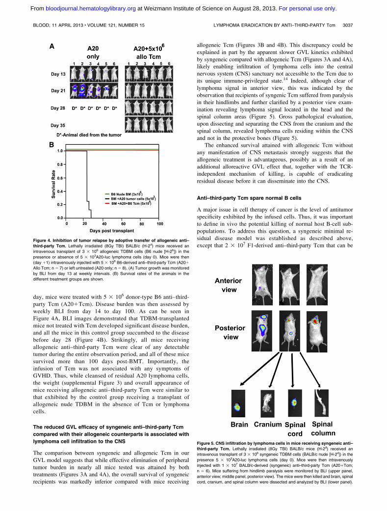
day, mice were treated with 5 3 106 donor-type B6 anti–third-party Tcm (A201Tcm). Disease burden was then assessed byweekly BLI from day 14 to day 100. As can be seen inFigure 4A, BLI images demonstrated that TDBM-transplantedmice not treated with Tcm developed significant disease burden,and all the mice in this control group succumbed to the diseasebefore day 28 (Figure 4B). Strikingly, all mice receivingallogeneic anti–third-party Tcm were clear of any detectabletumor during the entire observation period, and all of these micesurvived more than 100 days post-BMT. Importantly, theinfusion of Tcm was not associated with any symptoms ofGVHD. Thus, while cleansed of residual A20 lymphoma cells,the weight (supplemental Figure 3) and overall appearance ofmice receiving allogeneic anti–third-party Tcm were similar tothat exhibited by the control group receiving a transplant ofallogeneic nude TDBM in the absence of Tcm or lymphomacells.
The reduced GVL efficacy of syngeneic anti–third-party Tcm
compared with their allogeneic counterparts is associated with
lymphoma cell infiltration to the CNS
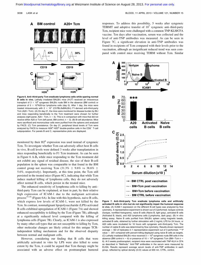
The comparison between syngeneic and allogeneic Tcm in ourGVL model suggests that while effective elimination of peripheraltumor burden in nearly all mice tested was attained by bothtreatments (Figures 3A and 4A), the overall survival of syngeneicrecipients was markedly inferior compared with mice receiving
allogeneic Tcm (Figures 3B and 4B). This discrepancy could beexplained in part by the apparent slower GVL kinetics exhibitedby syngeneic compared with allogeneic Tcm (Figures 3A and 4A),likely enabling infiltration of lymphoma cells into the centralnervous system (CNS) sanctuary not accessible to the Tcm due toits unique immune-privileged state.14 Indeed, although clear oflymphoma signal in anterior view, this was indicated by theobservation that recipients of syngenic Tcm suffered from paralysisin their hindlimbs and further clarified by a posterior view exam-ination revealing lymphoma signal located in the head and thespinal column areas (Figure 5). Gross pathological evaluation,upon dissecting and separating the CNS from the cranium and thespinal column, revealed lymphoma cells residing within the CNSand not in the protective bones (Figure 5).
The enhanced survival attained with allogeneic Tcm withoutany manifestation of CNS metastasis strongly suggests that theallogeneic treatment is advantageous, possibly as a result of anadditional alloreactive GVL effect that, together with the TCR-independent mechanism of killing, is capable of eradicatingresidual disease before it can disseminate into the CNS.
Anti–third-party Tcm spare normal B cells
A major issue in cell therapy of cancer is the level of antitumorspecificity exhibited by the infused cells. Thus, it was importantto define in vivo the potential killing of normal host B-cell sub-populations. To address this question, a syngeneic minimal re-sidual disease model was established as described above,except that 2 3 107 F1-derived anti–third-party Tcm that can be
Figure 4. Inhibition of tumor relapse by adoptive transfer of allogeneic anti–
third-party Tcm. Lethally irradiated (8Gy TBI) BALB/c (H-2d) mice received an
intravenous transplant of 3 3 106 allogeneic TDBM cells (B6 nude [H-2b]) in the
presence or absence of 5 3 103A20-luc lymphoma cells (day 0). Mice were then
(day 11) intravenously injected with 5 3 106 B6-derived anti–third-party Tcm (A201
Allo Tcm; n 5 7) or left untreated (A20 only; n 5 8). (A) Tumor growth was monitored
by BLI from day 13 at weekly intervals. (B) Survival rates of the animals in the
different treatment groups are shown.
Figure 5. CNS infiltration by lymphoma cells in mice receiving syngeneic anti–
third-party Tcm. Lethally irradiated (8Gy TBI) BALB/c mice (H-2d) received an
intravenous transplant of 3 3 106 syngeneic TDBM cells (BALB/c nude [H-2d]) in the
presence 5 3 103A20-luc lymphoma cells (day 0). Mice were then intravenously
injected with 1 3 107 BALB/c-derived (syngeneic) anti–third-party Tcm (A201Tcm;
n 5 6). Mice suffering from hindlimb paralysis were monitored by BLI (upper panel,
anterior view; middle panel, posterior view). The mice were then killed and brain, spinal
cord, cranium, and spinal column were dissected and analyzed by BLI (lower panel).
BLOOD, 11 APRIL 2013 x VOLUME 121, NUMBER 15 LYMPHOMA ERADICATION BY ANTI–THIRD-PARTY Tcm 3037
For personal use only. at Weizmann Institute of Science on August 28, 2013. bloodjournal.hematologylibrary.orgFrom
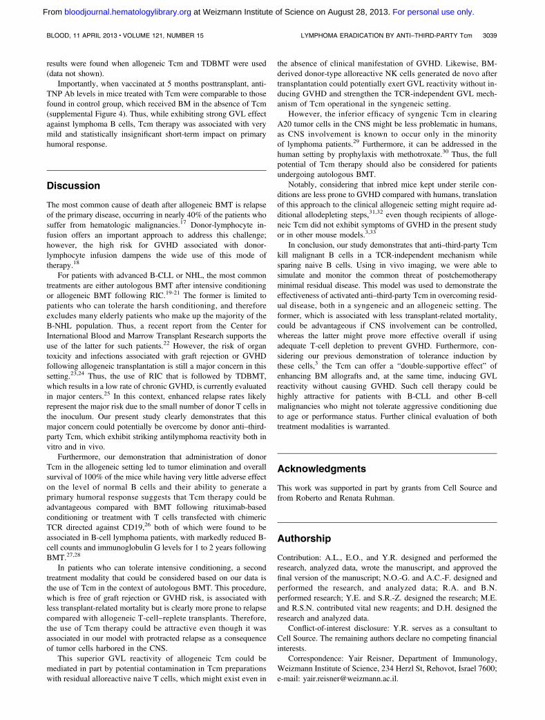
monitored by their H2b expression was used instead of syngeneicTcm. To investigate whether Tcm can adversely affect host B cellsin vivo, B-cell levels were defined 3 weeks after transplantation inmice responding beneficially to F1 Tcm treatment. As can be seenin Figure 6 A-B, while mice responding to the Tcm treatment didnot exhibit any signal of residual disease, the size of their B-cellpopulation in the spleen was comparable to that found in the BMcontrol group not receiving Tcm (31.3% 6 9.6% vs 38.6% 65.6%, respectively). Importantly, at this time point, the Tcm stillpersisted in the treated mice (Figure 6C), indicating that while Tcminduce marked killing of lymphoma cells, they do not adverselyaffect normal B cells, which persist in the treated mice.
The enhanced sensitivity of lymphoma cells to killing by anti–third-party Tcm can be explained, at least in part, by their relativehigh expression of ICAM-1 due to the malignant activationprocess15,16 (Figure 6A). In line with this hypothesis, naive B cells,which express low levels of ICAM-1, were not killed by theTcm. In contrast, nonmalignant lipopolysaccharide (LPS)-activatedB cells exhibited upregulation of ICAM-1 (Figure 7A) and showedenhanced susceptibility to killing by the Tcm (Figure 7B), althoughat a significantly reduced level compared with the killing oflymphoma cells (Figure 7B). Clearly, as ICAM-1 is also expressedby many other cell types that are not susceptible to killing by Tcm,other molecular changes are likely critical for this unique TCR-independent killing mechanism and for the observed disparitybetween normal and malignant cells.
However, considering our finding that B cells that wereartificially activated in vitro by LPS were also killed to someextent by the Tcm, it could be argued that Tcm therapy might beassociated with an adverse effect on posttransplant humoral
responses. To address this possibility, 5 weeks after syngeneicTDBMT and adoptive transfer of 107 syngeneic anti–third-partyTcm, recipient mice were challenged with a common TNP-KLH/CFAvaccine. Ten days after vaccination, serum was collected and thelevel of anti-TNP antibodies was measured. As can be seen inFigure 7C, a significant elevation in anti-TNP antibodies wasfound in recipients of Tcm compared with their levels prior to thevaccination, although an insignificant reduced trend was seen com-pared with control mice receiving TDBM without Tcm. Similar
Figure 6. Anti–third-party Tcm eradicate lymphoma cells while sparing normal
B cells in vivo. Lethally irradiated BALB/c mice (H-2d) received an intravenous
transplant of 3 3 106 syngeneic BALB/c nude BM in the absence (BM control) or
presence of 5 3 103A20-luc lymphoma cells (day 0). After 1 day, the mice were
treated intravenously with 2 3 107 (C57BL/6xBALB/c) F1-derived anti–third-party
Tcm (A201Tcm). (A) On day 21, the mice were monitored for disease burden by BLI
and mice responding beneficially to the Tcm treatment were chosen for further
analysis (right panel, A201 Tcm, n 5 3). This is in comparison with mice that did not
receive either A20 or Tcm (left panel, BM control, n 5 3). (B) B-cell abundance. Mice
were sacrificed and mononuclear cells were purified from the spleens and analyzed
by FACS. (C) Tcm persistence. On day 21, peripheral blood levels of Tcm were
analyzed by FACS to measure H2Kb H2Dd double-positive cells in the CD81 T-cell
subpopulation. For panels B and C, representative plots are displayed.
Figure 7. Anti–third-party Tcm eradicate lymphoma cells and artificially
activated B cells in vitro but do not significantly impair the humoral response
in vivo. (A) ICAM-1 expression on the different B-cell types was analyzed by flow
cytometry. A representative experiment is shown out of 3 performed. Isotype controls
(Isotype, nonfilled histograms), naive B cells (Naive B, light gray), activated B cells
(Activated B, black), and A20 lymphoma cells (Lymphoma, dark gray). (B) In vitro
killing assay. Naive B cells purified from BALB/c spleen using magnetic beads,
activated B cells, obtained by further stimulation with 2 mg/mL LPS for 24 hours, or
A20 cells were incubated for 16 hours with syngeneic anti–third-party Tcm. The
number of viable B cells was determined by flow cytometry. Results shown represent
average 6 SD of triplicates in 1 representative experiment out of 3 performed. ***P
value , .001 compared with percent killing displayed using naive B cells as targets.
(C) Lethally irradiated BALB/c mice received 3 3 106 syngeneic nude BM cells in the
absence (BM control n 5 6) or presence of 10 3 106 syngeneic Tcm (BM1Tcm n 5
6). At 5 weeks posttransplant, recipient mice were vaccinated with TNP-KLH in CFA
as described in “Methods.” Anti-TNP antibodies in the serum were measured by
ELISA. Results represent average serum levels of anti-TNP antibodies in each
group, reflected by optical density (O.D) values at 630 nm. CTRL, control.
3038 LASK et al BLOOD, 11 APRIL 2013 x VOLUME 121, NUMBER 15
For personal use only. at Weizmann Institute of Science on August 28, 2013. bloodjournal.hematologylibrary.orgFrom
results were found when allogeneic Tcm and TDBMT were used(data not shown).
Importantly, when vaccinated at 5 months posttransplant, anti-TNP Ab levels in mice treated with Tcm were comparable to thosefound in control group, which received BM in the absence of Tcm(supplemental Figure 4). Thus, while exhibiting strong GVL effectagainst lymphoma B cells, Tcm therapy was associated with verymild and statistically insignificant short-term impact on primaryhumoral response.
Discussion
The most common cause of death after allogeneic BMT is relapseof the primary disease, occurring in nearly 40% of the patients whosuffer from hematologic malignancies.17 Donor-lymphocyte in-fusion offers an important approach to address this challenge;however, the high risk for GVHD associated with donor-lymphocyte infusion dampens the wide use of this mode oftherapy.18
For patients with advanced B-CLL or NHL, the most commontreatments are either autologous BMT after intensive conditioningor allogeneic BMT following RIC.19-21 The former is limited topatients who can tolerate the harsh conditioning, and thereforeexcludes many elderly patients who make up the majority of theB-NHL population. Thus, a recent report from the Center forInternational Blood and Marrow Transplant Research supports theuse of the latter for such patients.22 However, the risk of organtoxicity and infections associated with graft rejection or GVHDfollowing allogeneic transplantation is still a major concern in thissetting.23,24 Thus, the use of RIC that is followed by TDBMT,which results in a low rate of chronic GVHD, is currently evaluatedin major centers.25 In this context, enhanced relapse rates likelyrepresent the major risk due to the small number of donor T cells inthe inoculum. Our present study clearly demonstrates that thismajor concern could potentially be overcome by donor anti–third-party Tcm, which exhibit striking antilymphoma reactivity both invitro and in vivo.
Furthermore, our demonstration that administration of donorTcm in the allogeneic setting led to tumor elimination and overallsurvival of 100% of the mice while having very little adverse effecton the level of normal B cells and their ability to generate aprimary humoral response suggests that Tcm therapy could beadvantageous compared with BMT following rituximab-basedconditioning or treatment with T cells transfected with chimericTCR directed against CD19,26 both of which were found to beassociated in B-cell lymphoma patients, with markedly reduced B-cell counts and immunoglobulin G levels for 1 to 2 years followingBMT.27,28
In patients who can tolerate intensive conditioning, a secondtreatment modality that could be considered based on our data isthe use of Tcm in the context of autologous BMT. This procedure,which is free of graft rejection or GVHD risk, is associated withless transplant-related mortality but is clearly more prone to relapsecompared with allogeneic T-cell–replete transplants. Therefore,the use of Tcm therapy could be attractive even though it wasassociated in our model with protracted relapse as a consequenceof tumor cells harbored in the CNS.
This superior GVL reactivity of allogeneic Tcm could bemediated in part by potential contamination in Tcm preparationswith residual alloreactive naive T cells, which might exist even in
the absence of clinical manifestation of GVHD. Likewise, BM-derived donor-type alloreactive NK cells generated de novo aftertransplantation could potentially exert GVL reactivity without in-ducing GVHD and strengthen the TCR-independent GVL mech-anism of Tcm operational in the syngeneic setting.
However, the inferior efficacy of syngenic Tcm in clearingA20 tumor cells in the CNS might be less problematic in humans,as CNS involvement is known to occur only in the minorityof lymphoma patients.29 Furthermore, it can be addressed in thehuman setting by prophylaxis with methotroxate.30 Thus, the fullpotential of Tcm therapy should also be considered for patientsundergoing autologous BMT.
Notably, considering that inbred mice kept under sterile con-ditions are less prone to GVHD compared with humans, translationof this approach to the clinical allogeneic setting might require ad-ditional allodepleting steps,31,32 even though recipients of alloge-neic Tcm did not exhibit symptoms of GVHD in the present studyor in other mouse models.3,33
In conclusion, our study demonstrates that anti–third-party Tcmkill malignant B cells in a TCR-independent mechanism whilesparing naive B cells. Using in vivo imaging, we were able tosimulate and monitor the common threat of postchemotherapyminimal residual disease. This model was used to demonstrate theeffectiveness of activated anti–third-party Tcm in overcoming resid-ual disease, both in a syngeneic and an allogeneic setting. Theformer, which is associated with less transplant-related mortality,could be advantageous if CNS involvement can be controlled,whereas the latter might prove more effective overall if usingadequate T-cell depletion to prevent GVHD. Furthermore, con-sidering our previous demonstration of tolerance induction bythese cells,3 the Tcm can offer a “double-supportive effect” ofenhancing BM allografts and, at the same time, inducing GVLreactivity without causing GVHD. Such cell therapy could behighly attractive for patients with B-CLL and other B-cellmalignancies who might not tolerate aggressive conditioning dueto age or performance status. Further clinical evaluation of bothtreatment modalities is warranted.
Acknowledgments
This work was supported in part by grants from Cell Source andfrom Roberto and Renata Ruhman.
Authorship
Contribution: A.L., E.O., and Y.R. designed and performed theresearch, analyzed data, wrote the manuscript, and approved thefinal version of the manuscript; N.O.-G. and A.C.-F. designed andperformed the research, and analyzed data; R.A. and B.N.performed research; Y.E. and S.R.-Z. designed the research; M.E.and R.S.N. contributed vital new reagents; and D.H. designed theresearch and analyzed data.
Conflict-of-interest disclosure: Y.R. serves as a consultant toCell Source. The remaining authors declare no competing financialinterests.
Correspondence: Yair Reisner, Department of Immunology,Weizmann Institute of Science, 234 Herzl St, Rehovot, Israel 7600;e-mail: [email protected].
BLOOD, 11 APRIL 2013 x VOLUME 121, NUMBER 15 LYMPHOMA ERADICATION BY ANTI–THIRD-PARTY Tcm 3039
For personal use only. at Weizmann Institute of Science on August 28, 2013. bloodjournal.hematologylibrary.orgFrom

References
1. Gale RP, Reisner Y. Graft rejection and graft-versus-host disease: mirror images. Lancet. 1986;1(8496):1468-1470.
2. Marmont AM, Horowitz MM, Gale RP, et al. T-celldepletion of HLA-identical transplants in leukemia.Blood. 1991;78(8):2120-2130.
3. Ophir E, Eidelstein Y, Afik R, Bachar-Lustig E,Reisner Y. Induction of tolerance to bone marrowallografts by donor-derived host nonreactive exvivo-induced central memory CD8 T cells. Blood.2010;115(10):2095-2104.
4. Ophir E. Ex vivo-induced donor central memoryCD8 T-cells induce tolerance toward T-cell-depleted BM allografts under reducedconditioning and mediate GVL reactivity againstB-cell malignancies, without causing GVHD. BoneMarrow Transplant. 2011;46(suppl 1):S22-S23.
5. Arditti FD, Aviner S, Dekel B, et al. Eradicationof B-CLL by autologous and allogeneic hostnonreactive anti-third-party CTLs. Blood. 2005;105(8):3365-3371.
6. Lask A, Goichberg P, Cohen A, et al. TCR-independent killing of B cell malignancies by anti-third-party CTLs: the critical role of MHC-CD8engagement. J Immunol. 2011;187(4):2006-2014.
7. Edinger M, Cao YA, Verneris MR, Bachmann MH,Contag CH, Negrin RS. Revealing lymphomagrowth and the efficacy of immune cell therapiesusing in vivo bioluminescence imaging. Blood.2003;101(2):640-648.
8. Aiuti A, Webb IJ, Bleul C, Springer T,Gutierrez-Ramos JC. The chemokine SDF-1is a chemoattractant for human CD341hematopoietic progenitor cells and provides a newmechanism to explain the mobilization of CD341progenitors to peripheral blood. J Exp Med. 1997;185(1):111-120.
9. Hodes RJ, Singer A. Cellular and genetic controlof antibody responses in vitro. I. Cellularrequirements for the generation of geneticallycontrolled primary IgM responses to solubleantigens. Eur J Immunol. 1977;7(12):892-897.
10. Godfrey DI, Hammond KJ, Poulton LD, Smyth MJ,Baxter AG. NKT cells: facts, functions andfallacies. Immunol Today. 2000;21(11):573-583.
11. Kong Y, Cao W, Xi X, Ma C, Cui L, He W. TheNKG2D ligand ULBP4 binds to TCRgamma9/delta2 and induces cytotoxicity to tumor cellsthrough both TCRgammadelta and NKG2D.Blood. 2009;114(2):310-317.
12. Raulet DH. Roles of the NKG2D immunoreceptorand its ligands. Nat Rev Immunol. 2003;3(10):781-790.
13. Maccalli C, Scaramuzza S, Parmiani G. TNK cells(NKG2D1 CD81 or CD41 T lymphocytes) in thecontrol of human tumors. Cancer ImmunolImmunother. 2009;58(5):801-808.
14. Barker CF, Billingham RE. Immunologicallyprivileged sites. Adv Immunol. 1977;25:1-54.
15. Padros MR, Noli MI, Fainboim L. Expression ofICAM-1 (CD54) on normal and leukaemic B cells:implication for the mixed lymphocyte reaction. ClinExp Immunol. 1992;88(2):329-334.
16. Terol MJ, Lopez-Guillermo A, Bosch F, et al.Expression of the adhesion molecule ICAM-1 innon-Hodgkin’s lymphoma: relationship with tumordissemination and prognostic importance. J ClinOncol. 1998;16(1):35-40.
17. CIBMTR. CIBMTR Summary Slides. http://www.cibmtr.org. 2008.
18. Frey NV, Porter DL. Graft-versus-host diseaseafter donor leukocyte infusions: presentation andmanagement. Best Pract Res Clin Haematol.2008;21(2):205-222.
19. Baron F, Maris MB, Sandmaier BM, et al.Graft-versus-tumor effects after allogeneichematopoietic cell transplantation withnonmyeloablative conditioning. J Clin Oncol.2005;23(9):1993-2003.
20. Deconinck E, Foussard C, Milpied N, et al;GOELAMS. High-dose therapy followed byautologous purged stem-cell transplantation anddoxorubicin-based chemotherapy in patients withadvanced follicular lymphoma: a randomizedmulticenter study by GOELAMS. Blood. 2005;105(10):3817-3823.
21. Gisselbrecht C, Vose J, Nademanee A, GianniAM, Nagler A. Radioimmunotherapy for stem celltransplantation in non-Hodgkin’s lymphoma: inpursuit of a complete response. Oncologist. 2009;14(suppl 2):41-51.
22. Hari P, Carreras J, Zhang MJ, et al. Allogeneictransplants in follicular lymphoma: higher risk ofdisease progression after reduced-intensitycompared to myeloablative conditioning. BiolBlood Marrow Transplant. 2008;14(2):236-245.
23. Mattsson J, Ringden O, Storb R. Graft failure afterallogeneic hematopoietic cell transplantation. BiolBlood Marrow Transplant. 2008;14(suppl 1):165-170.
24. Robinson SP, Goldstone AH, Mackinnon S, et al;Lymphoma Working Party of the European Groupfor Blood and Bone Marrow Transplantation.Chemoresistant or aggressive lymphoma predictsfor a poor outcome following reduced-intensityallogeneic progenitor cell transplantation: ananalysis from the Lymphoma Working Party of theEuropean Group for Blood and Bone MarrowTransplantation. Blood. 2002;100(13):4310-4316.
25. van Besien K. Allogeneic stem cell transplantationin follicular lymphoma: recent progress andcontroversy. Hematology Am Soc Hematol EducProgram. 2009;610-618.
26. Kalos M, Levine BL, Porter DL, Katz S, Grupp SA,Bagg A, June CH. T cells with chimeric antigenreceptors have potent antitumor effects and canestablish memory in patients with advancedleukemia. Sci Transl Med. 2011;3(95):95ra73.
27. Kasamon YL, Jones RJ, Brodsky RA, et al.Immunologic recovery following autologousstem-cell transplantation with pre- andposttransplantation rituximab for low-grade ormantle cell lymphoma. Ann Oncol. 2010;21(6):1203-1210.
28. Horwitz SM, Negrin RS, Blume KG, et al.Rituximab as adjuvant to high-dose therapy andautologous hematopoietic cell transplantation foraggressive non-Hodgkin lymphoma. Blood. 2004;103(3):777-783.
29. Hollender A, Kvaloy S, Nome O, Skovlund E, LoteK, Holte H. Central nervous system involvementfollowing diagnosis of non-Hodgkin’s lymphoma:a risk model. Ann Oncol. 2002;13(7):1099-1107.
30. Hill QA, Owen RG. CNS prophylaxis inlymphoma: who to target and what therapy to use.Blood Rev. 2006;20(6):319-332.
31. Barrett AJ, Rezvani K, Solomon S, et al. Newdevelopments in allotransplant immunology.Hematology Am Soc Hematol Educ Program.2003;350-371.
32. Perruccio K, Topini F, Tosti A, et al.Photodynamic purging of alloreactive T cells foradoptive immunotherapy after haploidentical stemcell transplantation. Blood Cells Mol Dis. 2008;40(1):76-83.
33. Ophir E, Or-Geva N, Gurevich I, et al. Murine anti-third-party central-memory CD81 T cells promotehematopoietic chimerism under mild conditioning:lymph-node sequestration and deletion of anti-
donor T cells. Blood. 2013;121(7):1220-1228.
3040 LASK et al BLOOD, 11 APRIL 2013 x VOLUME 121, NUMBER 15
For personal use only. at Weizmann Institute of Science on August 28, 2013. bloodjournal.hematologylibrary.orgFrom