transcription factor ii (tfii) tata box (hogness box) tataaa (-25, -35) tfii d ---- tata binding...
Post on 21-Dec-2015
216 views
TRANSCRIPT
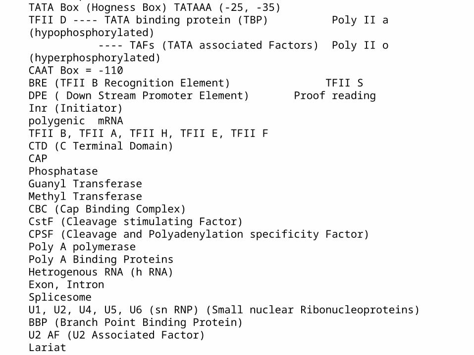
Transcription factor II (TFII)TATA Box (Hogness Box) TATAAA (-25, -35) TFII D ---- TATA binding protein (TBP) Poly II a (hypophosphorylated) ---- TAFs (TATA associated Factors) Poly II o (hyperphosphorylated)CAAT Box = -110BRE (TFII B Recognition Element) TFII S DPE ( Down Stream Promoter Element) Proof readingInr (Initiator) polygenic mRNATFII B, TFII A, TFII H, TFII E, TFII FCTD (C Terminal Domain)CAP PhosphataseGuanyl TransferaseMethyl TransferaseCBC (Cap Binding Complex)CstF (Cleavage stimulating Factor)CPSF (Cleavage and Polyadenylation specificity Factor)Poly A polymerasePoly A Binding ProteinsHetrogenous RNA (h RNA)Exon, IntronSplicesomeU1, U2, U4, U5, U6 (sn RNP) (Small nuclear Ribonucleoproteins)BBP (Branch Point Binding Protein)U2 AF (U2 Associated Factor)LariatSR Proteins (Serine, Argenine)
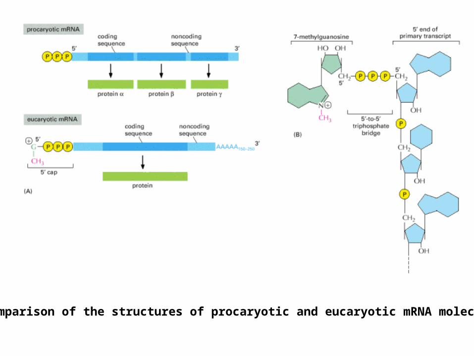
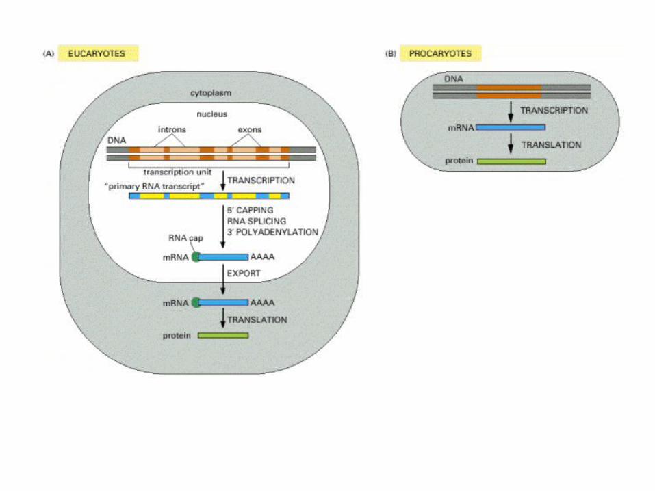
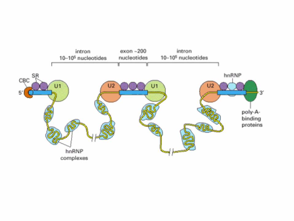
A comparison of the structures of procaryotic and eucaryotic mRNA molecules.
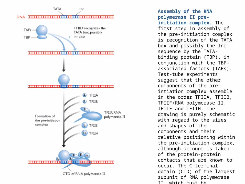
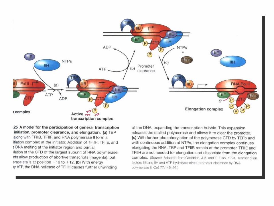
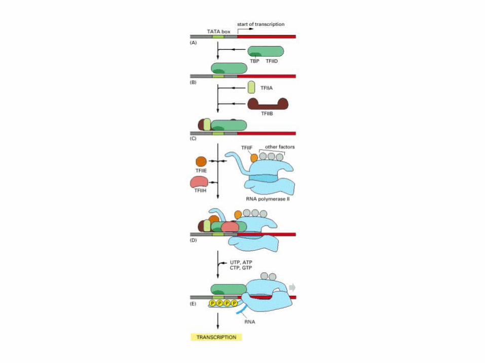
Assembly of the RNA polymerase II pre-initiation complex. The first step in assembly of the pre-initiation complex is recognition of the TATA box and possibly the Inr sequence by the TATA-binding protein (TBP), in conjunction with the TBP-associated factors (TAFs). Test-tube experiments suggest that the other components of the pre-intiation complex assemble in the order TFIIA, TFIIB, TFIIF/RNA polymerase II, TFIIE and TFIIH. The drawing is purely schematic with regard to the sizes and shapes of the components and their relative positioning within the pre-initiation complex, although account is taken of the protein-protein contacts that are known to occur. The C-terminal domain (CTD) of the largest subunit of RNA polymerase II, which must be phosphorylated before the polymerase can leave the promoter, is depicted as a spike projecting from the bottom of the complex.
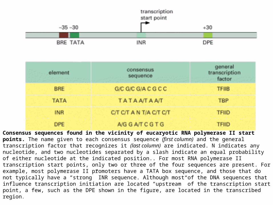
Consensus sequences found in the vicinity of eucaryotic RNA polymerase II start points. The name given to each consensus sequence (first column) and the general transcription factor that recognizes it (last column) are indicated. N indicates any nucleotide, and two nucleotides separated by a slash indicate an equal probability of either nucleotide at the indicated position.. For most RNA polymerase II transcription start points, only two or three of the four sequences are present. For example, most polymerase II promoters have a TATA box sequence, and those that do not typically have a “strong” INR sequence. Although most of the DNA sequences that influence transcription initiation are located “upstream” of the transcription start point, a few, such as the DPE shown in the figure, are located in the transcribed region.

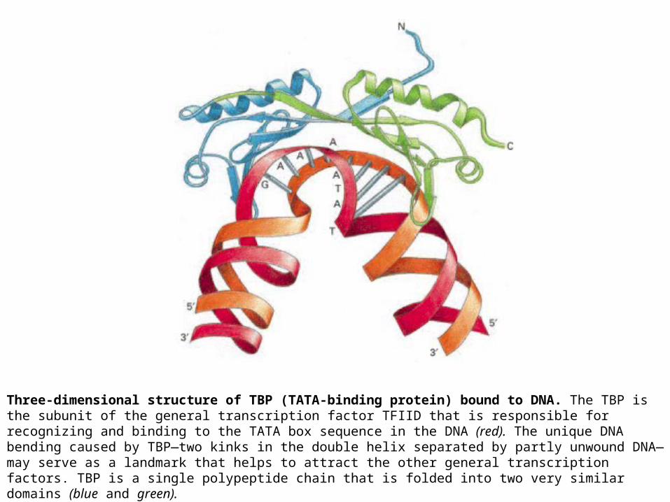
Three-dimensional structure of TBP (TATA-binding protein) bound to DNA. The TBP is the subunit of the general transcription factor TFIID that is responsible for recognizing and binding to the TATA box sequence in the DNA (red). The unique DNA bending caused by TBP—two kinks in the double helix separated by partly unwound DNA—may serve as a landmark that helps to attract the other general transcription factors. TBP is a single polypeptide chain that is folded into two very similar domains (blue and green).
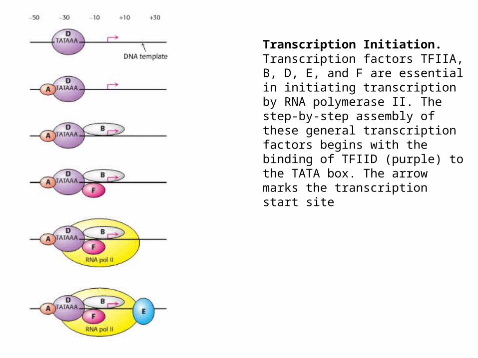
Transcription Initiation. Transcription factors TFIIA, B, D, E, and F are essential in initiating transcription by RNA polymerase II. The step-by-step assembly of these general transcription factors begins with the binding of TFIID (purple) to the TATA box. The arrow marks the transcription start site
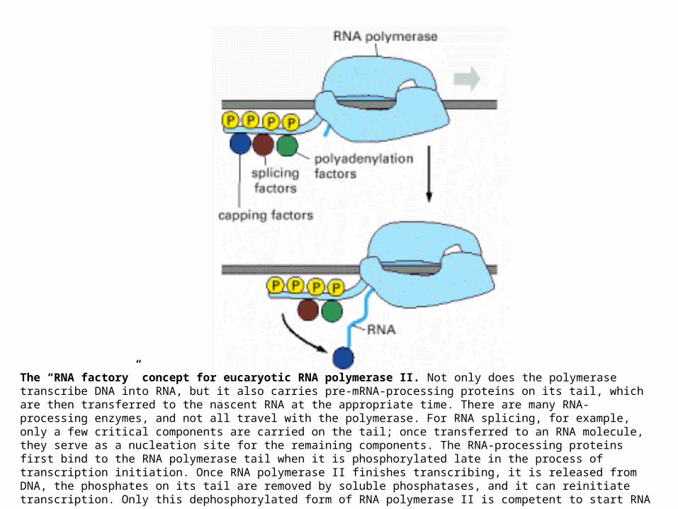
The “RNA factory” concept for eucaryotic RNA polymerase II. Not only does the polymerase transcribe DNA into RNA, but it also carries pre-mRNA-processing proteins on its tail, which are then transferred to the nascent RNA at the appropriate time. There are many RNA-processing enzymes, and not all travel with the polymerase. For RNA splicing, for example, only a few critical components are carried on the tail; once transferred to an RNA molecule, they serve as a nucleation site for the remaining components. The RNA-processing proteins first bind to the RNA polymerase tail when it is phosphorylated late in the process of transcription initiation. Once RNA polymerase II finishes transcribing, it is released from DNA, the phosphates on its tail are removed by soluble phosphatases, and it can reinitiate transcription. Only this dephosphorylated form of RNA
polymerase II is competent to start RNA synthesis at a promoter.
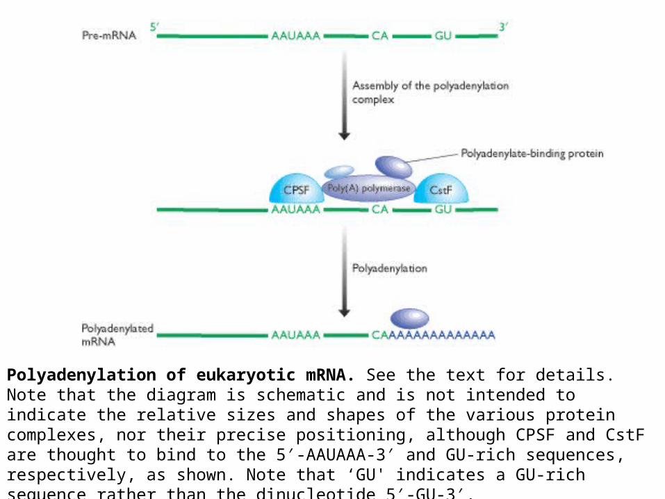
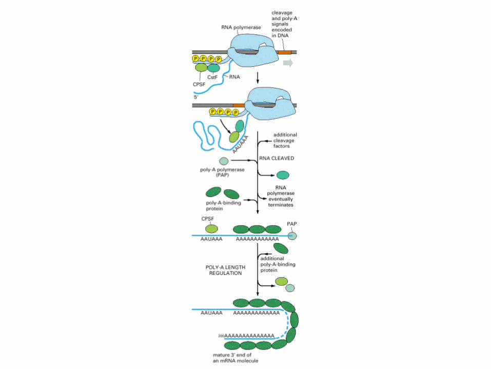
Polyadenylation of eukaryotic mRNA. See the text for details. Note that the diagram is schematic and is not intended to indicate the relative sizes and shapes of the various protein complexes, nor their precise positioning, although CPSF and CstF are thought to bind to the 5′-AAUAAA-3′ and GU-rich sequences, respectively, as shown. Note that ‘GU' indicates a GU-rich sequence rather than the dinucleotide 5′-GU-3′.

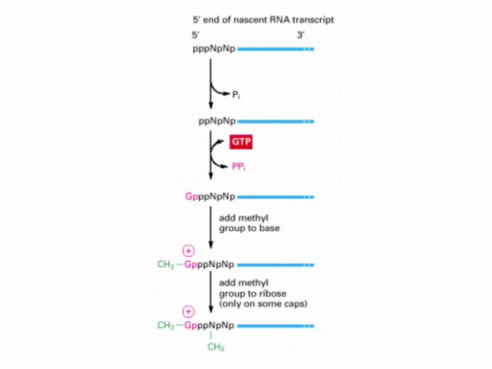
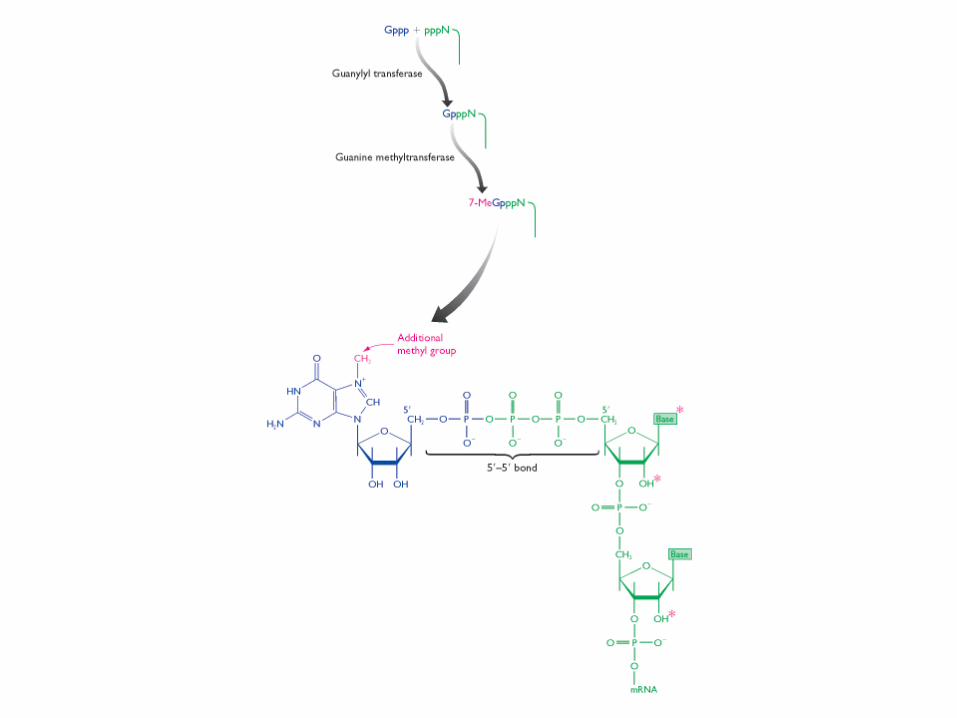
The reactions that cap the 5′ end of each RNA molecule synthesized by RNA polymerase II. The final cap contains a novel 5′-to-5′ linkage between the positively charged 7-methyl G residue and the 5′ end of the RNA transcript The letter N represents any one of the four ribonucleotides, although the nucleotide that starts an RNA chain is usually a purine
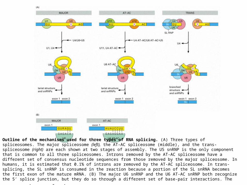
Outline of the mechanisms used for three types of RNA splicing. (A) Three types of spliceosomes. The major spliceosome (left), the AT-AC spliceosome (middle), and the trans-spliceosome (right) are each shown at two stages of assembly. The U5 snRNP is the only component that is common to all three spliceosomes. Introns removed by the AT-AC spliceosome have a different set of consensus nucleotide sequences from those removed by the major spliceosome. In humans, it is estimated that 0.1% of introns are removed by the AT-AC spliceosome. In trans-splicing, the SL snRNP is consumed in the reaction because a portion of the SL snRNA becomes the first exon of the mature mRNA. (B) The major U6 snRNP and the U6 AT-AC snRNP both recognize the 5′ splice junction, but they do so through a different set of base-pair interactions. The sequences shown are from
humans
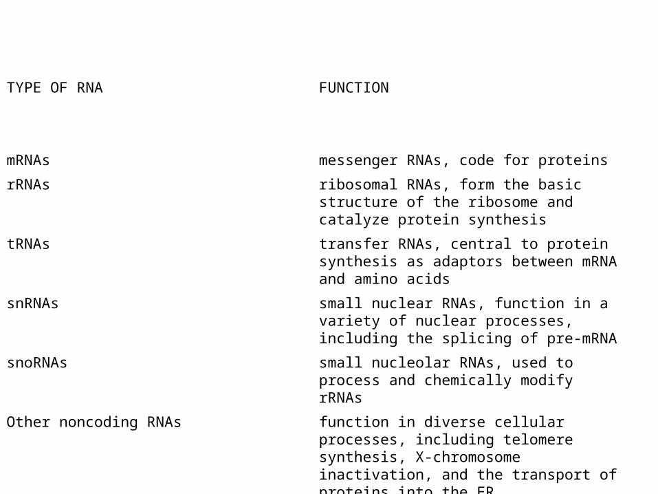
Principal Types of RNAs Produced in Cells
TYPE OF RNA FUNCTION
mRNAs messenger RNAs, code for proteins
rRNAs ribosomal RNAs, form the basic structure of the ribosome and catalyze protein synthesis
tRNAs transfer RNAs, central to protein synthesis as adaptors between mRNA and amino acids
snRNAs small nuclear RNAs, function in a variety of nuclear processes, including the splicing of pre-mRNA
snoRNAs small nucleolar RNAs, used to process and chemically modify rRNAs
Other noncoding RNAs function in diverse cellular processes, including telomere synthesis, X-chromosome inactivation, and the transport of proteins into the ER
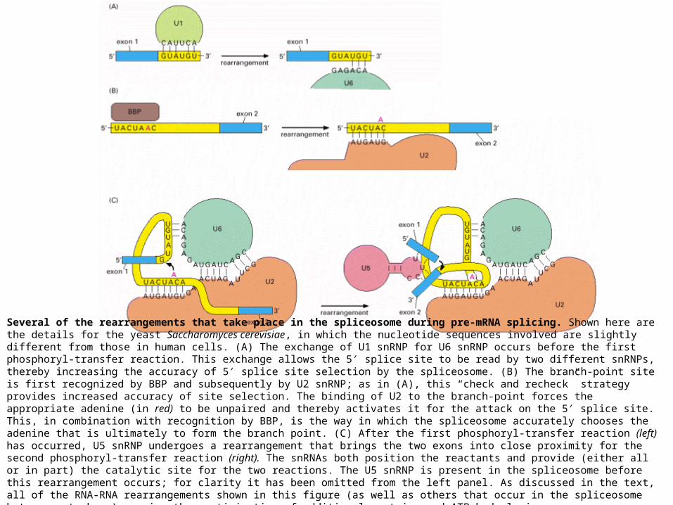
Several of the rearrangements that take place in the spliceosome during pre-mRNA splicing. Shown here are the details for the yeast Saccharomyces cerevisiae, in which the nucleotide sequences involved are slightly different from those in human cells. (A) The exchange of U1 snRNP for U6 snRNP occurs before the first phosphoryl-transfer reaction. This exchange allows the 5′ splice site to be read by two different snRNPs, thereby increasing the accuracy of 5′ splice site selection by the spliceosome. (B) The branch-point site is first recognized by BBP and subsequently by U2 snRNP; as in (A), this “check and recheck” strategy provides increased accuracy of site selection. The binding of U2 to the branch-point forces the appropriate adenine (in red) to be unpaired and thereby activates it for the attack on the 5′ splice site. This, in combination with recognition by BBP, is the way in which the spliceosome accurately chooses the adenine that is ultimately to form the branch point. (C) After the first phosphoryl-transfer reaction (left) has occurred, U5 snRNP undergoes a rearrangement that brings the two exons into close proximity for the second phosphoryl-transfer reaction (right). The snRNAs both position the reactants and provide (either all or in part) the catalytic site for the two reactions. The U5 snRNP is present in the spliceosome before this rearrangement occurs; for clarity it has been omitted from the left panel. As discussed in the text, all of the RNA-RNA rearrangements shown in this figure (as well as others that occur in the spliceosome but are not shown) require the participation of additional proteins and ATP hydrolysis.
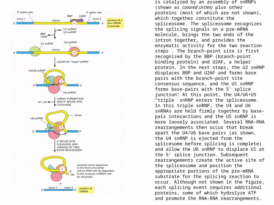
The RNA splicing mechanism. RNA splicing is catalyzed by an assembly of snRNPs (shown as colored circles) plus other proteins (most of which are not shown), which together constitute the spliceosome. The spliceosome recognizes the splicing signals on a pre-mRNA molecule, brings the two ends of the intron together, and provides the enzymatic activity for the two reaction steps . The branch-point site is first recognized by the BBP (branch-point binding protein) and U2AF, a helper protein. In the next steps, the U2 snRNP displaces BBP and U2AF and forms base pairs with the branch-point site consensus sequence, and the U1 snRNP forms base-pairs with the 5′ splice junction. At this point, the U4/U6•U5 “triple” snRNP enters the spliceosome. In this triple snRNP, the U4 and U6 snRNAs are held firmly together by base-pair interactions and the U5 snRNP is more loosely associated. Several RNA-RNA rearrangements then occur that break apart the U4/U6 base pairs (as shown, the U4 snRNP is ejected from the splicesome before splicing is complete) and allow the U6 snRNP to displace U1 at the 5′ splice junction. Subsequent rearrangements create the active site of the spliceosome and position the appropriate portions of the pre-mRNA substrate for the splicing reaction to occur. Although not shown in the figure, each splicing event requires additional proteins, some of which hydrolyze ATP and promote the RNA-RNA rearrangements.
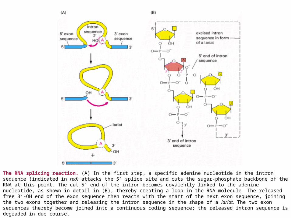
The RNA splicing reaction. (A) In the first step, a specific adenine nucleotide in the intron sequence (indicated in red) attacks the 5′ splice site and cuts the sugar-phosphate backbone of the RNA at this point. The cut 5′ end of the intron becomes covalently linked to the adenine nucleotide, as shown in detail in (B), thereby creating a loop in the RNA molecule. The released free 3′-OH end of the exon sequence then reacts with the start of the next exon sequence, joining the two exons together and releasing the intron sequence in the shape of a lariat. The two exon sequences thereby become joined into a continuous coding sequence; the released intron sequence is degraded in due course.

The two known classes of self-splicing intron sequences. The group I intron sequences bind a free G nucleotide to a specific site on the RNA to initiate splicing, while the group II intron sequences use an especially reactive A nucleotide in the intron sequence itself for the same purpose. The two mechanisms have been drawn to emphasize their similarities. Both are normally aided in the cell by proteins that speed up the reaction, but the catalysis is nevertheless mediated by the RNA in the intron sequence. Both types of self-splicing reactions require the intron to be folded into a highly specific three-dimensional structure that provides the catalytic activity for the reaction. The mechanism used by group II intron sequences releases the intron as a lariat structure and closely resembles the pathway of pre-mRNA splicing catalyzed by the spliceosome (compare with. The great majority of RNA splicing in eucaryotic cells is performed by the spliceosome, and self-splicing RNAs represent unusual cas
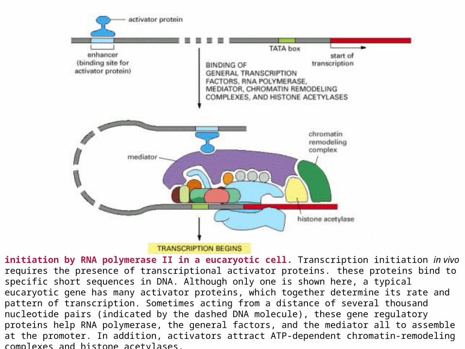
initiation by RNA polymerase II in a eucaryotic cell. Transcription initiation in vivo requires the presence of transcriptional activator proteins. these proteins bind to specific short sequences in DNA. Although only one is shown here, a typical eucaryotic gene has many activator proteins, which together determine its rate and pattern of transcription. Sometimes acting from a distance of several thousand nucleotide pairs (indicated by the dashed DNA molecule), these gene regulatory proteins help RNA polymerase, the general factors, and the mediator all to assemble at the promoter. In addition, activators attract ATP-dependent chromatin-remodeling complexes and histone acetylases.
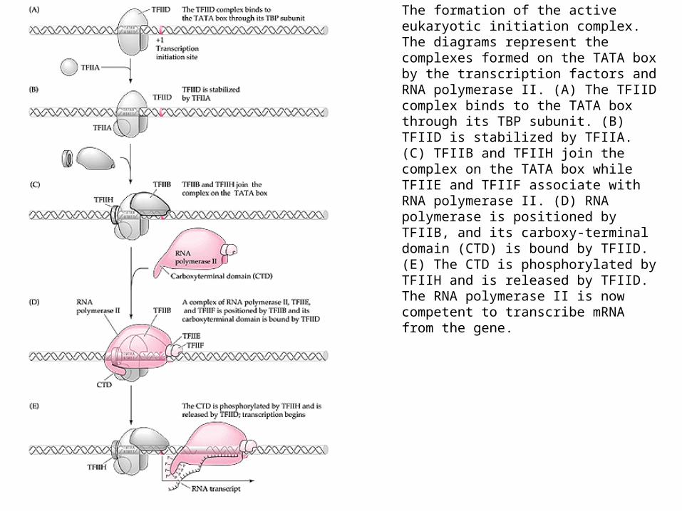
The formation of the active eukaryotic initiation complex. The diagrams represent the complexes formed on the TATA box by the transcription factors and RNA polymerase II. (A) The TFIID complex binds to the TATA box through its TBP subunit. (B) TFIID is stabilized by TFIIA. (C) TFIIB and TFIIH join the complex on the TATA box while TFIIE and TFIIF associate with RNA polymerase II. (D) RNA polymerase is positioned by TFIIB, and its carboxy-terminal domain (CTD) is bound by TFIID. (E) The CTD is phosphorylated by TFIIH and is released by TFIID. The RNA polymerase II is now competent to transcribe mRNA from the gene.
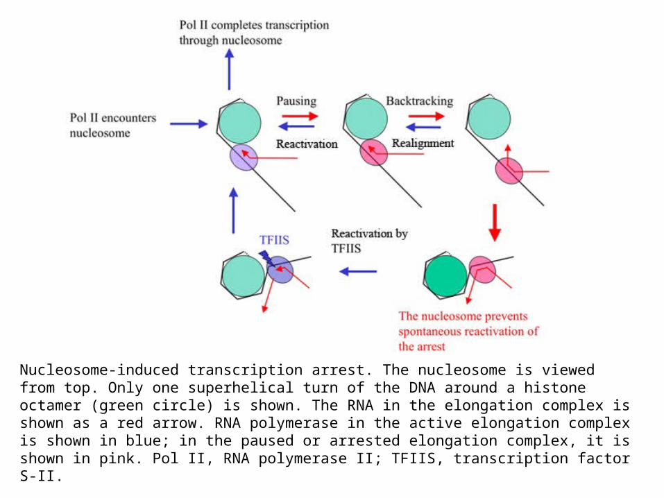
Nucleosome-induced transcription arrest. The nucleosome is viewed from top. Only one superhelical turn of the DNA around a histone octamer (green circle) is shown. The RNA in the elongation complex is shown as a red arrow. RNA polymerase in the active elongation complex is shown in blue; in the paused or arrested elongation complex, it is shown in pink. Pol II, RNA polymerase II; TFIIS, transcription factor S-II.


