treatment of calcific tendinitis of the rotator cuff with
TRANSCRIPT
Treatment of Calcific Tendinitis of the Rotator Cuff with Barbotage - A Case Study
1
MedDocs Publishers
Received: Oct 05, 2021Accepted: Nov 09, 2021Published Online: Nov 11, 2021Journal: Journal of Radiology and Medical ImagingPublisher: MedDocs Publishers LLCOnline edition: http://meddocsonline.org/Copyright: © Sneha NM (2021). This Article is distributed under the terms of Creative Commons Attribution 4.0 International License
*Corresponding Author(s): Sneha Narayana Murthy Junior Clinical Fellow in Surgery Queen’s hospital, 16 Hunt Court 3 Union Road Romford, UK. Tel: 07493454937; Email: [email protected]
Journal of Radiology and Medical ImagingOpen Access | Case Report
Cite this article: Sneha NM, Maheshwari S, Nagraj H, Vrizidou S. Treatment of Calcific Tendinitis of the Rotator Cuff with Barbotage - A Case Study. J Radiol Med Imaging. 2021; 4(2): 1059.
ISSN: 2637-885X
Sneha Murthy1*; Sagar Maheshwari2; Harish Nagraj2; Sofia Vrizidou3
1Junior Clinical Fellow in Surgery Queen’s hospital, Romford, UK.2FRCR, CCCT Consultant Radiologist, UK.3Consultant Radiologist, UK.
Introduction
Calcific tendinitis of the rotator cuff is a very common pa-thology. The pathophysiology involves deposition of hydroxyap-atite (form of calcium) crystals within the tendons of the rotator cuff [1,3]. The usual presentation could be pain, restriction of movements of the shoulder joint, swelling, redness and fever. Although the aetiology remains unclear, this condition needs immediate intervention especially in the acute phase of the condition. The incidence of this condition is nearly 7.5% in as-ymptomatic patients and nearly 20% in painful shoulders [1]. The following case report demonstrates the efficiency of sonog-raphy in the diagnosis of calcific tendinitis as well as its use in percutaneous treatment.
Case report
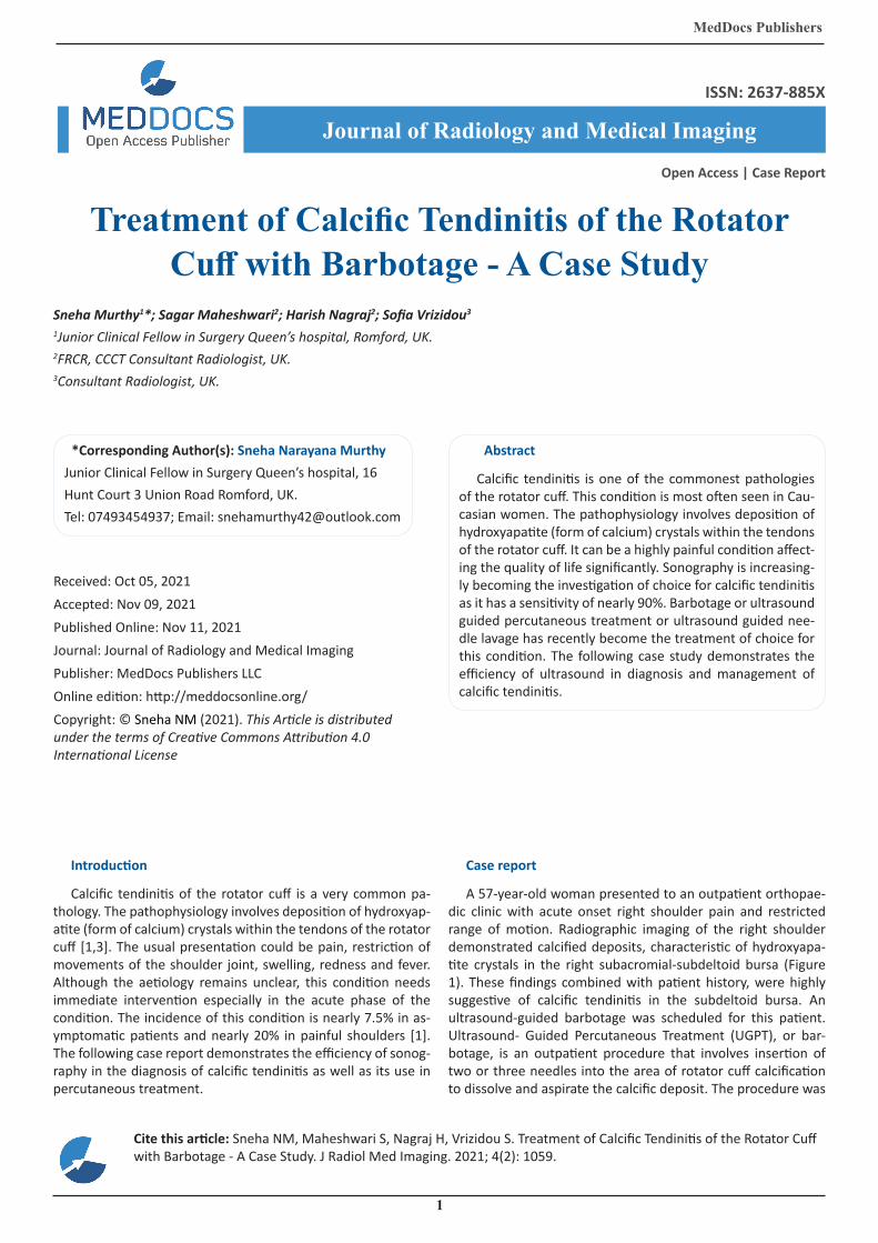
A 57-year-old woman presented to an outpatient orthopae-dic clinic with acute onset right shoulder pain and restricted range of motion. Radiographic imaging of the right shoulder demonstrated calcified deposits, characteristic of hydroxyapa-tite crystals in the right subacromial-subdeltoid bursa (Figure 1). These findings combined with patient history, were highly suggestive of calcific tendinitis in the subdeltoid bursa. An ultrasound-guided barbotage was scheduled for this patient. Ultrasound- Guided Percutaneous Treatment (UGPT), or bar-botage, is an outpatient procedure that involves insertion of two or three needles into the area of rotator cuff calcification to dissolve and aspirate the calcific deposit. The procedure was
Abstract
Calcific tendinitis is one of the commonest pathologies of the rotator cuff. This condition is most often seen in Cau-casian women. The pathophysiology involves deposition of hydroxyapatite (form of calcium) crystals within the tendons of the rotator cuff. It can be a highly painful condition affect-ing the quality of life significantly. Sonography is increasing-ly becoming the investigation of choice for calcific tendinitis as it has a sensitivity of nearly 90%. Barbotage or ultrasound guided percutaneous treatment or ultrasound guided nee-dle lavage has recently become the treatment of choice for this condition. The following case study demonstrates the efficiency of ultrasound in diagnosis and management of calcific tendinitis.
MedDocs Publishers
2Journal of Radiology and Medical Imaging
Figure 1: Radiographic imaging of the focal calcification.
performed with a high-frequency linear transducer. Post- proce-dure radiographs showed that the treated calcium deposit was nearly absent. The patient tolerated the procedure well and was sent home. At follow up in a month’s time, patient complained of no pain with near full range of motion of the shoulder joint and absent calcification upon radiographic and sonographic im-aging (Figure 6 & Figure 7).
Discussion
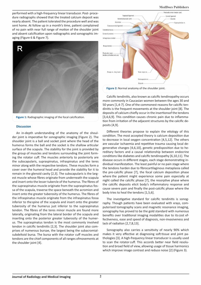
An in-depth understanding of the anatomy of the shoul-der joint is imperative for sonographic imaging (Figure 2). The shoulder joint is a ball and socket joint where the head of the humerus forms the ball and the socket is the shallow articular surface of the scapula. The stability for the joint is provided by the group of muscles and tendons surrounding the joint form-ing the rotator cuff. The muscles anteriorly to posteriorly are the subscapularis, supraspinatus, infraspinatus and the teres minor along with the respective tendons. These muscles form a cover over the humeral head and provide the stability for it to remain in the glenoid cavity [2,3]. The subscapularis is the larg-est muscle whose fibres originate from underneath the scapula and insert onto the lesser tubercle of the humerus. The fibres of the supraspinatus muscle originate from the supraspinatus fos-sa of the scapula, traverse the space beneath the acromion and insert onto the greater tuberosity of the humerus. The fibres of the infraspinatus muscle originate from the infraspinatus fossa inferior to the spine of the scapula and insert onto the greater tuberosity of the humerus just inferior to the supraspinatus tendon. The fibres of the teres minor muscle are found more laterally, originating from the lateral border of the scapula and inserting onto the posterior greater tuberosity of the humer-us. The supraspinatus tendon is the most commonly involved tendon in calcific tendinitis [2,3]. The shoulder joint also com-prises of numerous bursae, the largest being the subacromial-subdeltoid bursa. The bursa with the rotator cuff muscles and tendons are the chief components of all ranges of movements at the shoulder joint [4].
Figure 2: Normal anatomy of the shoulder joint.
Calcific tendinitis, also known as calcific tendinopathy occurs more commonly in Caucasian women between the ages 30 and 50 years [1,4-7]. One of the commonest reasons for calcific ten-dinitis is the frequent movements at the shoulder joint [8]. The deposits of calcium chiefly occur in the insertions of the tendons [3,4,6,9]. This condition causes chronic pain due to inflamma-tion from irritation of the adjacent structures by the calcific de-posits [4,9].
Different theories propose to explain the etiology of this condition. The most accepted theory is calcium deposition due to decrease in local oxygen concentration [4,5,12]. The others are vascular ischaemia and repetitive trauma causing local de-generative changes [4,6,10], genetic predisposition due to he-reditary factors and a causal relationship between endocrine conditions like diabetes and calcific tendinopathy [6,10,11]. The disease occurs in different stages, each stage demonstrating in-dividual manifestation. The least painful or no pain stage where the tendons harden due to fibrocartilaginous metaplasia called the pre-calcific phase [7], the focal calcium deposition phase where the patient might experience some pain especially at night called the calcific phase [7], the resorptive phase where the calcific deposits elicit body’s inflammatory response and cause severe pain and finally the post-calcific phase where the body tries to heal the tendons [1,5,6].
The investigative standard for calcific tendinitis is sonog-raphy. Though patients have been evaluated with xrays, com-puterised tomography scans and magnetic resonance imaging, sonography has proved to be the gold standard with numerous benefits over traditional imaging modalities due to its cost ef-fectiveness, ease and speed of diagnosis, non-invasiveness and lack of radiation [2,7,8,13].
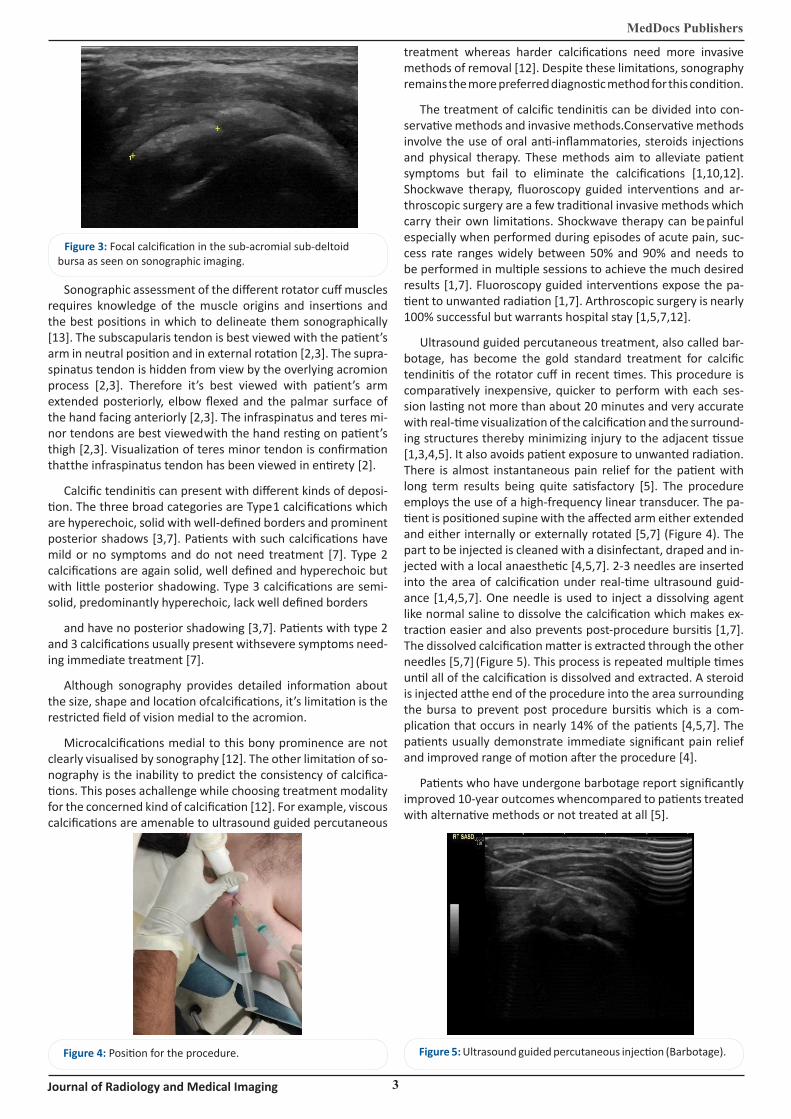
Sonography also carries a sensitivity of nearly 90% which makes it very effective at diagnosing soft tissue and joint pa-thologies [3]. A high-frequency linear transducer is usually used to scan the rotator cuff. This accords better near field resolu-tion and broad field of view, allowing usage of tissue harmonics which improve image contrast and reduce noise [2] (Figure 3).
MedDocs Publishers
3Journal of Radiology and Medical Imaging
Figure 3: Focal calcification in the sub-acromial sub-deltoid bursa as seen on sonographic imaging.
Sonographic assessment of the different rotator cuff muscles requires knowledge of the muscle origins and insertions and the best positions in which to delineate them sonographically [13]. The subscapularis tendon is best viewed with the patient’s arm in neutral position and in external rotation [2,3]. The supra-spinatus tendon is hidden from view by the overlying acromion process [2,3]. Therefore it’s best viewed with patient’s arm extended posteriorly, elbow flexed and the palmar surface of the hand facing anteriorly [2,3]. The infraspinatus and teres mi-nor tendons are best viewed with the hand resting on patient’s thigh [2,3]. Visualization of teres minor tendon is confirmation that the infraspinatus tendon has been viewed in entirety [2].
Calcific tendinitis can present with different kinds of deposi-tion. The three broad categories are Type 1 calcifications which are hyperechoic, solid with well-defined borders and prominent posterior shadows [3,7]. Patients with such calcifications have mild or no symptoms and do not need treatment [7]. Type 2 calcifications are again solid, well defined and hyperechoic but with little posterior shadowing. Type 3 calcifications are semi-solid, predominantly hyperechoic, lack well defined borders
and have no posterior shadowing [3,7]. Patients with type 2 and 3 calcifications usually present with severe symptoms need-ing immediate treatment [7].
Although sonography provides detailed information about the size, shape and location of calcifications, it’s limitation is the restricted field of vision medial to the acromion.
Microcalcifications medial to this bony prominence are not clearly visualised by sonography [12]. The other limitation of so-nography is the inability to predict the consistency of calcifica-tions. This poses a challenge while choosing treatment modality for the concerned kind of calcification [12]. For example, viscous calcifications are amenable to ultrasound guided percutaneous
treatment whereas harder calcifications need more invasive methods of removal [12]. Despite these limitations, sonography remains the more preferred diagnostic method for this condition.
The treatment of calcific tendinitis can be divided into con-servative methods and invasive methods. Conservative methods involve the use of oral anti-inflammatories, steroids injections and physical therapy. These methods aim to alleviate patient symptoms but fail to eliminate the calcifications [1,10,12]. Shockwave therapy, fluoroscopy guided interventions and ar-throscopic surgery are a few traditional invasive methods which carry their own limitations. Shockwave therapy can be painful especially when performed during episodes of acute pain, suc-cess rate ranges widely between 50% and 90% and needs to be performed in multiple sessions to achieve the much desired results [1,7]. Fluoroscopy guided interventions expose the pa-tient to unwanted radiation [1,7]. Arthroscopic surgery is nearly 100% successful but warrants hospital stay [1,5,7,12].
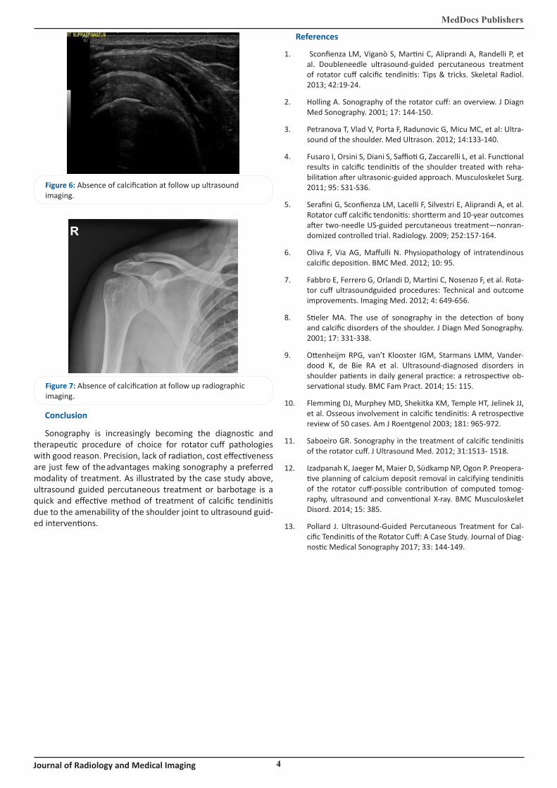
Ultrasound guided percutaneous treatment, also called bar-botage, has become the gold standard treatment for calcific tendinitis of the rotator cuff in recent times. This procedure is comparatively inexpensive, quicker to perform with each ses-sion lasting not more than about 20 minutes and very accurate with real-time visualization of the calcification and the surround-ing structures thereby minimizing injury to the adjacent tissue [1,3,4,5]. It also avoids patient exposure to unwanted radiation. There is almost instantaneous pain relief for the patient with long term results being quite satisfactory [5]. The procedure employs the use of a high-frequency linear transducer. The pa-tient is positioned supine with the affected arm either extended and either internally or externally rotated [5,7] (Figure 4). The part to be injected is cleaned with a disinfectant, draped and in-jected with a local anaesthetic [4,5,7]. 2-3 needles are inserted into the area of calcification under real-time ultrasound guid-ance [1,4,5,7]. One needle is used to inject a dissolving agent like normal saline to dissolve the calcification which makes ex-traction easier and also prevents post-procedure bursitis [1,7]. The dissolved calcification matter is extracted through the other needles [5,7] (Figure 5). This process is repeated multiple times until all of the calcification is dissolved and extracted. A steroid is injected at the end of the procedure into the area surrounding the bursa to prevent post procedure bursitis which is a com-plication that occurs in nearly 14% of the patients [4,5,7]. The patients usually demonstrate immediate significant pain relief and improved range of motion after the procedure [4].
Patients who have undergone barbotage report significantly improved 10-year outcomes when compared to patients treated with alternative methods or not treated at all [5].
Figure 4: Position for the procedure. Figure 5: Ultrasound guided percutaneous injection (Barbotage).
MedDocs Publishers
4Journal of Radiology and Medical Imaging
Figure 6: Absence of calcification at follow up ultrasound imaging.
Figure 7: Absence of calcification at follow up radiographic imaging.
Conclusion
Sonography is increasingly becoming the diagnostic and therapeutic procedure of choice for rotator cuff pathologies with good reason. Precision, lack of radiation, cost effectiveness are just few of the advantages making sonography a preferred modality of treatment. As illustrated by the case study above, ultrasound guided percutaneous treatment or barbotage is a quick and effective method of treatment of calcific tendinitis due to the amenability of the shoulder joint to ultrasound guid-ed interventions.
References
1. Sconfienza LM, Viganὸ S, Martini C, Aliprandi A, Randelli P, et al. Doubleneedle ultrasound-guided percutaneous treatment of rotator cuff calcific tendinitis: Tips & tricks. Skeletal Radiol. 2013; 42:19-24.
2. Holling A. Sonography of the rotator cuff: an overview. J Diagn Med Sonography. 2001; 17: 144-150.
3. Petranova T, Vlad V, Porta F, Radunovic G, Micu MC, et al: Ultra-sound of the shoulder. Med Ultrason. 2012; 14:133-140.
4. Fusaro I, Orsini S, Diani S, Saffioti G, Zaccarelli L, et al. Functional results in calcific tendinitis of the shoulder treated with reha-bilitation after ultrasonic-guided approach. Musculoskelet Surg. 2011; 95: S31-S36.
5. Serafini G, Sconfienza LM, Lacelli F, Silvestri E, Aliprandi A, et al. Rotator cuff calcific tendonitis: shortterm and 10-year outcomes after two-needle US-guided percutaneous treatment—nonran-domized controlled trial. Radiology. 2009; 252:157-164.
6. Oliva F, Via AG, Maffulli N. Physiopathology of intratendinous calcific deposition. BMC Med. 2012; 10: 95.
7. Fabbro E, Ferrero G, Orlandi D, Martini C, Nosenzo F, et al. Rota-tor cuff ultrasoundguided procedures: Technical and outcome improvements. Imaging Med. 2012; 4: 649-656.
8. Stieler MA. The use of sonography in the detection of bony and calcific disorders of the shoulder. J Diagn Med Sonography. 2001; 17: 331-338.
9. Ottenheijm RPG, van’t Klooster IGM, Starmans LMM, Vander-dood K, de Bie RA et al. Ultrasound-diagnosed disorders in shoulder patients in daily general practice: a retrospective ob-servational study. BMC Fam Pract. 2014; 15: 115.
10. Flemming DJ, Murphey MD, Shekitka KM, Temple HT, Jelinek JJ, et al. Osseous involvement in calcific tendinitis: A retrospective review of 50 cases. Am J Roentgenol 2003; 181: 965-972.
11. Saboeiro GR. Sonography in the treatment of calcific tendinitis of the rotator cuff. J Ultrasound Med. 2012; 31:1513- 1518.
12. Izadpanah K, Jaeger M, Maier D, Sϋdkamp NP, Ogon P. Preopera-tive planning of calcium deposit removal in calcifying tendinitis of the rotator cuff-possible contribution of computed tomog-raphy, ultrasound and conventional X-ray. BMC Musculoskelet Disord. 2014; 15: 385.
13. Pollard J. Ultrasound-Guided Percutaneous Treatment for Cal-cific Tendinitis of the Rotator Cuff: A Case Study. Journal of Diag-nostic Medical Sonography 2017; 33: 144-149.