val. no. 25, pp. 3, the journal of chemistry u. in s. a. … · hepatitis b virus envelope l...
TRANSCRIPT

THE JOURNAL OF BIOLOGICAL CHEMISTRY 0 1992 by The American Society for Biochemistry and Molecular Biology, Inc.
Val. 267, No. 3, Issue of January 25, pp. 1953-1961,1992 Printed in U. S. A.
Hepatitis B Virus Envelope L Protein Particles SYNTHESIS AND ASSEMBLY IN SACCHAROMYCES CEREVISIAE, PURIFICATION AND CHARACTERIZATION*
(Received for publication, August 5 , 1991)
Shun’ichi KurodaS, Sachiko Otaka, Takeshi Miyazaki, Masafumi Nakaoj, and Yukio Fujisawa From the Biotechnology Research Laboratories and the §Biology Research Laboratories, Takeda Chemical Industries, Ltd., Osaka 532, Japan
The hepatitis B virus envelope gene encodes three transmembrane proteins in frame; S, the product of S gene; M, the product of M (pre-S2 + S ) gene; and L, the product of L (pre-SI + pre-S2 + S ) gene. Unlike the S and M proteins, attempts to efficiently synthesize L proteins and assemble them into L protein particles in various eukaryotic cells have been unsuccessful, probably because of the presence of the pre-S1 peptide with an unknown function which appears to be inhib- itory to the host secretory apparatus. To investigate the role of the pre-S1 peptide, we constructed an L gene fused with a synthetic gene for chicken-lysozyme signal peptide (C-SIG) at the 5”terminal and placed the resultant gene under the control of the yeast glyc- eraldehyde-3-phosphate dehydrogenase gene pro- moter. After the fused-C-SIG peptide was correctly processed by the yeast secretory apparatus, a yeast transformant synthesized a protein with a molecular mass of approximately 52 kDa at a level of 42% of the total soluble protein. Electron micrographic observa- tion showed that the gene products assembled into 23- nm spherical and filamentous particles. The pre-S pep- tide of the gene product was deposited into the endo- plasmic reticulum (ER) lumen and well-glycosylated. It seemed that the gene products were accumulated as particles in certain specific membrane structures of the yeast secretory apparatus. Moreover, both the amount of mRNAs specific for the L gene and the in vivo stability of the synthesized L proteins did not change significantly by the addition of the C-SIG gene. These findings indicated that, if the pre-S1 peptide penetrates the ER membrane efficiently, the L proteins can be synthesized cotranslationally, translocate across the ER membrane with its S region, and then assemble by themselves into the particle form. There- fore, the pre-S1 peptide may involve weak or reduced signal peptide activity for recognition by the secretory apparatus and/or for the transport of the pre-S peptide into the ER lumen.
Hepatitis B virus (HBV)’ is an enveloped DNA virus of the hepadnavirus family. Human liver cells infected with HBV
* The costs of publication of this article were defrayed in part by the payment of page charges. This article must therefore be hereby marked “aduertisement” in accordance with 18 U.S.C. Section 1734 solely to indicate this fact.
6141 or 6037; Fax: 81-6-300-6276. 3 To whom correspondence should be addressed. Tel.: 81-6-300-
’ The abbreviations used are: HBV, hepatitis B virus; ER, endo- plasmic reticulum; C-SIG, chicken-lysozyme signal peptide; HBsAg, HBV surface antigens; SDS-PAGE, sodium dodecyl sulfate-polyacryl- amide gel electrophoresis; (/), plasmid-carrier state.
synthesize and release large amounts of empty subviral par- ticles (22-nm spherical and filamentous particles), along with 42-nm HBV virions (also called Dane particles). These sub- viral particles consist of the viral envelope glycoproteins bound to the lipid bilayer derived from the host cell (1, 2). The envelope glycoproteins consist of three transmembrane proteins; these proteins, termed surface antigens (HBsAg), are designated S, M, and L proteins. S protein is a major envelope protein, and M protein comprises the entire se- quence of S protein with an additional 55 amino acids at the amino-terminal (pre-S2 peptide). L protein contains the en- tire sequence of M protein and an additional 108 or 119 amino acids at the amino-terminal (pre-S1 peptide) (2, 3).
In mammalian cells, both S and M proteins are cotransla- tionally inserted into the endoplasmic reticulum (ER) mem- brane by the signal peptide within the amino-terminal region of S protein and span the ER membrane several times (4-6). These envelope proteins can also form subviral particles within the ER lumen by a nucleocapsid-independent process involving extrusion of the envelope proteins from the ER membrane (7-9); the resulting subviral particles are then exported from the cells via the constitutive pathway of vesic- ular transport (10).
Expressing either the S or the M gene in yeast cells results in similar 22-nm spherical particles. These particles however, are not found in culture broth but in cell extracts (11-18). The shape and size of these particles suggest that formation is not affected by either lipids or the secretory apparatus of different cell origins. In previous reports, we found that the pre-S2 peptide of the M particles purified from yeast cells are well glycosylated (19) and that the M proteins expressed in yeast cells accumulate as intracellular 22-nm particles (20). Therefore, it is indicated that the S and M proteins expressed in yeast cells are initially translocated across the ER mem- brane, then the proteins (at least M proteins) assemble them- selves into subviral particles in the yeast secretory apparatus.
When the L protein is expressed in either mammalian or yeast cells, the protein, unlike the S and M protein, does not aggregate into the 22-nm subviral particles, although the protein is efficiently expressed as a cellular membrane-asso- ciated form (17, 21-28). However, the native L protein is preferentially localized on HBV virions and filamentous par- ticles (3), and most of the pre-S (pre-S1 + pre-S2) peptide of native L protein is initially in the ER lumen, since (after budding) the peptide is displayed on the particle surface (3, 29). Therefore, these failures of particle formation in both mammalian and yeast cells prompted us to investigate whether the translocation of the flanking S protein is inhib- ited or suppressed by the retention of the pre-S1 peptide in the cytoplasm or ER membrane. If so, we speculate that, when the pre-S1 protein is actively transferred to the ER lumen by
1953

1954 HB V Envelope L Protein Particles
the action of an additive topogenic element, the flanking protein will span the ER membrane by the S protein-derived signal peptides (4-6) and that the L proteins will assemble into subviral particles without the other viral components (e.g. HBV core protein). In this study, we fused a gene encoding the chicken-lysozyme signal peptide (C-SIG), that is efficiently recognized by the yeast secretory apparatus (30- 32), to the 5'-terminal of the HBV envelope L gene in frame, and examined the expression of the fused gene in yeast cells.
EXPERIMENTAL PROCEDURES
Materials Zymolyase lOOT was purchased from Seikagaku Kogyo Co. (Tokyo,
Japan). Other enzymes were from Takara Shuzo Co. (Kyoto, Japan). Yeast-derived M particles (M-P3lc particles) were purified from Saccharomyces cereuisiae AH22R- cells carrying pGLD P31-RcT (18, 19). Auszyme I1 EIA kit (Abbott Laboratories, Chicago IL) was used to quantify the HBsAg particles (33). The serum in this kit reacts with particulate but not with free HBsAg. A mouse a n t i 4 protein monoclonal antibody (HBs 2-06), which reacts with HBsAg both in particle and free form (34), was generously provided by Nibon Seiyaku Co., Ltd. (Tokyo, Japan).
Strains and Media Plasmids were constructed in Escherichia coli K12 strain JM109
(35). L protein was expressed in S. cerevisiae strain AH22R- (a leu2 his4 can2 cir+ pho80) (13). Yeast transformation was performed using the method of Hinnen et al. (36). Transformants were selected by the Leu+ phenotype and cultured as described by Kitano et al. (20).
Plasmids and Synthetic DNAs The plasmid pGLD P31-RcT contains the HBV envelope M gene
carrying the modified pre-S2 gene (M-P3lc gene; subtype adr) be- tween the yeast GLD (glyceraldehyde-3-phosphate dehydrogenase gene; also called TDH3) promoter and the yeast PGK (3-phospho- glycerate kinase gene) terminator (18). The M-P3lc gene lacks the region encoding the peptide (Se~~'-Thr '~ of the pre-S2 peptide) that contains a trypsin-like proteinase-sensitive site (Arg4*-Thr4') (15). Plasmid pGLD 906-1 (16) is a cassette expression vector controlled by the GLD promoter, and contains the two-plasmid origin and arsl
length HBV (subtype adr) genomic DNA (37). The synthetic DNAs (autonomous replicating sequence). Plasmid pHBr 330 contains full-
were purchased from Operon Technologies, Inc. (Alameda, CA). The sequence of the C-SIG gene has been described previously (32).
Assays for the L Protein Expressed in Yeast Cells When the yeast cells reached the stationary growth phase (culti-
vation time 72 h), whole cell extract was prepared with glass beads in the presence of 7 M urea and 0.05% Triton X-100, as previously described (18). The levels of the HBsAg proteins (both particle and free form) in cell extracts were determined by the method of Wingfield et al. (38); this method is dependent on the densitometric intensity of the sample in silver-stained SDS-PAGE (39) using purified M- P31c particles as a standard. The amount of the HBsAg particles (particle form only) in cell extracts was determined with the Auszyme I1 EIA kit using human plasma-derived HBsAg (h-HBsAg) particles as a standard. The protein concentration was determined by the modified Lowry method (40) with crystalline bovine serum albumin as a standard. Western blotting analysis using the a n t i 3 protein monoclonal antibody (HBs 2-06) was performed as previously de- scribed (18).
Northern Blotting Analysis Yeast cells were grown to a density of 4.0 X lo7 cells/ml. Total
RNA was extracted using phenol and glass beads, then purified with an oligo(dT)-cellulose column (Pharmacia-LKB) (41). Five pg of the purified poly(A)+ RNAs were electrophoresed in 6% formaldehyde, 1.5% agarose gels, transferred onto nitrocellulose filters, and hybrid- ized with the S gene-specific probe (the 580-base pair XbaI-Aha111 fragment of pGLD P31-RcT) (42). After autoradiography, the filter was reprobed with the URA3 gene-specific probe (the 460-base pair StuI-PstI fragment from YEp24). To determine the ratio of the S gene-specific mRNA to that of the URA3 gene-specific mRNA (stand-
ard mRNA) (43), the densitometric intensities of both autoradi- ographic bands were measured by a densitometer model 620 (Bio- Rad). Sizes of the mRNAs were determined with RNA markers (Bethesda Research Laboratories).
In Vivo P5SlMethionine Pulse-labeling Assay The in vivo degradation rate of the expressed L protein was
determined as previously described (18). About 3.0 X lo4 cells carrying pGLD LIIP39-RcT growing in early exponential growth phase (24 h) were incubated with 1.11 MBq of [35S]methionine (Amersham Inter- national plc) at 30 "C for 60 min (pulse reaction), washed with 140 mM NaCl, resuspended in 20 mM methionine, then incubated at 30 "C for 0, 1,3,4, and 5 h (chase reaction). At the end of the chase reaction, whole cell extracts were immunoprecipitated with the anti-S protein monoclonal antibody (HBs 2-06) (44). Samples were separated by SDS-PAGE, then fluorographed using an intensifying screen (Fuji, Tokyo, Japan) (45). About 2.0 X 10' cells carrying pGLD P39-RcT were used to equalize the densitometric intensity between yeast cells a t 0 h.
Purification of L Particles from the Yeast (PGLD LZIP39-RcT) All operations were performed at 0-5 "C. Step I-The yeast cells (pGLD LIIP39-RcT) in stationary growth
phase (72 h) were harvested by centrifugation. The wet cells (26 g) were suspended in 100 ml of buffer A (7.5 M urea, 0.1 M sodium phosphate, pH 7.2, 15 mM EDTA, 2 mM phenylmethylsulfonyl fluo- ride, 0.1 mM (p-amidinophenyl) methanesulfonyl fluoride (APMSF) and 0.1% Tween 80), and disrupted with glass beads by using a BEAD-BEATER (Biospec Products, Bartlesville, OK). The whole cell extract was obtained by centrifugation at 12,000 X g for 30 min.
Step 2-The extract was mixed with a 0.75 volumes of polyethylene glycol (PEG) 6000 solution (33%, w/w), and then stirred for 30 min. The precipitated materials were collected by centrifugation at 13,900 X g for 30 min, and dissolved in 50 ml of buffer A without Tween 80.
Step 3-The solution was layered onto a discontinuous cesium chloride (CsCl) gradient (30 ml, concentration: 10-40% (w/v) in buffer A without Tween 80) and centrifuged at 141,000 X g (Beckman SW-Ti-28 rotor) for 16 h. The HBsAg particle fractions were col- lected, then dialyzed against buffer A without Tween 80 for 8 h. The precipitate formed during the dialysis was removed by centrifugation at 10,000 X g for 15 min, then the supernatant was concentrated with an ultrafiltration unit, Diaflow (Bio Engineering, Tokyo, Japan).
Step 4-The solution was separated repeatedly by CsCl density equilibrium centrifugation as described in Step 3.
Step 5-The solution was layered onto a discontinuous sucrose gradient (30 ml, concentration: 5-50% (w/v) in buffer A without urea and Tween 80 but with 0.85% NaCl) and centrifuged at 141,000 X g (Beckman SW-Ti-28 rotor) for 16 h. The HBsAg fractions were collected, then dialyzed against 0.85% NaCl and 10 mM sodium phosphate, pH 7.2, for 16 h. The dialyzed solution was filtered through Acrodiscs (Gelman Science, Ann Arbor, MI) and stored at 4 "C.
Assays for the Purified L Particles Electron micrographic analysis of the purified L particles was
performed as follows. The purified L particles (about 100 pg/ml) were adsorbed on collodium coated grids, stained with phosphotungstic acid, and examined using a JEM-1200 EX transmission electron microscope (JEOL Ltd., Tokyo, Japan). The amino acid sequence of
quencer model 470A (Applied Biosystems, Inc., CA) using purified L amino-terminal peptide was determined by a gas-phase protein se-
particles (2.20 nmol, as the L protein monomer). The carboxyl- terminal amino acids were determined by hydrazinolysis of the puri- fied L particles (3.66 nmol, as the L protein monomer) (46). The amino acid composition of the purified L particles was measured as follows. The L particles were dialyzed against 5 mM potassium phos- phate buffer, pH 7.2, and hydrolyzed in vacuo at 170 "C for 1 h in constantly boiling HCl containing 33.3% trifluoroacetic acid. The hydrolysates were evaporated to dryness under reduced pressure and examined using an amino acid analyzer model 835 (Hitachi CO., Ltd., Tokyo, Japan) (47). Half-cystine was determined as cysteic acid after performic acid oxidation and hydrolysis. Tryptophan and tyrosine were determined spectrophotometrically (48).
Digestion with Glycosidases Purified L particles (50 fig) were dialyzed against 0.1 M sodium
citrate, pH 4.5, and 0.1 mM ZnSO., then incubated at 37 "C with

HBV Envelope L Protein Particles 1955
either endo-p-N-acetylglucosaminidase (2 pg) or a-mannosidase (5 pg) for 2 or 4 h, respectively. The reacted materials were immunopre- cipitated with the anti-S protein monoclonal antibody (HBs 2-06) as described previously (18).
Digestion with Proteinases The purified L particles (50 pg) were dialyzed against 0.85% NaCl,
10 mM sodium phosphate, pH 7.2, with or without 0.5% SDS, then incubated with either L-1-tosylethylchloromethylketone-treated tryp- sin (1 pg) or Staphylococcus aureus V8 proteinase (50 pg) at 37 "C for 2 or 4 h, respectively. The reacted materials were immunoprecipitated with HBs 2-06 as described previously (18).
Thin Section Electron Microscopic Analyses of Yeast Celk
Yeast cells growing in late exponential growth phase (40 h) were prefixed with 2.5% glutaraldehyde in 50 mM cacodylate buffer, pH 7.4, for 3 h and post-fixed with cacodylate-buffered 1% osmium tetroxide for 2 h. The fixed cells were dehydrated with a graded series of ethanol and embedded in an Epon 812. Ultra thin sections were prepared using an Ultrotome I11 (Pharmacia-LKB), doubly stained with uranyl acetate and lead citrate, and observed under the JEM- 1200 EX transmission electron microscope.
RESULTS
Construction of the Lprotein Expression System-The HBV envelope L gene was fused at its 5'-terminal with a synthetic gene encoding 20 residues of the chicken-lysozyme signal peptide (C-SIG) (Met"'-Val*) and 3 residues from the 5'- noncoding region of the L gene (Ar$-Gly5) (Fig. 1). The fused gene was inserted in the cassette expression vector pGLD 906-1 to be placed between the yeast GLD promoter and the yeast PGK terminator (Fig. 2). The resulting plasmid (pGLD LIIP39-RcT) was used to transform yeast cells. To express the C-SIG-free L protein gene as a control, we constructed pGLD P39-RcT in a similar manner (Figs. 1 and 2) and used this plasmid to transform the same yeast cells.
Expression of the L Proteins in Yeast Cells-Cells carrying one of these plasmids (pGLD LIIP39-RcT, pGLD P39-RcT, pGLD 906-1) were cultivated in a synthetic medium as de- scribed previously (20). During the cultivation of the trans- formants, no significant difference in cell growth was ob- served. At the stationary growth phase of transformants, whole cell extracts were prepared by vortexing with glass beads in the presence of several proteinase inhibitors (7 M urea, 5.0 mM EDTA, and 0.5 mM phenylmethylsulfonyl fluo- ride), then assayed for HBsAg particles using the Auszyme 11 EIA kit. The yeasts (pGLD LIIP39-RcT) and (pGLD P39-
A, paD LUP2-9-M
r> - 18 M R
RcT) synthesized 296 and 0.4 pg of HBsAg particles (equiva- lent to h-HBsAg) in 5.0 X lo7 cells, respectively. This indicates that the synthesis of the HBsAg particles was enhanced 740- fold by the C-SIG gene.
L protein (both particle and free form) in the extracts was determined by silver-staining SDS-PAGE or Western blotting using the anti-S protein monoclonal antibody (HBs 2-06). In the yeast (pGLD LIIP39-RcT), we found that a protein of about 52 kDa with S-antigenicity was efficiently synthesized (Fig. 3, lunes 1 and 5 ) . By comparing the densitometric intensity of the 52-kDa band with those of the the yeast (pGLD 906-1) (lune 3 ) and the purified M-P3lc particles (lune 4 ) , the ratio of the 52-kDa protein was approximately 42% of the total soluble protein (38). The trace amount of about 28-kDa protein observed in lune 5 may have been generated by proteolysis. A similar degradation product of the HBsAg protein has been previously reported (17,49). On the other hand, the C-SIG-free yeast (pGLD P39-RcT) synthe- sized a protein of about 42 kDa with S-antigenicity. This protein accounted for 0.7% of the total soluble protein (lunes 2 and 6). Since this molecular mass is in complete agreement with that deduced from the DNA sequence, this 42-kDa protein may not be modified as observed by other researchers (17, 24). Thus, by additing the C-SIG gene to the 5"terminal of the L gene, the synthesis of L protein and L particle was elevated 60- and 740-fold, respectively. We estimated, using these values, that the assembly of the L proteins was also enhanced about 12-fold (740/60) by the addition of the C-SIG peptide.
The S and M proteins synthesized in yeast cells are recover- able as 22-nm particles similar in size to the h-HBsAg parti- cles. The proteins are assembled by yeast ER membrane (11- 20), as are particles from mammalian cells. To confirm that the L proteins synthesized in the yeast (pGLD LIIP39-RcT) are indeed assembled, the cell extract was sedimented through a 5-60% (w/w) sucrose gradient as previously described (19). The L proteins displayed the S-antigenicity peak, as measured by the Auszyme I1 EIA kit, at the position 40s (not shown). This value is slightly slower than that of h-HBsAg particles (42s). However, the L proteins in the C-SIG-free yeast (pGLD P39-RcT) had broad sedimentation profiles. These observa- tions confirmed that the L proteins are enhanced by the C- SIG gene to assemble themselves into similar particles.
Analyses of the Enhancement of L Protein Synthesis in the
Qlicken-lysozyme signal peptide (C-SIG) r' protein S L L I L V L C F L P L A A L G K V R Q G M G -1 +1 +6
FIG. 1. DNA sequences of the 5'- terminal junction region of the L gene in expression plasmids. A C- SIG gene-fused L gene (panel A, pGLD LIIP39-RcT) and a C-SIG gene-free L gene (panel B , pGLD P39-RcT) are rep- resented. B, pa0 P39-M

1956 HB V Envelope L Protein Particles
3.2 kb DNA
FIG. 2. Schematic representation of plasmid construction for the expression of the HBV envelope L genes. The plasmids pGLD LIIP39- RcT and pGLD P39-RcT allow the expression of the L gene fused with the C-SIG gene (hatched box) and the C- SIG-free L gene in S. cereuisiae cells, respectively. Transcription is driven by the constitutive glyceraldehyde-3-phos- phate dehydrogenase gene ( 0 ) pro- moter (GLDp) described previously (18). The transcriptional terminators in pGLD LIIP39-RcT and pGLD P39-RcT are the 3-phosphoglycerate kinase gene (PGK) terminator (PGKt) described elsewhere (18). The parent yeast vector pGLD 906-1 (16) contains the LEU2 gene for the selection of S. cereuisiae transformants under leucine-deficient conditions, Ap' gene for the selection of E. coli transformants under the presence of ampicillin, and arsl and ZR genes for the plasmid-replication in yeast cells.
TaqI-ECORI 1 t
0 . 3 4 kb DNA
71bp C-Jig DNA X" T
+ '
XhoI-Hind111 adaptor S'AGCTTGGCC3'
3'ACCGGAGCTS' f HindIII-EcoRI digested pVCl8
XhoI-ECoRI
TaqI
TI DNA pol + 4dNTPs
Sal1 linker S'GGTCGACC3'
Salx-EcoRI
0.34 kb DNA
1 pre-S1
I t t Sall-ECORI digested pUCl8
ECORI-Sa11
1.1 kb DNA SalI-ECORI
0.42kb DNA 0 3 4 kb DNA
1 9 . 4 k b ) p G L D 9 0 6 - I
( 9 . 4 k b l
1.4 kb DNA
Yeast (pGLD LlIP39-RcT)"In the C-SIG-free yeast (pGLD P39-RcT), the L proteins constituted only up to 0.7% of the total soluble protein, whereas the C-SIG-fused yeast (pGLD LIIP39-RcT) synthesized them at a ratio of 42% of the total soluble protein. To examine whether this was caused at the transcriptional level, we performed Northern blotting analysis by using an S gene-specific probe and a standard probe (URA3 gene). The URA3 gene is transcribed constantly in yeast cells (43). As shown in Fig. 4, the ratio of the amount of mRNAs encoding L proteins (1430 and 1350 nucleotides; lanes 1 and 2 ) to that of the URA3-specific mRNA (1000 nucleotides; lanes 4 and 5) were 1.02 and 1.00 in the yeasts (pGLD LIIP39- RcT) and (pGLD P39-RcT), respectively. This suggested that L protein synthesis in the yeast (pGLD LIIP39-RcT) was enhanced at the translational level and/or that the synthe- sized L protein became stable in the yeast cell.
To examine the possibility that the in vivo degradation rate of L proteins was decreased by the C-SIG peptide, we pulse-
pGLDP39-RcT I l O B X b I
labeled L proteins with ["SJmethionine in yeast cells then immunoprecipitated them with HBs 2-06 (18). Densitometric analyses of the immunoreactive bands (Fig. 5 ) indicated that the L proteins in the C-SIG-fused yeast (pGLD LIIP39-RcT) degraded slightly faster than those in the C-SIG-free yeast (pGLD P39-RcT). Therefore, it appears that the enhanced synthesis of L protein by the C-SIG peptide in the yeast (pGLD LIIP39-RcT) was due to an increased efficiency at the translational level.
Purification of L Particles from the Yeast (pGLD LIIP39- RcT)-Owing to the high expression level, the L particles in the yeast (pGLD LIIP39-RcT) could be purified without any chromatographic procedures. The whole cell extract from 26 g (wet weight) was fractionated with PEG 6000 (final concen- tration 19%, w/w), and separated twice by CsCl density equi- librium centrifugation (10-40%, w/v) and once by sucrose gradient centrifugation (5-50%, w/v). The profile of the first CsCl density equilibrium centrifugation (Fig. 6, left) and the

HB V Envelope L Protein Particles 1957 Silver stain Western blot
kDa 1 2 3 4 M 5 6 7 M
FIG. 3. Expression and characterization of the L proteins in recombinant yeast cells. Samples of whole cell extracts or yeast- derived M particles were applied to a 12.5% SDS-PAGE. The gel was stained with silver reagent (lanes 1-4) or Western blotted using anti- S protein monoclonal antibody (lanes 5-7). In the silver-stained gel and Western blot, each lane (except lanes 4 and M) contains 1 and 20 pg of the protein, respectively. Lanes 1 and 5, cell extract of the yeast (pGLD LIIP39-RcT); lanes 2 and 6, cell extracts of the yeast (pGLD P39-RcT); lanes 3 and 7, cell extracts of the yeast (pGLD 906-1); lane 4, 2 of the M-P3lc particles; lane M, molecular weight markers (Bio-Rad).
I
1 2 3 4 5 6 FIG. 4. Northern blot analyses of poly(A)+ RNAs from yeast
transformants. The poly(A)’ RNAs from yeast (pGLD LIIP39- RcT) (lanes I and 4) , yeast (pGLD P39-RcT) (lanes 2 and 5), or yeast (pGLD 906-1) (lanes 3 and 6 ) were analyzed using the S gene- specific probe (lanes 1-3) and the URA3-specific probe (control, lanes 4-6) as described under “Experimental Procedures.” The densito- metric intensities of the S gene-specific and URA3-specific mRNAs, indicated by arrowheads, were measured with a densitometer model 620 (Bio-Rad). Sizes of mRNAs are given in nucleotides (nt).
A 500. 0 1- .3 4 5 hrs
B 0 1 3 4 5 h r s
-42kDa i - C
0 hrs ii Z O O L A 100 0
0 1 2 3 4 5 Tim,: (tars)
FIG. 5. In vivo [‘?3]methionine pulse-labeling assay of the L proteins in yeast cells. After pulse-labeling with [35S]methionine for 1 h, the chase reactions were performed for 0, 1, 3, 4, and 5 h (lanes 0,1,3,4, and 5) as described under “Experimental Procedures.” The degradation patterns of the L protein in yeast (pGLD LIIP39- RcT) (pane lA) , yeast (pGLD P39-RcT) (pane lB) , and yeast (pGLD 906-1) (panel C) are shown. Based on the densitometric intensity of each fluorographs (left) , the curves of i n uiuo degradation of L proteins are also indicated (right).
Western blots of each fraction (Fig. 6, right) showed that the 52-kDa L protein and the S-antigenic activity comigrated as a single peak at a density of about 1.21 g/cc, which coincides with that reported for the h-HBsAg particles. Finally, we obtained 6 mg of pure L particles. A summary of the purifi- cation of L particles is shown in Table I. The purified L
Fraction number
11 12 13 14 15 M
Fraction number I O S r n l h c 1
FIG. 6. The profiles of first cesium chloride (CsCl) density equilibrium centrifugation in the purification steps. The L protein fraction precipitated with PEG 6000 (19%, w/w) was analyzed as described under “Experimental Procedures.” The profiles of S- antigenicity were determined by the Auszyme I1 EIA kit, and the density of each fraction are shown (left). The Western blotting profile using the anti-S protein monoclonal antibody of each fraction is shown (right). The number of each lane represents the fraction number of the first CsCl density equilibrium centrifugation.
TABLE I Purification of L Darticles from yeast cells (RGLD LIIP39-RcT)
SteD Protein L oarticle Purification Yield ~~
mg“ mgb -fold % Crude extract 790 23.8 1.00 100 PEG 6000 precipitate 320 13.7 1.42 57 1st CsCl (10-40%) equilib- 78.0 10.5 4.49 44
2nd CsCl (10-40%) equilib- 15.0 8.8 19.5 31
Sucrose (5-50%) gradient 6.0 5.7 31.6 24
rium centrifugation
rium centrifugation
centrifugation The protein concentration was determined by the Lowry method
*The L particle was quantitated by S-antigenicity using the as described under “Experimental Procedures.”
Auszyme I1 EIA kit using h-HBsAg particles as a standard.
1 2 1
- 93 -66
-45
4 31
- 22
4 14
k D o
FIG. 7. SDS-polyacrylamide gel electrophoretogram of pu- rified L particles under the reducing (lane I ) or nonreducing (lane 2 ) conditions. Samples (600 ng) of purified L protein were separated on 12.5% SDS-PAGE, then subjected to silver staining. Molecular weight markers are shown in lane M.
particles were separated by SDS-PAGE under reducing or nonreducing conditions, then stained with silver reagent. As shown in Fig. 7, the L particles consist of a major 52- and a minor 28-kDa polypeptide, which are linked by intermolecular disulfide bonds to form a multimeric protein.
Chemical Properties of the Purified L Particles-Electron micrographs of the purified materials directly indicated that the L proteins from the yeast (pGLD LIIP39-RcT) indeed assembled into particle form (Fig. 8). There are two distinct
1958 HB V Envelope L Protein Particles
FIG. 8. Electron micrographs of purified L particles. Mag- nification: panel A , X 100,000; panel R, X 300,000.
shapes; about 80% of the observed particles are spherical (23 nm in diameter), very similar to the h-HBsAg particles, and the rest are filamentous (40-120 nm in length and 23 nm in diameter). These findings are consistent with the observation that filamentous particles (up to 200 nm in length and 22 nm in diameter) obtained from the sera of HBV-infected individ- uals are abundant in L proteins and directly supported the hypothesis that the shape of the HBV-related particles may be influenced by the presence of the L protein (3). On the other hand, the C-SIG-free L proteins purified from the yeast (pGLD P39-RcT) were found not to form such an ordered structure (not shown). This observation is consistent with those by other researchers (17, 24, 27).
The amino-terminal amino acid sequence of the L protein was analyzed by automated Edman degradation. The amino- terminal sequence is Lys-Val-Arg-Gln-Gly-Met-Gly-Thr-X- Leu-Ser-Val-, indicating that the C-SIG peptide was correctly processed by the yeast secretory apparatus (between Gly" and Lys+'; see Fig. 1) (30-32). The carboxyl-terminal amino acid residue of the L protein is only Ile, which is consistent with that encoded by the L gene. The amino acid composition of the L protein is almost identical to that deduced from the L gene (not shown). Based upon these observations, the molecular weight calculated from the L gene was 42,394, whereas the observed molecular mass of the purified L protein was mainly 52 kDa. Therefore, it was suggested that this difference was attributable to post-translational glycosyla- tion.
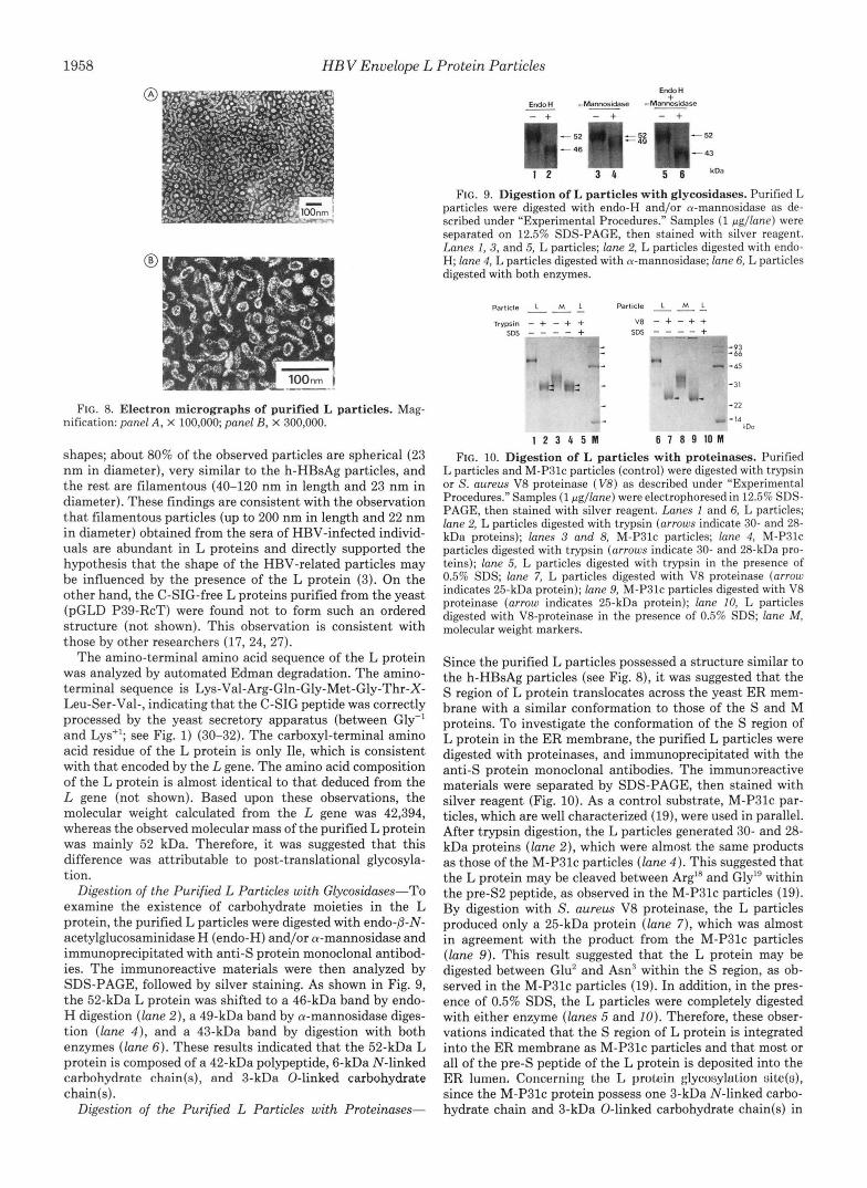
Digestion of the Purified L Particles with Glycosidases-To examine the existence of carbohydrate moieties in the L protein, the purified L particles were digested with endo-p-N- acetylglucosaminidase H (endo-H) and/or a-mannosidase and immunoprecipitated with anti-S protein monoclonal antibod- ies. The immunoreactive materials were then analyzed by SDS-PAGE, followed by silver staining. As shown in Fig. 9, the 52-kDa L protein was shifted to a 46-kDa band by endo- H digestion (lane 2 ) , a 49-kDa band by a-mannosidase diges- tion (lane 4) , and a 43-kDa band by digestion with both enzymes (lane 6). These results indicated that the 52-kDa L protein is composed of a 42-kDa polypeptide, 6-kDa N-linked carhohydrnte chnin(s), nnd 3-kDa 0-linked carbohydrate chain(s).
Digestion of the Purified L Particles with Proteinuses-
FIG. 9. Digestion of L particles with glycosidases. Purified L particles were digested with endo-H and/or a-mannosidase as de- scribed under "Experimental Procedures." Samples (1 pgllane) were separated on 12.5% SDS-PAGE, then stained with silver reagent. Lunes 1, 3, and 5, L particles; lane 2, L particles digested with endo- H; lane 4, L particles digested with a-mannosidase; lane 6, L particles digested with both enzymes.
Particle L M L Particle L M 1. Trypsin - + - + +
SDS T + SDS - - - - + V8 - + - + +
07 ki, 45
31
22
14 4C.a
1 2 3 4 5 1 6 7 8 9 1 0 M
FIG. 10. Digestion of L particles with proteinases. Purified L particles and M-P3lc particles (control) were digested with trypsin or S. aureus V8 proteinase (V8) as described under "Experimental Procedures." Samples (1 pgllane) were electrophoresed in 12.5% SDS- PAGE, then stained with silver reagent. Lunes 1 and 6, L particles; lane 2, L particles digested with trypsin (arrows indicate 30- and 28- kDa proteins); lunes 3 and 8, M-P3lc particles; lane 4, M-P3lc particles digested with trypsin (arrows indicate 30- and 28-kDa pro- teins); lane 5, L particles digested with trypsin in the presence of 0.5% SDS; lane 7, L particles digested with V8 proteinase (arrow indicates 25-kDa protein); lane 9, M-P3lc particles digested with V8 proteinase (arrow indicates 25-kDa protein); lane 10, L particles digested with V8-proteinase in the presence of 0.5% SDS; lane M , molecular weight markers.
Since the purified L particles possessed a structure similar to the h-HBsAg particles (see Fig. 8), it was suggested that the S region of L protein translocates across the yeast ER mem- brane with a similar conformation to those of the S and M proteins. To investigate the conformation of the S region of L protein in the ER membrane, the purified L particles were digested with proteinases, and immunoprecipitated with the anti-S protein monoclonal antibodies. The immunxeactive materials were separated by SDS-PAGE, then stained with silver reagent (Fig. 10). As a control substrate, M-P3lc par- ticles, which are well characterized (19), were used in parallel. After trypsin digestion, the L particles generated 30- and 28- kDa proteins (lane 2 ) , which were almost the same products as those of the M-P3lc particles (lane 4 ) . This suggested that the L protein may be cleaved between ArgIR and Gly" within the pre-S2 peptide, as observed in the M-P3lc particles (19). By digestion with S. aurezu V8 proteinase, the L particles produced only a 25-kDa protein (lane 7), which was almost in agreement with the product from the M-P3lc particles (lane 9 ) . This result suggested that the L protein may be digested between Glu' and Asn3 within the S region, as ob- served in the M-P3lc particles (19). In addition, in the pres- ence of 0.5% SDS, the L particles were completely digested with either enzyme (lanes 5 and 10). Therefore, these obser- vations indicated that the S region of L protein is integrated into the ER membrane as M-P3lc particles and that most or all of the pre-S peptide of the L protein is deposited into the ER lumen. Concerning tho L protoin glycosylulion site(ti), since the M-P3lc protein possess one 3-kDa N-linked carbo- hydrate chain and 3-kDa 0-linked carbohydrate chain(s) in

HB V Envelope L Protein Particles 1959
the pre-S2 peptide (19) and since the S region of the L protein had almost the same structure as that of the M-P3lc protein (see Fig. 10, lanes 7 and 9 ) , we confirmed that L protein has 6-kDa N-linked carbohydrate chain(s) and 3-kDa 0-linked carbohydrate chain(s) within the pre-S peptide.
Transmission Electron Microscopic Analysis-The forego- ing results indicated that the L proteins synthesized in the yeast (pGLD LIIP39-RcT) efficiently assemble into L parti- cles. Therefore, we examined thin sections of the yeast (pGLD LIIP39-RcT) using a transmission electron microscope. Fig. 11 shows an electron micrograph of thin sections of the yeast (pGLD 906-1) (panel A), the C-SIG-free yeast (pGLD P39- RcT) (panel B), and the C-SIG-fused yeast (pGLD LIIP39- RcT) (panel C). Many highly dense spots (-300 nm in di- ameter) were observed specifically in the yeast (pGLD LIIP39-RcT) (panel C), and the L particles seemed to be accumulated here. These spots were studied in more detail by expanding the magnification. As shown in panel D, the dense spots were full of particles of about 20-30 nm, since most of the L particles purified from the same yeast form 23-nm spherical particles (see Fig. 8), strongly suggesting that the L proteins accumulate as particles within the spots. The same phenomena were observed in the M protein-expressing yeast (20). Moreover, since most of the L proteins were both N - and O-glycosylated (see Fig. 9) and the spots seemed to be covered by a membranous structure, the spots may be part of the yeast secretory apparatus, perhaps downstream of the cis- Golgi compartment (50). However, conclusive proof must await detection techniques that utilize specifically labeled antibody probes.
DISCUSSION
Eble et aL(4-6) reported that the translocation of the S and M proteins across the ER membrane is initiated by its amino- terminal peptide and that these proteins finally span the ER membrane several times by its two S region-specific signal peptides. The L protein, which has an additional pre-S1 peptide on the amino-terminal of the M protein, is preferen- tially localized on HBV virions and filamentous subviral particles. This protein spans the ER membrane as do the S and M proteins, and its pre-S peptide is deposited into the ER lumen (3, 29). Therefore, it is highly likely that the translocation of the native L protein across the ER membrane
@
of yeast (pGLD 906-1) (panel A ) , yeast (pGLD P39-RcT) FIG. 11. Transmission electron micrographs of thin sections
(panel B) , and yeast (pGLD LIIP39-RcT) (panels C and D ) . Magnification: panel A, X 4,000; panels B and C, X 6,000; panel D, X 30,000. Bars indicate 500 nm (panels A-C) and 100 nm (panel D ) .
is initiated by its amino-terminal peptide. In this study, we found that the addition of the C-SIG gene
to the 5'-terminal of L gene enhanced the translational effi- ciency of the L gene-specific mRNA significantly. Thereafter, the synthesis level of the L protein reached up to about 42% of the total soluble protein. In addition, we demonstrated that the C-SIG peptide was correctly processed, that the flanking pre-S peptide was deposited into the ER lumen, that the pre- S peptide was well glycosylated, and that the C-SIG-free L protein was not efficiently synthesized (about 0.7% of total soluble protein) and not glycosylated. These observations indicated that the efficient recognition of the C-SIG peptide by the yeast secretory apparatus increased the transport of the flanking pre-S peptide into the ER lumen. As a result, the synthesis level of L protein might be enhanced significantly by a cotranslational effect. When human lysozyme is secreted by yeast cells, which utilize modified C-SIG peptides, the secretion level of the lysozyme is well correlated with a signal activity (e.g. hydrophobicity) of its modified C-SIG peptide (32,51). In addition, we previously found that the hydropho- bicity of the pre-S2 peptide controls the synthesis level of the M protein in yeast cells (18). These facts support that the synthesis level of L protein was determined by the signal activity of its amino-terminal peptide.
Recently, by expressing a chimeric HBsAg (p-lactamase signal peptide + LY globin + S protein) gene, Bruss and Ganem (52) demonstrated that the deposition of the globin domain into the ER lumen requires three signal peptides (p-lactamase signal peptide and two S protein-derived signal peptides), and that the correct transmembrane disposition of the amino- terminal peptide of the chimeric HBsAg is important for particle formation. Therefore, it is strongly suggested that the amino-terminal of the native pre-Sl peptide involves weak or reduced signal peptide activity for recognition by the secretory apparatus and/or for the depositing the flanking pre-S peptide into the ER lumen. As a result, the native L protein might fail to efficiently translocate across the ER membrane.
Concerning particle formation, in contrast with S and M proteins, L proteins previously failed to assemble into well- ordered structures (17, 21-28). Therefore, it has been pro- posed that the L proteins can form particles only by assem- bling with other viral components (e.g. HBsAg S and M proteins, HBV core protein) (17, 53). In this study, however, we demonstrated that the L proteins, if they translocate across the ER membrane depositing their pre-S peptide into the ER lumen, can assemble into the particle form without any other viral components. Therefore, in the secretory pathway of human hepatocytes, we speculate that there is a viral or cellular machinery which enhances the signal peptide activity of the amino-terminal region of pre-S1 peptide and that this may play a crucial role(s) in the morphogenesis of HBV virions and filamentous subviral particles. For example, the interaction with other viral components may trigger the ini- tiation of the translocation of L protein, or the lipid compo- sition of the human hepatocyte ER membrane may be more suitable for penetration by the pre-S1 peptide than those of any other ER membranes.
Recently, using a mammalian cells, the amino-terminal peptide of the L protein has been shown to involve a novel ER retention signal (54). Therefore, it has been speculated that the aggregation of L proteins is inhibited by the accu- mulated L proteins in the ER and requires interactions with other components to trigger the budding event of the L particles. In yeast cells, it remains unclear whether the novel ER retention signal in the pre-Sl peptide is active, although ER retention machinery in yeast cells has been identified (55,

1960 HB V Envelope L Protein Particles
56). In this study, the L particles, however, possess a complete ER retention signal within its pre-S1 peptide (Gly7-Pro13 in the C-SIG-fused L protein). Therefore, determining whether the C-SIG peptide upstream of the L protein abolishes the activity of the ER retention signal awaits further elucidation.
Although myristylation is a well-known modification of cytoplasmic proteins, the pre-S1 peptide is one of the few transmembrane proteins which harbors this modification (57). If the yeast cells modify the pre-S1 peptide (Gly' site) with myristic acid as a human immunodeficiency virus Gag precursor protein (58), then the modified L proteins may inhibit the formation of particle structure by their retention in the yeast ER. Recently, however, other researchers have reported that this modification alone does not determine the ER retention of the L protein (54). In addition, when the mutated C-SIG-free L gene, in which the Gly' codon is mu- tated to the Asn2 codon, was expressed in yeast cells, there was no significant change in the synthesis of L particles (not shown). Therefore, we considered that the myristylation of the pre-S1 peptide does not play a crucial role in the formation of the L particles.
When the L particles were compared with the h-HBsAg particles, three distinct structural differences were found. First, the Argl'' in the S region of the h-HBsAg protein is sensitive to trypsin digestion (3). Second, there is a complex carbohydrate chain in the S region of the h-HBsAg protein (3). These two differences are common characteristics among the yeast-derived S, M, and L particles (17, 19). Third, the pre-S peptide of the h-HBsAg L protein is not glycosylated (3). However, no attempts have been made to pursue the cause of these differences.
Currently available yeast-derived HBV vaccine contains the S and/or M proteins in the particle form. These vaccines have good immunogenicity and protective efficacy (59-61). However, a recent study employing synthetic peptides has demonstrated the presence of additional protective epitopes in the pre-S1 peptide (62). Using a congenic mouse model, nonresponsiveness to the S and M protein was circumvented when the pre-S1 peptide was included in the immunogen (63). Furthermore, many epitopes of the pre-S1 peptide seem to be conformational (64). If comparable mechanisms of immune responses are operative in humans, the use of the L particles as a HBV vaccine could broaden and enhance the protective response and possibly diminish the percentage of slow re- sponders or nonresponders.
Acknowledgments-We thank Drs. A. Kakinuma and K. Tsuka- mot0 of our laboratories for their encouragement throughout this work. We are grateful to Dr. R. Marumoto for supplying synthetic oligonucleotides, to K. Ohfune for taking electron micrographs, and to M. Shimizu-Utsunomiya and M. Kusunoki-Ozawa for their tech- nical support.
1.
2.
3.
4.
5.
6.
7.
8.
REFERENCES Tiollais, P., Pourcel, C., and Dejean, A. (1985) Nature 317 , 489-
Neurath, A. R., and Kent, S. B. H. (1988) Adu. Virus Res. 34 ,
Heerman, K. H., Goldmann, U., Schwartz, W., Seyffarth, T., Baumgarten, H., and Gerlich, W. H. (1984) J. Virol. 5 2 , 396- 402
Eble, B. E., Lingappa, V. R., and Ganem, D. (1986) Mol. Cell.
Eble, B. E., MacRae, D. R., Lingappa, V. R., and Ganem, D.
Eble, B. E., Lingappa, V. R., and Ganem, D. (1990) J. Virol. 6 4 ,
Gerber, M,, Hadziyannis, S., Vissoulis, C., Schaffner, F., Paro- netto, F., and Popper, H. (1974) Am. J. Pathol. 75,489-502
Patzer, E. J., Nakamura, G. R., Simonsen, C. C., Levinson, A. D.,
495
65-142
Biol. 6, 1453-1463
(1987) Mol. Cell. Biol. 7, 3591-3601
1414-1419
9.
10. 11.
12.
13.
14.
15.
16.
17.
18.
19.
20.
21.
22.
23.
24.
25.
26. 27.
28.
29.
30. 31.
32.
33.
34.
35. 36.
37.
38.
39. 40.
41.
42.
43.
44.
45.
and Brands, R. (1986) J. Virol. 58,884-892 Simon, K., Lingappa, V. R., and Ganem, D., (1988) J. Cell Bwl.
Kelly, R. (1985) Science 230, 25-31 Valenzuela, P., Medina, A., Rutter, W. J., Ammerer, G., and Hall,
B. D (1982) Nature 298 , 347-350 Valenzuela, P., Coit, D., and Kuo, C. H. (1985) BioTechnology 3,
Miyanohara, A., Toh-e, A., Nozaki, C., Hamada, F., Ohtomo, N., and Matsubara, K. (1983) Proc. Natl. Acud. Sci. U. S. A. 8 0 ,
Hitzeman, R. A., Chen, C. Y., Hagie, F. E., Patzer, E. J., Liu, C, -C., Estell, D. A., Miller, J. V., Yaffe, A., Kleid, D. G., Levinson, A. D., and Opperman, H. (1983) Nucleic Acids Res. 11 , 2745- 2763
Itoh, Y., andFuiisawa, Y. (1986) Biochem. Biophys. Res. Commun.
107 , 2163- 2168
317-320
1-5
141,.942-948 "
Itoh. Y.. Havakawa. T.. and Fuiisawa. Y. (1986) Biochem. Bio- , , ,
phys. Res. kommun. 138,2681274 Imamura, T., Araki, M., Miyanohara, A., Nakao, J., Yonemura,
H., Ohtomo, N., and Matsubara, K. (1987) J. Virol. 6 1 , 3543- 3549
Kuroda, S., Itoh, Y., Miyazaki, T., Otaka-Imai, S., and Fujisawa, Y (1989) Gene (Amst.) 78,297-308
Kobayashi, M., Asano, T., Utsunomiya, M., Itoh, Y., Fujisawa, Y., Nishimura, O., Kato, K., and Kakinuma, A. (1988) J. Bwtechnol. 8 , 1-22
Kitano, K., Nakao, M., Itoh, Y., and Fujisawa, Y. (1987) Bio- Technology 5 , 281-283
Persing, D. H., Varmus, H. E., and Ganem, D. (1986) Science 234,1388-1391
Cheng, K., Smith, G. L., and Moss, B. (1986) J. Virol. 6 0 , 337- 344
Standring, D. N., Ou, J.-H., and Rutter, W. J. (1986) Proc. Natl. Acud. Sci. U. S. A. 83,9338-9342
Dehoux, P., Ribes, V., Sobczak, E., and Streeck, R. E. (1986) Gene (Amst.) 48,155-163
McLachlan, A., Milich, D. R., Raney, A. K., Riggs, M. G., Hughes, J. L., Sorge, J., and Chisari, F. V. (1987) J. Virol. 61,683-692
Ou, J., and Rutter, W. J. (1987) J. Virol. 6 1 , 782-786 Kniskern, R. J., Hagopian, A., Burke, P., Dunn, N., Emini, E. A.,
Miller, W. J., Yamazaki, S., and Ellis, R. W. (1988) Hepatology
Molnar-Kimber, K. L., Jarocki-Witek, V., Dheer, S. K., Vernon, S. K., Conley, A. J., Davis, A. R., and Hung, P. P. (1988) J. Virol. 6 2 , 407-416
Kuroki, K., Floreani, M., Mimms, L., and Ganem, D. (1990) Virology 176,620-624
Oberto, J. and Davison, J. (1985) Gene (Amst.) 4 0 , 57-65 Jigami, Y., Muraki, M., Harada, N., and Tanaka, H. (1986) Gene
(Amst.) 43,273-279 Yoshimura, K., Toibana, A., Kikuchi, K., Kobayashi, M., Hay-
akawa, T., Nakahama, K., Kikuchi, M., and Ikehara, M. (1987) Biochem. Biophys. Res. Commun. 145 , 712-718
Budkowska, A., and Karwowska, S. (1982) J. Immunol. Methods
Fujisawa, Y., Ito, Y., Ikeyama, S., and Kikuchi, M. (1985) Gene
Messing, J. (1983) Methods Enzymol. 101 , 20-78 Hinnen, A., Hicks, J. B., and Fink, G. R. (1978) Proc. Natl. Acud.
Ono, Y., Onda, H., Sasada, R., Igarashi, K., Sugino, Y., and
Wingfeld, P. T., Graber, P., and Payton, M. A. (1987) Yeast 3 ,
Laemmli, U. K. (1970) Nature 227, 680-685 Lowry, 0. H., Rosebrough, N. J., Farr, A. L., and Randall, R. J.
(1951) J. Biol. Chem. 193, 265-275 Henikoff, S., Kelly, J. D., and Cohen, E. H. (1983) Cell 33 , 607-
614 Maniatis, T., Fritsch, E. F., and Sambrook, J. (1982) in Molecular
Cloning: A Laboratory Manual, pp. 197-207, Cold Spring Har- bor Laboratory, Cold Spring Harbor, NY
Bach, M.-L., Lacroute, F., and Botstein, D. (1979) Proc. Natl. Acd . Sci. U. S. A. 76 , 386-390
Haguenauer-Tsapis, R., and Hinnen, A. (1984) Mol. Cell. Biol. 4 ,
Chamberlain, G. P. (1979) Anal. Biochem. 98 , 132-135
8.82-87
51,341-346
(Amst.) 4 0 , 23-29
Sci U. S. A. 7 5 , 1929-1933
Nishioka, K. (1983) Nucleic Acids Res. 11, 1747-1757
43-49
2668-2675

HB V Envelope L Protein Particles 1961
46. Narita, K., Murakami, H., and Ikenaka, T. (1966) J. Biochem. 56. Lewis, M. J., Deborah, J . S., and Pelham, H. R. B. (1990) Cell
47. Tsugita, A., and Scheffler, J.-J. (1982) Eor. J. Biochem. 124, 57. Persing, D. H., Varmus, H. E., and Ganem, D. (1987) J. Virol.
48. Edelhoch, H. (1967) Biochemistry 6,1948-1954 58. Jacobs, E., Gheysen, D., Thines, D., Francotte, M., and de Wilde, 49. Langley, K. E., Egan, K. M., Barendt, J. M., Parker, C. G., and M. (1989) Gene (Amst.) 79, 71-81
Bitter, G. A. (1988) Gene (Amst.) 67, 229-245 59. Hazama, M., Takaoki, M., Ohfune, K., Hinuma, S., and Fujisawa, 50. Kukuruzinska, M. A., Bergh, M. L. E., and Jackson, B. J. (1987) Y. (1989) Vaccine 7 , 567-574
Annu. Rev. Biochem. 56, 915-944 60. Fujisawa, Y., Kuroda, S., Van Eerd, P. M. C. A., Schellekens, H., 51. Yamamoto, Y., Taniyama, Y., and Kikuchi, M. (1989) Biochem- and Kakinuma, A. (1990) Vaccine 8, 192-198
52. Bruss, V., and Ganem, D. (1991) J. Virol. 65,3813-3820 (1991) Vaccine 9 , 163-169 53. Bruss, V., and Ganem, D. (1991) Proc. Natl. Acad. Sci. U. S. A. 62. Neurath, A. R., Seto, B., and Strick, N. (1989) Vaccine 7, 234-
54. Kuroki, K., Russnak, R., and Ganem, D. (1989) Mol. Cell. Bwl. 63. Milich, D. R., McLachlan, A., Chisari, F. V., Kent, S. B. H., and
55. Semenza, J. C., Hardwik, K. G., Dean, N., and Pelham, H. R. B. 64. Heerman, K. H., Kruse, F., Seifer, M., and Gerlich, W. H. (1987)
(Tokyo) 69, 170-175 61,1359-1363
585-588 61,1672-1677
istu 28, 2728-2732 61. Kuroda, S., Fujisawa, Y., Iino, S., Akahane, Y., and Suzuki, H.
88, 1059-1063 236
9,4459-4466 Thornton, G. B. (1986) J. Zmmunol. 137,315-322
(1990) Cell 61 , 1349-1357 Intervirology 28 , 14-18