autolytic systemof staphylococcus 22: influence on …5454 bierbaum andsahl table 1. purification...
TRANSCRIPT

Vol. 169, No. 12JOURNAL OF BACTERIOLOGY, Dec. 1987, p. 5452-54580021-9193/87/125452-07$02.00/0Copyright © 1987, American Society for Microbiology
Autolytic System of Staphylococcus simulans 22: Influence ofCationic Peptides on Activity of N-Acetylmuramoyl-
L-Alanine AmidaseGABRIELE BIERBAUM AND HANS-GEORG SAHL*
Institut fur Medizinische Mikrobiologie und Immunologie der Universitat Bonn, D-5300 Bonn,Federal Republic of Germany
Received 28 May 1987/Accepted 13 August 1987
Pep 5 and nisin are cationic peptide antibiotics which in addition to their membrane-disruptive action induceautolysis in staphylococci. To investigate the mechanism of lysis induction, the influence of the peptides on theactivity of the N-acetylmuramoyl-L-alanine amidase of Staphylococcus simulans 22 was studied. In experimentswith isolated cell walls at low ionic strength, the amidase activity was stimulated by the addition of Pep 5 andnisin, as well as by polylysine, streptomycin, and mono- and divalent cations. The concentrations necessary foractivation depended on the nature of the cation and rahged from 5 ,uM for poly-L-lysine (n = 17) to 150 mMfor Na+ at a cell wail concentration of 100 ,ug of cell wails per ml. No effect was observed if the cell walls weredevoid of polyanionic constituents. Kinetic data suggested that the amidase bound to the teichoic andteichuronic acids of the cell wall and was thereby inhibited. Cationic molecules reversed this inhibition, mostlikely by displacing the enzyme from the polyanions. If the concentrations of the larger peptides were high inrelation to cell wall concentration, the activation turned into inhibition, presumably by interfering with theaccess of the enzyme to its substrate. These experiments demonstrate that the activity of the amidase ismodulated by basic peptides in vitro and help to explain how Pep 5 and nisin may cause lysis of treated cells.
Pep 5 and nisin are small cationic peptide antibiotics whichare produced by Staphylococcus epidermidis 5 and Strepto-coccus lactis, respectively. Both were reported to consist ofmixtures of closely related, structurally similar peptides (Mr,3,500; isoelectric point, >10.5) and to contain the rarethioether amino acid lanthionine (10, 24). Pep 5 adsorbs togram-positive bacteria via ionic interaction with negativelycharged cell wall polymers (teichoic, teichuronic, andlipoteichoic acids [LTA]) (25). The primary target of Pep 5and nisin is the cytoplasmic membrane, which becomespermeable for small molecules such as K+, amino acids, andATP (19, 20, 23). The concomitant breakdown of the mem-brane potential is the likely reason for cessation of biosyn-thesis. Additionally, lysis of treated cells sets in rapidly andinitiates a substantial amount of wall hydrolysis (2). Walldestruction occurs primarily in the area of the septum, asrevealed by electron microscopy (3).To elucidate the induction mechanism of lysis, we tried to
imitate the effect of Pep 5 on membrane potential by incu-bating cells in the presence of carbonyl cyanide m-chlorophenylhydrazone or valinomycin, which induce lysisin Bacillus subtilis (11). In contrast, we could not observe asimilar effect in the Pep 5 indicator organism Staphylococcussimulans 22 (formerly Staphylococcus cohnii 22) (2). How-ever, autolysin activity could be demonstrated in the super-natant of a cell suspension of S. simulans treated with Pep 5,including at least two different autolysins which hydrolyzedthe glycan chain and peptide bridges of the murein. Thestrongly basic peptide Pep 5 was shown to associate with thepolyanionic LTA (2, 25) which have been discussed aspotential regulators of autolysin activity (8, 18). Theautolysins of S. simulans were inhibited in the presence ofLTA and were reactivated by addition of a sufficient quantityof Pep 5. Unexpectedly, the activity of the partially purified
* Corresponding author.
autolysins was also stimulated by Pep 5 (2) in the absence ofLTA.To investigate this phenomenon in view of a possible
regulatory mechanismn, we isolated two autolysins of S.simulans 22 and report on the interaction among N-acetylmuramoyl-L-alanine amidase, cell walls, and basicpeptides. It is demonstrated that addition of small quantitiesof Pep 5, nisin, or poly-L-lysine at low ionic strength led to astimulation of enzyme activity if teichoic or teichuronicacids were present in the cell walls.
MATERIALS AND METHODS
Strains and growth media. The Pep 5-producing strain S.epidermidis 5 and the indicator strain S. simulans 22 (for-merly S. cohnii 22) are clinical isolates. Micrococcus luteusATCC 4698 was purchased from the American Type CultureCollection, Rockville, Md.
All strains were maintained routinely on blood agar plates.For preparation of purified cell walls or autolysin, strainswere grown at 36°C in 20-liter batches of tryptone soya broth(Oxoid Ltd., Basingstoke, England) under vigorous aera-tion.
Purification of Pep 5. Purification and activity testing ofPep 5 have been described previously (22). Pep 5 wasadditionally subjected to RP C18 high-pressure liquid chro-matography (HPLC); only the most prominent peak, P5, wasused for activity measurements of the amidase (24).
Analytical methods. Protein was determined as describedby Lowry et al. (14), with bovine serum albumin as astandard. Phosphate determination was performed by themethod of Fiske-Subbarow modified by Chen et al. (4) afterhydrolysis in 0.1 ml of HCl04 (70%) at 165°C for 2.5 h.D-alanine was estimated after hydrolysis of D-alanine estersin 0.1 M sodium hydroxide at 37°C for 3 h by the method ofGraBl (9). Free amino groups and reducing sugars in cell wall
5452
on February 28, 2020 by guest
http://jb.asm.org/
Dow
nloaded from
INFLUENCE OF BASIC PEPTIDES ON AUTOLYSIN ACTIVITY
hydrolysates were determined with 2,4-dinitrofluorobenzeneand by a modified Park-Johnson procedure, respectively (6).
Preparation of cell walls. Purified trypsinized cell wallswere prepared from S. simulans 22 and M. luteus as previ-ously described (25). Teichoic acids were extracted byhydrolysis with 70% HF for 3 h at 0°C (13). HF wasneutralized by addition of 6 N KOH, and the cell walls wereextensively washed with distilled water. They tended toaggregate and had to be resuspended by thorough ultrasonicvibration (Bransonic 12, Branson Sonic Power Co., Dan-bury, Conn.) after each washing.To prepare cell walls in which substitution of D-alanine
ester was maintained in teichoic acids, the cells were brokenin an MSK homogenizer with 0.1 M citrate-sodium hydrox-ide (pH 3). They were washed in the same buffer and stirredfor 2.5 h in the presence of 2% sodium dodecyl sulfate (SDS)at 40°C. After several washings with citrate buffer, they weredialyzed against sodium acetate buffer (pH 4.75), washedseveral times in the same buffer, and stored at -20°C(further referred to as SDS-walls). All cell walls were ho-mogenized with glass beads before use. They were assayedfor purity by hydrolysis and reversed-phase HPLC of aminoacids and amino sugars after derivatization with o-phthaldialdehyde (24).
Purification of autolysins. S. simulans 22 was grown aero-bically in tryptone soya broth containing 2 ml of siliconantifoam emulsion. The cells (70 to 80 g [wet weight]) wereharvested by centrifugation after 14 h of growth (A6w, 3),chilled on ice, and washed once in 50 mM Tris hydrochloride(pH 7) containing 20 ,uM phenylmethylsulfonyl fluoride.The autolysins were extracted by suspending the cells in
50 mM Tris hydrochloride (pH 7) containing 20 ,uM phenyl-methylsulfonyl fluoride, 2.5 M NaCl, and 5.7 x 10⁻³ U ofcarboxylic-ester hydrolase (EC 3.1.1.1) (Sigma ChemicalCo., Munich, Federal Republic of Germany). The cells werecollected by centrifugation (25 min, 22,100 x g) at 10°C, andthe extraction was repeated. The supernatant was dialyzedtwice against 10 liters of Tris hydrochloride (pH 7) at 6°C.After dialysis, insoluble material was removed by centrifu-gation (20 min, 22,100 x g) if necessary. All column chro-matographies were executed at 2°C.
Ecteola cellulose anion-exchange chromatography. The di-alysate then was loaded onto an Ecteola cellulose column(200 ml) equilibrated in 20 mM Tris hydrochloride (pH 7)containing 10 mM NaCl. The column was washed with 2 to3 bed volumes of the same buffer; for elution a lineargradient of NaCl (10 to 860 mM) in a 20 mM Tris hydrochlo-ride buffer (pH 7) was used. All fractions were monitored foractivity and organic phosphate to detect contamination ofthe enzyme preparation with LTA. Active fractions contain-ing no organic phosphate were pooled, desalted, and con-centrated by ultrafiltration (YM10 membrane; AmiconCorp., Lexington, Mass.) with 50 mM sodium acetate (pH 6)containing 1 mM MgCl2 and 0.5 M NaCl.
Peptidoglycan affinity chromatography. The peptidoglycanaffinity gel was prepared by coupling a lysate of purified cellwalls of S. simulans 22 to CNBr-activated Sepharose 4B.The cell walls had been trypsinized, treated with NaOH (0.5N, 3 h, 37°C) to reduce teichoic acid content, and digestedwith lysozyme (26).The sample was applied to a column of Sepharose 4B
peptidoglycan resin (9 ml) equilibrated in 50 mM sodiumacetate (pH 6) containing 1 mM MgCl2 and 0.5 M NaCl. Thecolumn was washed with 2 to 3 bed volumes of the followingbuffers: 50 mM sodium acetate buffer (pH 6)-i mMMgCl2-0.5 M NaCl; 50 mM sodium acetate buffer (pH 6)-i
mM MgCl2-3 M NaCl; 0.1 M glycine-sodium hydroxide (pH7)-i mM MgCl2. The activity was eluted by a combinedgradient of salt and pH with the use of 0.1 M glycine-sodiumhydroxide (pH 7) and 0.1 M sodium hydroxide (pH 10.6)-1.8M NaCl. All fractions (1 ml) were collected in tubes alreadycontaining 2 ml of 0.1 M sodium acetate buffer (pH 5.5)-0.2M NaCl, and active fractions were pooled. As removal ofsalt at this stage resulted in considerable loss of activity, theenzyme was stabilized by the addition of 0.1 mg of bovineserum albumin per ml, and the sample was desalted byultrafiltration with 50 mM sodium acetate (pH 6)-i mMMgCl2.Cation-exchange chromatography on CM Sepharose
CL-6B. The enzyme extract was loaded onto a CM Sepha-rose CL-6B column (31 ml) equilibrated in 50 mM sodiumacetate (pH 6)-i mM MgCl2. The column was washed withthe same buffer and eluted with a linear gradient of NaCl (0to 1 M) in the same buffer. Active fractions of the amidaseand glucosaminidase plus amidase were pooled, concen-trated by ultrafiltration, and diluted with 0.8 M sodiumacetate (pH 6). In this buffer the purified amidase could bestored at -20°C for several months without considerableloss of activity.
Estimation of molecular weight. SDS-polyacrylamide gelelectrophoresis was performed by the method of Weber etal. (27) in 0.8-mm horizontal slab gels containing 15%polyacrylamide, 0.38% diallyltartardiamide or bisacryl-amide, and 0.1% SDS. Protein was precipitated by theaddition of small samples of 20% trichloroacetic acid, col-lected by centrifugation, and taken up in 20 ,LI of samplebuffer containing 2% SDS. Gels were stained with Coomas-sie blue or (for carbohydrates) with periodic acid and Schiff'sreagent (15).Gel filtration was performed on a Sephacryl S200 column
(0.8 by 46.5 cm) in 0.1 M potassium phosphate buffer (pH7)-i mM MgCl2. The column was calibrated with ferritin(Mr, 440,000), aldolase (Mr, 158,000), ovalbumin (Mr,45,000), and cytochrome c (Mr, 12,384) as molecular refer-ence proteins.
Isoelectric focusing. Isoelectric focusing was carried out onPAG plates (LKB, Bromma, Sweden) (pH 3.5 to 9.5) at 4°Cfor 1.5 h according to the directions of the manufacturer. Theamidase was desalted by ultrafiltration (YM 10 membrane) in0.1% glycine and then concentrated by evaporation. Theplates were stained with silver stain (Bio-Rad, Munich,Federal Republic of Germany).
Specificity of enzymes. (i) Amidase. The amidase activitywas identified after dinitrophenylation of the N-terminalamino acid in the reaction product, hydrolysis, and subse-quent thin-layer chromatography as described by Ghuysenet al. (6), as well as after Edman degradation (1) of digestedcell walls and identification of the terminal amino acid as aphenylthiohydantoin derivative. Phenylthiohydantoin aminoacids were separated by reversed-phase HPLC on a C18column (5 ,um; Bischoff, Leonberg, Federal Republic ofGermany) with 10 mM sodium phosphate (pH 6.5) (A) andmethanol-3% (vol/vol) tetrahydrofuran (B) at 45°C and aflow rate of 1.5 ml/min (15 to 45% in 36 min). Amino acidswere detected at 254 nm.
(ii) Glycosidase. To specify the glycosidase activity, adigest of cell walls was dialyzed against distilled water, andonly those fragments that had passed the dialysis tube (Mrcut off, 10,000) were used for determinations. The fragmentswere reduced with freshly prepared 0.1 M NaBH4 (6). Thereaction products were hydrolyzed (16 h, 110°C, 4 N HCI)and separated after derivatization with o-phthaldialdehyde
VOL. 169, 1987 5453
on February 28, 2020 by guest
http://jb.asm.org/
Dow
nloaded from

5454 BIERBAUM AND SAHL
TABLE 1. Purification of N-acetylmuramoyl-L-alanineamidase of S. simulans 22
Purification Total Protein Sp act Yield Purfi-steps activity (mg) (U/mg of cation
(mg) protein) (od
Salt extract 28,900 186 155 100Dialyzed extract 27,400 123 223 95 1.4Anion-exchange 18,100 22 834 63 5.4chromatographyb
Peptidoglycan affinity 8,900 3.5 2,600 31 16.8chromatographyb
Cation-exchange 5,290 0.53 9,980 18 64.4chromatographyb
a Separation of the N-acetylmuramoyl-L-alanine amidase from the P-N-acetylglucosaminidase was achieved during cation-exchange chromatogra-phy. Therefore, the activity of glucosaminidase may have interfered with theactivity determination of the amidase in the first purification steps. However,it was noted throughout the purification that glucosamindase was probably notactive on cell walls of S. simulans 22 in the absence of amidase when theoptical test was conducted.
b Pooled fractions.
on an HPLC reversed-phase C18 column. O-Phthaldialde-hyde derivatization and chromatographical details have al-ready been published (24).
Assay of autolysin activity. The activity of the amidase wasroutinely measured at 37°C in a reaction mixture containing10 to 250 ,ug of purified cell walls in 20 mM sodium acetatebuffer (pH 6) in a total volume of 1 ml. The reaction wasstarted by the addition of enzyme (0.64 ,ug of protein) andmonitored by recording the optical density at 600 nm. AnA6w of 0.3 was not exceeded. Reaction rates were calculatedfrom initial velocity measurements, and for Lineweaver-Burk plots, a statistical evaluation (linear regression analy-sis) was undertaken. One unit of enzyme was defined as theamount of protein that decreased the A600 by 0.001/min witha suspension of purified cell walls of S. simulans 22 in 20 mMsodium acetate buffer (pH 6). As purified cell walls are aninsoluble substrate, all kinetic data are given in milligramsper milliliter.
It was noticed that addition of cations to purified cell wallsresulted in an increase of optical density depending on typeand concentration of the polycation and the cell walls.Therefore, the shift in optical density was determined for allpeptides, and streptomycin and substrate concentrationswere corrected accordingly.Enzyme activity was also assayed with cell walls which
had been coupled to fluorescamine (16). The reaction wasstopped after 2 min of incubation by the addition of 5 Rl of20% SDS. Residual cell walls were removed by centrifuga-tion, and fluorescence in the supernatant (0.7 ml) wasmeasured after dilution with 1.3 ml of 0.5 M sodium boratebuffer (pH 8.5) by using a Shimadzu spectrofluorometerRF-540 with the excitation wavelength set at 390 nm andemission wavelength set at 475 nm. The pH optimum of theamidase was determined in 40 mM sodium acetate buffers(pH 4.0 to 6.5) and 40 mM 3-(N-morpholino)-propane-sulfonic acid-sodium hydroxide buffers (pH 6.0 to 8.0).
Materials. Biochemicals were obtained from Sigma Chem-ical Co., Deisenhofen, Federal Republic of Germany.Ecteola cellulose, lysyl-lysine, poly-L-lysine (n = 5), andstreptomycin sulfate were purchased from Serva, Heidel-berg, Federal Republic of Germany. CM Sepharose CL6B,CNBr-activated Sepharose 4B, and Sephacryl S200 wereobtained from Pharmacia AB, Uppsala, Sweden. Nisin waspurchased from Koch&Light, Colnbrook, England. Hydro-gen fluoride, 72 to 75%, p.a., was a gift from Bayer,
Leverkusen, Federal Republic of Germany. Chemicals forSDS-polyacrylamide gel electrophoresis were supplied byBio-Rad Laboratories, Richmond, Calif.
RESULTSPurification of autolysins of S. simulans 22. A purification
protocol is shown in Table 1. The enzymes were extractedfrom the cells by treatment with 2.5 M NaCl. Despite thehigh isoelectric point of the amidase, the enzyme adsorbedto an anion exchange resin. This interaction is probablymediated by a complex of the enzyme with polyanions suchas LTA, which was found in enzyme-containing fractions.The autolysins bound specifically to the Sepharose 4B
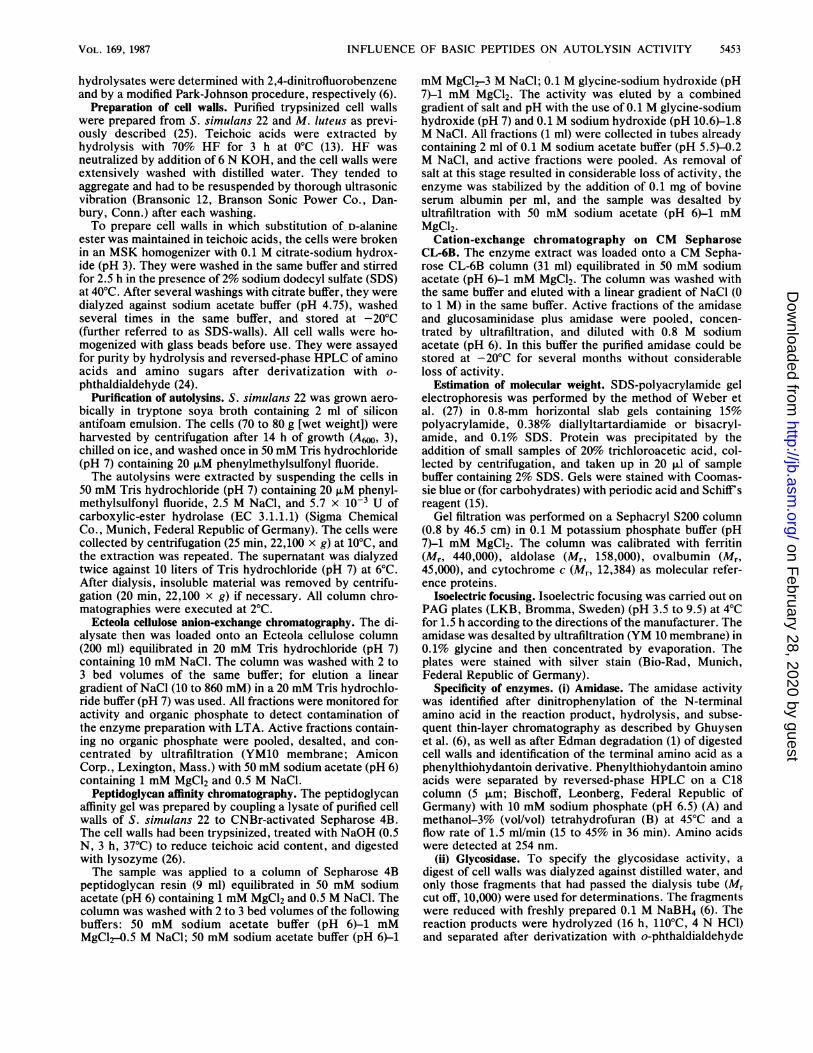
peptidoglycan resin and could only be eluted by alteration ofpH and ionic strength. The cation-!exphange chromatographyserved to separate the autolysins from each other (Fig. 1).The activity was eluted in two peaks. The enzyme that
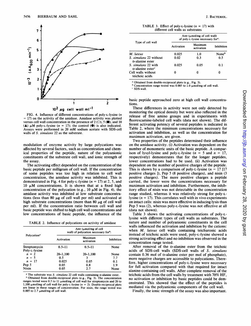
produced free amino groups in a digest of cell walls wasassociated with peak 1. In SDS-polyacrylamide gel electro-phoresis, this enzyme migrated at least in two bands (Mrs,-48,000 and -50,000), and in some preparation batches, upto six close bands were discernible (Afrs, -48,000 to-56,000) (Fig. 2). The higher-molecular-weight bands onlyoccurred when freshly prepared affinity gels were used.Attempts to separate the bands on an HPLC column byreversed-phase and ion-exchange chromatography or chro-matofocusing were unsuccessful. It seems possible thatrepeating units of the peptidoglycan were bound to theenzyme which could not be removed by SDS treatment, asthe enzyme could be stained with Schiffs reagent aftertreatment with periodic acid, which is specific for carbohy-drates.Peak 2 contained an enzyme that hydrolyzed the glycan
chain of the murein, liberating free reducing groups. It alsomigrated in several bands after peptidoglycan affinity chro-matography and displayed an Mr of -57,000 (Fig. 2).Mode of autolytic enzyme action. To identify the cleavage
site of the smaller enzyme, the amino groups released frommurein were labeled with 2,4-dinitrofluorobenzene (6). Afterhydrolysis and thin-layer chromatography, alanine was iden-tified as the N-terminal amino acid. An N-acetylmuramoyl-L-alanine amidase releases L-alanine, and a peptidase liber-ates D-alanine. To distinguish between amidase andpeptidase, the digest was subjected to Edman degradation,
E
ChU
In
cm
.-
C-
0
0 10 20 30 40 50frac tion
FIG. 1. Separation of the autolysins of S. simulans 22 on CMSepharose CL-6B. The enzymes were eluted by a gradient of 0 to 1M NaCl. Symbols: 0, total enzyme activity tested with M. luteuscell walls as substrate; *, release of reducing sugars from the cellwalls.
J. BACTERIOL.
on February 28, 2020 by guest
http://jb.asm.org/
Dow
nloaded from

INFLUENCE OF BASIC PEPTIDES ON AUTOLYSIN ACTIVITY
which again yielded alanine in cycle 1. In cycle 2, no aminoacid was released, which agrees with D-isoglutamine as thenext amino acid. In cell walls of staphylococci, D-iSo-glutamine is linked by its -y-carboxyl group to lysine, andonly its a-amino group reacts with phenylisothiocyanate (5).To verify this result, murein obtained after cycle 1 of Edmandegradation was again labeled with 2,4-dinitrofluoro-benzene, and glutamine (i.e., glutamate after acidic hydrol-ysis) was identified as second amino acid. This proved thatthe enzyme acts as N-acetylmuramoyl-L-alanine amidase(EC 3.5.1.28), which cleaves the bond between N-acetyl-muramic acid and D-alanine.The second enzyme liberated reducing groups. To specify
whether a glucosaminidase or a muramidase was involved,the location of the reducing group in N-acetylglucosamine orN-acetylmuramic acid had to be determined. Small frag-ments from peptidoglycan digests, mostly disaccharides,were reduced with sodium borohydride and characterizedafter hydrolysis and derivatization with o-phthaldialdehydeby chromatography on an HPLC reversed-phase C18 col-umn. Unreduced samples were shown to contain gluco-samine and muramic acid in a ratio of 1:1. After reduction,the glucosamine peak disappeared almost completely andwas replaced by a new peak of glucosaminitol, whereas theamount of muramic acid remained unchanged. This demon-strated that N-acetylglucosamine carried the reducing groupin the disaccharide that was produced by the glycosidase andthus characterized the enzyme as a ,-N-acetylgluco-saminidase.
Properties of N-acetylmuramoyl-L-alanine amidase. Themolecular weight of this enzyme as determined by SDS-polyacrylamide gel electrophoresis was -48,000. A value of-44,000 was calculated after gel filtration on Sephacryl 200under nondenaturing conditions, proving that the enzymeconsisted of one peptide chain only.When the isoelectric point was determined by isoelectric
focusing on PAG plates (pH 3.5 to 9.5), the enzyme focusedclose to the cathode strip. Therefore, it must be concludedthat the isoelectric point of the amidase is higher than 9.5.The amidase was active on S. simulans 22 cell walls, as wellas on purified cell walls of M. luteus ATCC 4698. The
- 92.5
* -66.2
-45.0
- 31.0
- 21.5
A B CFIG. 2. SDS-polyacrylamide gel electrophoresis of the auto-
lysins of S. simulans 22 in a 15% polyacrylamide gel. Lanes: A,P-N-acetylglucosaminidase (4 jig of protein); B, N-acetylmuramoyl-L-alanine amidase (25 p,g of protein); C, Mrs of standard peptidesgiven in thousands.
30
w-
w-
Ic 20._
aC
0
4
10-
p
C
S0
50 100
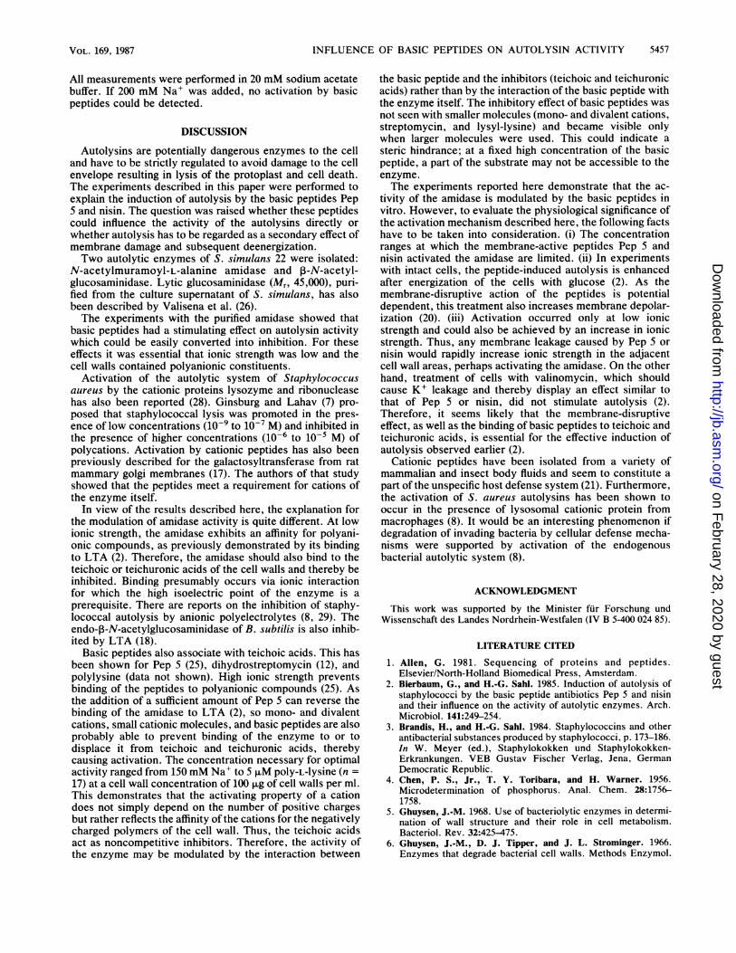
(mg cell woll m-11 )_ 1FIG. 3. Influence of polycations on the activity of amidase.
Shown is a double-reciprocal plot of SDS-cell wall hydrolysis. C,Control (20 mM sodium acetate, pH 6); S, addition of 0.1 mMstreptomycin; P, addition of 0.1 mM poly-L-lysine (n = 5).
amidase reached its maximum activity between pHs 5.5 and6.5.
Kinetics of amidase. To investigate whether the stimulationof autolysis by nisin and Pep 5 can be deduced from an effectof the basic peptides on the activity of the amidase, westudied the influence of inorganic and organic cations on thekinetics of this enzyme. The dependence of the reaction rateon substrate concentration corresponded to Michaelis-Menten kinetics. An apparent Km of 36.8 ,ug of cell walls perml and a corresponding Vmax of 4.79 mg of cell walls of S.simulans 22 per ml per min were determined for enzymeactivity in 20 mM sodium acetate (pH 6.0).
Influence of mono- and divalent cations. Addition ofmonovalent cations (Na+, K+) to the assay mixture in 20mM sodium acetate (pH 6.0) resulted in a mixed type ofactivation of the amidase; i.e., Km was lowered and Vmaxincreased. A broad range of maximum activity was obtainedbetween 100 and 150 mM KCl or NaCl.
Divalent cations activated the enzyme in the same way asmonovalent cations did, with maximum enzyme activitybetween 20 and 40 mM MgCl2. The amidase had no essentialrequirement for divalent cations because it was not inhibitedby the addition of EDTA. If ionic strength of the reactionmixture was raised beyond the range of maximum activity,the Km of the enzyme was decreased, whereas Vmax re-mained at its maximum value.
Influence of polycations. Addition of small organic cations(Mr, 300 to 500) with increasing numbers of positive charges,such as lysyl-lysine (5 mM) and streptomycin (0.1 mM),resulted only in a higher Vmax, whereas Km remained un-changed. Double-reciprocal plots were linear, as shown forstreptomycin in Fig. 3. However, when larger polycationssuch as poly-L-lysine (n = 5 or n = 17), nisin, and Pep 5 weretested, the pattern became more complex, as indicated bythe curved double-reciprocal plot for poly-L-lysine (n = 5) inFig. 3. Activation of the amidase occurred only in a certainrange of polycation concentration, and inhibition of theenzyme was observed when this range was exceeded. The
VOL. 169, 1987 5455
on February 28, 2020 by guest
http://jb.asm.org/
Dow
nloaded from
5456 BIERBAUM AND SAHL
/ _ _
TABLE 3. Effect of POIY-L-lysine (n = 17) withdifferent cell walls as substrates
Amt (,umol/mg of cell wall)of poly-L-lysine necessary fora:
Type of cell wallActivation Matimatuom Inhibition
M. luteus 0.025 1.0 NonebS. simulans 22 without 0.02 0.3 0.5
D-alanine esterS. simulans 22 with 0.025 0.05 0.1
D-alanine estercCell walls without 0 0 0
teichoic acids
aObtained from double-reciprocal plots (e.g., Fig. 3).b Concentration range tested was 0.005 to 1.0 ,umol/mg of cell wall.C SDS-wall.
1 2
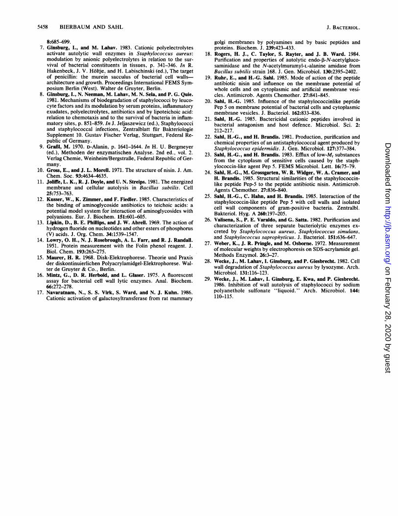
102 ,ig cell wall mV1IFIG. 4. Influence of different concentrations of pOly-L-lysine (n
= 17) on the activity of the amidase. Amidase activity was plottedversus cell wall concentration in the presence of 2 (0), 5 (i), and 10(A) JIM pOly-L-1ysine (n = 17); the control (0) is also indicated.Assays were performed in 20 mM sodium acetate with SDS-cellwalls of S. simulans 22 as the substrate.
modulation of enzyme activity by large polycations wasaffected by several factors, such as concentration and chem-ical properties of the peptide, nature of the polyanionicconstituents of the substrate cell wall, and ionic strength ofthe assay.The activating effect depended on the concentration of the
basic peptide per milligram of cell wall. If the concentrationof some peptides was too high in relation to cell wallconcentration, the amidase activity was inhibited. This isdemonstrated in Fig. 4 for poly-L-lysine (n = 17) at 2, 5, and10 ,uM concentrations. It is shown that at a fixed highconcentration of the polycation (e.g., 10 ,uM in Fig. 4), theamidase activity was inhibited at low substrate concentra-tions (less than 80 p.g of cell wall per ml) and activated athigh substrate concentrations (more than 80 ,ug of cell wallper ml). If the concentration ratio between cell wall andbasic peptide was shifted to high cell wall concentrations andlow concentrations of basic peptide, the influence of the
TABLE 2. Influence of polycations on activity of amidase
Amt (,mol/mg of cellwall) of polycation necessary forb:
PolycationaActivation Maximum Inhibition
Streptomycin 0.5-11 0.5-11 NonePoly-L-lysine
n = 2 20-1,100 20-1,100 Nonen = 5 0.5 3 7.7n = 17 0.025 0.05 0.1
Pep 5 0.05 0.8 1.9Nisin 0.05 2.7 None
a The substrate was S. simulans 22 cell walls containing D-alanine ester.b Obtained from double-reciprocal plots (e.g., Fig. 3). The concentration
ranges tested were 0.5 to 11 ,umol/mg of cell wall for streptomycin and 20 to1,100 ,umol/mg of cell wall for poly-L-lysine (n = 2). Double-reciprocal plotsare linear in these ranges of concentration. For nisin, the range tested was0.005 to 2.7 ,umol/mg of cell wall.
basic peptide approached zero at high cell wall concentra-tions.These differences in activity were not only detected by
monitoring the optical density but were also reflected in therelease of free amino groups and in experiments withfluorescamine-labeled cell walls (data not shown). The dif-ferent activating potency of several peptides is indicated inTable 2, where the minimum concentrations necessary foractivation and inhibition, as well as the concentration formaximum activation, are given.Two properties of the peptides determined their influence
on the amidase activity. (i) Activation was dependent on thenumber of monomeric units of the basic peptide. A compar-ison of lysyl-lysine and pOly-L-lysine (n = 5 and n = 17,respectively) demonstrates that for the longer peptides,lower concentrations had to be used. (ii) Activation wasdependent on the number of positive charges of the peptide.This is shown by a comparison of POIY-L-lysine (n = 17 [17positive charges ]), Pep 5 (8 positive charges), and nisin (3
positive charges). The more positive charges a peptidecarried, the lower were the concentrations necessary formaximum activation and inhibition. Furthermore, the inhib-itory effect of nisin was not detectable in the concentrationrange studied, whereas it was relatively strong for poly-L-lysine (n = 17). This correlates well with in vivo experimentson intact cells: nisin was more effective in inducing lysis thanPep 5 was (2), whereas poly-L-lysine was not effective at all(data not shown).
Table 3 shows the activating concentrations of poly-L-lysine with different types of cell walls as substrates. Thenature and number of polyanionic constituents in the cellwalls influenced the activation and inhibition by the cations:when M. luteus cell walls containing teichuronic acidsinstead of teichoic acids were used, poly-L-lysine showed astrong activating effect and no inhibition was observed in theconcentration range tested.
After removal of the D-alanine ester from the teichoicacids of SDS-cell walls (SDS-cell walls of S. simulanscontain 0.36 mol of D-alanine ester per mol of phosphate),more negative charges are accessible to polycations. There-fore, higher concentrations of poly-L-lysine were necessaryfor full activation compared with that required for intactalanine-containing cell walls. After complete removal of theteichoic acids from the cell walls by treatment with 70% HF,no activation or inhibition by basic peptides could be dem-onstrated. This showed that the effect of the peptides ismediated via the polyanionic components of the cell wall.
Finally, the ionic strength of the assay was also important.
7
6
5
3
2
._
cE(D4044
(Es0
J. BACTERIOL.
I
on February 28, 2020 by guest
http://jb.asm.org/
Dow
nloaded from
INFLUENCE OF BASIC PEPTIDES ON AUTOLYSIN ACTIVITY
All measurements were performed in 20 mM sodium acetatebuffer. If 200 mM Na+ was added, no activation by basicpeptides could be detected.
DISCUSSION
Autolysins are potentially dangerous enzymes to the celland have to be strictly regulated to avoid damage to the cellenvelope resulting in lysis of the protoplast and cell death.The experiments described in this paper were performed toexplain the induction of autolysis by the basic peptides Pep5 and nisin. The question was raised whether these peptidescould influence the activity of the autolysins directly orwhether autolysis has to be regarded as a secondary effect ofmembrane damage and subsequent deenergization.Two autolytic enzymes of S. simulans 22 were isolated:
N-acetylmuramoyl-L-alanine amidase and ,-N-acetyl-glucosaminidase. Lytic glucosaminidase (Mr, 45,000), puri-fied from the culture supernatant of S. simulans, has alsobeen described by Valisena et al. (26).The experiments with the purified amidase showed that
basic peptides had a stimulating effect on autolysin activitywhich could be easily converted into inhibition. For theseeffects it was essential that ionic strength was low and thecell walls contained polyanionic constituents.
Activation of the autolytic system of Staphylococcusaureus by the cationic proteins lysozyme and ribonucleasehas also been reported (28). Ginsburg and Lahav (7) pro-posed that staphylococcal lysis was promoted in the pres-ence of low concentrations (10-' to 10-7 M) and inhibited inthe presence of higher concentrations (10-6 to 10-5 M) ofpolycations. Activation by cationic peptides has also beenpreviously described for the galactosyltransferase from ratmammary golgi membranes (17). The authors of that studyshowed that the peptides meet a requirement for cations ofthe enzyme itself.
In view of the results described here, the explanation forthe modulation of amidase activity is quite different. At lowionic strength, the amidase exhibits an affinity for polyani-onic compounds, as previously demonstrated by its bindingto LTA (2). Therefore, the amidase should also bind to theteichoic or teichuronic acids of the cell walls and thereby beinhibited. Binding presumably occurs via ionic interactionfor which the high isoelectric point of the enzyme is aprerequisite. There are reports on the inhibition of staphy-lococcal autolysis by anionic polyelectrolytes (8, 29). Theendo-p-N-acetylglucosaminidase of B. subtilis is also inhib-ited by LTA (18).
Basic peptides also associate with teichoic acids. This hasbeen shown for Pep 5 (25), dihydrostreptomycin (12), andpolylysine (data not shown). High ionic strength preventsbinding of the peptides to polyanionic compounds (25). Asthe addition of a sufficient amount of Pep 5 can reverse thebinding of the amidase to LTA (2), so mono- and divalentcations, small cationic molecules, and basic peptides are alsoprobably able to prevent binding of the enzyme to or todisplace it from teichoic and teichuronic acids, therebycausing activation. The concentration necessary for optimalactivity ranged from 150 mM Na+ to 5 JIM poly-L-lysine (n =17) at a cell wall concentration of 100 ,ug of cell walls per ml.This demonstrates that the activating property of a cationdoes not simply depend on the number of positive chargesbut rather reflects the affinity of the cations for the negativelycharged polymers of the cell wall. Thus, the teichoic acidsact as noncompetitive inhibitors. Therefore, the activity ofthe enzyme may be modulated by the interaction between
the basic peptide and the inhibitors (teichoic and teichuronicacids) rather than by the interaction of the basic peptide withthe enzyme itself. The inhibitory effect of basic peptides wasnot seen with smaller molecules (mono- and divalent cations,streptomycin, and lysyl-lysine) and became visible onlywhen larger molecules were used. This could indicate asteric hindrance; at a fixed high concentration of the basicpeptide, a part of the substrate may not be accessible to theenzyme.The experiments reported here demonstrate that the ac-
tivity of the amidase is modulated by the basic peptides invitro. However, to evaluate the physiological significance ofthe activation mechanism described here, the following factshave to be taken into consideration. (i) The concentrationranges at which the membrane-active peptides Pep 5 andnisin activated the amidase are limited. (ii) In experimentswith intact cells, the peptide-induced autolysis is enhancedafter energization of the cells with glucose (2). As themembrane-disruptive action of the peptides is potentialdependent, this treatment also increases membrane depolar-ization (20). (iii) Activation occurred only at low ionicstrength and could also be achieved by an increase in ionicstrength. Thus, any membrane leakage caused by Pep 5 ornisin would rapidly increase ionic strength in the adjacentcell wall areas, perhaps activating the amidase. On the otherhand, treatment of cells with valinomycin, which shouldcause K+ leakage and thereby display an effect similar tothat of Pep 5 or nisin, did not stimulate autolysis (2).Therefore, it seems likely that the membrane-disruptiveeffect, as well as the binding of basic peptides to teichoic andteichuronic acids, is essential for the effective induction ofautolysis observed earlier (2).
Cationic peptides have been isolated from a variety ofmammalian and insect body fluids and seem to constitute apart of the unspecific host defense system (21). Furthermore,the activation of S. aureus autolysins has been shown tooccur in the presence of lysosomal cationic protein frommacrophages (8). It would be an interesting phenomenon ifdegradation of invading bacteria by cellular defense mecha-nisms were supported by activation of the endogenousbacterial autolytic system (8).
ACKNOWLEDGMENT
This work was supported by the Minister fur Forschung undWissenschaft des Landes Nordrhein-Westfalen (IV B 5-400 024 85).
LITERATURE CITED
1. Allen, G. 1981. Sequencing of proteins and peptides.Elsevier/North-Holland Biomedical Press, Amsterdam.
2. Bierbaum, G., and H.-G. Sahl. 1985. Induction of autolysis ofstaphylococci by the basic peptide antibiotics Pep 5 and nisinand their influence on the activity of autolytic enzymes. Arch.Microbiol. 141:249-254.
3. Brandis, H., and H.-G. Sahl. 1984. Staphylococcins and otherantibacterial substances produced by staphylococci, p. 173-186.In W. Meyer (ed.), Staphylokokken und Staphylokokken-Erkrankungen. VEB Gustav Fischer Verlag, Jena, GermanDemocratic Republic.
4. Chen, P. S., Jr., T. Y. Toribara, and H. Warner. 1956.Microdetermination of phosphorus. Anal. Chem. 28:1756-1758.
5. Ghuysen, J.-M. 1968. Use of bacteriolytic enzymes in determi-nation of wall structure and their role in cell metabolism.Bacteriol. Rev. 32:425-475.
6. Ghuysen, J.-M., D. J. Tipper, and J. L. Strominger. 1966.Enzymes that degrade bacterial cell walls. Methods Enzymol.
5457VOL. 169, 1987
on February 28, 2020 by guest
http://jb.asm.org/
Dow
nloaded from
5458 BIERBAUM AND SAHL
8:685-699.7. Ginsburg, I., and M. Lahav. 1983. Cationic polyelectrolytes
activate autolytic wall enzymes in Staphylococcus aureus:modulation by anionic polyelectrolytes in relation to the sur-vival of bacterial constituents in tissues, p. 341-346. In R.Hakenbeck, J. V. Holtje, and H. Labischinski (ed.), The targetof penicillin: the murein sacculus of bacterial cell walls-architecture and growth. Proceedings International FEMS Sym-posium Berlin (West). Walter de Gruyter, Berlin.
8. Ginsburg, I., N. Neeman, M. Lahav, M. N. Sela, and P. G. Quie.1981. Mechanisms of biodegradation of staphylococci by leuco-cyte factors and its modulation by serum proteins, inflammatoryexudates, polyelectrolytes, antibiotics and by lipoteichoic acid:relation to chemotaxis and to the survival of bacteria in inflam-matory sites, p. 851-859. In J. Jeljaszewicz (ed.), Staphylococciand staphylococcal infections, Zentralblatt fur BakteriologieSupplement 10. Gustav Fischer Verlag, Stuttgart, Federal Re-public of Germany.
9. GraBl, M. 1970. D-Alanin, p. 1641-1644. In H. U. Bergmeyer(ed.), Methoden der enzymatischen Analyse. 2nd ed., vol. 2.Verlag Chemie, Weinheim/BergstraBe, Federal Republic of Ger-many.
10. Gross, E., and J. L. Morell. 1971. The structure of nisin. J. Am.Chem. Soc. 93:4634-4635.
11. Joliffe, L. K., R. J. Doyle, and U. N. Streips. 1981. The energizedmembrane and cellular autolysis in Bacillus subtilis. Cell25:753-763.
12. Kusser, W., K. Zimmer, and F. Fiedler. 1985. Characteristics ofthe binding of aminoglycoside antibiotics to teichoic acids: apotential model system for interaction of aminoglycosides withpolyanions. Eur. J. Biochem. 151:601-605.
13. Lipkin, D., B. E. Phillips, and J. W. Abrell. 1969. The action ofhydrogen fluoride on nucleotides and other esters of phosphorus(V) acids. J. Org. Chem. 34:1539-1547.
14. Lowry, 0. H., N. J. Rosebrough, A. L. Farr, and R. J. Randall.1951. Protein measurement with the Folin phenol reagent. J.Biol. Chem. 193:265-275.
15. Maurer, H. R. 1968. Disk-Elektrophorese. Theorie und Praxisder diskontinuierlichen Polyacrylamidgel-Elektrophorese. Wal-ter de Gruyter & Co., Berlin.
16. Mintz, G., D. R. Herbold, and L. Glaser. 1975. A fluorescentassay for bacterial cell wall lytic enzymes. Anal. Biochem.66:272-278.
17. Navaratnam, N., S. S. Virk, S. Ward, and N. J. Kuhn. 1986.Cationic activation of galactosyltransferase from rat mammary
golgi membranes by polyamines and by basic peptides andproteins. Biochem. J. 239:423-433.
18. Rogers, H. J., C. Taylor, S. Rayter, and J. B. Ward. 1984.Purification and properties of autolytic endo-,-N-acetylgluco-saminidase and the N-acetylmuramyl-L-alanine amidase fromBacillus subtilis strain 168. J. Gen. Microbiol. 130:2395-2402.
19. Ruhr, E., and H.-G. Sahl. 1985. Mode of action of the peptideantibiotic nisin and influence on the membrane potential ofwhole cells and on cytoplasmic and artificial membrane vesi-cles. Antimicrob. Agents Chemother. 27:841-845.
20. Sahl, H.-G. 1985. Influence of the staphylococcinlike peptidePep 5 on membrane potential of bacterial cells and cytoplasmicmembrane vesicles. J. Bacteriol. 162:833-836.
21. Sahl, H.-G. 1985. Bactericidal cationic peptides involved inbacterial antagonism and host defence. Microbiol. Sci. 2:212-217.
22. Sahl, H.-G., and H. Brandis. 1981. Production, purification andchemical properties of an antistaphylococcal agent produced byStaphylococcus epidermidis. J. Gen. Microbiol. 127:377-384.
23. Sahl, H.-G., and H. Brandis. 1983. Efflux of low-Mr substancesfrom the cytoplasm of sensitive cells caused by the staph-ylococcin-like agent Pep 5. FEMS Microbiol. Lett. 16:75-79.
24. Sahl, H.-G., M. Grossgarten, W. R. Widger, W. A. Cramer, andH. Brandis. 1985. Structural similarities of the staphylococcin-like peptide Pep-5 to the peptide antibiotic nisin. Antimicrob.Agents Chemother. 27:836-840.
25. Sahl, H.-G., C. Hahn, and H. Brandis. 1985. Interaction of thestaphylococcin-like peptide Pep 5 with cell walls and isolatedcell wall components of gram-positive bacteria. Zentralbl.Bakteriol. Hyg. A 260:197-205.
26. Valisena, S., P. E. Varaldo, and G. Satta. 1982. Purification andcharacterization of three separate bacteriolytic enzymes ex-creted by Staphylococcus aureus, Staphylococcus simulans,and Staphylococcus saprophyticus. J. Bacteriol. 151:636-647.
27. Weber, K., J. R. Pringle, and M. Osborne. 1972. Measurementof molecular weights by electrophoresis on SDS-acrylamide gel.Methods Enzymol. 26:3-27.
28. Wecke, J., M. Lahav, I. Ginsburg, and P. Giesbrecht. 1982. Cellwall degradation of Staphylococcus aureus by lysozyme. Arch.Microbiol. 131:116-123.
29. Wecke, J., M. Lahav, I. Ginsburg, E. Kwa, and P. Giesbrecht.1986. Inhibition of wall autolysis of staphylococci by sodiumpolyanethole sulfonate "liquoid." Arch. Microbiol. 144:110-115.
J. BACTERIOL.
on February 28, 2020 by guest
http://jb.asm.org/
Dow
nloaded from