bmi booklet
DESCRIPTION
Brain Mind Institute BookletTRANSCRIPT
SCHOOL OF LIFE SCIENCESWhere life sciences meet technology
BRAIN MIND INSTITUTEBLUE BRAIN PROJECT
- 1 -
Brain Mind Institute. ...............................................................................................................2
Blue Brain Project. .................................................................................................................3
Teaching and NCCR ...............................................................................................................4
Neurodegenerative studies laboratory. ................................................................................5
Laboratory of cognitive neurosciences. ..............................................................................6
Laboratory of molecular and cellular biology of Alzheimer’s disease. .............................7
Laboratory of computational neuroscience. ........................................................................8
Laboratory of psychophysics. ..............................................................................................9
Laboratory of Molecular and Chemical Biology of Neurodegeneration ..........................10
Laboratory of functional neurogenomics. .........................................................................11
Laboratory of neuroenergetics and cellular dynamics. ....................................................12 Laboratory of neural microcircuitry....................................................................................13
Laboratory of molecular neurodegenerative research. ....................................................14
Laboratory of sensory processing. ....................................................................................15
Laboratory of behavioral genetics. .....................................................................................16
Laboratory of synaptic mechanisms. .................................................................................17
Social cognitive neurosciences...........................................................................................18
Overview of the Blue Brain Project ...................................................................................19
Affiliated groups and Artists-in-lab....................................................................................20
INTRODUCTION----------------------------------------------------------------------------------------------------------------------------------------Magistretti Pierre J.
Markram Henry
Teaching and NCCR
LABORATORIES----------------------------------------------------------------------------------------------------------------------------------------Aebischer Patrick
Blanke Olaf
Fraering Patrick
Gerstner Wulfram
Herzog Michael
Lashuel Hilal A.
Luthi-Carter Ruth
Magistretti Pierre J.
Markram Henry
Moore Darren J.
Petersen Carl
Sandi Carmen
Schneggenburger Ralf
RESEARCH GROUP----------------------------------------------------------------------------------------------------------------------------------------Hadjikhani Nouchine
BLUE BRAIN PROJECT (BBP)----------------------------------------------------------------------------------------------------------------------------------------Markram Henry
AFFILIATED GROUPS AND ARTISTS-IN-LAB----------------------------------------------------------------------------------------------------------------------------------------
CONTENTS
- 2 -
Pierre J. MagistrettiDirector
brain mind institute
The mission of the Brain Mind Institute (BMI) is to understand the fundamental principles of brain function in health and disease, by using and developing unique experimental, theoretical, technological and computational approaches. The scientific challenge addressed by the BMI consists in connecting different levels of analysis of brain activity, such that cog-nitive functions can be understood as a manifestation of specific brain processes; specific brain processes as emerging from the collective activity of thousands of cells and synapses; synaptic and neuronal activity in turn as emerging proper-ties of the biophysical and molecular mechanisms of cellular compartments.
Understanding information processing in the brain and its higher emerging properties is arguably one of the major chal-lenges in the life sciences. Research at the BMI focuses on three main areas: i) Molecular neurobiology and mechanisms of neurodegeneration ii) Molecular and cellular mechanisms of synapse and microcircuit function up to the behavioural level and including metabolic aspects; iii) Sensory perception and cognition in humans. In all areas, the BMI strives to inte-grate knowledge gained by multidisciplinary approaches and across different disciplines and research laboratories. Finally, underlying all levels of analysis, research at BMI is characterized by a sustained interest in pathological processes.
In order to achieve these scientific goals, the Brain Mind Institute benefits from a unique academic environment:
An institute organized as a network of independent laboratories reflecting complementary technological approaches; • each laboratory collaborates with several others within the institute in addition to cross-disciplinary interactions on campus.A campus that stands out as a premier technological university in engineering, computer science and basic sci-• ences.An intimate collaboration with the Blue Brain Project which stands out as one of the most challenging neuroscience • simulation and databasing projects worldwide.A proximity to and joint affiliations of our faculty with top university hospitals in Lausanne and Geneva in particular for • projects related to cognition and neurodegenerative diseases.A new initiative in neuroprosthetics to which the BMI is strongly committed that will further the collaboration with engi-• neering sciences by a host of inspiring common projects.
A feature of the Brain Mind Institute is that several faculty members have strong expertise in physics or mathematics; this holds not only for theoretical but also for experimental neuroscience. In this way the Brain Mind Institute reflects the mis-sion of the School of Life Science: to provide a life science curriculum with a strong emphasis on quantitative approaches. As far as teaching is concerned, the BMI Faculty is committed to provide a comprehensive and formal training in neurosci-ence from the undergraduate to the graduate levels.
BMI
- 3 -
Henry MarkramDirector
The Blue Brain Project (BBP) began in July 2005 as a collaboration between Professor Henry Markram from the Brain Mind Institute at the Ecole Polytechnique Fédérale de Lausanne (EPFL) and International Business Machines (IBM), aimed at modeling the neocortical column. The neocortical column represents the basic functional unit of the cerebral cortex in mammals that underlies nearly all sensory and cognitive processing. These units are repeated millions of times across the cortex, with the basic structure remaining the same from the mouse to man. From its origins – IBM’s BlueGene/L super-computer and 10 years of experimental data from Professor Markram’s laboratory and – the project has grown to include an international multidisciplinary team of experimentalists, modelers and computer scientists.
KEYWORDSHIGH PERFORMANCE COMPUTING
SIMULATION-BASED RESEARCHREVERSE ENGINEERING
NEOCORTEX
By the end of 2007 a simulation-based research process has been developed to generate a cellular-level model of a 2-week-old rat somatosensory neocortex based on extensive experimental data. The 10,000 threedimensional model neurons were placed in the neocortical volume (550 μm in diameter by 1,200 μm high) and arranged in 6 layers according to their morphological and electrical properties. The cells have model ion channels, determined by gene expression data, distributed along their membrane surfaces to reproduce the experimentally observed diversity of electrical firing proper-ties seen in cortical pyramidal cells and interneurons. Potential synaptic locations between cells are identified through a touch detection process that searches for axons and dendrites in proximity to each other. Functional synapses are placed according to experimental observations of the probability of synaptic interactions between different cell types. The entire circuit, once constructed, is run through a barrage of tests to calibrate the network circuitry according to experimental data. This includes systematic checks of the ion channels, electrical behavior of neurons, composition of the column, patterns of connectivity and behavior of monosynaptic and polysynaptic pathways. The model network is then run through a series of test protocols evaluating its capacity for replicating network level phenomena.
FIND MORE ON PAGE 19 !
blue brain project
- 4 -
TEACHING AND NCCRTeaching - Master
The Brain Mind Institute Faculty organizes a neuroscience curriculum as part of the master in Life Sciences and Technology. The curriculum includes a systematic introduction to the neurosciences ranging from molecular neu-roscience (Neuroscience I) to electrophysiology and cellular neuroscience (Neuroscience II) and cognitive neu-roscience (Neuroscience III); students can choose from a long list of optional courses related to the neurosciences (Fundamentals of Biomedical Imaging, Brain Imaging, Neural Networks and Biological Modeling, Pharmacology and Pharmacokinetics) taught by the BMI faculty, as well as Theory and Signal Processing related courses. There is also a range of optional lab work. The master program finishes with a master thesis in one of the laboratories of the BMI.
For further information see:Neuroscience Curriculum http://bmi.epfl.ch/page-9290.html and http://ssv.epfl.ch/page-33502-en.htmlMaster in Life Sciences and Technology http://ssv.epfl.ch/page-25109-en.html
Teaching - Phd
The Faculty of the Brain Mind Institute is responsible for the Doctoral Program in the Neurosciences.
For further information see: http://phd.epfl.ch/page-19787.html
NCCR - Synapsy “The synaptic bases of mental diseases”
In a strong nationwide competition the EPFL has received in 2010 nationwide recognition by the award of two big grants for cutting edge research as a ‘National Center for competence in Research’ (NCCR). One of these NCCR is in the field of Synaptic Bases of Mental Diseases and lead by the Brain Mind Institute, in collaboration with the universities of Lausanne and Geneva. The NCCR will run until 2014 with a funding of about 5 Mio per year. NCCR Synapsy is an instrument of the Swiss National Science Foundation involving high-end research teams and com-petencies from Universities of Geneva, Lausanne and Basel, EPFL as the leading house, as well as clinicians from the Departments of Psychiatry at Geneva and Lausanne. NCCR Synapsy aims to discover the synaptic mechanisms from the molecular to network and integrative aspects underlying mental and cognitive diseases, such as depression, addiction, anxiety disorders or development disorders. From a long-term perspective, scientific advances should benefit directly to the patient, by elaborating novel therapeutic approaches and eventually curative treatments, in order to improve its quality of life.
For further information see: http://www.nccr-synapsy.ch
- 5 -
Full ProfessorPatrick Aebischer
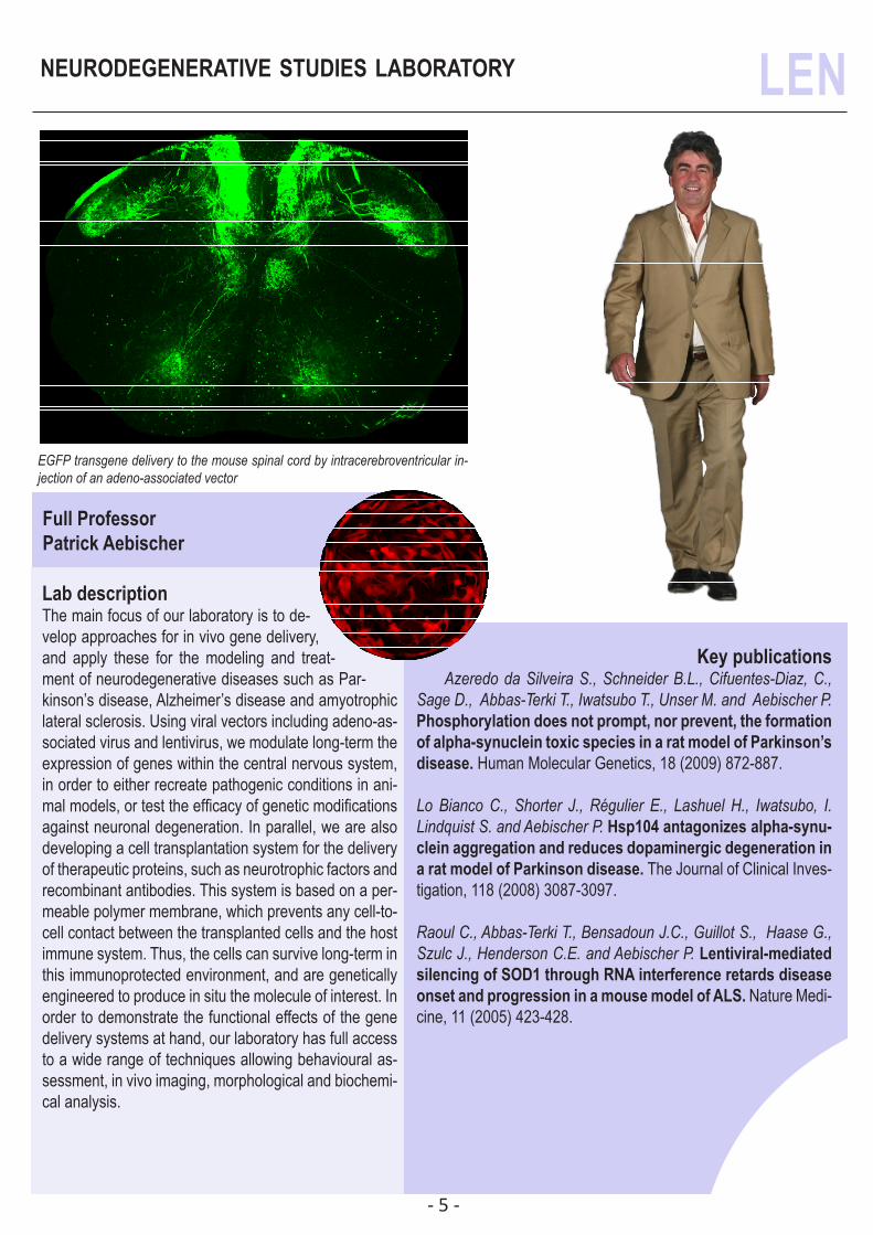
Lab description The main focus of our laboratory is to de-velop approaches for in vivo gene delivery, and apply these for the modeling and treat-ment of neurodegenerative diseases such as Par-kinson’s disease, Alzheimer’s disease and amyotrophic lateral sclerosis. Using viral vectors including adeno-as-sociated virus and lentivirus, we modulate long-term the expression of genes within the central nervous system, in order to either recreate pathogenic conditions in ani-mal models, or test the efficacy of genetic modifications against neuronal degeneration. In parallel, we are also developing a cell transplantation system for the delivery of therapeutic proteins, such as neurotrophic factors and recombinant antibodies. This system is based on a per-meable polymer membrane, which prevents any cell-to-cell contact between the transplanted cells and the host immune system. Thus, the cells can survive long-term in this immunoprotected environment, and are genetically engineered to produce in situ the molecule of interest. In order to demonstrate the functional effects of the gene delivery systems at hand, our laboratory has full access to a wide range of techniques allowing behavioural as-sessment, in vivo imaging, morphological and biochemi-cal analysis.
Key publicationsAzeredo da Silveira S., Schneider B.L., Cifuentes-Diaz, C.,
Sage D., Abbas-Terki T., Iwatsubo T., Unser M. and Aebischer P. Phosphorylation does not prompt, nor prevent, the formation of alpha-synuclein toxic species in a rat model of Parkinson’s disease. Human Molecular Genetics, 18 (2009) 872-887.
Lo Bianco C., Shorter J., Régulier E., Lashuel H., Iwatsubo, I. Lindquist S. and Aebischer P. Hsp104 antagonizes alpha-synu-clein aggregation and reduces dopaminergic degeneration in a rat model of Parkinson disease. The Journal of Clinical Inves-tigation, 118 (2008) 3087-3097.
Raoul C., Abbas-Terki T., Bensadoun J.C., Guillot S., Haase G., Szulc J., Henderson C.E. and Aebischer P. Lentiviral-mediated silencing of SOD1 through RNA interference retards disease onset and progression in a mouse model of ALS. Nature Medi-cine, 11 (2005) 423-428.
LEN neurodegenerative studies laboratory
EGFP transgene delivery to the mouse spinal cord by intracerebroventricular in-jection of an adeno-associated vector
- 6 -
laboratory of cognitive neurosciences LNCO
Associate ProfessorOlaf Blanke
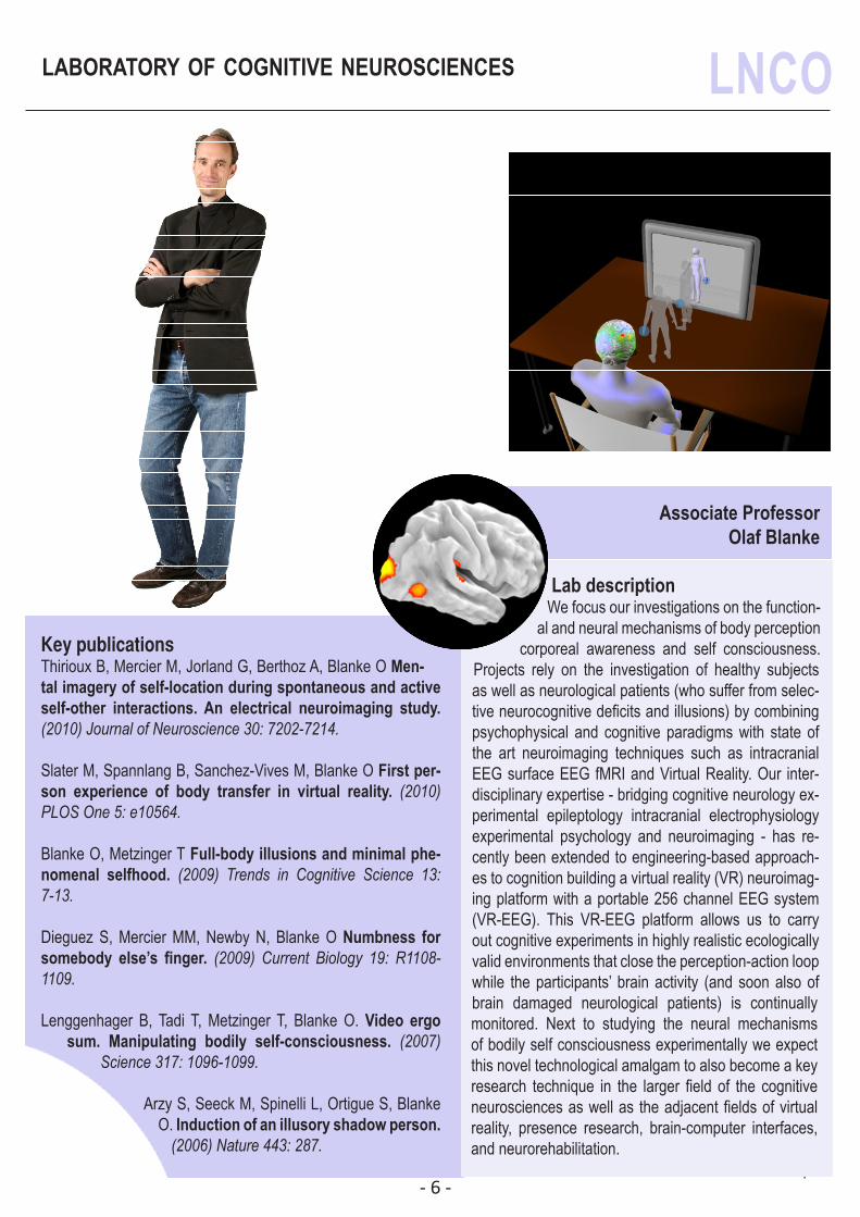
Lab descriptionWe focus our investigations on the function-
al and neural mechanisms of body perception corporeal awareness and self consciousness.
Projects rely on the investigation of healthy subjects as well as neurological patients (who suffer from selec-tive neurocognitive deficits and illusions) by combining psychophysical and cognitive paradigms with state of the art neuroimaging techniques such as intracranial EEG surface EEG fMRI and Virtual Reality. Our inter-disciplinary expertise - bridging cognitive neurology ex-perimental epileptology intracranial electrophysiology experimental psychology and neuroimaging - has re-cently been extended to engineering-based approach-es to cognition building a virtual reality (VR) neuroimag-ing platform with a portable 256 channel EEG system (VR-EEG). This VR-EEG platform allows us to carry out cognitive experiments in highly realistic ecologically valid environments that close the perception-action loop while the participants’ brain activity (and soon also of brain damaged neurological patients) is continually monitored. Next to studying the neural mechanisms of bodily self consciousness experimentally we expect this novel technological amalgam to also become a key research technique in the larger field of the cognitive neurosciences as well as the adjacent fields of virtual reality, presence research, brain-computer interfaces, and neurorehabilitation.
Key publicationsThirioux B, Mercier M, Jorland G, Berthoz A, Blanke O Men-tal imagery of self-location during spontaneous and active self-other interactions. An electrical neuroimaging study. (2010) Journal of Neuroscience 30: 7202-7214.
Slater M, Spannlang B, Sanchez-Vives M, Blanke O First per-son experience of body transfer in virtual reality. (2010) PLOS One 5: e10564.
Blanke O, Metzinger T Full-body illusions and minimal phe-nomenal selfhood. (2009) Trends in Cognitive Science 13: 7-13.
Dieguez S, Mercier MM, Newby N, Blanke O Numbness for somebody else’s finger. (2009) Current Biology 19: R1108-1109.
Lenggenhager B, Tadi T, Metzinger T, Blanke O. Video ergo sum. Manipulating bodily self-consciousness. (2007)
Science 317: 1096-1099.
Arzy S, Seeck M, Spinelli L, Ortigue S, Blanke O. Induction of an illusory shadow person.
(2006) Nature 443: 287.
PICTURE
- 7 -
laboratory of molecular & cellular biology of CMSN
Tenure-Track Assistant ProfessorPatrick Fraering
Key publicationsF. Wu, C. Schweizer, N. Rudinskiy, D. M. Taylor, A. Kazantsev, R. Lu-thi-Carter and PC. Fraering. Novel γ-secretase inhibitors uncover a common nucleotide-binding site in JAK3, SIRT2 and PS1. (2010) FASEB, Jul;24(7):2464-74
Osenkowski P, Li H, Ye W, Li D, Aeschbach L, Fraering PC, Wolfe MS, Selkoe DJ, Li H. Cryoelectron Microscopy Structure of Purified gamma-Secretase at 12 A Resolution. (2009) J Mol Biol 385: 642-652.
Kukar TL, Ladd TB, Bann MA, Fraering PC, et al. Substrate-targeting gamma-secretase modulators. (2008) Nature. Jun 12;453(7197):925-9.
M. Cacquevel, L. Aeschbach, P. Osenkowski, D. Li, W. Ye, MS. Wolfe, H. Li, DJ. Selkoe and PC. Fraering. Rapid purification of active γ-secretase, an intramembrane-cleaving protease involved in Al-zheimer’s disease. (2008) Journal of Neurochemistry, 104(1): 210-20.
VK. Lazarov*, PC. Fraering*, W. Ye, MS. Wolfe, DJ. Selkoe and H. Li. Electron microscopic structure of purified, ac-tive γ-secretase reveals an aqueous intramembrane chamber and two pores. (2006) Proc Natl Acad Sci USA, 103:6889-6894.
Lab descriptionWe focus our research on the molecular and biological mechanisms of Alzheimer’s disease (AD), by far the most frequent age relat-ed neurological disorder that impairs memory, thinking and behavior. Studies of the pathological changes that characterize AD have implicated the amyloid-β peptides (Aβ) as potential causative agents in the pathogenesis of AD. Because γ-secretase, the founding member of an emerging class of intramembrane-cleaving proteases, catalyzes the final cleavage during the neuronal pro-duction of the toxic Aβ peptides, partially inhibiting its enzymatic activity is an attractive therapeutic strategy to safely treat AD. Toward advancing the biochemistry of γ-secretase with attendant therapeutic implications, the main goals of our laboratory are to understand (1) the structure and function of the protease complex and (2) how mutations in the presenilin genes causing familial, early-onset AD, alter its activity and lead to neurode-generation and AD. Besides providing details about the intramembrane proteolysis of membrane proteins, such understanding ultimately offers to the laboratory prom-ising targets for developing new therapeutic strategies and new drugs having the potential to slow down the progression of AD. The efficacy and potency of these are tested in controlled cell-free and cell-based assays, and in vivo by using AD transgenic models.
alzheimer’s disease - merck serono chair in neuroscience
- 8 -
laboratory of computational neuroscience LCN
Full ProfessorWulfram Gerstner
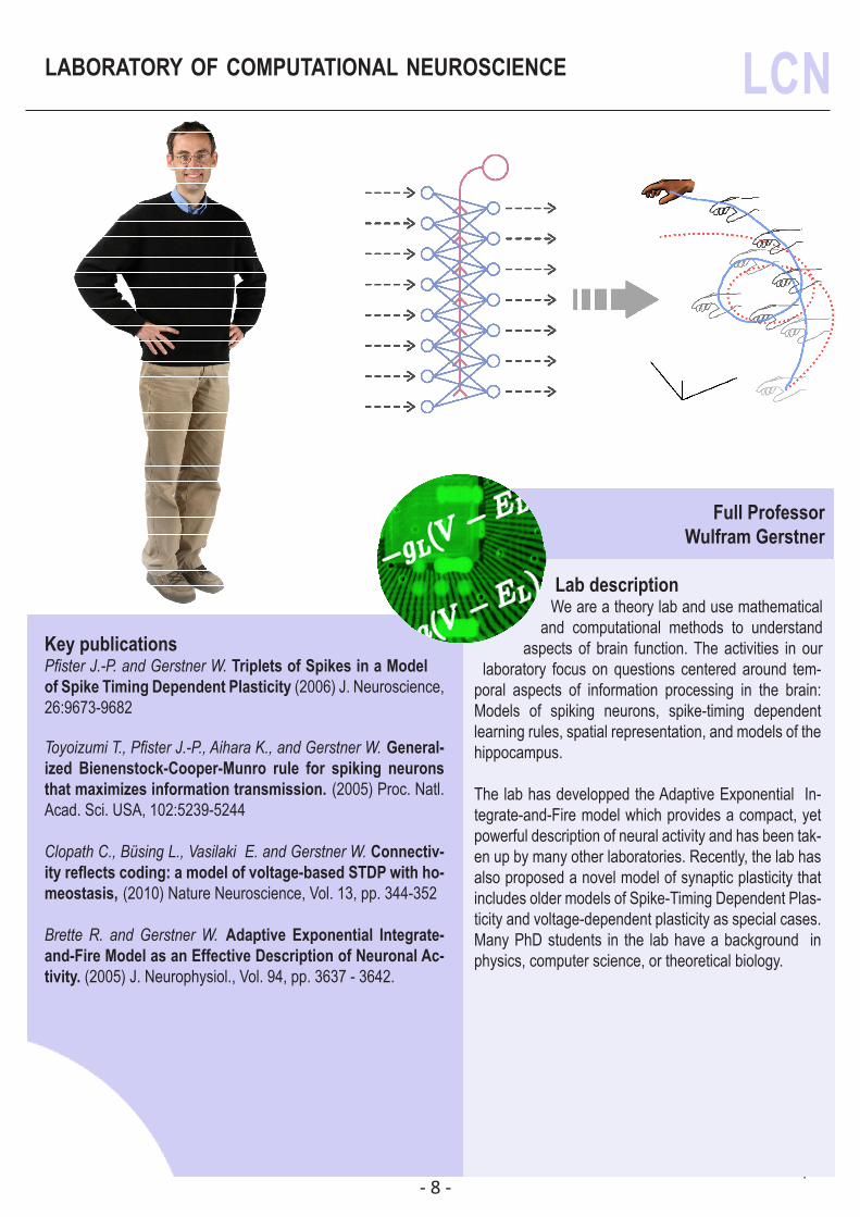
Lab descriptionWe are a theory lab and use mathematical
and computational methods to understand aspects of brain function. The activities in our
laboratory focus on questions centered around tem-poral aspects of information processing in the brain: Models of spiking neurons, spike-timing dependent learning rules, spatial representation, and models of the hippocampus.
The lab has developped the Adaptive Exponential In-tegrate-and-Fire model which provides a compact, yet powerful description of neural activity and has been tak-en up by many other laboratories. Recently, the lab has also proposed a novel model of synaptic plasticity that includes older models of Spike-Timing Dependent Plas-ticity and voltage-dependent plasticity as special cases. Many PhD students in the lab have a background in physics, computer science, or theoretical biology.
Key publicationsPfister J.-P. and Gerstner W. Triplets of Spikes in a Model of Spike Timing Dependent Plasticity (2006) J. Neuroscience, 26:9673-9682
Toyoizumi T., Pfister J.-P., Aihara K., and Gerstner W. General-ized Bienenstock-Cooper-Munro rule for spiking neurons that maximizes information transmission. (2005) Proc. Natl. Acad. Sci. USA, 102:5239-5244
Clopath C., Büsing L., Vasilaki E. and Gerstner W. Connectiv-ity reflects coding: a model of voltage-based STDP with ho-meostasis, (2010) Nature Neuroscience, Vol. 13, pp. 344-352
Brette R. and Gerstner W. Adaptive Exponential Integrate-and-Fire Model as an Effective Description of Neuronal Ac-tivity. (2005) J. Neurophysiol., Vol. 94, pp. 3637 - 3642.
PICTURE
- 9 -
laboratory of psychophysics LPSY
Associate ProfessorMichael Herzog
Key publicationsTartaglia E.M., Bamert L., Mast F.W., Herzog M.H. Human
perceptual learning by mental imagery. (2009) Current Biology, 19(24), 2081-2085.
Plomp G., Mercier M.R., Otto T.U., Blanke O., Herzog M.H. Non-retinotopic feature integration decreases response-locked brain activity as revealed by electrical neuroimaging. (2009) NeuroImage, 48, 405-414.
Scharnowski F., Rüter J., Jolij J., Hermens F., Kammer T., Herzog M.H. Long lasting modulation of feature integration by tran-scranial magnetic stimulation. (2009) Journal of Vision, 9(6):1, 1-10. (see also: Faculty of 1000 Biology Evaluation).
Boi M., Öğmen H., Krummenacher J., Otto T.U., Herzog M.H. A (fascinating) litmus test for human retino- vs. non-retinotopic processing. (2009) Journal of Vision, 9(13):5, 1-11.
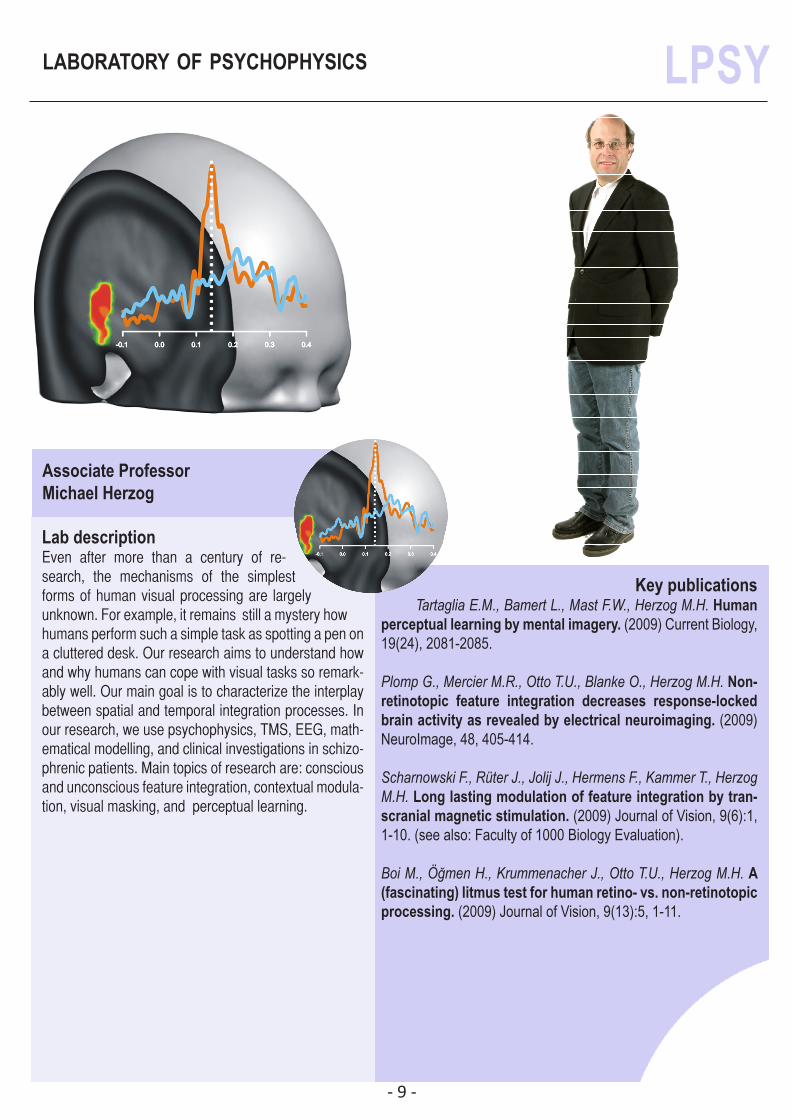
Lab descriptionEven after more than a century of re-search, the mechanisms of the simplest forms of human visual processing are largely unknown. For example, it remains still a mystery how humans perform such a simple task as spotting a pen on a cluttered desk. Our research aims to understand how and why humans can cope with visual tasks so remark-ably well. Our main goal is to characterize the interplay between spatial and temporal integration processes. In our research, we use psychophysics, TMS, EEG, math-ematical modelling, and clinical investigations in schizo-phrenic patients. Main topics of research are: conscious and unconscious feature integration, contextual modula-tion, visual masking, and perceptual learning.
- 10 -
laboratory of molecular and chemical biology LMCBN
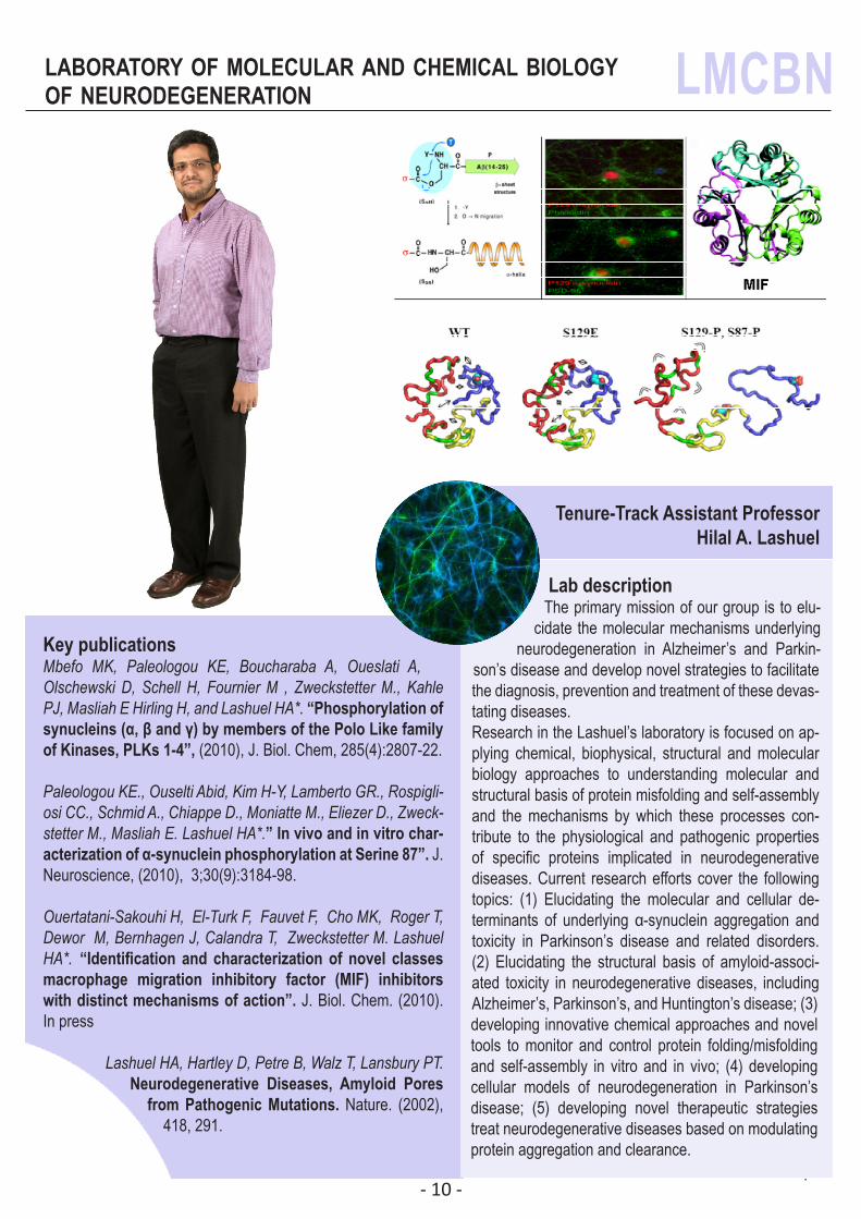
Lab descriptionThe primary mission of our group is to elu-
cidate the molecular mechanisms underlying neurodegeneration in Alzheimer’s and Parkin-
son’s disease and develop novel strategies to facilitate the diagnosis, prevention and treatment of these devas-tating diseases.Research in the Lashuel’s laboratory is focused on ap-plying chemical, biophysical, structural and molecular biology approaches to understanding molecular and structural basis of protein misfolding and self-assembly and the mechanisms by which these processes con-tribute to the physiological and pathogenic properties of specific proteins implicated in neurodegenerative diseases. Current research efforts cover the following topics: (1) Elucidating the molecular and cellular de-terminants of underlying α-synuclein aggregation and toxicity in Parkinson’s disease and related disorders. (2) Elucidating the structural basis of amyloid-associ-ated toxicity in neurodegenerative diseases, including Alzheimer’s, Parkinson’s, and Huntington’s disease; (3) developing innovative chemical approaches and novel tools to monitor and control protein folding/misfolding and self-assembly in vitro and in vivo; (4) developing cellular models of neurodegeneration in Parkinson’s disease; (5) developing novel therapeutic strategies treat neurodegenerative diseases based on modulating protein aggregation and clearance.
Key publicationsMbefo MK, Paleologou KE, Boucharaba A, Oueslati A, Olschewski D, Schell H, Fournier M , Zweckstetter M., Kahle PJ, Masliah E Hirling H, and Lashuel HA*. “Phosphorylation of synucleins (α, β and γ) by members of the Polo Like family of Kinases, PLKs 1-4”, (2010), J. Biol. Chem, 285(4):2807-22.
Paleologou KE., Ouselti Abid, Kim H-Y, Lamberto GR., Rospigli-osi CC., Schmid A., Chiappe D., Moniatte M., Eliezer D., Zweck-stetter M., Masliah E. Lashuel HA*.” In vivo and in vitro char-acterization of α-synuclein phosphorylation at Serine 87”. J. Neuroscience, (2010), 3;30(9):3184-98.
Ouertatani-Sakouhi H, El-Turk F, Fauvet F, Cho MK, Roger T, Dewor M, Bernhagen J, Calandra T, Zweckstetter M. Lashuel HA*. “Identification and characterization of novel classes macrophage migration inhibitory factor (MIF) inhibitors with distinct mechanisms of action”. J. Biol. Chem. (2010). In press
Lashuel HA, Hartley D, Petre B, Walz T, Lansbury PT. Neurodegenerative Diseases, Amyloid Pores
from Pathogenic Mutations. Nature. (2002), 418, 291.
Tenure-Track Assistant ProfessorHilal A. Lashuel
of neurodegeneration
- 11 -
laboratory of functional neurogenomics LNGF
Tenure-Track Assistant ProfessorRuth Luthi-Carter
Key publicationsKuhn A., Hodges A., Strand. A., Hunter J.M., Goldstein D.R.,
Aragaki A.K., Hughes G., Sengstag T., Kooperberg C., Cha J.-H.J., Dunnet S.B., Ferrante R.J., Albin R., Bates G., Shelbourne P., Detloff P., Delorenzi M., Augood S.J., Faull R.L.M., Olson J.M., Jones L. and Luthi-Carter R. Mutant huntingtin’s effects on stri-atal gene expression in mice recapitulate changes observed in human Huntington’s disease brain and do not differ with mutant huntingtin length or wild-type huntingtin dosage. (2007) Hum. Mol. Genet. 16:1845-1861
Luthi-Carter, R., Taylor., D., Pallos, J., Lambert, E., Amore, A., Parker, A., Moffitt, H., Smith, D.L., Runne, H., Gokce, O., Kuhn, A., Xiang, Z., Maxwell, M.M., Reeves, S.A., Bates, G.P., Neri, C., Thompson, L.M., Marsh, J.L., Kazantsev, A.G. SIRT2 inhibition achieves neuroprotection by decreasing sterol biosynthesis. (2010) Proc. Natl. Acad. Sci. U.S.A. 107, 7927-7932.
Runne, H., Régulier, E., Kuhn, A., Zala, D., Gokce, O., Perrin, V., Sick, B., Aebischer, P., Déglon, N. and Luthi-Carter, R. Dysregulation of gene expression in primary neu-ron models of Huntington’s disease shows that polyglutamine-related effects on the striatal transcriptome may not be dependent on brain circuitry. (2008) J. Neurosci. 28, 9723-9731.
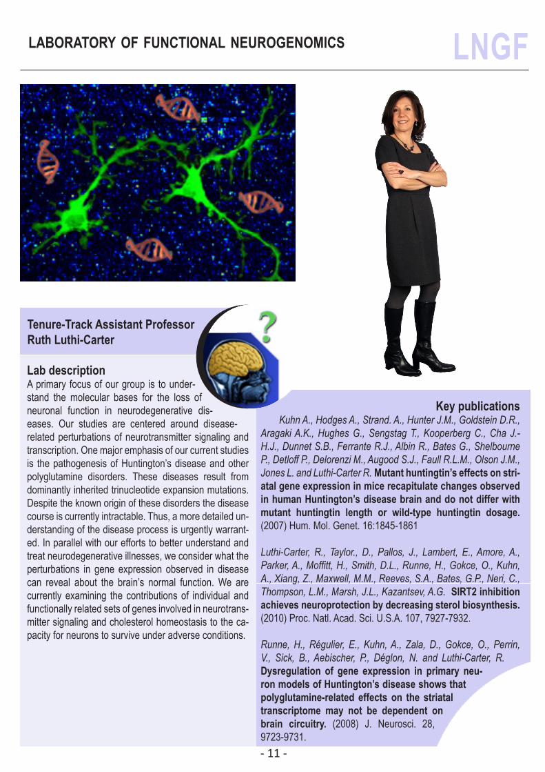
Lab descriptionA primary focus of our group is to under-stand the molecular bases for the loss of neuronal function in neurodegenerative dis-eases. Our studies are centered around disease-related perturbations of neurotransmitter signaling and transcription. One major emphasis of our current studies is the pathogenesis of Huntington’s disease and other polyglutamine disorders. These diseases result from dominantly inherited trinucleotide expansion mutations. Despite the known origin of these disorders the disease course is currently intractable. Thus, a more detailed un-derstanding of the disease process is urgently warrant-ed. In parallel with our efforts to better understand and treat neurodegenerative illnesses, we consider what the perturbations in gene expression observed in disease can reveal about the brain’s normal function. We are currently examining the contributions of individual and functionally related sets of genes involved in neurotrans-mitter signaling and cholesterol homeostasis to the ca-pacity for neurons to survive under adverse conditions.
- 12 -
laboratory of neuroenergetics and cellular LNDC
Full ProfessorPierre J. Magistretti
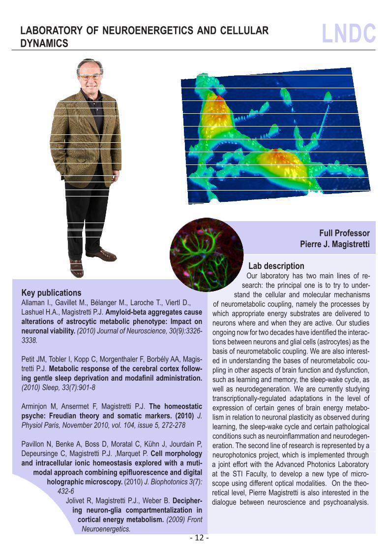
Lab descriptionOur laboratory has two main lines of re-
search: the principal one is to try to under-stand the cellular and molecular mechanisms
of neurometabolic coupling, namely the processes by which appropriate energy substrates are delivered to neurons where and when they are active. Our studies ongoing now for two decades have identified the interac-tions between neurons and glial cells (astrocytes) as the basis of neurometabolic coupling. We are also interest-ed in understanding the bases of neurometabolic cou-pling in other aspects of brain function and dysfunction, such as learning and memory, the sleep-wake cycle, as well as neurodegeneration. We are currently studying transcriptionally-regulated adaptations in the level of expression of certain genes of brain energy metabo-lism in relation to neuronal plasticity as observed during learning, the sleep-wake cycle and certain pathological conditions such as neuroinflammation and neurodegen-eration. The second line of research is represented by a neurophotonics project, which is implemented through a joint effort with the Advanced Photonics Laboratory at the STI Faculty, to develop a new type of micro-scope using different optical modalities. On the theo-retical level, Pierre Magistretti is also interested in the dialogue between neuroscience and psychoanalysis.
Key publicationsAllaman I., Gavillet M., Bélanger M., Laroche T., Viertl D., Lashuel H.A., Magistretti P.J. Amyloid-beta aggregates cause alterations of astrocytic metabolic phenotype: Impact on neuronal viability. (2010) Journal of Neuroscience, 30(9):3326-3338.
Petit JM, Tobler I, Kopp C, Morgenthaler F, Borbély AA, Magis-tretti P.J. Metabolic response of the cerebral cortex follow-ing gentle sleep deprivation and modafinil administration. (2010) Sleep, 33(7):901-8
Arminjon M, Ansermet F, Magistretti P.J. The homeostatic psyche: Freudian theory and somatic markers. (2010) J. Physiol Paris, November 2010, vol. 104, issue 5, 272-278
Pavillon N, Benke A, Boss D, Moratal C, Kühn J, Jourdain P, Depeursinge C, Magistretti P.J. ,Marquet P. Cell morphology and intracellular ionic homeostasis explored with a muti-
modal approach combining epifluorescence and digital holographic microscopy. (2010) J. Biophotonics 3(7):
432-6Jolivet R, Magistretti P.J., Weber B. Decipher-
ing neuron-glia compartmentalization in cortical energy metabolism. (2009) Front
Neuroenergetics.
PICTURE
dynamics
- 13 -
laboratory of neural microcircuitry LNMC
Full ProfessorHenry Markram
Key publicationsCali C., Berger T.K., Pignatelli M., Carleton A., Markram H. and Giugliano M. Inferring connection proximity in networks of electrically coupled cells by subthreshold frequency re-sponse analysis, J Comput Neurosci, (2008), 24, 330-45.
Gawad S., Giugliano M., Heuschkel M.O., Wessling B., Markram H., Schnakenberg V,. Renaud P. and Morgan H., Substrate ar-rays of iridium oxide microelectrodes for in-vitro neuronal interfacing, Frontiers in Neuroengineering, (2009), 2, 1.1-1.7.
Rinaldi T., Silberberg G. and Markram H., Hyperconnectivity of Local Neocortical Microcircuitry Induced by Prenatal Expo-sure to Valproic Acid, Cereb Cortex, (2008), 18, 763-70.
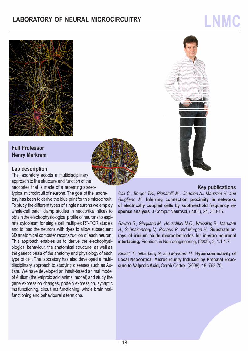
Lab descriptionThe laboratory adopts a multidisciplinary approach to the structure and function of the neocortex that is made of a repeating stereo-typical microcircuit of neurons. The goal of the labora-tory has been to derive the blue print for this microcircuit. To study the different types of single neurons we employ whole-cell patch clamp studies in neocortical slices to obtain the electrophysiological profile of neurons to aspi-rate cytoplasm for single cell multiplex RT-PCR studies and to load the neurons with dyes to allow subsequent 3D anatomical computer reconstruction of each neuron. This approach enables us to derive the electrophysi-ological behaviour, the anatomical structure, as well as the genetic basis of the anatomy and physiology of each type of cell. The laboratory has also developed a multi-disciplinary approach to studying diseases such as Au-tism. We have developed an insult-based animal model of Autism (the Valproic acid animal model) and study the gene expression changes, protein expression, synaptic malfunctioning, circuit malfunctioning, whole brain mal-functioning and behavioural alterations.
- 14 -
laboratory of molecular neurodegenerative LNMR
Tenure-Track Assistant ProfessorDarren J. Moore
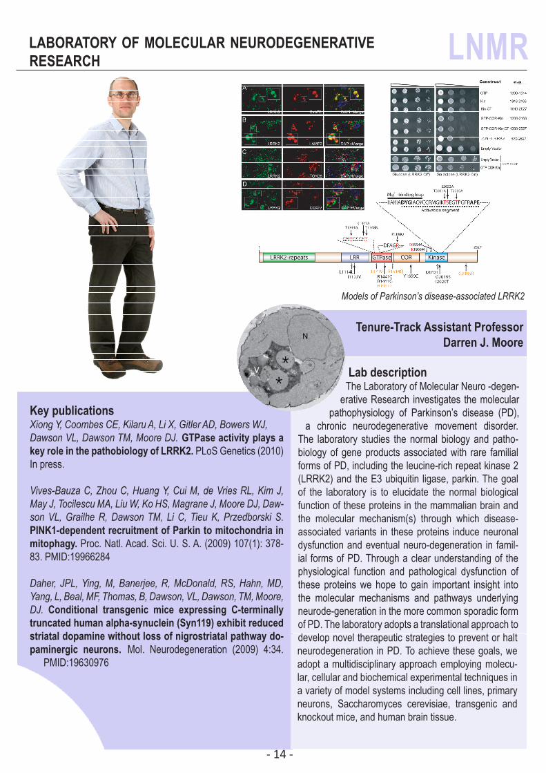
Lab descriptionThe Laboratory of Molecular Neuro -degen-
erative Research investigates the molecular pathophysiology of Parkinson’s disease (PD),
a chronic neurodegenerative movement disorder. The laboratory studies the normal biology and patho-biology of gene products associated with rare familial forms of PD, including the leucine-rich repeat kinase 2 (LRRK2) and the E3 ubiquitin ligase, parkin. The goal of the laboratory is to elucidate the normal biological function of these proteins in the mammalian brain and the molecular mechanism(s) through which disease-associated variants in these proteins induce neuronal dysfunction and eventual neuro-degeneration in famil-ial forms of PD. Through a clear understanding of the physiological function and pathological dysfunction of these proteins we hope to gain important insight into the molecular mechanisms and pathways underlying neurode-generation in the more common sporadic form of PD. The laboratory adopts a translational approach to develop novel therapeutic strategies to prevent or halt neurodegeneration in PD. To achieve these goals, we adopt a multidisciplinary approach employing molecu-lar, cellular and biochemical experimental techniques in a variety of model systems including cell lines, primary neurons, Saccharomyces cerevisiae, transgenic and knockout mice, and human brain tissue.
Key publicationsXiong Y, Coombes CE, Kilaru A, Li X, Gitler AD, Bowers WJ, Dawson VL, Dawson TM, Moore DJ. GTPase activity plays a key role in the pathobiology of LRRK2. PLoS Genetics (2010) In press.
Vives-Bauza C, Zhou C, Huang Y, Cui M, de Vries RL, Kim J, May J, Tocilescu MA, Liu W, Ko HS, Magrane J, Moore DJ, Daw-son VL, Grailhe R, Dawson TM, Li C, Tieu K, Przedborski S. PINK1-dependent recruitment of Parkin to mitochondria in mitophagy. Proc. Natl. Acad. Sci. U. S. A. (2009) 107(1): 378-83. PMID:19966284
Daher, JPL, Ying, M, Banerjee, R, McDonald, RS, Hahn, MD, Yang, L, Beal, MF, Thomas, B, Dawson, VL, Dawson, TM, Moore, DJ. Conditional transgenic mice expressing C-terminally truncated human alpha-synuclein (Syn119) exhibit reduced striatal dopamine without loss of nigrostriatal pathway do-paminergic neurons. Mol. Neurodegeneration (2009) 4:34.
PMID:19630976
Models of Parkinson’s disease-associated LRRK2
research
- 15 -
laboratory of sensory processing LSENS
Associate ProfessorCarl Petersen
Key publicationsGentet LJ, Avermann M, Matyas F, Staiger JF, Petersen CCH Membrane potential dynamics of GABAergic neurons in the barrel cortex of behaving mice. (2010) Neuron 65: 422-435.
Lefort S, Tomm C, Sarria JCF, Petersen CCH The excitatory neuronal network of the C2 barrel column in mouse primary somatosensory cortex. (2009) Neuron 61: 301-316.
Poulet JFA, Petersen CCH Internal brain state regulates mem-brane potential synchrony in barrel cortex of behaving mice. (2008) Nature 454: 881-885.
Ferezou I, Haiss F, Gentet LJ, Aronoff R, Weber B, Petersen CCH Spatiotemporal dynamics of cortical sensorimotor integra-tion in behaving mice. (2007) Neuron 56: 907-923.
Crochet S, Petersen CCH Correlating whisker behavior with membrane potential in barrel cortex of awake mice. (2006) Nat Neurosci 9: 608-610.
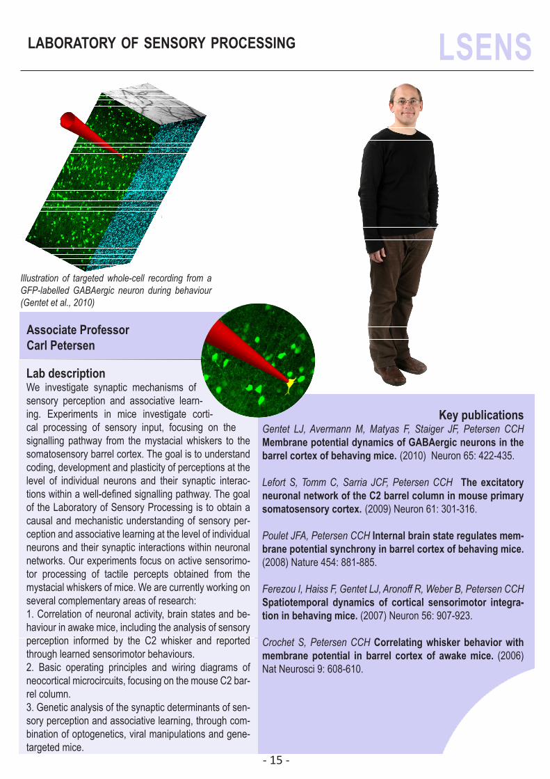
Lab descriptionWe investigate synaptic mechanisms of sensory perception and associative learn-ing. Experiments in mice investigate corti-cal processing of sensory input, focusing on the signalling pathway from the mystacial whiskers to the somatosensory barrel cortex. The goal is to understand coding, development and plasticity of perceptions at the level of individual neurons and their synaptic interac-tions within a well-defined signalling pathway. The goal of the Laboratory of Sensory Processing is to obtain a causal and mechanistic understanding of sensory per-ception and associative learning at the level of individual neurons and their synaptic interactions within neuronal networks. Our experiments focus on active sensorimo-tor processing of tactile percepts obtained from the mystacial whiskers of mice. We are currently working on several complementary areas of research:1. Correlation of neuronal activity, brain states and be-haviour in awake mice, including the analysis of sensory perception informed by the C2 whisker and reported through learned sensorimotor behaviours.2. Basic operating principles and wiring diagrams of neocortical microcircuits, focusing on the mouse C2 bar-rel column.3. Genetic analysis of the synaptic determinants of sen-sory perception and associative learning, through com-bination of optogenetics, viral manipulations and gene-targeted mice.
Illustration of targeted whole-cell recording from a GFP-labelled GABAergic neuron during behaviour (Gentet et al., 2010)
- 16 -
Laboratory of behavioral genetics LGC
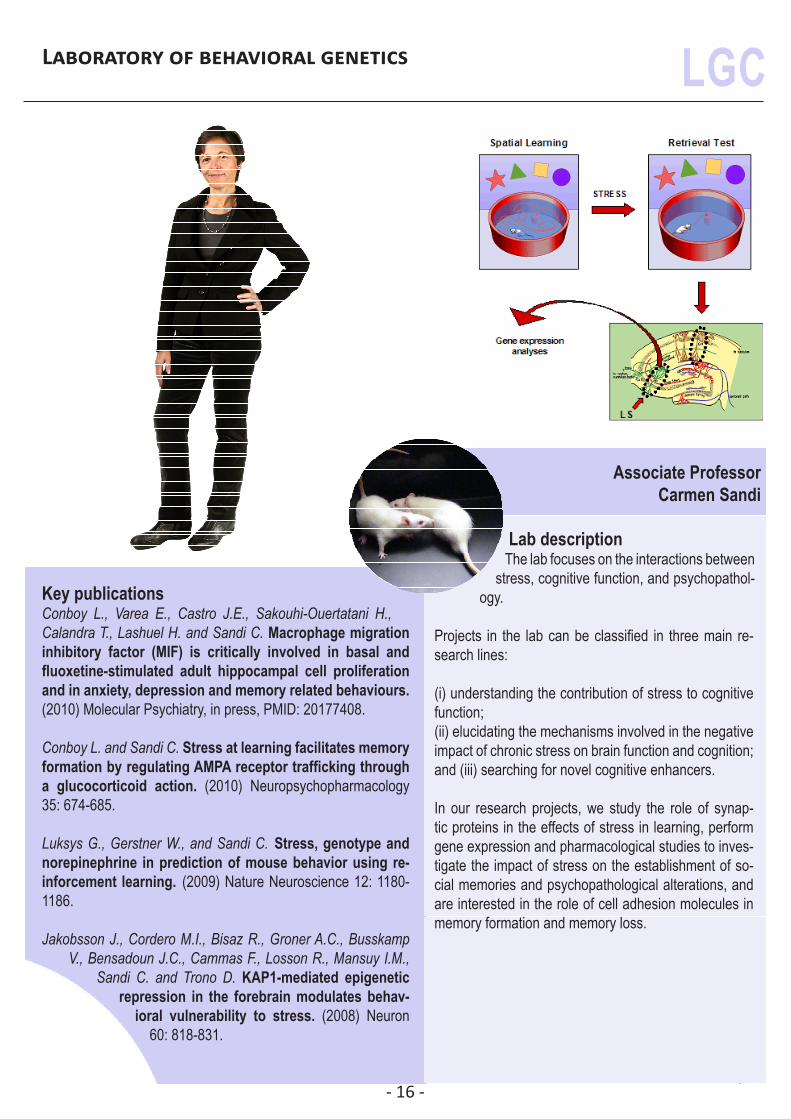
Associate ProfessorCarmen Sandi
Lab descriptionThe lab focuses on the interactions between
stress, cognitive function, and psychopathol-ogy.
Projects in the lab can be classified in three main re-search lines:
(i) understanding the contribution of stress to cognitive function;(ii) elucidating the mechanisms involved in the negative impact of chronic stress on brain function and cognition; and (iii) searching for novel cognitive enhancers.
In our research projects, we study the role of synap-tic proteins in the effects of stress in learning, perform gene expression and pharmacological studies to inves-tigate the impact of stress on the establishment of so-cial memories and psychopathological alterations, and are interested in the role of cell adhesion molecules in memory formation and memory loss.
Key publicationsConboy L., Varea E., Castro J.E., Sakouhi-Ouertatani H., Calandra T., Lashuel H. and Sandi C. Macrophage migration inhibitory factor (MIF) is critically involved in basal and fluoxetine-stimulated adult hippocampal cell proliferation and in anxiety, depression and memory related behaviours. (2010) Molecular Psychiatry, in press, PMID: 20177408.
Conboy L. and Sandi C. Stress at learning facilitates memory formation by regulating AMPA receptor trafficking through a glucocorticoid action. (2010) Neuropsychopharmacology 35: 674-685.
Luksys G., Gerstner W., and Sandi C. Stress, genotype and norepinephrine in prediction of mouse behavior using re-inforcement learning. (2009) Nature Neuroscience 12: 1180-1186.
Jakobsson J., Cordero M.I., Bisaz R., Groner A.C., Busskamp V., Bensadoun J.C., Cammas F., Losson R., Mansuy I.M.,
Sandi C. and Trono D. KAP1-mediated epigenetic repression in the forebrain modulates behav-
ioral vulnerability to stress. (2008) Neuron 60: 818-831.
- 17 -
laboratory of synaptic mechanisms LSYM
Associate ProfessorRalf Schneggenburger
Key publicationsMüller, M., Goutman, J., Kochubey O., Schneggenburger R. In-teraction between facilitation and depression at a large CNS synapse reveals mechanisms of short-term plasticity. (2010) J. Neuroscience, 30: 2007 - 2016.
Korogod N., Lou X. and Schneggenburger R. Posttetanic poten-tiation critically depends on an enhanced Ca2+ sensitivity of vesicle fusion mediated by presynaptic PKC. (2007) Proc Natl Acad Sci U S A 104, 15923-15928.
Lou X., Scheuss V. and Schneggenburger R. Allosteric modula-tion of the presynaptic Ca2+ sensor for vesicle fusion. (2005) Nature 435, 497-501.
Schneggenburger, R. & Neher, E. Presynaptic Calcium and control of vesicle fusion. (2005) Curr. Op. Neurobiol. 44: 266 - 274. (Review)
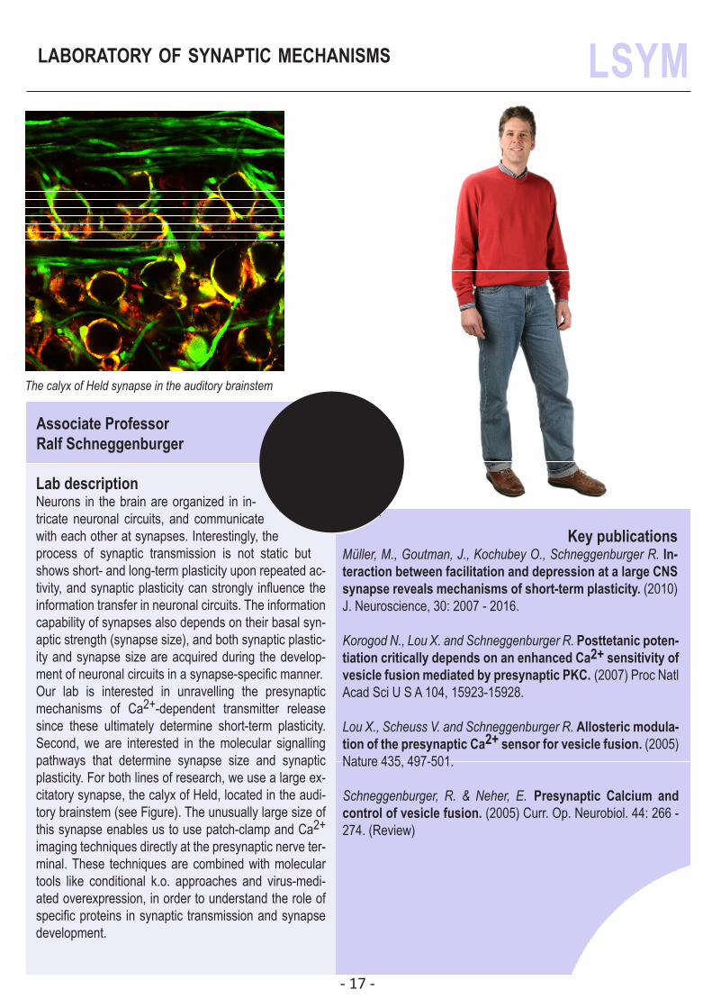
Lab descriptionNeurons in the brain are organized in in-tricate neuronal circuits, and communicate with each other at synapses. Interestingly, the process of synaptic transmission is not static but shows short- and long-term plasticity upon repeated ac-tivity, and synaptic plasticity can strongly influence the information transfer in neuronal circuits. The information capability of synapses also depends on their basal syn-aptic strength (synapse size), and both synaptic plastic-ity and synapse size are acquired during the develop-ment of neuronal circuits in a synapse-specific manner.Our lab is interested in unravelling the presynaptic mechanisms of Ca2+-dependent transmitter release since these ultimately determine short-term plasticity. Second, we are interested in the molecular signalling pathways that determine synapse size and synaptic plasticity. For both lines of research, we use a large ex-citatory synapse, the calyx of Held, located in the audi-tory brainstem (see Figure). The unusually large size of this synapse enables us to use patch-clamp and Ca2+ imaging techniques directly at the presynaptic nerve ter-minal. These techniques are combined with molecular tools like conditional k.o. approaches and virus-medi-ated overexpression, in order to understand the role of specific proteins in synaptic transmission and synapse development.
The calyx of Held synapse in the auditory brainstem
- 18 -
research group: social cognitive neuroscience
SNSF - funded ProfessorNouchine Hadjikhani
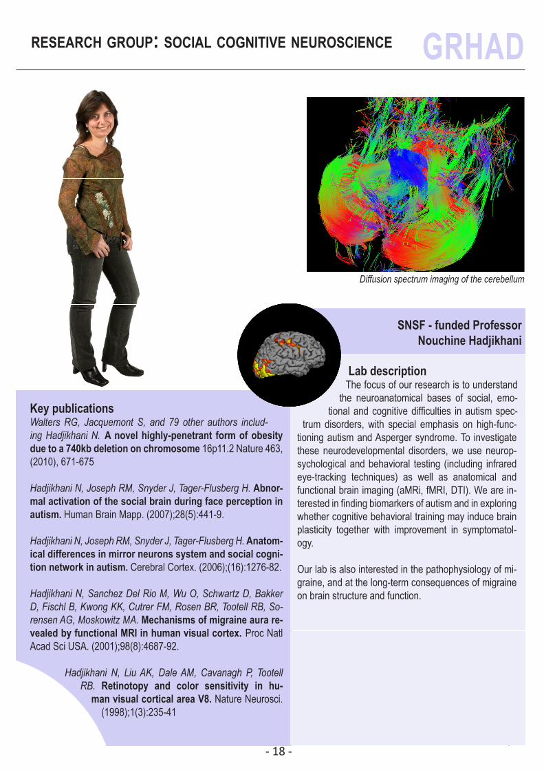
Lab descriptionThe focus of our research is to understand
the neuroanatomical bases of social, emo-tional and cognitive difficulties in autism spec-
trum disorders, with special emphasis on high-func-tioning autism and Asperger syndrome. To investigate these neurodevelopmental disorders, we use neurop-sychological and behavioral testing (including infrared eye-tracking techniques) as well as anatomical and functional brain imaging (aMRi, fMRI, DTI). We are in-terested in finding biomarkers of autism and in exploring whether cognitive behavioral training may induce brain plasticity together with improvement in symptomatol-ogy.
Our lab is also interested in the pathophysiology of mi-graine, and at the long-term consequences of migraine on brain structure and function.
Key publicationsWalters RG, Jacquemont S, and 79 other authors includ-ing Hadjikhani N. A novel highly-penetrant form of obesity due to a 740kb deletion on chromosome 16p11.2 Nature 463, (2010), 671-675
Hadjikhani N, Joseph RM, Snyder J, Tager-Flusberg H. Abnor-mal activation of the social brain during face perception in autism. Human Brain Mapp. (2007);28(5):441-9.
Hadjikhani N, Joseph RM, Snyder J, Tager-Flusberg H. Anatom-ical differences in mirror neurons system and social cogni-tion network in autism. Cerebral Cortex. (2006);(16):1276-82.
Hadjikhani N, Sanchez Del Rio M, Wu O, Schwartz D, Bakker D, Fischl B, Kwong KK, Cutrer FM, Rosen BR, Tootell RB, So-rensen AG, Moskowitz MA. Mechanisms of migraine aura re-vealed by functional MRI in human visual cortex. Proc Natl Acad Sci USA. (2001);98(8):4687-92.
Hadjikhani N, Liu AK, Dale AM, Cavanagh P, Tootell RB. Retinotopy and color sensitivity in hu-
man visual cortical area V8. Nature Neurosci. (1998);1(3):235-41
Diffusion spectrum imaging of the cerebellum
GRHAD
- 19 -
blue brain project
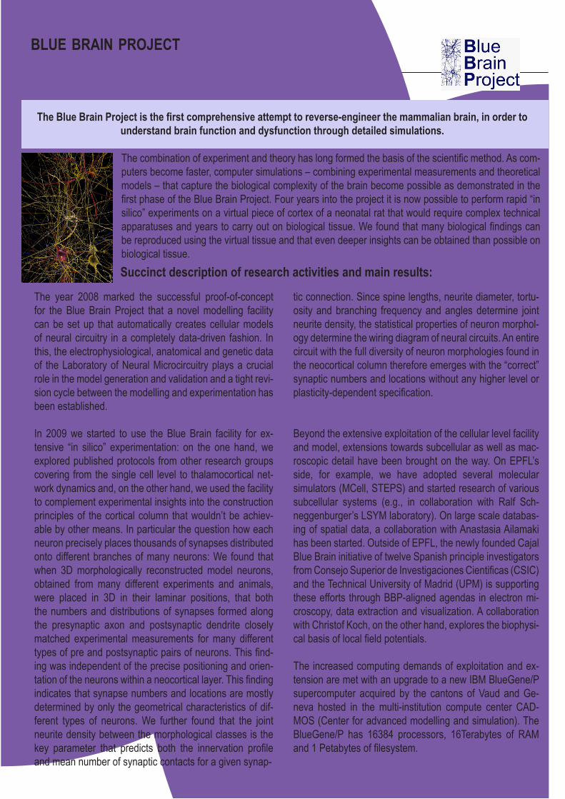
The combination of experiment and theory has long formed the basis of the scientific method. As com-puters become faster, computer simulations – combining experimental measurements and theoretical models – that capture the biological complexity of the brain become possible as demonstrated in the first phase of the Blue Brain Project. Four years into the project it is now possible to perform rapid “in silico” experiments on a virtual piece of cortex of a neonatal rat that would require complex technical apparatuses and years to carry out on biological tissue. We found that many biological findings can be reproduced using the virtual tissue and that even deeper insights can be obtained than possible on biological tissue.
The Blue Brain Project is the first comprehensive attempt to reverse-engineer the mammalian brain, in order to understand brain function and dysfunction through detailed simulations.
The year 2008 marked the successful proof-of-concept for the Blue Brain Project that a novel modelling facility can be set up that automatically creates cellular models of neural circuitry in a completely data-driven fashion. In this, the electrophysiological, anatomical and genetic data of the Laboratory of Neural Microcircuitry plays a crucial role in the model generation and validation and a tight revi-sion cycle between the modelling and experimentation has been established.
In 2009 we started to use the Blue Brain facility for ex-tensive “in silico” experimentation: on the one hand, we explored published protocols from other research groups covering from the single cell level to thalamocortical net-work dynamics and, on the other hand, we used the facility to complement experimental insights into the construction principles of the cortical column that wouldn’t be achiev-able by other means. In particular the question how each neuron precisely places thousands of synapses distributed onto different branches of many neurons: We found that when 3D morphologically reconstructed model neurons, obtained from many different experiments and animals, were placed in 3D in their laminar positions, that both the numbers and distributions of synapses formed along the presynaptic axon and postsynaptic dendrite closely matched experimental measurements for many different types of pre and postsynaptic pairs of neurons. This find-ing was independent of the precise positioning and orien-tation of the neurons within a neocortical layer. This finding indicates that synapse numbers and locations are mostly determined by only the geometrical characteristics of dif-ferent types of neurons. We further found that the joint neurite density between the morphological classes is the key parameter that predicts both the innervation profile and mean number of synaptic contacts for a given synap-
Succinct description of research activities and main results:
tic connection. Since spine lengths, neurite diameter, tortu-osity and branching frequency and angles determine joint neurite density, the statistical properties of neuron morphol-ogy determine the wiring diagram of neural circuits. An entire circuit with the full diversity of neuron morphologies found in the neocortical column therefore emerges with the “correct” synaptic numbers and locations without any higher level or plasticity-dependent specification.
Beyond the extensive exploitation of the cellular level facility and model, extensions towards subcellular as well as mac-roscopic detail have been brought on the way. On EPFL’s side, for example, we have adopted several molecular simulators (MCell, STEPS) and started research of various subcellular systems (e.g., in collaboration with Ralf Sch-neggenburger’s LSYM laboratory). On large scale databas-ing of spatial data, a collaboration with Anastasia Ailamaki has been started. Outside of EPFL, the newly founded Cajal Blue Brain initiative of twelve Spanish principle investigators from Consejo Superior de Investigaciones Cientificas (CSIC) and the Technical University of Madrid (UPM) is supporting these efforts through BBP-aligned agendas in electron mi-croscopy, data extraction and visualization. A collaboration with Christof Koch, on the other hand, explores the biophysi-cal basis of local field potentials.
The increased computing demands of exploitation and ex-tension are met with an upgrade to a new IBM BlueGene/P supercomputer acquired by the cantons of Vaud and Ge-neva hosted in the multi-institution compute center CAD-MOS (Center for advanced modelling and simulation). The BlueGene/P has 16384 processors, 16Terabytes of RAM and 1 Petabytes of filesystem.
- 20 -
Groups affiliated to the Brain Mind Institute
Dr Miguel Nicolelis group at Duke UniversityDr Gregor Rainer group at University FribourgDr Jean-Philippe Thiran at EPFL, School of Engineering, Signal Processing LaboratoryDr José del R. Millán at EPFL, STI, Defitech Foundation Chair in Non-Invasive Brain-Machine InterfaceDr Rolf Gruetter at the Center for Biomedical Imaging (CIBM)
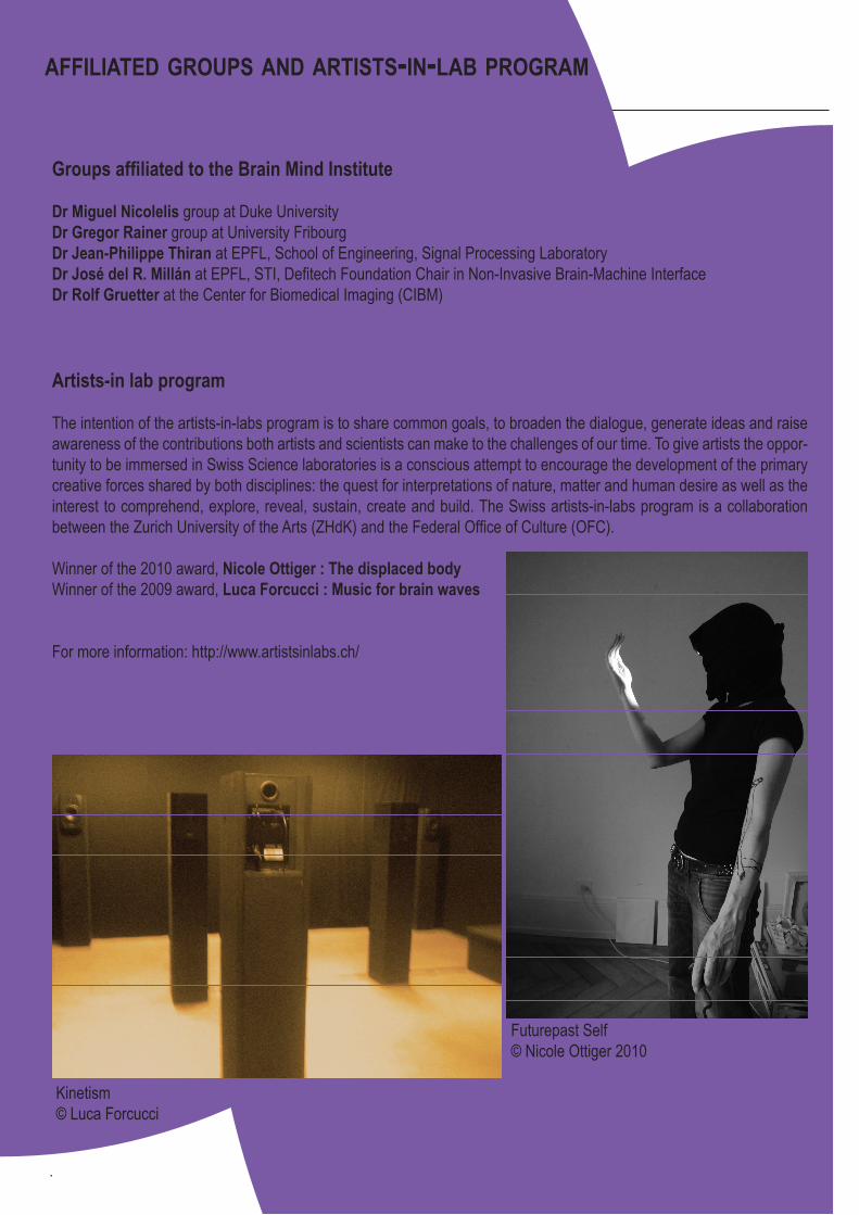
Artists-in lab program
The intention of the artists-in-labs program is to share common goals, to broaden the dialogue, generate ideas and raise awareness of the contributions both artists and scientists can make to the challenges of our time. To give artists the oppor-tunity to be immersed in Swiss Science laboratories is a conscious attempt to encourage the development of the primary creative forces shared by both disciplines: the quest for interpretations of nature, matter and human desire as well as the interest to comprehend, explore, reveal, sustain, create and build. The Swiss artists-in-labs program is a collaboration between the Zurich University of the Arts (ZHdK) and the Federal Office of Culture (OFC).
Winner of the 2010 award, Nicole Ottiger : The displaced bodyWinner of the 2009 award, Luca Forcucci : Music for brain waves
For more information: http://www.artistsinlabs.ch/
Futurepast Self © Nicole Ottiger 2010
Kinetism © Luca Forcucci
affiliated groups and artists-in-lab program
- 21 -
Produced and edited by the EPFL - Brain Mind InstituteNovember, 2010
Editor: Emilie PralongPrinted at EPFL “Atelier de Reprographie”
With many thanks to Egizia Carbone, Igor Allaman,Gabriele Grenningloh and all Professors
for their help and support!
© Alain Herzog for Professors portraits © Yves Burdet for Prof. Ruth Luthi-Carter portrait © EPFL / Brain Mind Institute for all other material published in this booklet