differential diagnosis between low-grade and high-grade...
TRANSCRIPT

Clinical StudyDifferential Diagnosis between Low-Grade and High-GradeAstrocytoma Using System A Amino Acid Transport PETImaging with C-11-MeAIB: A Comparison Study withC-11-Methionine PET Imaging
Ryuichi Nishii ,1,2,3 Tatsuya Higashi ,1,3 Shinya Kagawa,3 Maya Arimoto,4
Yoshihiko Kishibe,3 Masaaki Takahashi,3 Shigeki Yamada,5 Masaaki Saiki,6
Yoshiki Arakawa,7 Hiroshi Yamauchi,3 Chio Okuyama,3 Masato Hojo,8
Toshihiro Munemitsu,8 Masahiro Sawada,8 Masato Kobayashi,9 Keiichi Kawai,10
Shigeki Nagamachi,2,11 Toshinori Hirai,2 and Susumu Miyamoto7
1Department of Molecular Imaging and eranostics, National Institute of Radiological Sciences (NIRS), QST, 4-9-1 Anagawa,Inage-ku, Chiba 263-8555, Japan2Department of Radiology, Faculty of Medicine, University of Miyazaki, 5200 Kihara Kiyotake-cho, Miyazaki 889-1692, Japan3Division of PET Imaging, Shiga Medical Center Research Institute, 5-4-30 Moriyama, Moriyama, Shiga 524-8524, Japan4Department of Diagnostic Imaging and Nuclear Medicine, Graduate School of Medicine, Kyoto University,54 Shogoin Kawahara-cho, Sakyo-ku, Kyoto 606-8507, Japan5Department of Neurosurgery, Rakuwakai Otowa Hospital, 2 Otowachinjicho, Yamashina-ku, Kyoto 607-8062, Japan6Department of Radiology, Japanese Red Cross Otsu Hospital, 1-1-35 Nagara, Otsu, Shiga 520-8511, Japan7Department of Neurosurgery, Graduate School of Medicine, Kyoto University, 54 ShogoinKawahara-cho, Sakyo-ku,Kyoto 606-8507, Japan8Department of Neurosurgery, Shiga Medical Center, 5-4-30 Moriyama, Moriyama, Shiga 524-8524, Japan9Wellness Promotion Science Center, Institute of Medical, Kanazawa University, 5-10-80 Kodatsuno, Kanazawa,Ishikawa 920-0942, Japan10Graduate School of Health Sciences, College of Medical, Pharmaceutical and Health Sciences, Kanazawa University,5-10-80 Kodatsuno, Kanazawa, Ishikawa 920-0942, Japan
11Department of Radiology, Faculty of Medicine, Fukuoka University, 7-45-1 Nanakuma, Jonan-ku, Fukuoka 814-0180, Japan
Correspondence should be addressed to Tatsuya Higashi; [email protected]
Received 29 December 2017; Accepted 6 May 2018; Published 20 June 2018
Academic Editor: Guoqiang Shao
Copyright © 2018 Ryuichi Nishii et al.is is an open access article distributed under the Creative Commons Attribution License,which permits unrestricted use, distribution, and reproduction in any medium, provided the original work is properly cited.
Introductions. [N-methyl-C-11]α-Methylaminoisobutyric acid (MeAIB) is an arti�cial amino acid radiotracer used for PETstudy,which is metabolically stable in vivo. In addition, MeAIB is transported by system A neutral amino acid transport, which isobserved ubiquitously in all types of mammalian cells. It has already been shown that MeAIB-PET is useful for malignantlymphoma, head and neck cancers, and lung tumors. However, there have been no reports evaluating the usefulness of MeAIB-PET in the diagnosis of brain tumors. e purpose of this study is to investigate the e�cacy of system A amino acid transport PETimaging, MeAIB-PET, in clinical brain tumor diagnosis compared to [S-methyl-C-11]-L-methionine (MET)-PET. Methods.irty-one consecutive patients (male: 16, female: 15), who were suspected of having brain tumors, received bothMeAIB-PETandMET-PET within a 2-week interval. All patients were classi�ed into two groups: Group A as a benign group, which includedpatients who were diagnosed as low-grade astrocytoma, grade II or less, or other low-grade astrocytoma (n � 12) and Group B asa malignant group, which included patients who were diagnosed as anaplastic astrocytoma, glioblastoma multiforme (GBM), orrecurrent GBM despite prior surgery or chemoradiotherapy (n � 19). PET imaging was performed 20min after the IV injection ofMeAIB and MET, respectively. Semiquantitative analyses of MeAIB and METuptake using SUVmax and tumor-to-contralateral
HindawiContrast Media & Molecular ImagingVolume 2018, Article ID 1292746, 9 pageshttps://doi.org/10.1155/2018/1292746

normal brain tissue (T/N) ratio were evaluated to compare these PET images. ROC analyses for the diagnostic accuracy of MeAIB-PET and MET-PETwere also calculated. Results. In MeAIB-PET imaging, the SUVmax was 1.20± 1.29 for the benign group and2.94± 1.22 for the malignant group (p< 0.005), and the T/N ratio was 3.77± 2.39 for the benign group and 16.83± 2.39 for themalignant group (p< 0.001). In MET-PET, the SUVmax was 3.01± 0.94 for the benign group and 4.72± 1.61 for the malignantgroup (p< 0.005), and the T/N ratio was 2.64± 1.40 for the benign group and 3.21± 1.14 for the malignant group (n.s.). For theanalysis using the T/N ratio, there was a significant difference between the benign and malignant groups with MeAIB-PET withp< 0.001. -e result of ROC analysis using the T/N ratio indicated a better diagnosis accuracy for MeAIB-PET for brain tumorsthan MET-PET (p< 0.01). Conclusions. MeAIB, a system A amino acid transport-specific radiolabeled agents, could providebetter assessments for detecting malignant type brain tumors. In a differential diagnosis between low-grade and high-gradeastrocytoma, MeAIB-PET is a useful diagnostic imaging tool, especially in evaluations using the T/N ratio. Clinical trial reg-istration. -is trial was registered with UMIN000032498.
1. Introduction
Positron emission tomography (PET) imaging with aminoacid analogs has been focused greatly on clinical applica-tions, as it targets increased amino acid transport by tumors[1, 2]. Especially for detecting brain tumors, PETstudies withamino acid analogs have been developed [3, 4] to overcomethe drawbacks of F-18 FDG (FDG) PET, such as physio-logical uptake by the brain [5, 6].
As methionine, an essential sulfur amino acid, is nec-essary for the growth and development of cells, radiolabelled[S-methyl-C-11]-L-methionine (MET), mainly transportedby system L amino acid transporters [7, 8], has been clin-ically used as a tumor-seeking agent for PET imaging forseveral decades [9]. MET-PET images can visualize not onlythe population and activity of amino acid transport but alsometabolic events inside the body, such as active cellmembrane transport, cellular protein synthesis, polyaminesynthesis, and trans-methylation reactions [10, 11]. How-ever, MET-PET is known to have several drawbacks whendiagnosing tumors. MET is unstable in vivo due to theaminotransfer reaction [10] and is excreted into the bile andintestines. In addition, MET-PET shows faint physiologicaluptake in the brain, strong physiological uptake in the liverand bone marrow, and uptake in certain types of in-flammatory changes [12, 13].
[N-methyl-C-11]α-Methylaminoisobutyric acid (MeAIB)is an artificial amino acid radiotracer used for PET study,which is metabolically stable in vivo [14]. Although MET istransported mainly by system L neutral amino acid transport,MeAIB is transported by system A neutral amino acidtransport, which is observed ubiquitously in all types ofmammalian cells [11, 15]. It has already been shown thatMeAIB is useful for amino acid uptake measurements inskeletal muscle and for the diagnosis of malignant lym-phoma and head and neck cancers [14, 16, 17]. We havealso been investigating system A amino acid PETmolecularimaging withMeAIB to detect tumors and have reported itsusefulness in the differential diagnosis of pulmonary andmediastinal mass lesions [18] and prostate cancer [19] inclinical practice.
However, there have been no reports evaluating theusefulness of MeAIB-PET in the diagnosis of brain tumors.
-e purpose of this study is to investigate the efficacy ofsystem A amino acid transport PET imaging, MeAIB-PET,in clinical brain tumor diagnosis compared to MET-PET.
2. Materials and Methods
2.1. Patient Characteristics. From March 2009 to December2011, 31 consecutive patients (male: 16, female: 15), whowere suspected of having brain tumors, received bothMeAIB-PET and MET-PET within a 2-week interval. Pa-tients’ ages ranged from 5 to 71 years with a mean age of44.2± 18.5, as shown in Table 1. Inclusion criteria for thestudy were as follows: (1) patients were suspected of havingan intraaxial brain tumor (newly detected or recurrent le-sions 6 months or more after successful treatment) by CTand MRI (both were performed as routine clinical studies),(2) each patient gave written informed consent and receivedMeAIB-PET and MET-PET, and (3) results were confirmedpathologically, or by clinical follow-up more than 6 monthsafter the PET studies. Exclusion criteria were as follows: (1)patients with extra-axial tumors such as tumors of themeninges, pituitary tumors, pineal parenchymal tumors, orcranial nerve schwannomas, (2) patients with metastaticbrain tumors or lymphoma, and (3) patients who refused toreceive MeAIB-PET or MET-PET. Of the 52 patients whoreceived MeAIB-PET with suspected brain tumors fromMarch 2009 to December 2011, 31 patients were included inthe present study, while the others were excluded because ofthe exclusion criteria. According to final diagnosis aftersurgery or biopsy, all patients who met the criteria wereclassified into the following two groups: Group A (benign),which included patients who were diagnosed as low-gradeastrocytoma, grade II including a case of recurrent grade IIglioma or less, or other low-grade astrocytomas (n � 12;ranging from 5 to 46 years, mean age 32.2± 10.0 years; sevenmales and five females); Group B (malignant), which in-cluded patients who were diagnosed as anaplastic astrocy-toma, glioblastoma multiforme (GBM), or recurrent GBMdespite prior surgery or chemoradiotherapy (n � 19; rangingfrom 14 to 71 years, mean age 56.7± 16.8 years; nine malesand ten females).
-is prospective clinical study was approved by ourinstitutional review boards, the Human Study Committee(approval number: #36-04, March 25, 2009) and by theCommittee for the Clinical Use of Short-Half Life Ra-dioactive Materials (approval number: #2008-01, Novem-ber 28, 2008). All enrolled patients or their parents if thepatient was under 20 years old received explanations, andthen they provided written informed consent regarding thisstudy.
2 Contrast Media & Molecular Imaging

2.2. Radiotracers. Production of MeAIB followed a previouslydescribed procedure [18]. -e radiosynthesis method wasbased on that proposed by Nagren et al. [20]. Chemicals andsolvents were of analytical grade and purchased commercially.[11C]MeOTf was bubbled into the reactor of an automatedremotely controlled synthesizer module C-11-BII (SHI, Tokyo,Japan) filled with 1mg of methyl α-aminoisobutyrate hydro-chloride (6.5mmol) dissolved in 0.4ml of methanol/acetone(1/1, v/v) and 3.2μl of 2,2,6,6,N-pentamethyl-piperidine (PMP)at −20°C. -en, the reactor was heated to 80°C for 1min. Aftercooling to 25°C, 400μl of 2M NaOH was then loaded into thereactor. After heating the mixture for 3min at 60°C, the hy-drolyzed product was diluted with 0.5ml of HPLC eluent andsubsequently transferred to a preparative radio HPLC systemconsisting of a preparative HPLC pump (PU-980, JASCO), anautomated flow-detector-controlled injection systemwith a 2mlinjection loop, a semipreparative HPLC column (hydro-philic interaction chromatography: HILIC column, NacalaiTesque, 250 ×10mm2, 5 μm; mobile phase: MeCN/10mMCH3COONH4, 4/1, v/v; flow: 8ml/min), a UV detector(254 nm), and an NaI(Tl) radioactivity detector.-e product-containing fractionwas then dilutedwith 10ml of isotonic saline.-e radiochemical purity of the MeAIB was more than 99%.
MET was synthesized based on the method described ina previous report [21], by the reaction of C-11 methyltriflatewith an aqueous solution of L-homocysteine thiolactone ina Sep-Pak tC18 cartridge, followed by purification with ion-exchange cartridges. -e radiochemical purity of MET wasalso more than 99%.
2.3. PET Study. All patients were examined with a whole-body PETscanner, GE Advance (GE Healthcare, Waukesha,WI, USA), or with a whole-body PET/CT scanner, Siemens
True Point Biograph 16 (Siemens/CTI, Erlangen, Germany).All subjects received an intravenous injection of MeAIB(513.6± 65.6MBq) or MET (533.9± 35.0MBq). BrainPET/CT images were acquired 20min after the radiotracerinjection in 1 bed position in both study. Emission imageswere acquired for 5min per bed position. -e data werereconstructed using the ordered subsets expectation-maximization method using eight subsets, two iterations,and an array size of 256× 256. For the attenuation correctionof PET/CTfusion images, the CTcomponent was performedaccording to a standard protocol with the following pa-rameters: 140 kV; 50mAs; tube rotation time, 0.5 s per ro-tation; slice thickness, 5mm; and gap, 2mm. An E-softworkstation (Siemens, Nashville, TN, USA) was used toconstruct PET/CT fusion images.
Table 2: SUVmax and T/N ratios of MeAIB- and MET-PET studyin patients with brain tumors.
Group A (benign group)
DiagnosisMeAIB MET
SUVmax T/Nratio SUVmax T/N
ratio1 Low-grade glioma 0.58 3.22 1.98 1.78
2 Astrocytoma gradeII 2.83 8.09 2.83 1.35
3 Low-grade glioma 0.24 1.64 1.80 1.044 Glioma grade II 0.15 1.25 3.03 2.375 Low-grade glioma 0.62 1.11 3.13 1.826 Low-grade glioma 0.85 2.07 3.89 2.547 Brain stem glioma 3.40 4.86 3.03 1.89
8 Glioma grade II,rec. 3.49 6.13 1.96 2.68
9 Low-grade glioma 0.32 4.00 4.82 4.3810 Low-grade glioma 0.4 6.67 2.19 3.2211 Low-grade glioma 1.34 4.96 4.08 6.0712 Low-grade glioma 0.12 1.20 3.41 2.54Ave. 1.20 3.77 3.01 2.64Group B (malignant group)13 GBM 4.81 32.07 5.82 4.4414 GBM, rec. 2.95 14.89 3.49 3.4515 GBM, rec. 2.94 26.72 3.63 3.6716 GBM, rec. 2.33 8.96 5.39 2.5117 GBM, rec. 2.84 12.91 5.54 2.5518 GBM, rec. 2.83 15.00 4.27 3.3019 GBM 1.60 4.00 3.10 1.2420 GBM 2.89 22.23 6.47 3.8721 GBM, rec. 4.51 25.10 6.14 3.2322 GBM, rec. 2.89 17.00 3.40 2.9123 GBM 2.81 17.56 6.07 6.0724 GBM 5.85 15.81 4.57 3.4125 GBM, rec. 1.22 17.43 1.46 1.7626 GBM, rec. 4.58 21.73 5.90 2.7127 GBM, rec. 2.83 15.00 4.27 3.3028 GBM 2.89 22.23 7.82 4.68
29 Anaplasticastrocytoma 1.95 10.26 5.94 4.01
30 Anaplasticastrocytoma 1.73 7.86 2.18 1.79
31 GBM 1.43 13.00 4.27 2.12Ave. 2.94 16.83 4.72 3.21
Table 1: Patient characteristics.Total (n � 31)Age (years)Mean± SD 44.2± 18.5Median 44Range 5–71
Male : female 16 :15Group A (benign) (n � 12)
Age (years)Mean± SD 32.2± 10.0Median 33.5Range 5–46
Male : female 7 : 5DiagnosisAstrocytoma grade II or less/low-grade glioma 11Brain stem glioma 1
Group B (malignant) (n � 19)Age (years)Mean± SD 56.7± 16.8Median 60Range 14–71
Male : female 9 :10DiagnosisGlioblastoma multiforme 7Glioblastoma multiforme, recurrence 10Anaplastic astrocytoma, grade 3 2
Contrast Media & Molecular Imaging 3

2.4. Image Analysis. PET images were interpreted and an-alyzed by two experienced nuclear medicine physicians withall the available clinical information, and then a �nal di-agnosis was made in agreement. All PET images were fusedwith the MRI of each subject using the PMOD software,version 3.1 (PMOD; Zurich, Switzerland). We manuallyplaced an irregular region of interest (ROI) on the cor-egistered MRI image of each patient, and then these ROIswere transferred to the PET image for the interpretation andcalculation of the uptake of each radiotracer. e maximumstandardized uptake value (SUVmax) was calculated forsemiquantitative analysis of MeAIB and METuptake by thelesion.e tumor-to-contralateral normal brain tissue (T/N)ratio was determined by dividing the tumor SUVmax by theSUVmean of the contralateral hemisphere.
2.5. Statistics. All values are expressed as mean± SD. All thestatistical analyses were performed using statistical software,JMP version 12 (SAS Institute, Cary, NC, USA), in whichp values< 0.05 were considered to be statistically signi�cant.A comparison between each group was analyzed with theWilcoxon score for the unpaired data.
3. Results
3.1. Characteristics of Patients and Lesions. Final diagnosiswas con�rmed pathologically by surgical resection, stereo-tactic biopsy, or by follow-ups of at least more than 6months. In the benign group of 12 patients, there were 11astrocytoma grade II or less and one brain stem glioma. Inthe malignant group of 19 patients, there were 7 with newlydiagnosed GBM, 10 with recurrent GBM, and 2 with ana-plastic astrocytoma (Table 1).
3.2. Visual and Semiquantitative Analysis ofMeAIB andMETUptake. Table 2 summarizes the SUVmax and T/N ratio ofMeAIB- and MET-PET in all patients. In MeAIB-PET im-aging, the average SUVmax was 1.20± 1.29 for the benigngroup and 2.94± 1.22 for the malignant group (p< 0.005),
and the average T/N ratio was 3.77± 2.39 for the benigngroup and 16.83± 2.39 for the malignant group (p< 0.001).In MET-PET, the average SUVmax was 3.01± 0.94 for thebenign group and 4.72± 1.61 for the malignant group(p< 0.005), and the average T/N ratio was 2.64± 1.40 for thebenign group and 3.21± 1.14 for the malignant group (n.s.).
e average SUVmax of tumors with MeAIB-PET wassigni�cantly lower than that with MET-PET. However,MeAIB uptake in the tumors by the malignant group and thebenign group showed signi�cant statistical di©erences withp< 0.005 (Figure 1(a)). e average SUVmax of MET in thetumors of the malignant group was signi�cantly higher thanthat of the benign group p< 0.005; however, there was a wideoverlap in MET uptake between the benign and malignantgroups, resulting in many false positive cases withMET-PET(Figure 1(b)).
For the analysis using the T/N ratio, there was a signif-icant di©erence between the benign and malignant groupswith MeAIB-PET with p< 0.001, while no signi�cant dif-ference was observed with MET-PET (Figure 2).
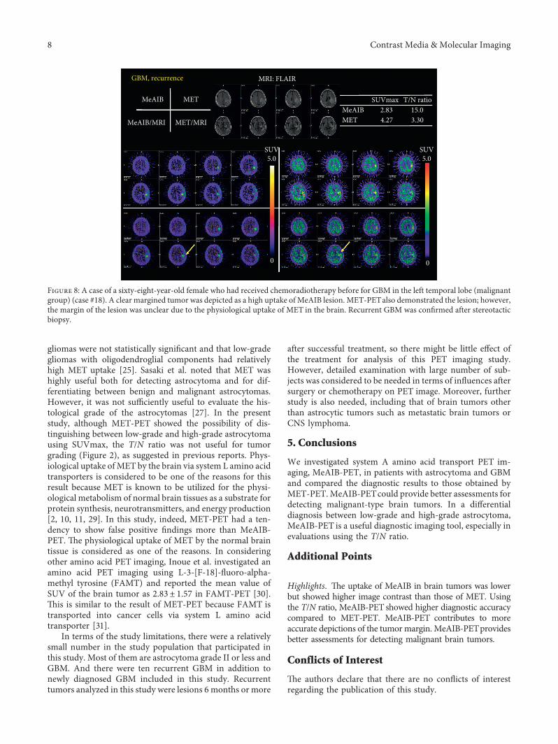
Figures 3 and 4 show typical cases in the benign group,which were diagnosed as astrocytoma grade II and low-grade glioma after surgery or stereotactic biopsy. Highuptake of METwas in the tumor, while no signi�cant uptakeof MeAIB was noted in both cases. In addition, other typicalcases in the malignant group are shown in Figures 5 and 8,which were diagnosed as GBM and recurrent GBM; a clearmargined tumor was depicted as a high uptake of MeAIBlesion. MET-PET also demonstrated the lesion with thephysiological uptake. Higher T/N ratio was noted inMeAIB-PET image, respectively.
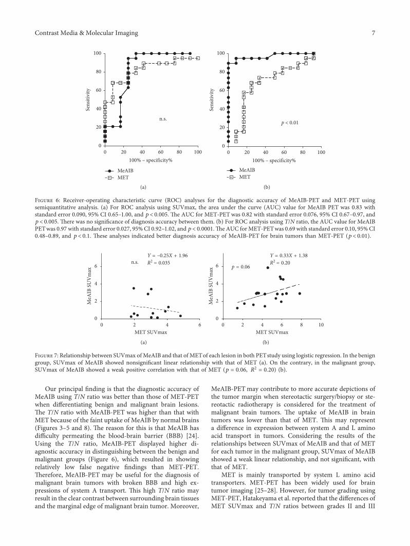
3.3. Diagnostic Accuracies of MeAIB- and MET-PET. As forthe di©erential diagnosis of brain tumors between the benignand malignant groups, receiver operating characteristiccurve (ROC) analyses for the diagnostic accuracy of MeAIB-PET and MET-PET using a semiquantitative analysis wereassessed (Figure 6). For ROC analysis using SUVmax, thearea under curve (AUC) value for MeAIB-PETwas 0.83 with
8p < 0.005
6
2
4SUVm
ax
0Benign Malignant
(a)
8p < 0.005
6
2
4SUVm
ax
0Benign Malignant
(b)
Figure 1: Comparison between benign and malignant groups by SUVmax of the lesions in MeAIB-PET (a) and MET-PET (b).
4 Contrast Media & Molecular Imaging

standard error 0.090, 95% CI 0.65–1.00, and p< 0.005. eAUC for MET-PETwas 0.82 with standard error 0.076, 95%CI 0.67–0.97, and p< 0.005. ere was no signi�cant dif-ference in diagnosis accuracy between them (Figure 6(a)).For ROC analysis using the T/N ratio, the AUC value forMeAIB-PET was 0.97 with standard error 0.027, 95% CI0.92–1.02, and p< 0.0001. e AUC for MET-PET was 0.69with standard error 0.10, 95% CI 0.48–0.89, and p< 0.1.ese analyses indicated a better diagnosis accuracy forMeAIB-PET for brain tumors than MET-PET (p< 0.01)(Figure 6(b)).
When the cuto© value was set as SUVmax� 2.0 forMeAIB-PET, the sensitivity, speci�city, and accuracy were73.7%, 91.7%, and 80.6%, respectively, while if the cuto©value was set as SUVmax� 3.5 for MET-PET, the sensitivity,
speci�city, and accuracy were 73.7%, 75.0%, and 74.2%,respectively. When the cuto© value was set as T/N ratio� 7.0for MeAIB-PET, the sensitivity, speci�city, and accuracywere 94.7%, 91.7%, and 93.5%, respectively, while if thecuto© value was set as T/N ratio� 3.0 for MET-PET, thesensitivity, speci�city, and accuracy were 57.9%, 75.0%, and64.5%, respectively.
3.4. Relationship of SUVmax in the Lesion with MeAIB andMET. Relationships between SUVmax ofMeAIB and that ofMET of each lesion in both PET studies using logistic re-gression are shown in Figure 7. In the benign group, theSUVmax of MeAIB showed a nonsigni�cant linear re-lationship with that of MET (Figure 7(a)). On the contrary,
30p < 0.001
20
10
T/N
ratio
0Benign Malignant
(a)
8
6
n.s.
4
2
T/N
ratio
0Benign Malignant
(b)
Figure 2: Comparison between benign and malignant groups by T/N ratio in MeAIB-PET (a) and MET-PET (b).
MRI: FLAIRGlioma, grade II
SUV
MET/MRIMeAIB/MRI
SUVmax T/N ratioMeAIB 0.15 1.25MET 3.03 2.37
SUV5.0
SUV5.0
0 0
MeAIB MET
Figure 3: A case of a forty-year-old male who had a di©usely irregular-shaped mass in the left frontal lobe, which was diagnosed asastrocytoma, grade II after surgery (benign group) (case #4). High uptake of METwas in the tumor, while no signi�cant uptake of MeAIBwas noted.
Contrast Media & Molecular Imaging 5

in themalignant group, the SUVmax ofMeAIB showed a weakpositive correlation with that of MET (p � 0.06, R2 � 0.20)(Figure 7(b)).
4. Discussion
System A amino acid transport is Na+- and energy-dependent, highly concentrative, and a putative regulatorof cell growth. Malignant transformation is associated withenhanced system A activity [22]. System A is specifically
capable of transporting N-methylated amino acids [23]. -eamino acid analogMeAIBwas developed as an ideal tracer forin vivo transport measurements, as the compound is non-metabolizable and concentrated in cells only via system Atransport [15, 20]. -ere are several reports regarding clinicalMeAIB-PET in patients with lymphoma [14], head and neckcancer [16], in addition to our previous study on pulmonaryand mediastinal mass lesions [18], and prostate cancer [19].However, there have been no reports evaluating the usefulnessof MeAIB-PET for the diagnosis of brain tumors.
MRI: FLAIRLow-grade glioma
MET
MET/MRI
MeAIB
MeAIB/MRI
SUVmaxMeAIB 0.85 2.07MET 3.89 2.54
SUV6.0
SUV6.0
0 0
T/N ratio
Figure 4: A case of a thirty-three-year-old male having newly diagnosed low-grade glioma in the right frontal lobe by stereotactic biopsy(benign group) (case #6). High uptake of MET was in the tumor, while no significant uptake of MeAIB was noted.
MRIGBM
MET
FLAIR Gd-DTPAT1WI
MET/MRI
MeAIB
MeAIB/MRI
SUVmaxMeAIB 4.81 32.07MET 5.82 4.44
SUV5.0
SUV5.0
0 0
T/N ratio
Figure 5: A case of a seventy-one-year-male who had newly diagnosed GBM in the left corpus callosum by stereotactic biopsy (malignantgroup) (case #13). A clear margined tumor was depicted as a high uptake of MeAIB lesion. MET-PETalso demonstrated the lesion with thephysiological uptake. Higher T/N ratio was noted in MeAIB-PET image.
6 Contrast Media & Molecular Imaging
Our principal �nding is that the diagnostic accuracy ofMeAIB using T/N ratio was better than those of MET-PETwhen di©erentiating benign and malignant brain lesions.e T/N ratio with MeAIB-PET was higher than that withMET because of the faint uptake of MeAIB by normal brains(Figures 3–5 and 8). e reason for this is that MeAIB hasdi�culty permeating the blood-brain barrier (BBB) [24].Using the T/N ratio, MeAIB-PET displayed higher di-agnostic accuracy in distinguishing between the benign andmalignant groups (Figure 6), which resulted in showingrelatively low false negative �ndings than MET-PET.erefore, MeAIB-PET may be useful for the diagnosis ofmalignant brain tumors with broken BBB and high ex-pressions of system A transport. is high T/N ratio mayresult in the clear contrast between surrounding brain tissuesand the marginal edge of malignant brain tumor. Moreover,
MeAIB-PETmay contribute to more accurate depictions ofthe tumor margin when stereotactic surgery/biopsy or ste-reotactic radiotherapy is considered for the treatment ofmalignant brain tumors. e uptake of MeAIB in braintumors was lower than that of MET. is may representa di©erence in expression between system A and L aminoacid transport in tumors. Considering the results of therelationships between SUVmax of MeAIB and that of METfor each tumor in the malignant group, SUVmax of MeAIBshowed a weak linear relationship, and not signi�cant, withthat of MET.
MET is mainly transported by system L amino acidtransporters. MET-PET has been widely used for braintumor imaging [25–28]. However, for tumor grading usingMET-PET, Hatakeyama et al. reported that the di©erences ofMET SUVmax and T/N ratios between grades II and III
METMeAIB
n.s.
0
20
40
60
80
100
Sens
itivi
ty
20 40 60 80 1000100% – specificity%
(a)
100
80
40
60
METMeAIB
20p < 0.01
Sens
itivi
ty
010040 60 80200
100% – specificity%
(b)
Figure 6: Receiver-operating characteristic curve (ROC) analyses for the diagnostic accuracy of MeAIB-PET and MET-PET usingsemiquantitative analysis. (a) For ROC analysis using SUVmax, the area under the curve (AUC) value for MeAIB PET was 0.83 withstandard error 0.090, 95% CI 0.65–1.00, and p< 0.005. e AUC for MET-PET was 0.82 with standard error 0.076, 95% CI 0.67–0.97, andp< 0.005. ere was no signi�cance of diagnosis accuracy between them. (b) For ROC analysis using T/N ratio, the AUC value for MeAIBPETwas 0.97 with standard error 0.027, 95%CI 0.92–1.02, and p< 0.0001.e AUC forMET-PETwas 0.69 with standard error 0.10, 95%CI0.48–0.89, and p< 0.1. ese analyses indicated better diagnosis accuracy of MeAIB-PET for brain tumors than MET-PET (p< 0.01).
6
Y = –0.25X + 1.96R2 = 0.035n.s.
4
4 60
2
2
MeA
IB S
UV
max
MET SUVmax0
(a)
6
Y = 0.33X + 1.38R2 = 0.20
4
MeA
IB S
UV
max
MET SUVmax
0
2
4 86 1020
p = 0.06
(b)
Figure 7: Relationship between SUVmax ofMeAIB and that of METof each lesion in both PETstudy using logistic regression. In the benigngroup, SUVmax of MeAIB showed nonsigni�cant linear relationship with that of MET (a). On the contrary, in the malignant group,SUVmax of MeAIB showed a weak positive correlation with that of MET (p � 0.06, R2 � 0.20) (b).
Contrast Media & Molecular Imaging 7
gliomas were not statistically significant and that low-gradegliomas with oligodendroglial components had relativelyhigh MET uptake [25]. Sasaki et al. noted that MET washighly useful both for detecting astrocytoma and for dif-ferentiating between benign and malignant astrocytomas.However, it was not sufficiently useful to evaluate the his-tological grade of the astrocytomas [27]. In the presentstudy, although MET-PET showed the possibility of dis-tinguishing between low-grade and high-grade astrocytomausing SUVmax, the T/N ratio was not useful for tumorgrading (Figure 2), as suggested in previous reports. Phys-iological uptake ofMET by the brain via system L amino acidtransporters is considered to be one of the reasons for thisresult because MET is known to be utilized for the physi-ological metabolism of normal brain tissues as a substrate forprotein synthesis, neurotransmitters, and energy production[2, 10, 11, 29]. In this study, indeed, MET-PET had a ten-dency to show false positive findings more than MeAIB-PET. -e physiological uptake of MET by the normal braintissue is considered as one of the reasons. In consideringother amino acid PET imaging, Inoue et al. investigated anamino acid PET imaging using L-3-[F-18]-fluoro-alpha-methyl tyrosine (FAMT) and reported the mean value ofSUV of the brain tumor as 2.83± 1.57 in FAMT-PET [30].-is is similar to the result of MET-PET because FAMT istransported into cancer cells via system L amino acidtransporter [31].
In terms of the study limitations, there were a relativelysmall number in the study population that participated inthis study. Most of them are astrocytoma grade II or less andGBM. And there were ten recurrent GBM in addition tonewly diagnosed GBM included in this study. Recurrenttumors analyzed in this study were lesions 6 months or more
after successful treatment, so there might be little effect ofthe treatment for analysis of this PET imaging study.However, detailed examination with large number of sub-jects was considered to be needed in terms of influences aftersurgery or chemotherapy on PET image. Moreover, furtherstudy is also needed, including that of brain tumors otherthan astrocytic tumors such as metastatic brain tumors orCNS lymphoma.
5. Conclusions
We investigated system A amino acid transport PET im-aging, MeAIB-PET, in patients with astrocytoma and GBMand compared the diagnostic results to those obtained byMET-PET.MeAIB-PETcould provide better assessments fordetecting malignant-type brain tumors. In a differentialdiagnosis between low-grade and high-grade astrocytoma,MeAIB-PET is a useful diagnostic imaging tool, especially inevaluations using the T/N ratio.
Additional Points
Highlights. -e uptake of MeAIB in brain tumors was lowerbut showed higher image contrast than those of MET. Usingthe T/N ratio, MeAIB-PET showed higher diagnostic accuracycompared to MET-PET. MeAIB-PET contributes to moreaccurate depictions of the tumormargin. MeAIB-PETprovidesbetter assessments for detecting malignant brain tumors.
Conflicts of Interest
-e authors declare that there are no conflicts of interestregarding the publication of this study.
MRI: FLAIRGBM, recurrence
MET
MET/MRIMeAIB/MRI
SUVmaxMeAIB 2.83 15.0MET 4.27 3.30
SUV5.0
SUV5.0
0 0
MeAIB T/N ratio
Figure 8: A case of a sixty-eight-year-old female who had received chemoradiotherapy before for GBM in the left temporal lobe (malignantgroup) (case #18). A clear margined tumor was depicted as a high uptake of MeAIB lesion. MET-PETalso demonstrated the lesion; however,the margin of the lesion was unclear due to the physiological uptake of MET in the brain. Recurrent GBM was confirmed after stereotacticbiopsy.
8 Contrast Media & Molecular Imaging

References
[1] G. Antoni and B. Langstrom, “Radiopharmaceuticals: molecularimaging using positron emission tomography,” Handbook ofExperimental Pharmacology, vol. 185, no. 1, pp. 177–201, 2008.
[2] P. L. Jager, W. Vaalburg, J. Pruim et al., “Radiolabeled aminoacids: basic aspects and clinical applications in oncology,”Journal of Nuclear Medicine, vol. 42, no. 3, pp. 432–445, 2001.
[3] B. Gulyas and C. Halldin, “New PET radiopharmaceuticalsbeyond FDG for brain tumor imaging,” Quarterly Journal ofNuclear Medicine and Molecular Imaging, vol. 56, no. 2,pp. 173–190, 2012.
[4] W. Wadsak and M. Mitterhauser, “Basics and principles ofradiopharmaceuticals for PET/CT,” European Journal ofRadiology, vol. 73, no. 3, pp. 461–469, 2010.
[5] U.Metser and E. Even-Sapir, “Increased (18)F-fluorodeoxyglucoseuptake in benign, nonphysiologic lesions found on whole-bodypositron emission tomography/computed tomography (PET/CT):accumulated data from four years of experience with PET/CT,”Seminars in Nuclear Medicine, vol. 37, no. 3, pp. 206–222, 2007.
[6] P. D. Shreve, Y. Anzai, and R. L. Wahl, “Pitfalls in oncologic di-agnosis with FDG PET imaging: physiologic and benign variants,”Radiographics, vol. 19, no. 1, pp. 61–77, quiz 150-151, 1999.
[7] Y. Kuang, F. Wang, D. J. Corn, H. Tian, and Z. Lee, “In vitrocharacterization of uptake mechanism of L-[methyl-(3)H]-methionine in hepatocellular carcinoma,” Molecular Imagingand Biology, vol. 16, no. 4, pp. 459–468, 2014.
[8] M. Yoshimoto, H. Kurihara, N. Honda et al., “Predominantcontribution of L-type amino acid transporter to 4-borono-2-(18)F-fluoro-phenylalanine uptake in human glioblastoma cells,”Nuclear Medicine and Biology, vol. 40, no. 5, pp. 625–629, 2013.
[9] S. Leskinen-Kallio, U. Ruotsalainen, K. Nagren, M. Teras, andH. Joensuu, “Uptake of carbon-11-methionine and fluo-rodeoxyglucose in non-Hodgkin’s lymphoma: a PET study,”Journal of Nuclear Medicine, vol. 32, no. 6, pp. 1211–1218, 1991.
[10] Y. Fujibayashi, K. Kawai, Y. Yonekura et al., “Problems of[S-methyl-11C]-L-methionine as a protein synthesis markerin the pancreas,” Annals of Nuclear Medicine, vol. 4, no. 1,pp. 29–33, 1990.
[11] H. N. Christensen, “Role of amino acid transport andcountertransport in nutrition and metabolism,” PhysiologicalReviews, vol. 70, no. 1, pp. 43–77, 1990.
[12] A. van Waarde, P. L. Jager, K. Ishiwata, R. A. Dierckx, andP. H. Elsinga, “Comparison of sigma-ligands and metabolicPET tracers for differentiating tumor from inflammation,”Journal of Nuclear Medicine, vol. 47, no. 1, pp. 150–154, 2006.
[13] Y. Yamada, Y. Uchida, K. Tatsumi et al., “Fluorine-18-fluorodeoxyglucose and carbon-11-methionine evaluationof lymphadenopathy in sarcoidosis,” Journal of NuclearMedicine, vol. 39, no. 7, pp. 1160–1166, 1998.
[14] E. Sutinen, S. Jyrkkio, T. Gronroos et al., “Biodistribution of[11C] methylaminoisobutyric acid, a tracer for PETstudies onsystem A amino acid transport in vivo,” European Journal ofNuclear Medicine, vol. 28, no. 7, pp. 847–854, 2001.
[15] S. Kagawa, R. Nishii, T. Higashi et al., “Relationship between[14C]MeAIB uptake and amino acid transporter family geneexpression levels or proliferative activity in a pilot study inhuman carcinoma cells: Comparison with [3H]methionineuptake,”Nuclear Medicine and Biology, vol. 49, pp. 8–15, 2017.
[16] E. Sutinen, S. Jyrkkio, K. Alanen, K. Nagren, and H. Minn,“Uptake of [N-methyl-11C]alpha-methylaminoisobutyricacid in untreated head and neck cancer studied by PET,”European Journal of NuclearMedicine andMolecular Imaging,vol. 30, no. 1, pp. 72–77, 2003.
[17] M. R. Asola, K. A. Virtanen, P. Peltoniemi et al., “Amino aciduptake in the skeletal muscle measured using [11C]methyl-aminoisobutyrate (MEAIB) and PET,” European Journal ofNuclear Medicine and Molecular Imaging, vol. 29, no. 11,pp. 1485–1491, 2002.
[18] R. Nishii, T. Higashi, S. Kagawa et al., “Diagnostic usefulness of anamino acid tracer, alpha-[N-methyl-(11)C]-methylaminoisobutyricacid ((11)C-MeAIB), in the PET diagnosis of chest malignancies,”Annals of Nuclear Medicine, vol. 27, no. 9, pp. 808–821, 2013.
[19] M. K. Arimoto, T. Higashi, R. Nishii et al., “(11)C-methylaminoisobutyric acid (MeAIB) PET for evaluation ofprostate cancer: compared with (18)F-fluorodeoxyglucose PET,”Annals of Nuclear Medicine, vol. 30, no. 8, pp. 553–562, 2016.
[20] K. Nagren, E. Sutinen, and S. Jyrkkio, “[N-methyl-11C]MeAIB, a tracer for system A amino acid transport: prepa-ration from [11C]methyl triflate and HPLC metaboliteanalysis of plasma samples after intravenous administration inman,” Journal of Labelled Compounds and Radiopharma-ceuticals, vol. 43, no. 10, pp. 1013–1021, 2000.
[21] M.Morooka, K. Kubota, H. Kadowaki et al., “11C-methioninePET of acute myocardial infarction,” Journal of NuclearMedicine, vol. 50, no. 8, pp. 1283–1287, 2009.
[22] J. McConathy and M. M. Goodman, “Non-natural aminoacids for tumor imaging using positron emission tomographyand single photon emission computed tomography,” Cancerand Metastasis Reviews, vol. 27, no. 4, pp. 555–573, 2008.
[23] H. N. Christensen, D. L. Oxender, M. Liang, and K. A. Vatz,“-e use of N-methylation to direct route of mediatedtransport of amino acids,” Journal of Biological Chemistry,vol. 240, no. 9, pp. 3609–3616, 1965.
[24] R. G. Blasberg, J. D. Fenstermacher, and C. S. Patlak,“Transport of alpha-aminoisobutyric acid across brain cap-illary and cellular membranes,” Journal of Cerebral Blood Flowand Metabolism, vol. 3, no. 1, pp. 8–32, 1983.
[25] T. Hatakeyama, N. Kawai, Y. Nishiyama et al., “11C-methionine (MET) and 18F-fluorothymidine (FLT) PET inpatients with newly diagnosed glioma,” European Journal ofNuclear Medicine and Molecular Imaging, vol. 35, no. 11,pp. 2009–2017, 2008.
[26] A. H. Jacobs, A. -omas, L. W. Kracht et al., “18F-fluoro-L-thymidine and 11C-methylmethionine as markers of in-creased transport and proliferation in brain tumors,” Journalof Nuclear Medicine, vol. 46, no. 12, pp. 1948–1958, 2005.
[27] M. Sasaki, Y. Kuwabara, T. Yoshida et al., “A comparativestudy of thallium-201 SPET, carbon-11 methionine PET andfluorine-18 fluorodeoxyglucose PET for the differentiation ofastrocytic tumours,” European Journal of Nuclear Medicine,vol. 25, no. 9, pp. 1261–1269, 1998.
[28] J. Hatazawa, K. Ishiwata, M. Itoh et al., “Quantitative eval-uation of L-[methyl-C-11] methionine uptake in tumor usingpositron emission tomography,” Journal of Nuclear Medicine,vol. 30, no. 11, pp. 1809–1813, 1989.
[29] T. K. Narayanan, S. Said, J. Mukherjee et al., “A comparativestudy on the uptake and incorporation of radiolabeled methi-onine, choline and fluorodeoxyglucose in human astrocytoma,”Molecular Imaging and Biology, vol. 4, no. 2, pp. 147–156, 2002.
[30] T. Inoue, T. Shibasaki, N. Oriuchi et al., “18F alpha-methyltyrosine PETstudies in patients with brain tumors,” Journal ofNuclear Medicine, vol. 40, no. 3, pp. 399–405, 1999.
[31] G. Miyashita, T. Higuchi, N. Oriuchi et al., “(1)(8)F-FAMTuptake correlates with tumor proliferative activity in oralsquamous cell carcinoma: comparative study with (1)(8)F-FDG PET and immunohistochemistry,” Annals of NuclearMedicine, vol. 24, no. 8, pp. 579–584, 2010.
Contrast Media & Molecular Imaging 9

Stem Cells International
Hindawiwww.hindawi.com Volume 2018
Hindawiwww.hindawi.com Volume 2018
MEDIATORSINFLAMMATION
of
EndocrinologyInternational Journal of
Hindawiwww.hindawi.com Volume 2018
Hindawiwww.hindawi.com Volume 2018
Disease Markers
Hindawiwww.hindawi.com Volume 2018
BioMed Research International
OncologyJournal of
Hindawiwww.hindawi.com Volume 2013
Hindawiwww.hindawi.com Volume 2018
Oxidative Medicine and Cellular Longevity
Hindawiwww.hindawi.com Volume 2018
PPAR Research
Hindawi Publishing Corporation http://www.hindawi.com Volume 2013Hindawiwww.hindawi.com
The Scientific World Journal
Volume 2018
Immunology ResearchHindawiwww.hindawi.com Volume 2018
Journal of
ObesityJournal of
Hindawiwww.hindawi.com Volume 2018
Hindawiwww.hindawi.com Volume 2018
Computational and Mathematical Methods in Medicine
Hindawiwww.hindawi.com Volume 2018
Behavioural Neurology
OphthalmologyJournal of
Hindawiwww.hindawi.com Volume 2018
Diabetes ResearchJournal of
Hindawiwww.hindawi.com Volume 2018
Hindawiwww.hindawi.com Volume 2018
Research and TreatmentAIDS
Hindawiwww.hindawi.com Volume 2018
Gastroenterology Research and Practice
Hindawiwww.hindawi.com Volume 2018
Parkinson’s Disease
Evidence-Based Complementary andAlternative Medicine
Volume 2018Hindawiwww.hindawi.com
Submit your manuscripts atwww.hindawi.com