duhok medical - uod
TRANSCRIPT
1
University of DuhokCollege of Medicine
VOLUME 7 ISSUE 1 DECEMBER 2013
The Official Journal of Duhok College of Medicine
DuhokMedical
Journal
ISSN: 2071-7326Duhok University Press

This page is left intentionally
Duhok Medical Journal Volume 7, Number 1, 2013
Duhok Med J
PATRON
Dr. ARIF Y. BALATAY, MBChB, Ph.D (Ophthalmology)
Dean, Faculty of Medical Sciences, University of Duhok
EDITOR-IN-CHIEF
Prof. SAMIM A. AL-DABBAGH, MBChB, DTM&H, D. Phil, FFPH
Head, Department of Family and Community Medicine, Duhok College of Medicine
MEMBER
Prof. DHIA J. AL-TIMIMI, BSc (pharm), Mphil, PhD
Head, Department of Clinical Biochemistry, Duhok College of Medicine
MEMBER
Prof. NASIR A. AL-ALLAWI, MBChB, MSc, PhD
Head, Department of Pathology, Duhok College of Medicine
MEMBER
Dr. FARHAD K. SULAYVANI, MBChB, CABS, FRCS
Assistant professor, Department of Surgery, Duhok College of Medicine
MEMBER
Dr. MAIDA Y. SHAMDEEN, MBChB, MRCOG, RECOG
Assistant professor, Department of Obstetrics and Gynecology, Duhok College of
Medicine
EDITORIAL BOARD
Duhok Medical Journal Volume 7, Number 1, 2013
MEMBER
Dr. MOHAMMED T. RASOOL, MBChB, FRCPG, FRCP (London)
Assistant professor, Head, Department of Internal Medicine, Duhok College of
Medicine
MEMBER
Dr. ABDULGHAFOOR S. ABDULKAREEM, MBChB, FICMS
Assistant professor of Urology, Department of Surgery, Duhok College of Medicine
EDITORIAL ASSISTANT
Dr. ABDULLA J. RAJAB, MBChB, MPH, PhD
Director of Department of Continuing Medical Education, Duhok Directorate of Health
Dr. HUSHYAR M. SULAIMAN, MBChB, MSc, MHS (Health Policy)
Department of Continuing Medical Education, Duhok Directorate of Health
Submission of Manuscript:
Manuscripts should be submitted to:
The Editor,
Duhok Medical Journal,
Duhok College of Medicine,
Post address: Nakhoshkhana Road 9, 1014, AM, Duhok, Iraq.
Telephone No.: 00964-62-7224268 EXT 115
E-mail: [email protected]
Electronic submission of articles is also accepted
Duhok Medical Journal Volume 7, Number 1, 2013
Duhok Med J
Prof. GAZI ZIBARI, MD, FACS, FICS
Director of W.K./L.S.U. Regional Transplant Program, Louisiana, USA
Prof. AHMAD MB. AL-KAFAJEI, MBChB, DTM&H, PhD, MFCM
Head, Department of Public Health, Jordanian College of Medical Sciences
Prof. FAYSIL A. ALNASIR, FPC, FRCGP, MICGP, PhD
Vice President, Arabian Gulf University, Bahrain
Dr. ASAD A. ZOMA FRCP, FRCPG, FACR
Consultant Physician in Rheumatology and Senior Clinical Lecturer
Lanarkshire Health Board and Glasgow University, Scotland, United Kingdom
Dr. NADA J. AL-WARD, MBChB, MFCM
Public Health Specialist, WHO, Geneva
Dr. CHRISTINE M. EVANS, MBChB, MD Ed, FRCS, FRCS Ed
Urologist, North Wales, United Kingdom
Dr. FARHAD U. HUWEZ, MBChB, PhD, MRCPI, FRCP, FRCPG
Consultant Physician / Lead Physician of Stroke Services, Basildon & Thurrock NHS
Trust, Basildon Hospital, United Kingdom
Dr. ABDULBAGHI AHMAD, MD, PhD
Consultant Child Psychiatrist and Director of Studies, Department of Neuroscience,
Child and Adolescence Psychiatry, Uppsala University Hospital, Sweden
ADVISORY BOARD

This page is left intentionally
Duhok Medical Journal Volume 7, Number 1, 2013
Duhok Med J
Aims and Scope Duhok Medical Journal is a peer reviewed journal issued bi – annually by Duhok College
of Medicine. Scientific and clinical researches are the main issues. The journal also publishes short articles,
letters to editors, review articles and case reports.
General The Duhok Medical Journal is a signatory journal to the uniform requirement for manuscripts
submitted to biomedical journals, February 2006 [updated 2009] (http://www.icmje.org).
To present your original work for consideration three manuscript copies written in English together
with Kurdish and Arabic abstracts should be submitted to the editor. All authors are required to provide the
manuscript on a CD labeled with the name and title of the paper.
Preparation of the manuscript The manuscript should be typed double spaced as normal text on one side
of the paper in single column format, font size 14 pt, paper type A4, 1″ margin at each side and each of the
following sections should begin on a new page in the following sequence:
1- Title page; should include the following: title, font size 16 pt, each author's full name,
academic degree(s), scientific title (if available), institutional affiliation, full contact
information including emails. If there are more than one author, article should include author to
whom correspondence should be addressed including the scientific title (if available),
institution affiliation, address, email, telephone.
2- Structured abstract; of no more than 250 words including background and objectives,
methods, results, and conclusions.
3 – 10 keywords or phrases should be put at the end of each abstract (Printed in bold font;
size12 pt).
3- Body of the text; structured in an IMRAD style;
(Introduction, Methods, Results and Discussion).
4- Acknowledgment (if any.)
5- References.
6- Tables with legends.
7- Illustrations with legends.
8- Structured Kurdish abstract including title in Kurdish.
ôØó“Žïq ì@wäbàŠb÷L@æŽïÙŽîŠ@ŽôåïÜíØóÄL@ãb−ó÷L@ãb−ó÷Šò†
9- Structured Arabic abstract including title in Arabic.
اتالاستنتاج ،النتائج البحث، طرقو اهداف البحث، خلفية
Tables Each table must be typed on separate page and should follow the reference list. All the tables must
be numbered consecutively in the order of their first citation in the text. Supply a brief title for each on top
and place explanatory matter in foot notes not in the heading (if needed). Tables should be simple and not
duplicated in the text. Percentages are included with numbers in the same cells but in brackets.
Illustrations Graphs, line drawing, photographs, printed x rays and other illustrations are accepted only if
they add to the evidence of the text. They should be of a high quality and suitable for reproduction. They
should be numbered consecutively according to the order in which they have been first cited in the text.
Supply a brief title beneath each illustration. Graphs should have white background; should be colored and
non 3-dimensional figure; and should have labels for X and Y axis.
INSTRUCTIONS FOR AUTHORS
Duhok Medical Journal Volume 7, Number 1, 2013
Numbers and Units Measurements of length, height, weight and volume should be reported in metric
units. Temperature in degrees Celsius, blood pressure should be expressed in mmHg and all hematologic
and clinical chemistry measurements in SI units.
Abbreviations should be defined on first use and then applied consistently throughout the article. Avoid
abbreviations in the title and abstract.
References should be numbered both in text and in the list of references in the order in which they appear
in the text. The punctuation of the Vancouver style should be followed; if the original reference is not
verified by the author, it should be given in the list of references followed by (cited by) and the paper it
was referring to. The titles of journals should be abbreviated according to the style used in Index Medicus.
This can be obtained from website (http://www.nlm.nih.gov/). The author is responsible for the accuracy of
references. The following are examples of the three most common types of citations:
The article citation: if six authors or fewer list all; if seven or more authors list the first six and then add "et
al":
1- Nuwayhid IA, Yamout B, Azar G, Kambris MA. Narghile (hubble bubble) smoking, low birth weight,
and other pregnancy outcomes. Am J Epidemiol. 1998;148(4):375-83.
Book citation, noting chapter and authors:
2- Arevalo JA, Nesbitt TS. Medical problems during pregnancy. In: Taylor RB, editor. Family medicine:
principles and practice. 6th ed. New York: Springer – Verlag; 2003. p. 109-16.
Electronic source:
3- Garfinkel PE, Lin E, Goering P. Should amenorrhoea be necessary for the diagnosis of anorexia
nervosa? Br J Psych [Internet]. 1996 [cited 1999 Aug 17];168(4):500-6. Available from:
URL:http://biomed.niss.ac.uk
Authorship and consent form All authors must give signed consent (Form No.1- Submission Form),
which should accompany the manuscript. The letter should say "this manuscript is an unpublished work,
which is not under consideration elsewhere in the record. Authors are requested to state an approximate
estimate of their contribution in the study, sign the form and send it with the manuscript.
Authors must declare if they have any competing interests in the study and to specify any funds given to
conduct the study.
Ethical considerations When experiments on humans are being reported the whole work in the
manuscript should conform to the ethical standards of the responsible committee on human
experimentation.
Submission of manuscript
Manuscripts should be submitted to:
The Editor,
Duhok Medical Journal,
Duhok College of Medicine,
Post address: Nakhoshkhana Road 9, 1014, AM, Duhok, Iraq.
Telephone no.: 00964-62-7224268 EXT 115
E-mail: [email protected]
Electronic submission of articles is also accepted
N.B. * Accepted manuscripts may be altered by the editorial board of Duhok Medical Journal to conform
to details of the journal publication style.
** The Editorial Board of Duhok Medical Journal accepts no responsibility for statement made by
authors in articles published by the journal.
Duhok Medical Journal Volume 7, Number 1, 2013
Duhok Med J
VALIDITY OF CLINICAL FEATURES IN THE DIAGNOSIS OF MITRAL VALVE
PROLAPSE
AMJED S. FARES, LARS A. PESCHKE, QAYSER S. HABEEB ………..…..….….... 1-10
PULMONARY HYDATID CYST IN DUHOK PROVINCE
MOHAMMED SALIH AL-ANI, ASHUR Y. IZAC, AHMED M.S. TAHIR ……...…. 11-19
ANGIOGRAPHIC VARIATIONS OF RENAL ARTERY AMONG DONORS OF
KIDNEY IN DUHOK CITY
AMEEN M. MOHAMMAD, MOHAMMED A. ABDULRAHMAN, SHAKER S.
JABALY…......................................................................................................................... 20-28
HEPATITIS B VACCINATION AMONG HEALTH CARE WORKERS IN ERBIL
CITY, IRAQ
SAMIR M. OTHMAN, KAMERAN H. ISMAIL ……...............................……………. 29-36
THE EPIDEMIOLOGICAL, CLINICAL AND LABORATORY CHARACTERISTICS
OF TYPHOID FEVER OUTBREAK IN SULAIMANI GOVERNORATE DURING
2007-2008
MOHAMMED O. MOHAMMED, HEMN M. MUSTAFA, MOHAMMED A.
ALSHEIKHANI ……….………………………………………………...……..…….... 37-48
VITAMIN D STATUS IN PREGNANT AND NON-PREGNANT WOMEN IN A
KURDISTAN REGION-NORTH IRAQ
DHIA J. AL-TIMIMI, FERWERDIN SH. BARZINGI, NARIN A. MOSSA ..…...…. 49-56
HYPERURICEMIA AMONG PATIENTS WITH METABOLIC SYNDROME
ATTENDING DUHOK DIABETES CENTER
IDRIS H. AHMED, DHIA J. AL-TIMIMI , MOHAMMED T. RASOOL ..…………. 57-64
PHYTOSTEROLS AND PHYTOSTANOLS IN PALM OILS
KAMAL A. KETULY ……..…............................................………………………...…. 65-81
CONTENTS
Duhok Medical Journal Volume 7, Number 1, 2013
CARDIAC ANGIOSARCOMA WITH HAEMOPERICARDIUM
HATEM L. AL-FARHAN, AFTAB A. SIDDIQUI, YASSER W. SHAREF, ADIL F. AL-
LAWATI ..…................................................................................................................…. 81-85
PARAGANGLIOMA OF THE MIDDLE EAR. A CASE REPORT IN DUHOK - IRAQ
ALAA H. RAZAK, JENA WARDA ……………...…………................………………. 86-92
ERRATUM: A CYTOPATHOLOGICAL STUDY OF THE EFFECT OF SMOKING
ON THE ORAL EPITHELIAL CELLS IN RELATION TO ORAL HEALTH STATUS
BY THE MICRONUCLEUS ASSAY. Duhok Med J 2012;6 Suppl 3:170-177
SAEED H. SAEED, WASEN H. YOUNIS ……………................………………. 93-94
1
Duhok Medical Journal Volume 7, Number 1, 2013
VALIDITY OF CLINICAL FEATURES IN THE DIAGNOSIS OF MITRAL VALVE
PROLAPSE
AMJED S. FARES, MBChB, Higher Diploma in Family Medicine*
LARS A. PESCHKE, M.D., ABFM (American Board of Family Medicine)**
QAYSER S. HABEEB, MBChB, MSc, DIM***
Submitted 7 Jun 2012; accepted 5 Sep 2013
ABSTRACT
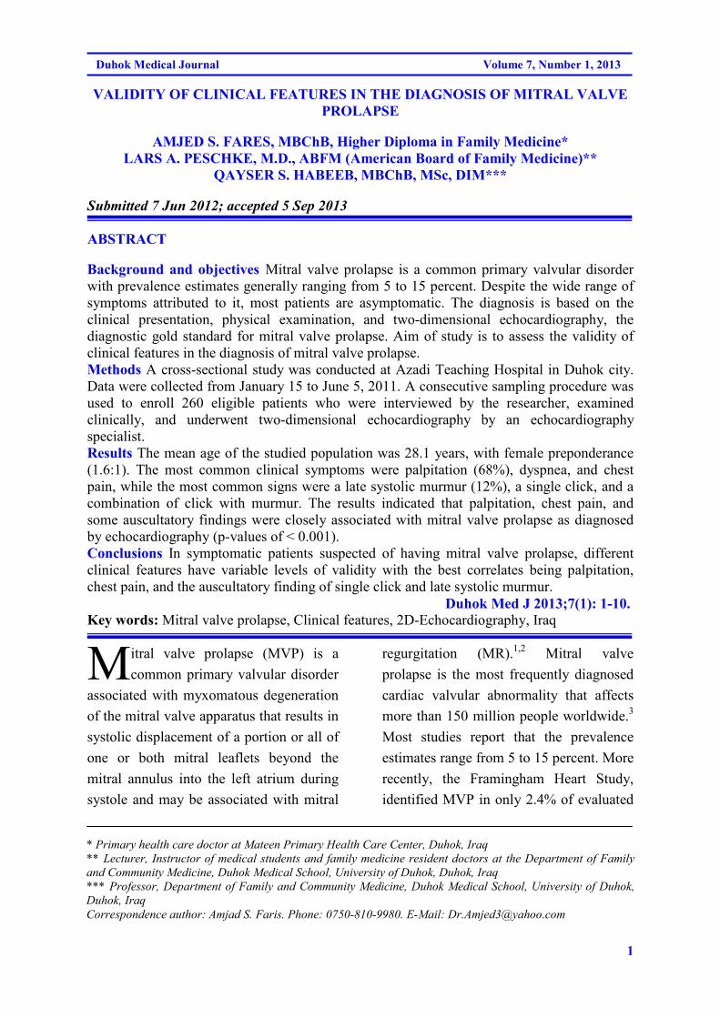
Background and objectives Mitral valve prolapse is a common primary valvular disorder
with prevalence estimates generally ranging from 5 to 15 percent. Despite the wide range of
symptoms attributed to it, most patients are asymptomatic. The diagnosis is based on the
clinical presentation, physical examination, and two-dimensional echocardiography, the
diagnostic gold standard for mitral valve prolapse. Aim of study is to assess the validity of
clinical features in the diagnosis of mitral valve prolapse.
Methods A cross-sectional study was conducted at Azadi Teaching Hospital in Duhok city.
Data were collected from January 15 to June 5, 2011. A consecutive sampling procedure was
used to enroll 260 eligible patients who were interviewed by the researcher, examined
clinically, and underwent two-dimensional echocardiography by an echocardiography
specialist.
Results The mean age of the studied population was 28.1 years, with female preponderance
(1.6:1). The most common clinical symptoms were palpitation (68%), dyspnea, and chest
pain, while the most common signs were a late systolic murmur (12%), a single click, and a
combination of click with murmur. The results indicated that palpitation, chest pain, and
some auscultatory findings were closely associated with mitral valve prolapse as diagnosed
by echocardiography (p-values of < 0.001).
Conclusions In symptomatic patients suspected of having mitral valve prolapse, different
clinical features have variable levels of validity with the best correlates being palpitation,
chest pain, and the auscultatory finding of single click and late systolic murmur.
Duhok Med J 2013;7(1): 1-10. Key words: Mitral valve prolapse, Clinical features, 2D-Echocardiography, Iraq
itral valve prolapse (MVP) is a
common primary valvular disorder
associated with myxomatous degeneration
of the mitral valve apparatus that results in
systolic displacement of a portion or all of
one or both mitral leaflets beyond the
mitral annulus into the left atrium during
systole and may be associated with mitral
regurgitation (MR).1,2 Mitral valve
prolapse is the most frequently diagnosed
cardiac valvular abnormality that affects
more than 150 million people worldwide.3
Most studies report that the prevalence
estimates range from 5 to 15 percent. More
recently, the Framingham Heart Study,
identified MVP in only 2.4% of evaluated
M
* Primary health care doctor at Mateen Primary Health Care Center, Duhok, Iraq
** Lecturer, Instructor of medical students and family medicine resident doctors at the Department of Family and Community Medicine, Duhok Medical School, University of Duhok, Duhok, Iraq
*** Professor, Department of Family and Community Medicine, Duhok Medical School, University of Duhok, Duhok, Iraq
Correspondence author: Amjad S. Faris. Phone: 0750-810-9980. E-Mail: [email protected]
2
VALIDITY OF CLINICAL FEATURES IN THE DIAGNOSIS OF MITRAL VALVE PROLAPSE ..
subjects.4 MVP is a disease of the young
but it is uncommon before the adolescent
growth spurt occurs. Although it was once
believed that MVP was more common in
women, it is now believed that it affects
men and women equally.5
In the majority of cases, MVP is
harmless and does not cause symptoms nor
does it need to be treated. When
symptomatic, the most common
complaints are palpitations, chest
discomfort, and shortness of breath. Other
clinical features include paroxysmal
supraventricular tachycardia, presyncope,
nocturnal dyspnea, and fatigue.6 The most
characteristic clinical finding is a
midsystolic click and late systolic murmur
detected on cardiac auscultation.
As electrocardiographic (ECG)
findings, such as ST segment depression
and supraventricular tachycardia, are
nonspecific, two-dimensional
echocardiography (2D-Echocardiography)
remains the gold standard for the diagnosis
of MVP, displaying one or both leaflets
prolapsing behind mitral annulus and into
left atrium in systole.5,7
Study Rationale
Abdulla8 conducted a study in Hawler to
assess the presentations of symptomatic
patients with MVP. Intensive search by the
author did not reveal any other study in
Kurdistan Region thus raising the need for
local studies to explore the problem.
The aim of the study was to assess the
validity of clinical features in the diagnosis
of MVP in Duhok city. Specifically,
sensitivity, specificity, positive and
negative predictive values (PPV and
NPV), and likelihood ratios (LR) for the
relevant presenting signs and symptoms.
METHODS
The study was conducted at the
echocardiography unit of Azadi Teaching
Hospital where data were collected from
January 15 to June 5, 2011. A cross-
sectional study design was adopted with a
consecutive sampling procedure to enroll
patients who presented for 2D-
Echocardiography for clinically suspected
MVP. Patients were referred from
outpatient departments at Azadi Hospital,
other hospitals and primary health care
centers as well as from private clinics in
Duhok. Eligible were those who were 15
to 40 years old. Patients with secondary
MVP due to e.g. coronary artery disease,
and younger children were not included in
order to have a more uniform study
population for more accurate data
evaluation. Furthermore, patients with the
following criteria were excluded:
ischemic, rheumatic, or congenital heart
disease, mitral valve repair,
cardiomyopathy, severe left ventricular
systolic dysfunction, and patients with an
established diagnosis of mitral valve
prolapse.
A questionnaire was designed to
record pertinent data, which included
demographic characteristics of the sample;
duration of relevant symptoms, such
as palpitations, dizziness, fatigue, dyspnea,
and chest pain; auscultatory findings like
single click, multiple clicks, late systolic
murmur, pansystolic murmur, or click plus
3
Duhok Medical Journal Volume 7, Number 1, 2013
murmur. Electrocardiographic data
(namely ST-changes, arrhythmias, and
conduction defects) and echocardiographic
findings (mitral valve prolapse, involved
leaflets in positive cases, mitral
regurgitation, and myxomatous leaflet
thickening) were recorded as well.
Data entry and analysis were carried
out using Microsoft Excel 2003, SPSS16,
and Open-Epi. Descriptive data analysis
was carried out to describe the distribution
of patients by age and sex.
RESULTS
The study sample included 260 patients,
100 (38.5%) men and 160 (61.5%) women
with a mean age of 28.1 years (SD 7.3).
Most of the patients (47.7%) were 21-30
years old and 38.1% were 31-40 years of
age.
The most common clinical symptoms
were palpitation, dyspnea, and chest pain.
Auscultatory findings were less common
than clinical symptoms, and included late
systolic murmur, single click, and click
with murmur. Electrocardiographic
changes were mainly seen as ST-T
changes and arrhythmias, as is shown in
Table 1.
Less than half of the patients who
were referred for clinical suspicion of
MVP were echocardiographically
confirmed where the anterior mitral leaflet
was most commonly affected. Mitral
regurgitation and myxomatous changes
occurred in less than ten percent, as is
demonstrated in Table 2.
While palpitation and chest pain had
high sensitivity and negative predictive
values, other symptoms were not as well
correlated with echocardiographically
confirmed MVP. Auscultatory findings
showed high sensitivity and positive
predictive values with positive likelihood
ratios that correlated well with MVP. In
case of an auscultatory single click, the
positive likelihood ratio reached clinical
significance. ECG changes did not show
any statistically or clinically significant
correlation, as is shown in Table 3.
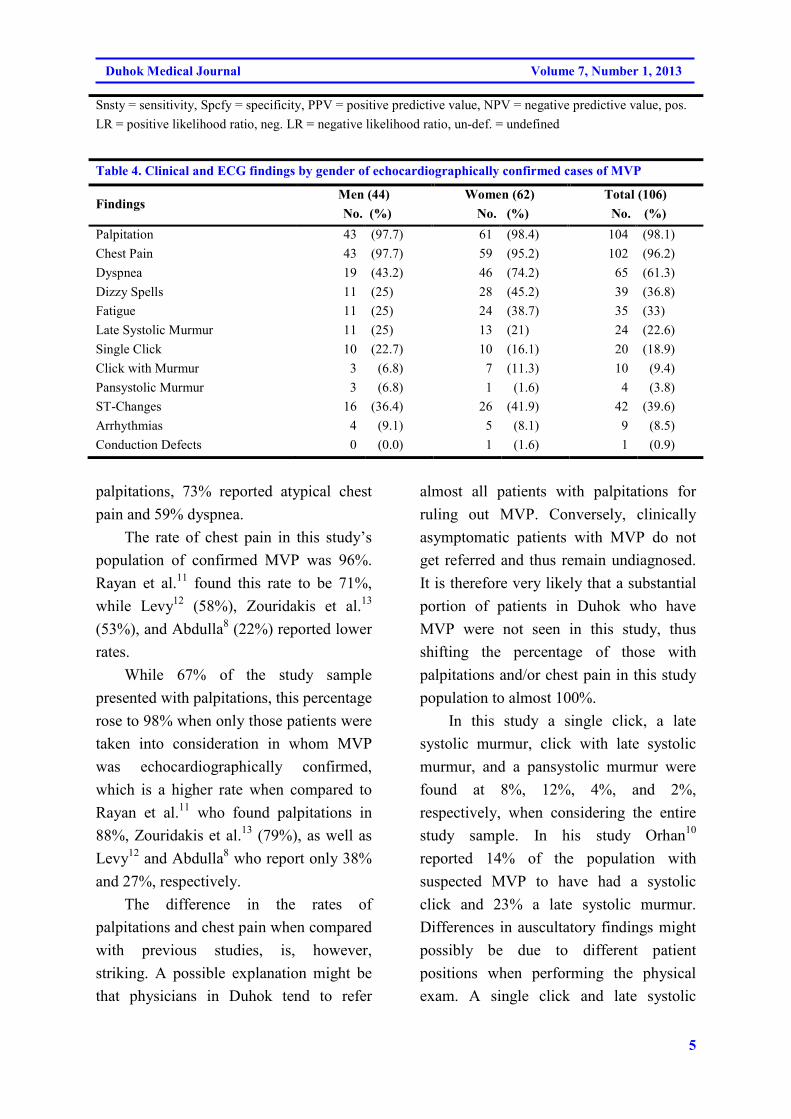
Among patients with
echocardiographically confirmed MVP,
palpitation, chest pain, and dyspnea were
the most prevalent symptoms and a late
systolic murmur and a single click on
auscultation the most common
auscultatory findings, as is shown in Table
4.
DISCUSSION
Primary mitral valve prolapse is a genetic
connective tissue disorder with an
autosomal dominant inheritance resulting
in anatomic abnormalities of the mitral
valve apparatus with a prevalence estimate
that ranges from less than 1% to 38%.9 As
the study population in this study
represents referrals to the
echocardiography unit at Azadi Teaching
Hospital, this study cannot estimate the
prevalence of MVP in Duhok.
About two thirds of all cases in this
study who were referred for suspected
MVP complained of palpitations, chest
pain, and dyspnea, which compares
roughly with a study done by Orhan10
who
found that 64% of patients with clinical
suspicion of MVP complained of
4
VALIDITY OF CLINICAL FEATURES IN THE DIAGNOSIS OF MITRAL VALVE PROLAPSE ..
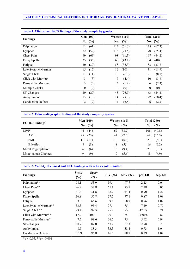
Table 1. Clinical and ECG findings of the study sample by gender
Findings Men (100)
..No. (%)
Women (160)
..No. (%)
Total (260)
.No. (%)
Palpitation 61 (61) 114 (71.3) 175 (67.3)
Dyspnea 52 (52) 118 (73.8) 170 (65.4)
Chest Pain 69 (69) 98 (61.3) 167 (64.2)
Dizzy Spells 35 (35) 69 (43.1) 104 (40)
Fatigue 30 (30) 58 (36.3) 88 (33.8)
Late Systolic Murmur 15 (15) 16 (10) 31 (11.9)
Single Click 11 (11) 10 (6.3) 21 (8.1)
Click with Murmur 3 (3) 7 (4.4) 10 (3.8)
Pansystolic Murmur 3 (3) 3 (1.9) 6 (2.3)
Multiple Clicks 0 (0) 0 (0) 0 (0)
ST-Changes 20 (20) 43 (26.9) 63 (24.2)
Arrhythmias 13 (13) 14 (8.8) 27 (10.4)
Conduction Defects 2 (2) 4 (2.5) 6 (2.3)
Table 2. Echocardiographic findings of the study sample by gender
ECHO-Findings Men (100)
...No. (%)
Women (160)
...No. (%)
Total (260)
..No. (%)
MVP 44 (44) 62 (38.7) 106 (40.8)
AML 25 (25) 44 (27.5) 69 (26.5)
PML 11 (11) 10 (6.3) 21 (8.1)
Bileaflet 8 (8) 8 (5) 16 (6.2)
Mitral Regurgitation 6 (6) 15 (9.4) 21 (8.1)
Myxomatous Changes 9 (9) 9 (5.6) 18 (6.9)
Table 3. Validity of clinical and ECG findings with echo as gold standard
Findings Snsty
(%)
Spcfy
(%) PPV (%) NPV (%) pos. LR neg. LR
Palpitation** 98.1 53.9 59.4 97.7 2.13 0.04
Chest Pain** 96.2 57.8 61.1 95.7 2.28 0.07
Dyspnea 61.3 31.8 38.2 54.4 0.90 1.22
Dizzy Spells 36.8 57.8 37.5 57.1 0.87 1.09
Fatigue 33.0 65.6 39.8 58.7 0.96 1.02
Late Systolic Murmur** 33.3 95.4 77.4 75.0 7.19 0.70
Single Click** 29.4 99.3 95.2 75.0 42.650 0.71
Click with Murmur** 17.2 100.00 100.00 75.0 undef. 0.82
Pansystolic Murmur* 07.7 98.6 66.7 75.0 5.62 0.94
ST-Changes 38.7 87.0 67.2 67.3 2.98 0.70
Arrhythmias 08.5 88.3 33.3 58.4 0.73 1.04
Conduction Defects `00.9 96.8 16.7 58.7 0.29 1.02
*p < 0.05, **p < 0.001
5
Duhok Medical Journal Volume 7, Number 1, 2013
Snsty = sensitivity, Spcfy = specificity, PPV = positive predictive value, NPV = negative predictive value, pos.
LR = positive likelihood ratio, neg. LR = negative likelihood ratio, un-def. = undefined
Table 4. Clinical and ECG findings by gender of echocardiographically confirmed cases of MVP
Findings Men (44)
..No. (%)
Women (62)
..No. (%)
Total (106)
..No. (%)
Palpitation 43 (97.7) 61 (98.4) 104 (98.1)
Chest Pain 43 (97.7) 59 (95.2) 102 (96.2)
Dyspnea 19 (43.2) 46 (74.2) 65 (61.3)
Dizzy Spells 11 (25) 28 (45.2) 39 (36.8)
Fatigue 11 (25) 24 (38.7) 35 (33)
Late Systolic Murmur 11 (25) 13 (21) 24 (22.6)
Single Click 10 (22.7) 10 (16.1) 20 (18.9)
Click with Murmur 3 (6.8) 7 (11.3) 10 (9.4)
Pansystolic Murmur 3 (6.8) 1 (1.6) 4 (3.8)
ST-Changes 16 (36.4) 26 (41.9) 42 (39.6)
Arrhythmias 4 (9.1) 5 (8.1) 9 (8.5)
Conduction Defects 0 (0.0) 1 (1.6) 1 (0.9)
palpitations, 73% reported atypical chest
pain and 59% dyspnea.
The rate of chest pain in this study’s
population of confirmed MVP was 96%.
Rayan et al.11
found this rate to be 71%,
while Levy12
(58%), Zouridakis et al.13
(53%), and Abdulla8 (22%) reported lower
rates.
While 67% of the study sample
presented with palpitations, this percentage
rose to 98% when only those patients were
taken into consideration in whom MVP
was echocardiographically confirmed,
which is a higher rate when compared to
Rayan et al.11
who found palpitations in
88%, Zouridakis et al.13
(79%), as well as
Levy12
and Abdulla8 who report only 38%
and 27%, respectively.
The difference in the rates of
palpitations and chest pain when compared
with previous studies, is, however,
striking. A possible explanation might be
that physicians in Duhok tend to refer
almost all patients with palpitations for
ruling out MVP. Conversely, clinically
asymptomatic patients with MVP do not
get referred and thus remain undiagnosed.
It is therefore very likely that a substantial
portion of patients in Duhok who have
MVP were not seen in this study, thus
shifting the percentage of those with
palpitations and/or chest pain in this study
population to almost 100%.
In this study a single click, a late
systolic murmur, click with late systolic
murmur, and a pansystolic murmur were
found at 8%, 12%, 4%, and 2%,
respectively, when considering the entire
study sample. In his study Orhan10
reported 14% of the population with
suspected MVP to have had a systolic
click and 23% a late systolic murmur.
Differences in auscultatory findings might
possibly be due to different patient
positions when performing the physical
exam. A single click and late systolic
6
VALIDITY OF CLINICAL FEATURES IN THE DIAGNOSIS OF MITRAL VALVE PROLAPSE ..
murmur can usually be heard most clearly
with the patient standing up from the
sitting position. Conversely, they cannot
be heard as clearly or cannot be heard at
all when the patient is sitting, squatting, or
lying down in a supine position.
In regard to the validity of clinical
features of MVP, this study found chest
pain to have a sensitivity of 96%, NPV of
96%, and negative LR of 0.07. Clinically,
this means that in patients without chest
pain, MVP can be ruled out with more
than 95% probability.
In patients with palpitations the
sensitivity in this study sample is high with
98%, as is the NPV (98%) with a very
good negative LR of 0.04. This means that
a patient can be ruled out to have MVP
with a probability of almost 98% if he or
she does not have palpitations.
When palpitations and chest pain are
combined, the accuracy reaches almost
88% with highly significant results. The
sensitivity and NPV are 94% and 96%,
respectively, yet, even the specificity
(83%) and PPV (79%) are reasonably
good. Clinically, the absence of the
combination of palpitations and chest pain
can be used to rule out MVP with more
than 95% probability, while its presence
gives an indication that MVP may be
present.
In regard to clinical signs, an
auscultatory single click on physical
examination is very specific (99%) for the
diagnosis of MVP where the PPV (95%) is
also very good and the positive LR very
high (42.7). Clinically, this means that a
single click on auscultation can be used to
rule in MVP with a probability of more
than 95%; however, the absence of a
single click does not rule out MVP.
Yet, these numbers are not
generalizable, particularly not to the
primary health care setting, because the
study sample comprised a selected number
of patients, thus is limited by referral bias.
Different clinical symptoms and signs
have variable levels of validity with the
best correlates being palpitation and chest
pain and the auscultatory finding of single
click and late systolic murmur.
Symptomatic patients presenting with
palpitation, chest pain, single click, and
late systolic murmur, singularly or in
combination, should alert the physician to
the need for follow up investigations to
verify the high probability of MVP.
Further studies are needed to assess the
prevalence of MVP in the region and
especially to define the groups of
asymptomatic individuals with a high
probability of a positive yield.
AUTHORSHIP AND CONSENT
FORM
This manuscript is an unpublished work,
which is not under consideration elsewhere
in the record. The author’s estimated
contribution in the study is as follows:
Dr. Amjed S. Fares: developing the design
of the study; conducting the patient
interviews; collecting and interpreting the
data; translation of abstract into Kurdish
and Arabic
Dr. Lars A. Peschke: assisting in data
processing, statistical analysis;
interpretation of the results ; writing of this
journal article manuscript

7
Duhok Medical Journal Volume 7, Number 1, 2013
Dr. Qayser S. Habeeb original idea of the
study; guidance and input along the study
None of the authors have any competing
interests in the study. The authors
themselves did not receive any funds for
conducting the study.
ACKNOWLEDGMENTS
Our gratitude goes to Prof. Samim Ahmad
Al-Dabbagh, the head of the De-partment
of Community and Family Medicine, for
his kind support as well as to the Dean of
the Faculty of Medical Sciences of the
University of Duhok, Dr. Arif Younis
Salih. We also wish to thank the former
Director General of Health in Duhok, Dr.
Abdulla Sa’eed Abdulla, as well as the
current Director General, Dr. Nizar Esmat,
for their great support of family medicine.
Thanks to Dr. Saad Younis Saeed and
Dr. Hushyar Musa Sulaiman for their
support in the analysis of the data. Thanks
also to Dr. Ibtesam Salih for her kind
support and useful comments as well as to
the staff at the echocardiography unit and
the Duhok Cardiac Center in Azadi
General Teaching Hospital, who enabled
me to conduct this study.
REFERENCES
1. Stouffer GA, Sheahan RG, Lenihan
DJ. Mitral Valve Prolapse: A Review
of the Literature. Am J Med Sci.
2001;320(6):401-10.
2. Rosas-Munive E, Valenzuela-Flores
AG, Valenzuela-Flores AA. Mitral
valve prolapse. A review. Circulation.
2004;72(5):415-20.
3. Avierinos JF, Inamo J, Grigioni F,
Gersh B, Shub C, and Maurice
Enriquez-Sarano M, et al. Sex
Differences in the Morphology and
outcomes of mitral valve prolapse.
Ann Inter Med 2. 2008;149(11):787-
95.
4. Freed LA, Levy D, Levin RA, Larson
MG, Evans JC, Fuller DL, et al.
Prevalence and clinical outcome of
MVP. N Eng J Med. 1999;341:1-7.
5. Sims JM, Miracle VA. An Overview
of Mitral Valve Prolapse. Dimens Crit
Care Nurs. 2007;26(4):145-9.
6. Tamam L, Nurgul O, Mustafa SB.
Association of Idiopathic Mitral Valve
Prolapse and Panic Disorder. Croat
Med J. 2000;41(4):410-6.
7. McLachlan J, Reddy PC, Ratts TE.
Mitral valve prolapse. A common
cardiac diagnosis in women. J La State
Med. 1998;150(2):92-6.
8. Abdulla AA. A Study on Presentations
of Symptomatic Patients with Mitral
Valve Prolapse in Erbil City. [Higher
Diploma in Internal Medicine
Dissertation]. University of
Salahaddin, College of Medicine.
Erbil, Iraq. 1999.
9. Theal M, Sleik K, Anand S, Yi Q,
Yusuf S, Lonn E. Prevalence of MVP
in ethnic groups. Can J Cardiol.
2004;20(5):511-5.
10. Orhan AL, Sayar N, Nurkalem Z, Uslu
N, Erdem I, Erdem EC, et al.
Assessment of autonomic dysfunction
and anxiety levels in patients with
mitral valve prolapse. Turk Kardiyol
Dern Arş - Arch Turk Soc Cardiol.
2009;37(4):226-33.
8
VALIDITY OF CLINICAL FEATURES IN THE DIAGNOSIS OF MITRAL VALVE PROLAPSE ..
11. Rayan M, Samir S, Nagwa E, Fouad
M. In Mild Mitral Valve Prolapse,
Sticking to 2D Rules is the Rule, a 3D
Echocardiographic Study. Heart Mirror
J. 2010;4(2):200-4.
12. Levy S. Arrhythmias in the mitral
valve prolapse syndrome: Clinical
significance and management. PACE
(Pacing Clin Electrophysiol).
1992;15:1080-8.
13. Zouridakis EG, Parthenakis FI,
Kochiadakis GE, Kanoupakis EM and
Vardas PE. QT dispersion in patients
with mitral valve prolapse is related to
the echocardiographic degree of the
prolapse and mitral leaflet thickness.
Europace. 2001;3:292-8.
9
Duhok Medical Journal Volume 7, Number 1, 2013
ón‚íqón‚íqón‚íqón‚íq@@@@@@@@ôÄýíØ@Žôîóm‹�Šò†@bäìíiŠŽí’@bä‹Ùäb“ïån�ò†@íi@ôÙïåïÝØ@æŽïäb“ïä@båmbè‹ŽïÙi@bä‡äb!äó�ÜóèôÄýíØ@Žôîóm‹�Šò†@bäìíiŠŽí’@bä‹Ùäb“ïån�ò†@íi@ôÙïåïÝØ@æŽïäb“ïä@båmbè‹ŽïÙi@bä‡äb!äó�ÜóèôÄýíØ@Žôîóm‹�Šò†@bäìíiŠŽí’@bä‹Ùäb“ïån�ò†@íi@ôÙïåïÝØ@æŽïäb“ïä@båmbè‹ŽïÙi@bä‡äb!äó�ÜóèôÄýíØ@Žôîóm‹�Šò†@bäìíiŠŽí’@bä‹Ùäb“ïån�ò†@íi@ôÙïåïÝØ@æŽïäb“ïä@båmbè‹ŽïÙi@bä‡äb!äó�Üóè@@@@@@@@
��
wäbàŠb÷@ì@ôØó“ŽïqwäbàŠb÷@ì@ôØó“ŽïqwäbàŠb÷@ì@ôØó“ŽïqwäbàŠb÷@ì@ôØó“ŽïqZZZZ@ôÄýíØ@ Žôîóm‹�Šò†@bäìíiŠŽí’@ Žõì@a‰ŽîŠ@íØ@óä@¶†@æŽïîóm‹�Šò†@æŽïï’í‚óä@æî‹mó“à@ˆ@@aŠójÄbä†5@@bmóè15@@Žõ†ó�ˆóî@Næäb“ïä@ Žôi†@çb’í‚óä@bïäa‹q@ Ž¶ói@ Žõì@æŽïäb“ïäˆ@ÛóÜó�@bØò‰ŽîŠ@õaŠòŠó�@N@ì@ôÙïåïÝØ@æŽïäb“ïä@bÙŽî‹i@ Žôï’í‚óä@ ŽôÄ@bä‹Ùäb“ïån�ò†
Žôm‹Ùäb“ïån�ò†@íi@ôØòŠó�@bä‹ÙïÔbm@Ûòì@æmŠbà‰è@ónŽïè†@ò‰Žïàˆ@íØ@óî@¶†@bî@íÙï÷@ì@ôÙïåïÝØ@b�yóÐ@NØóÄ@Óó÷@´�ƒÙŽîŠ@ómbè@óåïÜí¶†@ Žôî@ôÄýíØ@ Žôîóm‹�Šò†@bäìíiŠŽí’@bä‹Ùäb“ïån�ò†@íi@ŠíuìòŠíu@æŽïî@ôÙïåïÝØ@æŽïäb“ïä@bï!ä‹�@bä‹ØŠbî†@íiˆN@@bä‡äb!äó�Üóè@wäbàŠb÷
ôÄýíØ@Žôîóm‹�Šò†@bäìíiŠŽí’@bä‹Ùäb“ïån�ò†@íi@ôÙïåïÝØ@æŽïäb“ïä@båmbè‹ŽïÙi.@@ŽôåïÜíØóÄ@æŽïÙôåïÜíØóÄ@æŽïÙôåïÜíØóÄ@æŽïÙôåïÜíØóÄ@æŽïÙîŠZZZZ@ôîó�‹i@ båïÜíØóÄ@bèça‡àb−ó÷@ óm@ N@ íØ@ÚŽï÷@Òîì‡Ü@ÚŽï÷@æŽïäí¹@ bÙŽî‹i@æm‹�Šòì@ óåmbè@•í‚óä260@@óåmbè@•í‚óä
íÙï÷@b�yóÐ@õŠói@bmóÝ‚b�@Òîì‡Ü@æm‹�Šòì@N@bØóàbå�‹q@Òîì‡Ü@óÄ@õŠóÜíØóÄ@Žôîýˆ@ŽôånÐóÙŽïrÄbš@ôn“q@ç‹Ù�yóÐ@óåmbè@ó’í‚óä@Äó÷ôn�ƒÙŽîŠ@Nýˆ@ç‹Ø@ómbè†@íÙï÷@b�yóÐ@ôÙŽï÷@Šóè@íi@a‡Ñîì‡ÜŽôîíÙï÷@ŽôØò‡äó¸ójîbm@Žôî.@@
ãb−ó÷ãb−ó÷ãb−ó÷ãb−ó÷ZZZZ@@ çb’í‚óä@ Žôîˆ@ Žôîa‹ÙŽïm28.1@@ çìíi@�q@ Žôà@ ì@ ìíi@ ßb�I1Œ6:1@ NH@ ìíi@ çbmíÕ܆@ çb’í‚óä@Óò‡Ü@ çb“ïä@æî‹mó“àI68@HEô!å�@bäb“Žï÷@ì@ô!äónåéŽïi@a‡Ñîì‡Üì@N@ìíi@¶†@Žíî@ì‹îí�@Žôî@ò‡ŽîŒ@ Žô!äò†@ôÙïåïÝØ@æŽïäb“ïä@æî‹mó“à@ìI12@HE@a‡Ñîì‡Ü@ì@ì@Âäò†@óÙÑ�
aì솊óè@bäìíjÜóÙŽïm@NŒaìbïu@æŽïäb“ïä@ßó�†@çìíióè@Œaìbïu@æŽïåmbè‹ŽïÙi@íØ@‹ØŠbî†@ Žôåmbè‹ŽïÙi@bä‡äb!äó�Üóè@N@íØ@‹ØŠbî†@bàb−ó÷@ôn“!iŽî‹i@íØ@Žôï’í‚óä@bä‹Ùäb“ïån�ò†@ßó�†@ìíióè@âØíà@bØóî‡äòíîóq@¶†@bäìíjŽïÝèí�@æŽïäb“ïä@Ûò‡åè@ì@ô!å�@bäb“Žï÷@ì@ŽôäbmíÕ܆@ŽôîíÙŽï÷@bÙ
ç‹Ø@óïmbè.@@ãb−ó÷Šò†ãb−ó÷Šò†ãb−ó÷Šò†ãb−ó÷Šò†ZZZZ@æîóè@ çb“ïä@æŽïî@ ôÄýíØ@ Žôîóm‹�Šò†@ bäìíiŠŽí’@æŽï’í‚óä@ ~@Žôåmbè‹ŽïÙi@ æŽïî@ Œaìbïu@æŽïn�b÷@ Œaìbïu@æŽïî@ôÙïåïÝØ@æŽïäb“ïä
ìíióè@¶†@æŽïî@ò‡ŽîŒ@æŽï!äò†@ì@ô!å�@bäb“Žï÷@ì@çbmíÕ܆@ßó�†@âØíà@bî‡äòíîóq@ì@çìíióèN@@
10
VALIDITY OF CLINICAL FEATURES IN THE DIAGNOSIS OF MITRAL VALVE PROLAPSE ..
الخلاصة
اض و العلامات السريرية فى تشخيص تهدل الصمام الاكليلىمصداقية الاعر
ان تهدل الصمام الاكليلي من امراض الصمامات الاولية الشائعة ، وعموما يقدر معدل شيوعة :خلفية واهداف البحث
ن من بالرغم من اتساع نطاق الاعراض التي تعزى اليه الا ان اغلب المرضى لا يعانو %. 15و % 5بنسبة تتراوح بين
ان التشخيص يعتمد على الاعراض والعلامات السريرية وفحص صدى القلب ثنائي الابعاد الذي . اى اعراض
لقد صممت هذه الدراسة لتقصي مدى الاعتماد على مختلف الاعراض و العلامات . يعتبرالفحص المرجعى المعتمد
تقصي مصداقية الاعراض والعلامات السريرية فى الهدف من البحث هو . السريرية فى تشخيص تهدل الصمام الاكليلى
.تشخيص تهدل الصمام الاكليلى
وحدة صدى القلب فى مدينة دهوك خلال الفترة / دراسة مقطعية اجريت فى مستشفى ازادى العام التعليمى :طرق البحث
مريض من 260 استخدمت طريقة الاعتيان المتعاقب لضم . 2011حزيران من عام 5كانون الثانى الى 15من
تم فحص الجميع سريريا بعد مقابلتهم من قبل . المحالين لفحص صدى القلب واللذين استوفوا ضوابط الادخال للدراسة
بعدها اجري لهم جميعا فحص صدى القلب ثنائى . الباحث وفقا لاستمارة الاستبيان المعدة خصيصا لغرض الدراسة
.لبالابعاد من قبل اختصاصي فحص صدى الق
وان أكثر الاعراض ) . 1.6:1(سنة ، اغلبيتهم من الاناث 28.1اظهرت الدراسة ان معدل العمرللمرضى :النتائج
يليه ضيق التنفس والم الصدر بينما كانت اكثر العلامات السريرية شيوعا هي النفحة % 68السريرية شيوعا هي الخفقان
click)( ثم الطقة مع النفحة الانقباضية المتاخرة single clickالمنفردة ، تليها صوت الطقة %)12(الانقباضية المتاخرة
with murmur .وبشكل عام .كما اظهرت الدراسه تفاوت في درجات مصداقية الاعراض والعلامات السريرية المختلفة
فحص صدى القلب ثنائي بينت الدراسة وجود ترابط بين الخفقان و الم الصدر وبعض اصوات القلب بالنتائج الموجبة ل
.الابعاد بدليل احصائي معنوي
ان الاعراض والعلامات السريرية المختلفة لدى المرضى المشتبه اصابتهم بتهدل الصمام الاكليلى لها :اتالاستنتاج
والبعض درجات مصداقية مختلفة الا ان اكثرها ارتباطا بالنتائج الموجبة لفحص صدى القلب هي الخفقان، الم الصدر
.click with murmur)( والطقة مع النفحة الانقباضية المتاخرة single clickالطقة المنفردة (من اصوات القلب
11
Duhok Medical Journal Volume 7, Number 1, 2013
PULMONARY HYDATID CYST IN DUHOK PROVINCE
MOHAMMED SALIH AL-ANI, F.R.C.S, F.A.C.S*
ASHUR Y. IZAC, F.I.B.M.S**
AHMED M.S. TAHIR, M.B.Ch.B***
Submitted 5 Feb 2012; accepted 28 Mar 2013
ABSTRACT
Background and objectives Thoracic surgery unit in Duhok was established in 2006 since
then the majority of our thoracotomy was for treating pulmonary hydatid cysts and its
complications. It is evident that pulmonary hydatid cyst is major problem in Duhok province.
Hydatid cyst disease is known to be endemic in many parts of Iraq and Duhok province is
one of them. As pulmonary hydatid cyst are common in Duhok, the study aims to discuss its
incidence , the way they present and the variation of symptoms from being asymptomatic to
severe complications of ruptured cysts , beside the methods used for treatment.
Methods Duhok is the centre of the province and is the only place in the province where
thoracic surgery is available, nearly all cases of pulmonary hydatid cysts from all over the
province are referred to Duhok centre. These cases are studied and evaluated before and after
surgical management in order to find what is the best way of treating them. The types of
surgery used in these cases are discussed as well as their complications. It’s a retro and
prospective study including 100 cases of pulmonary hydatid cyst operated upon January 2007
and April 2011. The study include the age of the patient the size of cyst ,the compliant of the
patient and whether the cyst was complicated or not, the types of surgery performed with its
morbidity and mortality.
Results Hundred patients had been operated by the authors in hospitals of Duhok province
(public and private). Majority of patients is aged between 11-40 years, all of them through
thoracotomy incision, cyst(s) removed with preserving lung tissue in 86 % where resection
done in 14 % of cases, re exploration in 2% and no mortality recorded.
Conclusions Surgical management is only proved curable therapy for pulmonary hydatid,
multiplicity of cysts is not uncommon surgery is highly successful with no or very low
mortality rate.
Duhok Med J 2013;7(1): 11-19. Key words: Hydatid cyst, Pulmonary , Albendazole, Thoracotomy
he genus Echinococcus contain three
species for which humans are host to
larval stage, or hydatid.1 The adult tape
warm lives in the gut of dogs and other
carnivores (as a definitive host). The ova
of these warms contaminate the grass and
vegetables in the fields. Man is
accidentally infected when he/she eats
improperly washed vegetables and
uncooked green leaves contaminated by
the eggs of the tape warm as (intermediate
host). The ova will hatch in the sheep and
human intestine and pass through the
portal system to the liver where it is the
first filter (the commonest site for hydatid
cyst disease).2 If the parasite is able to pass
T
* Assistant professor, Department of Surgery, College of Medicine- Duhok University
** Lecturer-department of Surgery, College of Medicine-Duhok University
*** S.H.O Azadi hospital
Correspondence author: Dr. Ashur Y. Izac, Department of Surgery, College of Medicine, Duhok University,
Duhok, Kurdistan Region, Iraq, E-mail: [email protected] phone: +9647504177277.
12
PULMONARY HYDATID CYST IN DUHOK PROVINCE
this filter, it will reach the pulmonary
circulation where it is trapped in the
second capillary system (the lung is the
second commonest site),hydatid cyst of the
lung grows faster than that of the liver as
the surrounding lung tissue is softer and
spongy in comparison to that of liver,
symptoms of the patient vary according to
the size and site of the cysts and whether
there are any complications.3
Diagnosis of pulmonary hydatid cyst
is generally based on clinical and
radiological findings, which vary
according to the state of the cyst.
Uncomplicated cyst seen as a rounded or
oval opaq lesions in plane chest X-Ray
while infection or rapture changes the
radiological appearance. Computed
tomography ( CT) may be helpful in
establishing the diagnosis, exclude other
pathology , guiding the surgeon for the
surgical approach. Although several
clinical laboratory studies including fiber
optic bronchoscope, Casoni’s intradermal
test , and the indirect hemaglutination test
are not used routinely for diagnosis. Ultra
sound of abdomen to detect or exclude the
presence of cysts in the abdominal cavity
(e.g. liver), especially in cases of right
pulmonary hydatid cyst as both can be
dealt with during the same surgery.4
Early attempts to treat hydatid cyst with
Mebendazole (Vermox) was used in the
Medical City in Baghdad in 1970s without
much benefit (personal communications).
Albendazole was recently used in treating
hydatid cyst disease. It is helpful and
effective in treating hepatic hydatid cysts.5
In the case of pulmonary hydatid cyst
the condition is completely different,
preoperative use of Albendazole may
enhance rupture of the cyst, possibly due
to Interference with cyst wall and / or
Interference with the intracystic pressure.6
As it is one of the rare helminthes
infections, which has not benefited from
progress in chemotherapy. Definitive
treatment still remains surgery although
there had always been a need for medical
treatment when the cyst ruptured with
chances of dissemination. Albendazole has
been used with promising results.7
METHODS
Since the establishment of the
cardiothoracic unit in Duhok early 2007,
till April 2011, Hundred patients with
pulmonary hydatid cysts are dealt with.
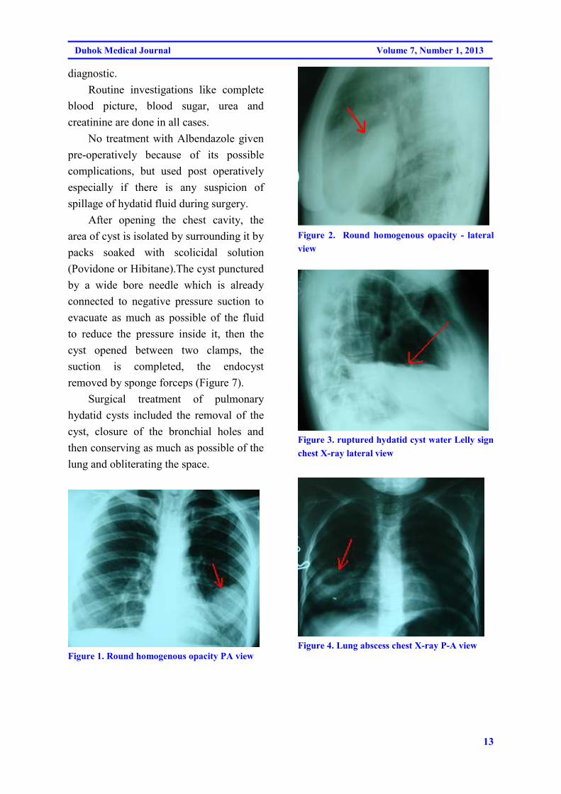
Diagnosis made mainly by chest X-
ray (2 views) is the item relied on
diagnosing the cases. Uncomplicated
pulmonary hydatid cyst will appear as a
rounded or an oval opacity surrounded by
the normal blackish appearance of the
lungs. Figure (1 & 2 ) Complicated
hydatid cyst shows a different appearance
according to the type of complication and
its state, Intrabronchial ruptured cyst might
show fluid level, water lily appearance
(Figure 3) or appears as lung abscess
(Figure 4). Rapture of he cyst to the
pleural space leads to hydropnaemothorax.
CT scan done but not as a routine
investigation, it is used to show more
details (Figure 5, 6) of the cyst.
Serological tests done only on limited
number of patients where it was done
before admission to the surgical unit as
they are only suggestive and not
13
Duhok Medical Journal Volume 7, Number 1, 2013
diagnostic.
Routine investigations like complete
blood picture, blood sugar, urea and
creatinine are done in all cases.
No treatment with Albendazole given
pre-operatively because of its possible
complications, but used post operatively
especially if there is any suspicion of
spillage of hydatid fluid during surgery.
After opening the chest cavity, the
area of cyst is isolated by surrounding it by
packs soaked with scolicidal solution
(Povidone or Hibitane).The cyst punctured
by a wide bore needle which is already
connected to negative pressure suction to
evacuate as much as possible of the fluid
to reduce the pressure inside it, then the
cyst opened between two clamps, the
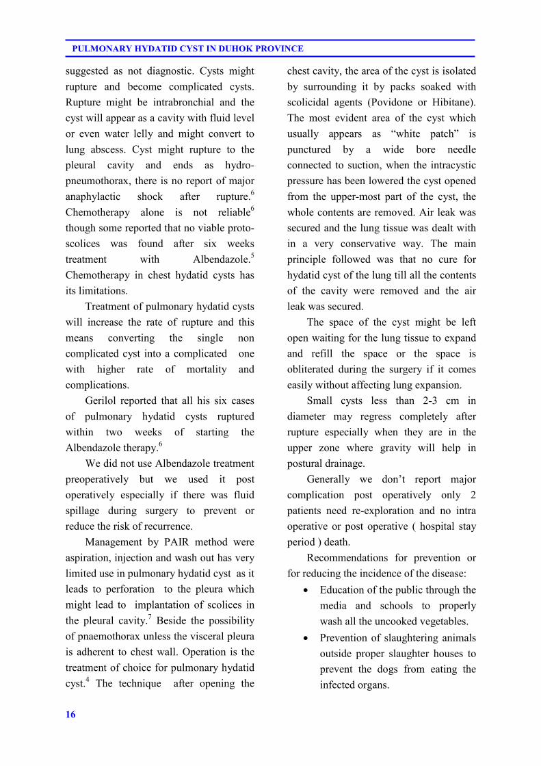
suction is completed, the endocyst
removed by sponge forceps (Figure 7).
Surgical treatment of pulmonary
hydatid cysts included the removal of the
cyst, closure of the bronchial holes and
then conserving as much as possible of the
lung and obliterating the space.
Figure 1. Round homogenous opacity PA view
Figure 2. Round homogenous opacity - lateral
view
Figure 3. ruptured hydatid cyst water Lelly sign
chest X-ray lateral view
Figure 4. Lung abscess chest X-ray P-A view
14
PULMONARY HYDATID CYST IN DUHOK PROVINCE
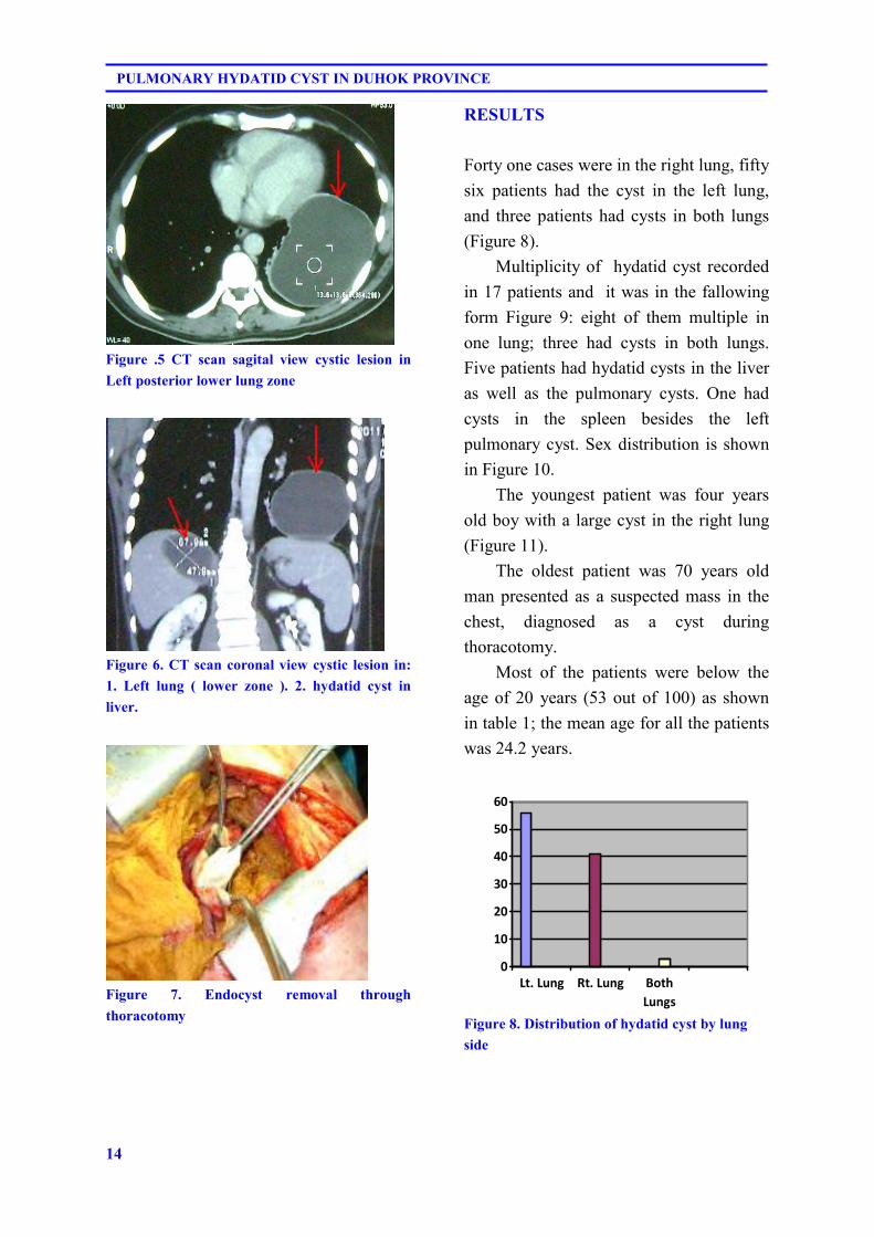
Figure 5. CT scan sagital view cystic lesion in
Left posterior lower lung zone
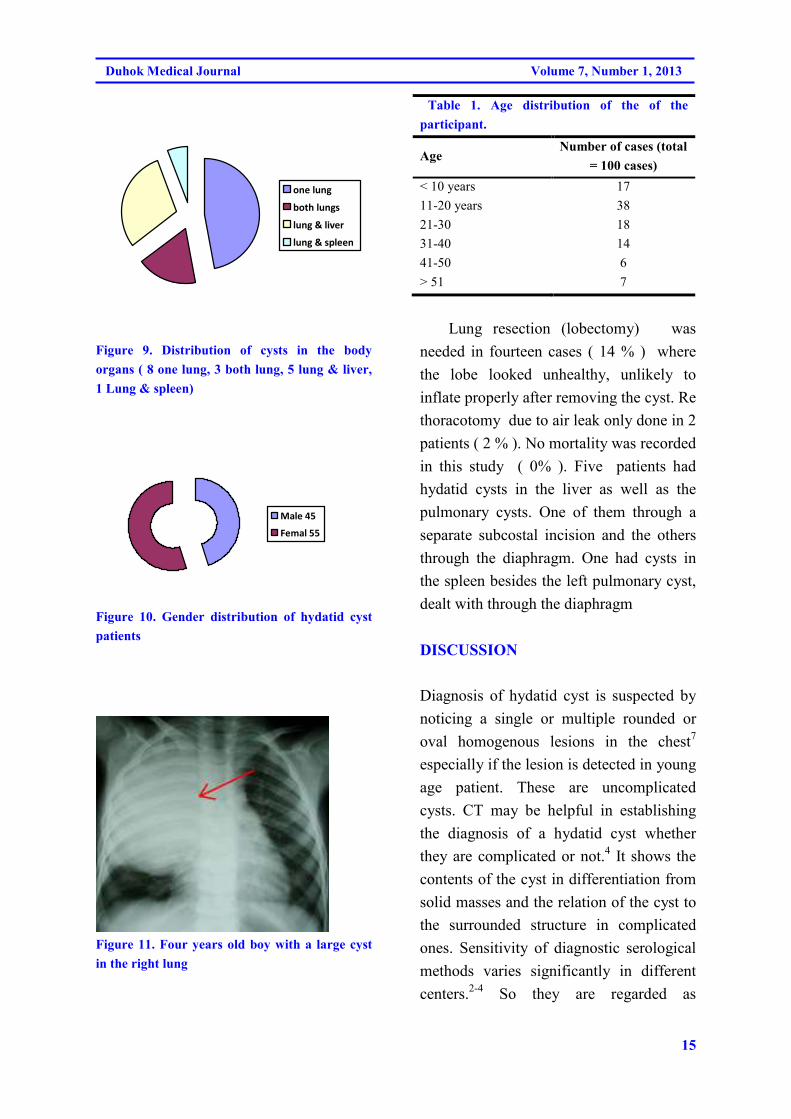
Figure 6. CT scan coronal view cystic lesion in:
1. Left lung ( lower zone ). 2. hydatid cyst in
liver.
Figure 7. Endocyst removal through
thoracotomy
RESULTS
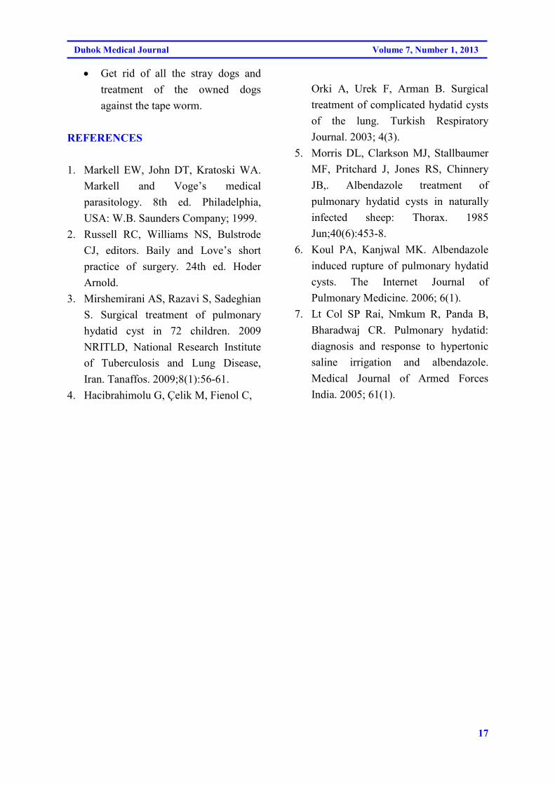
Forty one cases were in the right lung, fifty
six patients had the cyst in the left lung,
and three patients had cysts in both lungs
(Figure 8).
Multiplicity of hydatid cyst recorded
in 17 patients and it was in the fallowing
form Figure 9: eight of them multiple in
one lung; three had cysts in both lungs.
Five patients had hydatid cysts in the liver
as well as the pulmonary cysts. One had
cysts in the spleen besides the left
pulmonary cyst. Sex distribution is shown
in Figure 10.
The youngest patient was four years
old boy with a large cyst in the right lung
(Figure 11).
The oldest patient was 70 years old
man presented as a suspected mass in the
chest, diagnosed as a cyst during
thoracotomy.
Most of the patients were below the
age of 20 years (53 out of 100) as shown
in table 1; the mean age for all the patients
was 24.2 years.
0
10
20
30
40
50
60
Lt. Lung Rt. Lung Both
Lungs
Figure 8. Distribution of hydatid cyst by lung
side
15
Duhok Medical Journal Volume 7, Number 1, 2013
one lung
both lungs
lung & liver
lung & spleen
Figure 9. Distribution of cysts in the body
organs ( 8 one lung, 3 both lung, 5 lung & liver,
1 Lung & spleen)
Male 45
Femal 55
Figure 10. Gender distribution of hydatid cyst
patients
Figure 11. Four years old boy with a large cyst
in the right lung
Table 1. Age distribution of the of the
participant.
Age Number of cases (total
= 100 cases)
< 10 years 17
11-20 years 38
21-30 18
31-40 14
41-50 6
> 51 7
Lung resection (lobectomy) was
needed in fourteen cases ( 14 % ) where
the lobe looked unhealthy, unlikely to
inflate properly after removing the cyst. Re
thoracotomy due to air leak only done in 2
patients ( 2 % ). No mortality was recorded
in this study ( 0% ). Five patients had
hydatid cysts in the liver as well as the
pulmonary cysts. One of them through a
separate subcostal incision and the others
through the diaphragm. One had cysts in
the spleen besides the left pulmonary cyst,
dealt with through the diaphragm
DISCUSSION
Diagnosis of hydatid cyst is suspected by
noticing a single or multiple rounded or
oval homogenous lesions in the chest7
especially if the lesion is detected in young
age patient. These are uncomplicated
cysts. CT may be helpful in establishing
the diagnosis of a hydatid cyst whether
they are complicated or not.4 It shows the
contents of the cyst in differentiation from
solid masses and the relation of the cyst to
the surrounded structure in complicated
ones. Sensitivity of diagnostic serological
methods varies significantly in different
centers.2-4 So they are regarded as
16
PULMONARY HYDATID CYST IN DUHOK PROVINCE
suggested as not diagnostic. Cysts might
rupture and become complicated cysts.
Rupture might be intrabronchial and the
cyst will appear as a cavity with fluid level
or even water lelly and might convert to
lung abscess. Cyst might rupture to the
pleural cavity and ends as hydro-
pneumothorax, there is no report of major
anaphylactic shock after rupture.6
Chemotherapy alone is not reliable6
though some reported that no viable proto-
scolices was found after six weeks
treatment with Albendazole.5
Chemotherapy in chest hydatid cysts has
its limitations.
Treatment of pulmonary hydatid cysts
will increase the rate of rupture and this
means converting the single non
complicated cyst into a complicated one
with higher rate of mortality and
complications.
Gerilol reported that all his six cases
of pulmonary hydatid cysts ruptured
within two weeks of starting the
Albendazole therapy.6
We did not use Albendazole treatment
preoperatively but we used it post
operatively especially if there was fluid
spillage during surgery to prevent or
reduce the risk of recurrence.
Management by PAIR method were
aspiration, injection and wash out has very
limited use in pulmonary hydatid cyst as it
leads to perforation to the pleura which
might lead to implantation of scolices in
the pleural cavity.7 Beside the possibility
of pnaemothorax unless the visceral pleura
is adherent to chest wall. Operation is the
treatment of choice for pulmonary hydatid
cyst.4 The technique after opening the
chest cavity, the area of the cyst is isolated
by surrounding it by packs soaked with
scolicidal agents (Povidone or Hibitane).
The most evident area of the cyst which
usually appears as “white patch” is
punctured by a wide bore needle
connected to suction, when the intracystic
pressure has been lowered the cyst opened
from the upper-most part of the cyst, the
whole contents are removed. Air leak was
secured and the lung tissue was dealt with
in a very conservative way. The main
principle followed was that no cure for
hydatid cyst of the lung till all the contents
of the cavity were removed and the air
leak was secured.
The space of the cyst might be left
open waiting for the lung tissue to expand
and refill the space or the space is
obliterated during the surgery if it comes
easily without affecting lung expansion.
Small cysts less than 2-3 cm in
diameter may regress completely after
rupture especially when they are in the
upper zone where gravity will help in
postural drainage.
Generally we don’t report major
complication post operatively only 2
patients need re-exploration and no intra
operative or post operative ( hospital stay
period ) death.
Recommendations for prevention or
for reducing the incidence of the disease:
• Education of the public through the
media and schools to properly
wash all the uncooked vegetables.
• Prevention of slaughtering animals
outside proper slaughter houses to
prevent the dogs from eating the
infected organs.
17
Duhok Medical Journal Volume 7, Number 1, 2013
• Get rid of all the stray dogs and
treatment of the owned dogs
against the tape worm.
REFERENCES
1. Markell EW, John DT, Kratoski WA.
Markell and Voge’s medical
parasitology. 8th ed. Philadelphia,
USA: W.B. Saunders Company; 1999.
2. Russell RC, Williams NS, Bulstrode
CJ, editors. Baily and Love’s short
practice of surgery. 24th ed. Hoder
Arnold.
3. Mirshemirani AS, Razavi S, Sadeghian
S. Surgical treatment of pulmonary
hydatid cyst in 72 children. 2009
NRITLD, National Research Institute
of Tuberculosis and Lung Disease,
Iran. Tanaffos. 2009;8(1):56-61.
4. Hacibrahimolu G, Çelik M, Fienol C,
Orki A, Urek F, Arman B. Surgical
treatment of complicated hydatid cysts
of the lung. Turkish Respiratory
Journal. 2003; 4(3).
5. Morris DL, Clarkson MJ, Stallbaumer
MF, Pritchard J, Jones RS, Chinnery
JB,. Albendazole treatment of
pulmonary hydatid cysts in naturally
infected sheep: Thorax. 1985
Jun;40(6):453-8.
6. Koul PA, Kanjwal MK. Albendazole
induced rupture of pulmonary hydatid
cysts. The Internet Journal of
Pulmonary Medicine. 2006; 6(1).
7. Lt Col SP Rai, Nmkum R, Panda B,
Bharadwaj CR. Pulmonary hydatid:
diagnosis and response to hypertonic
saline irrigation and albendazole.
Medical Journal of Armed Forces
India. 2005; 61(1).
18
PULMONARY HYDATID CYST IN DUHOK PROVINCE
ón‚íqón‚íqón‚íqón‚íq@@@@@@@@ŽôÄb÷@oŽï�ïØ@b“Žï÷ŽôÄb÷@oŽï�ïØ@b“Žï÷ŽôÄb÷@oŽï�ïØ@b“Žï÷ŽôÄb÷@oŽï�ïØ@b“Žï÷@@@@ŽôØíè†@ß@ô�bäóè@Žôàa‡äó÷íØ@ߎôØíè†@ß@ô�bäóè@Žôàa‡äó÷íØ@ߎôØíè†@ß@ô�bäóè@Žôàa‡äó÷íØ@ߎôØíè†@ß@ô�bäóè@Žôàa‡äó÷íØ@ß@@@@@@@@
@@@@@@@@wäbàŠb÷@ì@ôØó“ŽïqwäbàŠb÷@ì@ôØó“ŽïqwäbàŠb÷@ì@ôØó“ŽïqwäbàŠb÷@ì@ôØó“ŽïqZZZZ@@@@@@@@@
ýb�@ˆ@Šóè 2006 @oŽïî@ ŽôÄb÷@oŽï�ïØ@bä‹ØŠó�òŠbu@üi@bîŠó�Šón“ä@çbÄ@ˆ@�qaŠbi@LôÙå�@bä‹ØóÄ@oŽïîŠó�Šón“ä@bä‹ÙŽïrn�ò†@ Žôàò†@Œ
ì@ Žô“q@@pbÑÈa�àîí‚@íØ@Lçìíi@çaíi@ Žô“q@bäìíj’ìím@ ŽõŠó�ó÷ˆ@oŽîŽôØíè†@bèó��ŽîŠbq@ß@óïn�ì슇äóm@bØó“ŽïØ@óï‚b�ä@Äó÷@íØ@ìíji@b@N
óäbéu@çaìˆ@ÚŽï÷@Ûíè†@LŽôÔa�È@ß@çbéu@Ûò‡åè@ß@óÄýóiŠói@bØó“Žï÷@Žô“q@bî@ŽôÄb÷@oŽï�ïØ@b“Žï÷N@@ŽôÄb÷@oŽï�ïØ@b“Žï÷@Læmìí�@óïmbè@ôØòì
@ßìóè@ óåïÜìíØóÄ@ Äó÷@ LŽôØíè†@ bèó��ŽîŠbq@ ß@ óÄýóiŠói@ bØó“Žï÷@ Žô“q@ bî@LŽô“Žï÷@ ŽôĈ@ Žõ‡äŒb�@ bäìíiŠbî†@ bïäaìbšì@ Žôäìíj’ìím@ a‰ŽîŠ@pò††
µäaŒ@pò‡i@ŽôîŠó�òŠbu@oŽïÙŽîŠ@æî�’biì@N@óÄ@ŽôØóÙ“ïä@ˆ@Žôï’ü‚óä@bä‹Ùäb“ïån�ò†@ˆ@LòŠíuìòŠíu@Žôî@´î†@ónŽï÷†@Žôi@•ü‚óä@ŽõíŽï’
@üi@Læióä@ŠaŒb÷@�@ô’ü‚óä@íØIpbÑÈb›àH�ïØ@båïÔóq@aŠó�ó÷@ˆ@Ša숆@ÛóÜóØ@oŽîŽôÄb÷@Žô.@@
ŽôåïÜíØóÄ@æŽïÙôåïÜíØóÄ@æŽïÙôåïÜíØóÄ@æŽïÙôåïÜíØóÄ@æŽïÙîŠZZZZ@@bàìóÜ@Lç‹Ø@óåŽï÷†@ Ž¶@ôÙå�@oŽïîŠó�Šón“ä@ ŽôÄó÷@ìíi@êu@ìó÷@LŽôèó��ŽîŠbq@ ŽõŠónäó�@Ûòì@LÛíè†IôÙî�Žïä@l@H@ôàóè
ç‹ØóäaìòŠ@óåmbè†@üi@æîìíi@Žô“q@oŽïî@ŽôÄb÷@oŽï�ïØ@ô’ìím@oŽï’ü‚óä@N@çaì@üi@õŠó�Šón“ä@ôn“qì@õŠói@µÜìíØóÄ@óåmbè†@ó’ü‚óä@Äó÷
è†ç‹Ø@ ónŽïi@ ŽôîŠó�òŠbš@ oŽïÙŽîŠ@ æî�’bi@ bä‡äaí‚@ a†@ Lç‹Ø@ óåmb@ N@Lç‹Øó’óIäó�@ ómbè@ çb’ü‚óä@ üi@ ç‹Ø@ ómbè†@ bîŠó�Šón“ä@ ŽõŠíu
@ bèòìŠóèIpbÑÈb›à@ Hõˆ@ N@ ò‡äóšŠóè@ LŽôîŠó�Šón“ä@ õŠói@ çbåï÷ŠbÙi@ óåmbèóä@ ˆ‡äbîˆ@ oŽïäbàŠò†@ �I‘ŠíØH@‡äóš@ üi@ ça†@ ìíjmbè@ Ûò
ŽôîŠó�Šón“ä@ôn“q@çb’ìí‚óäN@ß@çbî@LŽôï’ìí‚óä@bä�åiˆ@üi@õŠb�ˆìíàb÷@LŽôïèbºì†M@ôáŽïØ@lM@@óäýb�@bî@ Žôäìíj’ìím@a‰ŽîŠ@bä‹ÙáŽïØ
ç‹Øó’óIäó�@ómbè.@@ýb�@ˆ@Šóè@LŽôîŠó�Šón“ä@bÙŽî‹i@ç‹ØŠó�òŠbš@óåïmbè@æŽîìó÷@õóè@ŽôÄb÷@oŽï�ïØ@b“Žï÷@æŽï’ü‚óä@ìó÷2007@ôåîaŒ@Žõ
@ ýb�@ bäb�ïä@ bÅîóè@ bm2011íØóÄ@ óåmbè@ LŽõµÜì@ N@Žôï’ü‚óäˆ@ô’ü‚óä@ aŠaŒb÷@ LŽôÄb÷@ Žô�ïØ@ Žôéu@ Lô’ü‚óä@ Žôîˆ@ a†@ ŽôåïÜìíØóÄ@ ŽôÄ@ †
@l@Žôî@ŽôÄb÷@Žô�ïØ@óîb÷@bèòìŠóè@LoIpbÑÈb›à@Hæm‹�Šòì@óåmbè@ôàóè@òŠbØíè@Äó÷@Lóä@çbî@ìíiN@@@
ãb−ó÷ãb−ó÷ãb−ó÷ãb−ó÷ZZZZ@@ oŽî†ŠíÙîŠ@ bäìí›Ñîì†@ ì@ ç‹ØóÄìì‹’100@ŽôîŠó�Šón“ä@ ôn“qì@ õŠói@ Lç‹Ø@ ómbè@ çb’ü‚óä@ôàóè@ oŽïî@ ó’ü‚óä@ Äó÷@ L
çìíi@ŽôØíè†@bèó��ŽîŠbq@oŽïäbƒ’ü‚óä.@@Žôîˆ@†@b’í‚óä@bî�q@aŠbi11M40@çìíia†@¶b�@N@Ú�ïØ@ì@ç‹Ø@ómbè@ôIå�@bîŠó�Šón“ä@bïàóè@íi
@ ß@ ç‹ØaŠ@ ómbè86@ E@ ß@ ç‹ØaŠ@ ómbè@ô�bäóè@ Žôàa‡äó÷@ ì14@ NE@ ß@ ì2@ Eånèbä@ Žôä‹à@ æŽïmóÜby@ Žôš@ ì@ òŠbiìì†@õŠó�ón“ä@ b’í‚óä@ ˆ@ó
ç‹ØŠbàímN@@
ãb−ó÷Šò†ãb−ó÷Šò†ãb−ó÷Šò†ãb−ó÷Šò†@ZóÙŽîŠ@æŽî�’bi@ŽôîŠó�Šón“ä@bÙŽîŠ@l@ô�bäóè@Žôàa‡äó÷íØ@ß@ŽôÄb÷@æŽïÙ�ïØ@bîŠó�òŠbšN@@

19
Duhok Medical Journal Volume 7, Number 1, 2013
الخلاصة
الاكياس المائية الرئوية في محافظة دهوك
عندما بوشرت عمليات فتح الصدر في دهوك اغلبية الحالات المرضية التي اجري 2006منذ عام :خلفية واهداف البحث
لها فتح الصدر كانت لمعالجة الاكياس المائية الرئوية والمضاعفات الناتجة عن الاصابة بها حيث لوحظ انه مشكلة
محافضة دهوك . في بعض أجزاء العراق مرض مستوطنالمائية هو الرئوية مرضية في محافظة دهوك مرض الاكياس
تبغي الدراسة , كما ذكر ان الاكياس الرئوية المائية هو مرض مستوطن في محافظة دهوك .واحدة من هذه المناطق
اعراض المرض تتراوح من كونه غير , ظهور اعراض المرض والطرق الافضل للعلاج وكيفية لمعرفة نسب الاصابة
دهوك مركز المحافضة المكان التي تتم فيه .التي تتبع انفجار الكيس الى المضاعفات الشديدة) يكتشف بالصدفة (ظاهر
هذه الحالات تمت دراستها , جراحة الصدر ولهذا تقريبا كل حالات الاكياس المائية في المحافظة يتم احالتها الى المركز
, المتبعة نوقشت مع المضاعفات انواع التداخل الجراحي , وتقييمها قبل وبعد التداخل الجراحي لتقييم افضل طرق العلاج
أخيرآ نوقشت التوصيات , لم تستعمل مضادات الطفيليات قبل العملية ولكن استعملت بعد العملية في بعض الحالات
. لاستئصال او على الاقل لتقليل الاصابة بهذا المرض
جراحيآ في الفترة ما بين كانون هذه الدراسة لحالات الاكياس المائية في محافضة دهوك التي تم علاجها :طرق البحث
الشكوى الرئيسية للمريض وكون الكيس , الدراسة تضمنت العمر مكان الاصابة في الرئة , 2011ونيسان 2007الثاني
.بسيط او مع اختلاطات
معظم , مريض وتضمن الحالات الموجودة في كافة مستشفيات دهوك 100تم اجراء التداخل الجراحي ل :النتائج
استئصلت ) أو ألاكياس( الكيس , كل المرضى تم علاجهم عن طريق فتح الصدر , سنة 40-11ضى بعمر ما بين المر
من % 2أعادة فتح الصدر في , من الحالات % 14وتم الاستئصال في % 86مع الحفاظ على النسيج الرئوي في
.المرضى مع عدم تسجيل اي حالة وفاة
تعدد الاكياس ليس بغير , هو الحل الوحيد المثبت للشفاء من الاكياس المائية الرئوية العملية الجراحية :اتالاستنتاج
.التداخل الجراحي ناجح بنسبة كبيرة مع نسبة معدومة أو قليلة جدا للوفاة, شائع
20
ANGIOGRAPHIC VARIATIONS OF RENAL ARTERY AMONG DONORS OF KIDNEY …..
ANGIOGRAPHIC VARIATIONS OF RENAL ARTERY AMONG DONORS OF
KIDNEY IN DUHOK CITY
AMEEN M. MOHAMMAD, MBCHB, FICMS*
MOHAMMED A. ABDULRAHMAN, MBCHB, DM**
SHAKER S. JABALY, MBCHB, FICMS***
Submitted 17 Oct 2012; accepted 3 Nov 2013
ABSTRACT
Background and objectives Renal artery variations are not uncommon. It can be
investigated by many means; so far renal conventional angiography is dependable in the
disclosure of the anatomical variations of the renal artery. In order to take all the steps of
successful transplantation, the kidney of potential donor should be studied thoroughly mainly
its function and anatomy. From our side, we tried to study the prevalence of anatomical
variations of renal artery by angiography in donors of kidney in Duhok city.
Methods Case-series study involved eighty three (83) kidney donors; aged between 18- 47
years (24+-2.1), 76 of them were males, referred from Duhok transplantation unit and
assumed to be healthy after screening for any medical illness. In Cath-Lab of Azadi Heart
Center in Duhok renal artery angiograms were obtained. Each angiogram consists of right
and left renal artery images with a central aortogram. The data were collected from the 1st of
January 2010 to 1st of July 2011. This study was approved by the Scientific Review
Committee at Duhok College of Medicine.
Results Fifty four 54(65.1%) of them had normal single right and left renal arteries, but the
remaining 29(34.9%) of supposed donors had anatomical variations of renal artery. Right
renal artery: 14(16%) out of 83 showed variations (Double was 11,Trifurcate was 3 cases).
Left renal artery: 21(25%) out of 83 had variations (Double was 17, Trifurcate was 4 cases.
5(6%) were bilateral. Main right and left renal artery originated mainly between L1 and L2 in
97.5%, 99% respectively.
Conclusions The results of this study indicated that the anatomical variations of renal artery
is common and the anatomy of renal artery ultimately is necessary to be known prior of
transplantation.
Duhok Med J 2013;7(1): 20-28. Key words: Kidney, Renal artery variations, Kidney donors, Angiography
he anomalies of the kidneys; both
structural and vascular are various
and the congenital variations of urogenital
system are relatively higher when
compared to other systems, because the
developmental stages of this system are
more complicated. Some of these
anomalies do not even cause clinical
symptoms and don’t require treatment.
However some of them are predisposing to
some pathological disorders because of the
decrease in the blood supply or urine
T
*Lecturer, Department of Medicine, Duhok College of Medicine, Kurdistan Region, Iraq; Cardiologist and
fellow in Interventional Cardiology, Azadi Heart Center, Azadi Teaching Hospital, Duhok Governorate, Iraq
**Assistant lecturer, Department of Medicine, Duhok College of Medicine, Kurdistan Region, Iraq;
Cardiologist and fellow in Interventional Cardiology, Azadi Heart Center, Azadi Teaching Hospital, Duhok
Governorate, Iraq
***Assistant professor, Department of Surgery, Duhok College of Medicine, Kurdistan Region, Iraq;
Consultant Urologist and Director, Azadi Teaching Hospital, Duhok Governorate, Iraq
Correspondence author: Ameen M. Mohammad, Telephone number: +9647504586259; e-mail:
21
Duhok Medical Journal Volume 7, Number 1, 2013
flow.1
Renal artery variations are common in
the general population and the prevalence
of these variations shows social, ethnic and
racial differences. The frequency of
multiple renal arteries shows variability
from 9% to 76%, and is generally changes
between 28 and30%.2
Renal angiography is dependable in
the disclosure of this anatomical variation
of the renal artery. By successful renal
transplantation the course of chronic renal
failure, which is common in our area and
its cardiovascular sequels will be changed
and even might reverse to normal. In
order to take all the steps of successful
transplantation, the kidney of donor should
be studied thoroughly mainly its function
and anatomy, so that, from our side, we
tried to study the prevalence of anatomical
variation of renal vasculature by
angiography in donors of kidney in Duhok
city. Up to our knowledge studies on renal
artery variation in our area are absent.
METHODS
The study involved eighty three (83)
kidney donors; aged between 18- 47 years
(24+-2.1), 76(91.5%) of them were
males,7(8.5%) were females; referred from
Duhok Transplantation Unit and assumed
to be healthy after screening for any
evidence of medical illness and those who
were not healthy deferred from the
donation of kidney. In the Cathlab of
Azadi Heart Centre the interventional
cardiologist prepared the patient and
through trans femoral approach, renal
artery angiography were obtained. Each
angiogram consists of right and left renal
artery images with a central aortogram
image; in order not to miss any anatomical
variation of renal artery then documenting
the results of renal angiography by reports
and sending the patients to the transplant
unit. The data were collected from the 1st
of January 2010 to 1st of July 2011. The
angiogram reports and images were
archived in the Cath-Lab unit as a
document. The anatomical characteristics
of renal arteries and their distribution
according to their originating levels to
vertebrae were studied.
RESULTS
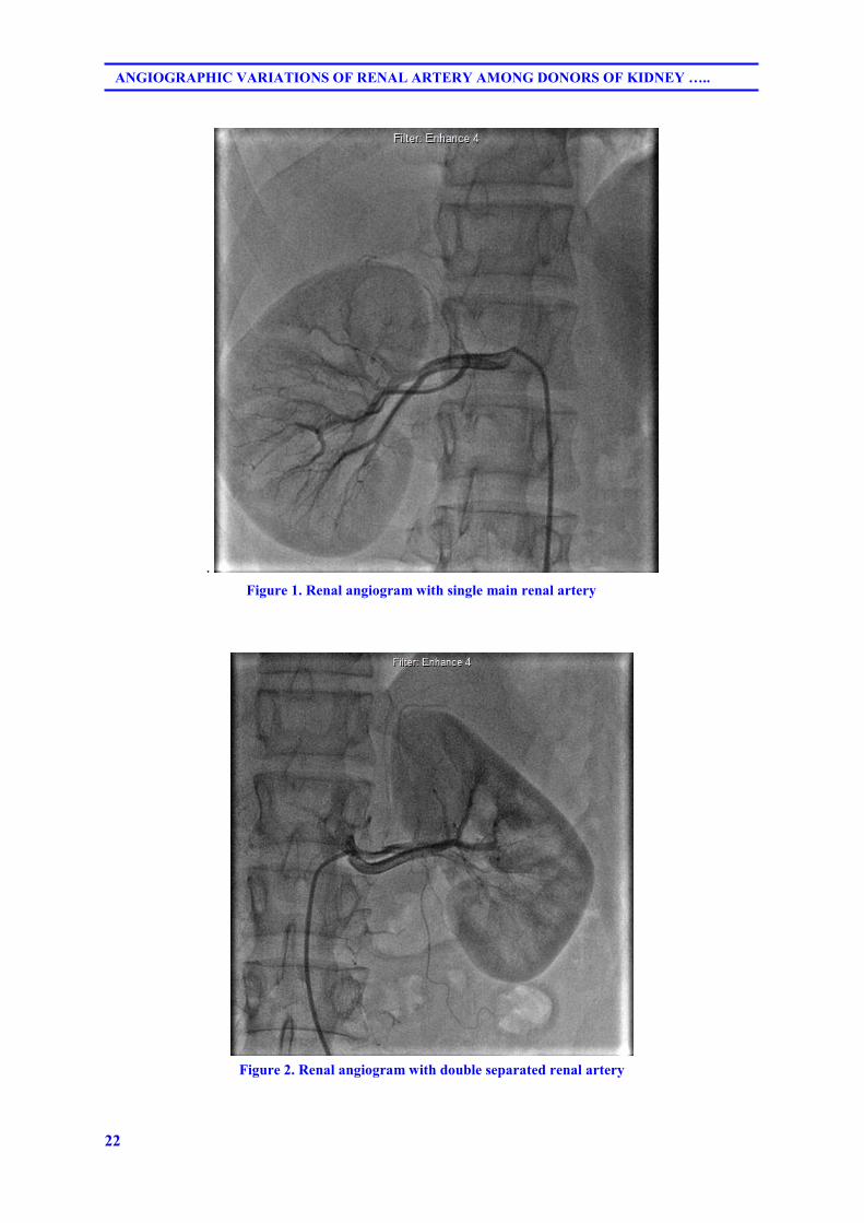
Fifty four 54 (65.1%) of them had normal
single right and left renal artery supplying
kidneys, but the remaining 29 (34.9%)
supposed donors had anatomical variation
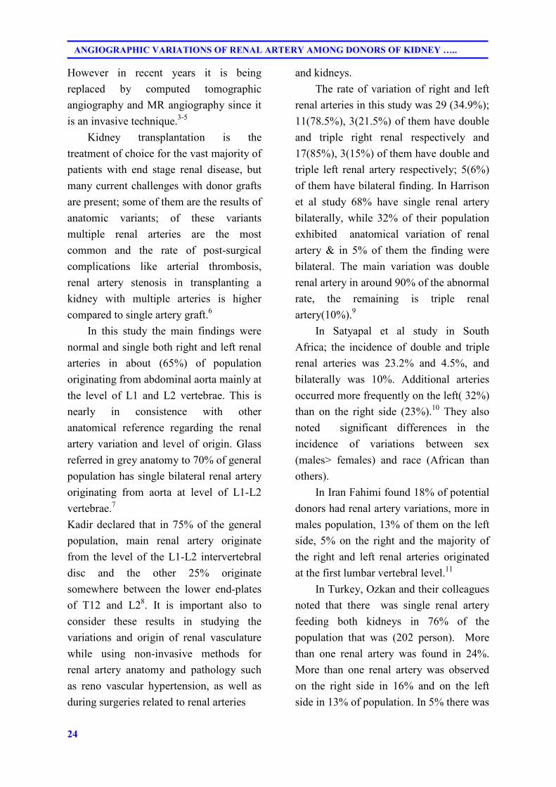
of renal artery. 5(6%) bilateral renal artery
variations. Right renal artery: {(double=
11 persons, 7 of them were of separate
origin from aorta & 4 were of single
origin), (Trifurcate right= 3 persons, two
of them from separate origin, one was of
single origin)}. Left renal artery:
{(double=17, 10 of them were of separate
origin, 7 were of single origin),
(Trifurcate=3, two of them from separate
origin, one was of single origin)} as shown
in figure 1 and 2.
Right renal artery origins: In 97.5% of
the patients, main renal artery originated
between the L1 and the L2 vertebrae.
Right main renal artery originated at the
level of the L1 vertebra in 23%, from the
level of the L1-L2 intervertebral disc in
66%, and at the level of the L2 vertebra in
22
ANGIOGRAPHIC VARIATIONS OF RENAL ARTERY AMONG DONORS OF KIDNEY …..
.
Figure 1. Renal angiogram with single main renal artery
Figure 2. Renal angiogram with double separated renal artery
23
Duhok Medical Journal Volume 7, Number 1, 2013
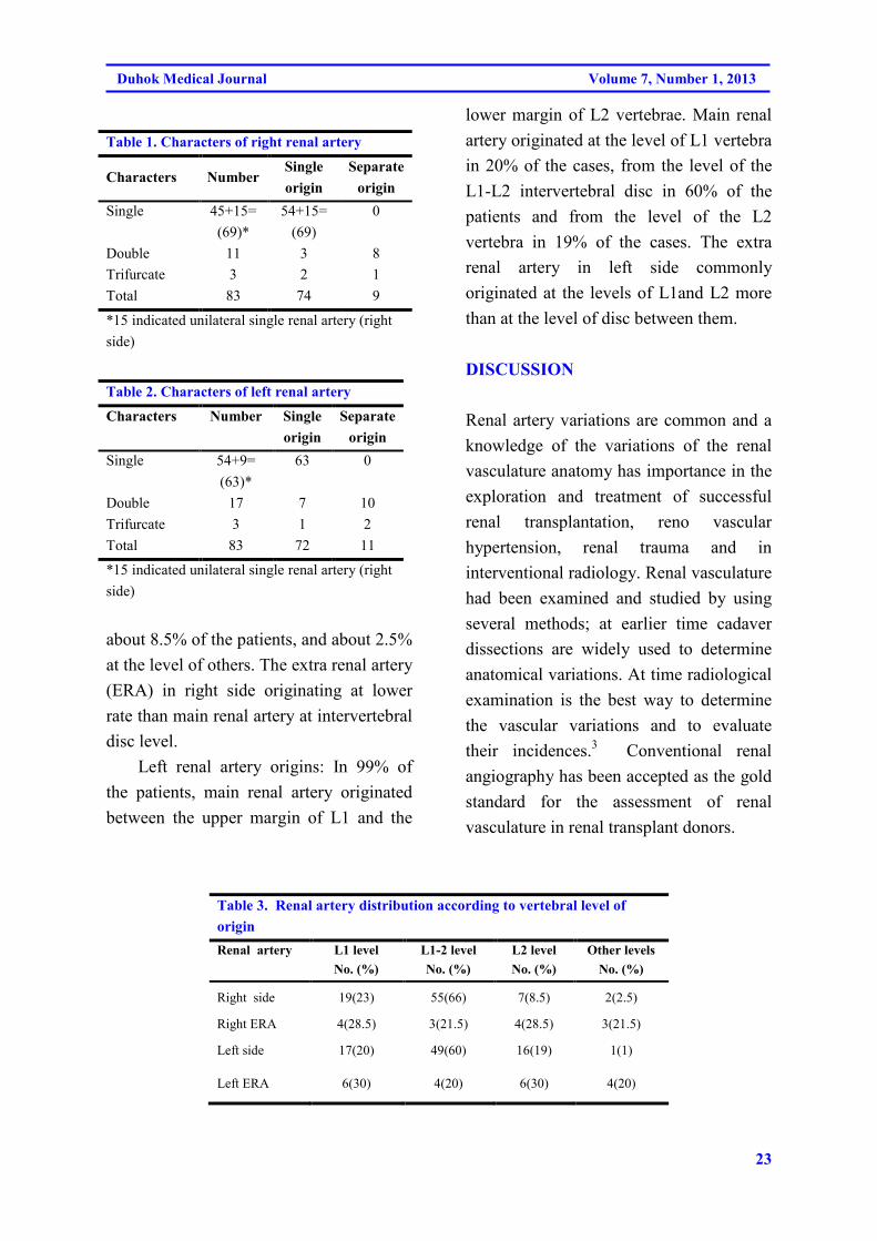
Table 1. Characters of right renal artery
Characters Number Single
origin
Separate
origin
Single 45+15=
(69)*
54+15=
(69)
0
Double 11 3 8
Trifurcate 3 2 1
Total 83 74 9
*15 indicated unilateral single renal artery (right
side)
Table 2. Characters of left renal artery
Characters Number Single
origin
Separate
origin
Single 54+9=
(63)*
63 0
Double 17 7 10
Trifurcate 3 1 2
Total 83 72 11
*15 indicated unilateral single renal artery (right
side)
about 8.5% of the patients, and about 2.5%
at the level of others. The extra renal artery
(ERA) in right side originating at lower
rate than main renal artery at intervertebral
disc level.
Left renal artery origins: In 99% of
the patients, main renal artery originated
between the upper margin of L1 and the
lower margin of L2 vertebrae. Main renal
artery originated at the level of L1 vertebra
in 20% of the cases, from the level of the
L1-L2 intervertebral disc in 60% of the
patients and from the level of the L2
vertebra in 19% of the cases. The extra
renal artery in left side commonly
originated at the levels of L1and L2 more
than at the level of disc between them.
DISCUSSION
Renal artery variations are common and a
knowledge of the variations of the renal
vasculature anatomy has importance in the
exploration and treatment of successful
renal transplantation, reno vascular
hypertension, renal trauma and in
interventional radiology. Renal vasculature
had been examined and studied by using
several methods; at earlier time cadaver
dissections are widely used to determine
anatomical variations. At time radiological
examination is the best way to determine
the vascular variations and to evaluate
their incidences.3 Conventional renal
angiography has been accepted as the gold
standard for the assessment of renal
vasculature in renal transplant donors.
Table 3. Renal artery distribution according to vertebral level of
origin
Renal artery
L1 level
No. (%)
L1-2 level
No. (%)
L2 level
No. (%)
Other levels
No. (%)
Right side 19(23) 55(66) 7(8.5) 2(2.5)
Right ERA 4(28.5) 3(21.5) 4(28.5) 3(21.5)
Left side 17(20) 49(60) 16(19) 1(1)
Left ERA 6(30) 4(20) 6(30) 4(20)
24
ANGIOGRAPHIC VARIATIONS OF RENAL ARTERY AMONG DONORS OF KIDNEY …..
However in recent years it is being
replaced by computed tomographic
angiography and MR angiography since it
is an invasive technique.3-5
Kidney transplantation is the
treatment of choice for the vast majority of
patients with end stage renal disease, but
many current challenges with donor grafts
are present; some of them are the results of
anatomic variants; of these variants
multiple renal arteries are the most
common and the rate of post-surgical
complications like arterial thrombosis,
renal artery stenosis in transplanting a
kidney with multiple arteries is higher
compared to single artery graft.6
In this study the main findings were
normal and single both right and left renal
arteries in about (65%) of population
originating from abdominal aorta mainly at
the level of L1 and L2 vertebrae. This is
nearly in consistence with other
anatomical reference regarding the renal
artery variation and level of origin. Glass
referred in grey anatomy to 70% of general
population has single bilateral renal artery
originating from aorta at level of L1-L2
vertebrae.7
Kadir declared that in 75% of the general
population, main renal artery originate
from the level of the L1-L2 intervertebral
disc and the other 25% originate
somewhere between the lower end-plates
of T12 and L28. It is important also to
consider these results in studying the
variations and origin of renal vasculature
while using non-invasive methods for
renal artery anatomy and pathology such
as reno vascular hypertension, as well as
during surgeries related to renal arteries
and kidneys.
The rate of variation of right and left
renal arteries in this study was 29 (34.9%);
11(78.5%), 3(21.5%) of them have double
and triple right renal respectively and
17(85%), 3(15%) of them have double and
triple left renal artery respectively; 5(6%)
of them have bilateral finding. In Harrison
et al study 68% have single renal artery
bilaterally, while 32% of their population
exhibited anatomical variation of renal
artery & in 5% of them the finding were
bilateral. The main variation was double
renal artery in around 90% of the abnormal
rate, the remaining is triple renal
artery(10%).9
In Satyapal et al study in South
Africa; the incidence of double and triple
renal arteries was 23.2% and 4.5%, and
bilaterally was 10%. Additional arteries
occurred more frequently on the left( 32%)
than on the right side (23%).10
They also
noted significant differences in the
incidence of variations between sex
(males> females) and race (African than
others).
In Iran Fahimi found 18% of potential
donors had renal artery variations, more in
males population, 13% of them on the left
side, 5% on the right and the majority of
the right and left renal arteries originated
at the first lumbar vertebral level.11
In Turkey, Ozkan and their colleagues
noted that there was single renal artery
feeding both kidneys in 76% of the
population that was (202 person). More
than one renal artery was found in 24%.
More than one renal artery was observed
on the right side in 16% and on the left
side in 13% of population. In 5% there was
25
Duhok Medical Journal Volume 7, Number 1, 2013
variation bilaterally.2
In Colombia, a study composed of
196 cases (85.4% were males, aged 33.8
years+_ 15.6), an additional renal artery
found in 22.3% of population, two
additional renal artery in 2.6%. The
variation was more common on left side.12
In this study there were no errors in
the prediction of arterial number when
compared to their surgical findings during
transplantation, and this indicated that the
technique of angiography was correct in
detecting the abnormalities of renal artery.
In conclusion the renal artery
variation in our population is not
uncommon. The rate nearly is close to the
population of other areas. The influence of
this variation should be taken in
consideration during renal vascular
surgeries in general and in renal
transplantation specifically. Here it is
worthy to recommend further studies to
compare such results with more
noninvasive test results such as MRA or
MSCT angio of renal artery anatomy in
order to use more noninvasive test for the
purpose of investigating the anatomy and
even pathology of renal vasculature.
COMPETING INTERESTS
The authors declare that they have no
competing interests.
AUTHORS' CONTRIBUTIONS
Ameen M Mohammad (AMM),
contributed the concept and design, data
collection; interpretation with analysis and
drafting with revision of the manuscript;
Mohammed A Abdulrahman (MAA),
contributed to the larger part of the
angiographic work ; Shakir S Jabaly (SSJ),
contributed to the larger part of the
surgical and transplantation work. All
authors approved the final submitted
version of the manuscript.
REFERENCES
1. Campell MF. Anomalies of kidney. In:
Campell MF, Harrison JS, editors.
Urology. 3rd ed. Vol. 2. Philadelphia:
W.B. Saunders; 1970. p. 1416-86.
2. Ozkan U, Oguzkurt L, Tercan F,
Kizilikilic O, Koc Z, Koca N. Renal
artery origins and variations:
angiographic evaluation of 855
consecutive patients. Diagn Interv
Radiol. 2006; 124: 183-6.
3. Ragiba Z, Can P, Ayla K. A
retrospective study on multiple renal
arteries in Turkish population.
Anatomy. 2009;3:35-9.
4. Firat A, Akin O, Agildere AM,
Aytekin C, Haberal M. Contrast-
enhanced resonance angiography:
evaluation of renal arteries in living
transplant donors. Eur J Radiol.
2004;521: 84-93.
5. Fox M, Yalin R. Renal transplantation
with multiple arteries. Br J Urol. 1979;
55:333-6
6. Benedetti E, Troppmann C,
Gillingham K, Sutherland DE, Payne
WD, Dunn DL, Matas AJ, Najarian JS,
Grussner RW. Short and long term
outcomes of kidney transplants with
multiple renal arteries. Ann.
Surg.1995;221(4):406-14.
26
ANGIOGRAPHIC VARIATIONS OF RENAL ARTERY AMONG DONORS OF KIDNEY …..
7. Glass J. Kidney and ureter. In:
Standring S, editor. Gray,s anatomy.
39th ed. Churchill Livingstone: 2005. p.
1274-6.
8. Kadir S. Kidneys. In: Kidar S, editor.
Atlas of normal and variant
angiographic anatomy. Philadelphia:
W.B. Saunders Company;1991.p. 387-
429.
9. Harrison Jr, Flye MW, Seigler HF.
Incidence of anatomical variants in
renal vasculature in the presence of
normal renal function. Ann Surg.
1978;188(1):83-9
10. Satyapal KS, Haffejee AA, Singh B,
Ramsaroop L, Robbs JV, Kalideen JM.
Additional renal arteries: incidence and
morphometry. Surg Radiol Anat. 2001.
23(1):33-8.
11. Fahimi F, Saba M. Evaluation of the
renal arteries in kidney donors by
conventional angiography. Journal of
Qazvin University of Medical
Sciences. 2003; 7(3):43-7
12. Saldarriaga B, Perez AF, Ballesteros
LE. A direct anatomical study of
additional renal arteries in a
Colombian mestizo population. Folia
Morphol (Warsz) 2008;67(2):129-34.
27
Duhok Medical Journal Volume 7, Number 1, 2013
ón‚íqón‚íqón‚íqón‚íq@@@@@@@@Ù�ï§íØ@bäa†@µï“‚@êií‚@ã@êu@ß@ôÙ�ï§íØ@båîí‚@bè@ëŠ@bïÐa‹Øíï−@ê÷@bîŒaìbïuÙ�ï§íØ@bäa†@µï“‚@êií‚@ã@êu@ß@ôÙ�ï§íØ@båîí‚@bè@ëŠ@bïÐa‹Øíï−@ê÷@bîŒaìbïuÙ�ï§íØ@bäa†@µï“‚@êií‚@ã@êu@ß@ôÙ�ï§íØ@båîí‚@bè@ëŠ@bïÐa‹Øíï−@ê÷@bîŒaìbïuÙ�ï§íØ@bäa†@µï“‚@êií‚@ã@êu@ß@ôÙ�ï§íØ@båîí‚@bè@ëŠ@bïÐa‹Øíï−@ê÷@bîŒaìbïuôØíè†@õ‹îŒbi@ß@ôôØíè†@õ‹îŒbi@ß@ôôØíè†@õ‹îŒbi@ß@ôôØíè†@õ‹îŒbi@ß@ô@@@@@@@@
@@@@@@@@wäbàŠb÷@ì@ôØó“ŽïqwäbàŠb÷@ì@ôØó“ŽïqwäbàŠb÷@ì@ôØó“ŽïqwäbàŠb÷@ì@ôØó“ŽïqZZZZ@@ë†@Œ@Š@êè@L¶†@bïÐa‹Øíï−@ê÷@õŠ@ênä@ê�@ß@ôØíè†@ß@õ†aŒb÷@bäbƒ’í‚@êä@ß@ça‡àb−@ê÷@êïmbè@bî@êåïÜíØ@êÐ@Ó@ê÷
@bÙïjn�2010 @bnÐ@êy@bÑî@êè@bm@êè2011. @íi@Œ@ça‡àb−@ê÷@êïmbè@bî@ôÐa‹Øíï−@ê÷83 ã@êÝ‚b�@µïÐ@ê÷@ôÙï�§íØ@µï“‚@êií‚@@Œõ†aŒb÷@bäbƒ’í‚@êä@ß@êÐ@ôÙ�§íØ@@bä‡äbu@õŠ@ênä@ê�@ôîý@Œ@ç‹Ø@êäaì@ëŠ@êåmbèì@LêÐ@õŠa†Œíä@µïÜb÷@ôà@êè. @ê÷@µï�@êØ@ì@ê÷@ôà@êè
@µîímíÐì@Lµåîìí£@ôåîí‚@µî@çaì@µîŠbà@ë†@l@ôÙ�§íØ@µî@l@êu@ôîýì@o�aŠ@ôîý@a††@ßíè@êà@Lça‡àb−@ê÷@êmbè†@íi@ôÐa‹Øíïuaì@íi@ôЇïi@l@ôàím@êä@ê÷@ôà@êè@a†@Læm‹Ø@êmbè@†@õì@õŠ@ênä@ê�@ß@ôî@êmŠì@ê÷@bè@ëŠ@íi@ë‡îŒ@ôØ@êîímíÐì@Læm‹ØŠ@ëì@êåmbè@†@çbè@ëŠ@ç
oïjiŠbî†@ôîa‹Ù’b÷.@@ŽôåïÜíØóÄ@æŽïÙôåïÜíØóÄ@æŽïÙôåïÜíØóÄ@æŽïÙôåïÜíØóÄ@æŽïÙîŠZZZZ@@íi@ça‡àb−@ê÷@êmbè@ôÐa‹Øíï−@ê÷83 @LôÙ�§íØ@bä‡äb“£í‚@íi@Ó@ëŠ@êi@çb�@êØ7 @êy@ì@Lìíi@�ä@õŒ@êØ@ëŠ@çaì@Œ
Ð@Œ@çaì@ôîŒ@Lìíi@ôà@õŒ@êÙèŠ@çaì@Œ@o18-47 ìíi@¶b�. @õŠ@ênä@ê�@ß@ça‡àb−@ê÷@êäìíjmbè@çb�@êØ@çbÐ@ìíi@ôЇïi@µïåÙ“i@ôà@êèôÙ�§íØ@bä‡äbu, ôÙ�§íØ@bî@ôåîí‚@bè@ëŠ@bïÐa‹Ø@íï−@ê÷@aŠbî‹Ø@bäa‡àb−@ê÷@íi@Œ@¶†@õŠ@ênä@ê�@íi@ç‹Ø@êäaì@ëŠ@êäìíjmbè@•bi@ì. @ß
a‹Øíïu@ê÷@aŠìŒça‡àb−@ê÷@êmbè@bïïÜb÷@ìì†@Š@êè@íi@ôЇïi@µîímíÐ@ôïi@bî@ôåîí‚@bè@ëŠ@bïîŠ@l@ôÐ.@@ãb−ó÷ãb−ó÷ãb−ó÷ãb−ó÷ZZZZ@@Œ83 @Lçb�@êØ69 ì@@ôn�aŠ@bÙ�§íØ@båîí‚@bè@ëŠ@çaì@Œ63 æmóØŠ@ë†@†@Š@ë‡îŒ@Úï÷@Œ@ôi@êu@bî. 54 @ôåîí‚@bè@ëŠ@Úï÷@çaì@Œ
çìíi@êè@ôi@êuì@o�aŠ@ôîý@bÙ�ï§íØ@ß,@¶29 òŠ@Úï÷@Œ@çb�@êØçìíi@êè@�i@ôè. @ôn�aŠ@ôîý@ß10 @çìíióè@êèŠ@ìì†@çb�@óØ( @Œ@ì@bäaì7 æmbè@†@Šò‡îŒ@Úï÷@Œ,@ì3 æmbè@†@aŠò‡îŒ@ìì†@Œ)@ì@L3 çìíióè@ëòŠ@ô�@çb�óØ. @ôióu@ôîý@ß17 @ì@çìíióè@ëòŠ@ìì†@çb�óØ2
çìíióè@ëòŠ@ô�@çb�óØ. óÝ‚b�@bî@êä@ôåîí‚@bèòŠ@ôi@êu@ôîý@ß@Úï÷ì@ôn�aŠ@ôîý@ß@Úï÷ìíi@ã.@@ãb−ó÷Šò†ãb−ó÷Šò†ãb−ó÷Šò†ãb−ó÷Šò†ZZZZ@@çaì@båïäaŒ@íi@Œ@ça‡àb−@ê÷@óåïè@l@ôЇïi@µïåÙ“i@êïЇïi@ôÙ�§íØ@bõåîí‚@µèòŠ@ß@ôàím@êä@ê÷@µîŒaìbïu@bäìíióè@Šói@Œ
ôÙ�§íØ@bä‡äbu@µïàb−@ê÷@Šó�@ß@çaì@bä‹Ùïm@ŠbØ@Š@êi@Œ@µîŒaìbïuN@@
28
ANGIOGRAPHIC VARIATIONS OF RENAL ARTERY AMONG DONORS OF KIDNEY …..
الخلاصة
رعين بالكلية في مدينة دهوكدراسة الاختلافات القسطارية للشريان الكلوي لدى المتب
ويمكن الكشف عن ذلك بواسطة العديد من ,الاختلافات التشريحية للشريان الكلوي ليست قليلة :خلفية واهداف البحث
الوسائل، وتصوير الأوعية عن طريق القسطرة يمكن الاعتماد عليها في الكشف عن الاختلافات التشريحية للشريان
كافة الخطوات من اجل زراعة ناجحة للكلية، ومن جانبنا، حاولنا دراسة انتشار الاختلافات وينبغي اتخاذ .الكلوي
التشريحية من الأوعية الدموية في الكلى عن طريق القسطرة للأشخاص المتبرعين بالكلية في مركز زراعة الكلية بمدينة
. دهوك
شخصا يفترض ان يكونوا من المتبرعين بالكلية، الذين خضعوا لتصوير الأوعية للشريان 83الدراسة شملت :طرق البحث
الكلوي في وحدة قسطرة القلب والأوعية الدموية في مركز آزادي للقلب، وذلك بالتعاون مع وحدة زراعة الكلى في مستشفى
نهم من الذكور، ويفترض أن يكونوا م 76، و (2.1- + 24)سنة 47 - 18وتتراوح أعمارهم ما بين .آزادي في دهوك
ومن خلال طريقة شريان الفخذ تم الحصول على تصوير القسطاري للشريان .اصحاء بعد اجراء الفحوصات اللازمة
وتاخذ الصورة لجهة اليمين واليسار للشريان الكلوي مع صورة واحدة للشريان الأبهر لكي لا تفوت أي اختلاف .الكلوي
.2011 يوليو 1إلى 2010 يناير 1وقد تم جمع البيانات من .لكلويتشريحي للشريان ا
( 34% 29)منهم لديهم شريان كلوي واحد طبيعي في الجهة اليمنى واليسرى للكلية، ولكن البقية (٪65.1) 54 :النتائج
م من أصل مستقل من منهم ه 7، (٪12) 11 =مزدوج )} :الشريان الكلوي اليمين .لديهم اختلاف تشريحي للشريان الكلوي
الشريان .{،(واحدة من أصل واحد، اثنان منهم من أصل منفصلة، 3 =ثلاثي الفروع )، (أصل واحد 3الشريان الأورطي،
، 3 =ثلاثي الفروع )، (من أصل واحد 7منهم هم من أصل مستقل، و 10، و (٪20) 17 =مزدوج )} :الكلوي اليسار
.ن أصل واحدواحد من أصل مستقل، والثاني هو م
أن الاختلاف التشريحي للشريان الكلوي هو شائع، ومعرفة هذه الاختلافات التشريحية هي في نهاية المطاف :اتالاستنتاج
ضرورية قبل عملية الزرع الكلوي، من اجل ان تكون عملية زراعة الكلية ناجحة اكثر وبمعدل اقل للمضاعفات
.الجراحية
29
Duhok Medical Journal Volume 7, Number 1, 2013
HEPATITIS B VACCINATION AMONG HEALTH CARE WORKERS IN ERBIL
CITY, IRAQ
SAMIR M. OTHMAN, M.B.Ch.B, M.Sc., Ph.D.*
KAMERAN H. ISMAIL, M.B.Ch.B, M.Sc., Ph.D.*
Submitted 12 Jan 2013; accepted 3 Nov 2013
ABSTRACT
Background and objectives In developing countries, 40-60% of Hepatitis B Virus infection
in Health care workers was attributed to professional hazard. This study was carried out to
calculate the vaccination rate among health care workers, and to assess the presence of
association between vaccination rate and socio-demographic characteristics of health care
workers.
Methods A cross-sectional study was conducted in Erbil City between September 1st, 2011
and March 1st, 2012 involving a convenient sample of health care workers from different
departments in Erbil Teaching Hospital.
Results The sample included 300 health care workers (57% were males and 43% were
females); their mean ± SD age was 30.43 ± 6.79 years (ranged from 20 to 55 years) with a
male: female ratio of 1.33:1. Results revealed that 56.7% of participants have received
vaccination and amongst this group 63.5% had completed their vaccination schedule of three
doses and 36.5% were partially vaccinated. Vaccination uptake among males was
significantly higher than females (64.3% vs. 46.5%) (P=0.002) and there was significant
(P=0.001) association between type of employment and vaccination coverage which was
highest among doctors.
Conclusions The vaccination rate was 56.7%, and the rate of vaccination was higher in males
and doctors had the highest rate of vaccination. The highest vaccination coverage is in those
who work in the emergency department followed by laboratory department than other work
areas.
Duhok Med J 2013;7(1): 29-36. Key words: Hepatitis B vaccine, rate, Erbil City
mong the blood borne pathogens,
hepatitis B virus (HBV) has gained
the status of global public health threat by
being the 10th
major deaths causing
disease. HBV infects more than 2 billion
peoples worldwide, of which over 350
million peoples are chronic carrier.1
Hepatitis B virus is the leading issue of
concern in society and medicine
particularly in under-resourced health care
system which lacks the safety measures
necessary to avert the risks of infection.2,3
Different wide hospital-based and
population-based HBV surveys (individual
researchers) estimated a prevalence rate of
2-7%. During the past two decades this
risk has become even more significant as
the prevalence of HBV has increased
significantly4, and risk of contracting
hepatitis B by Health Care Workers
(HCWs) is four fold higher as compared to
general adult population. 1,5,6
Worldwide
annual proportion of HCWs exposed to
HBV infection were about 5.9%.
A
* 1. Lecturer in Community Medicine Department, College of Medicine, Hawler Medical University, Erbil,
Iraq
Correspondence author: Samir M. Othman. Email: [email protected]
30
HEPATITIS B VACCINATION AMONG HEALTH CARE WORKERS IN ERBIL CITY, IRAQ
In developing countries, 40-60% of HBV
infection in HCWs was attributed to
professional hazard while in developed
countries the attributed fraction was less
than 10% due to vaccination coverage.1
The risk of HBV infection in an
unvaccinated person from a single HBV
infected needle stick injury ranges from 6-
30%.5
Considering the importance of health
care personnel and scarcity of significant
report in HCWs from Erbil city, this study
was designed to calculate the coverage rate
of HBV vaccination in different
occupational groups of HCWs, and to fine
main barriers to vaccination. In addition to
assessing its association with socio-
demographic characteristics of
participants.
METHODS
Population and data collection
A cross-sectional study was conducted in
Erbil City between September 1st, 2011
and March 1st, 2012 involving a
convenient sample of 300 HCWs from
different departments in Erbil Teaching
Hospital, Erbil city, Iraq. Verbal informed
consent was obtained from all participants
and they were assured that their
participations were voluntary and their
responses were anonymous and
confidential. The official permission for
carrying out this study was obtained from
Erbil Teaching Hospital. Health care
workers with more than 6 months of job
experience and having frequent blood
contacts were included in the study. Data
collection was done via self administered
questionnaire, including socio-
demographic characteristics; age, sex,
marital status, employment type,
employment place, and vaccination
coverage status. The questionnaire also
contained questions related to reasons for
non-vaccination. The vaccination status of
participants categorized into incomplete
vaccination (less than three doses) and
complete vaccination (three doses). The
usual schedule for adult is at 0,1, 6
months.
Data analysis
Data entry and analysis was done by using
Statistical Package for Social Sciences
(SPSS, version 18.0). P value ≤ 0.05
regarded as statistically significant.
Statistical tests included Chi-square (χ2)
test to compare between the proportions of
different “characteristics” among those
who received vaccination with the same
proportions among those who did not.
RESULTS
The sample included 300 HCWs (57%
were males and 43% were females); their
mean ± SD age was 30.43 ± 6.79 years
(ranged from 20 to 55 years) with a male:
female ratio of 1.33:1. The mean ± SD
ages of HCWs who received the vaccine
was 30.86 ±6.74 while the mean of those
who did not receive was 29.87 ±6.84
years. Table 1 shows that 56.7% of
participants have received vaccination and
vaccination rate among males was
significantly higher than females (64.3%
vs. 46.5%) (P=0.002). There was a
significant (P=0.001) association between
employment status where vaccination
coverage was highest among doctors. Out
of the 170 vaccinated participants, 63.5%
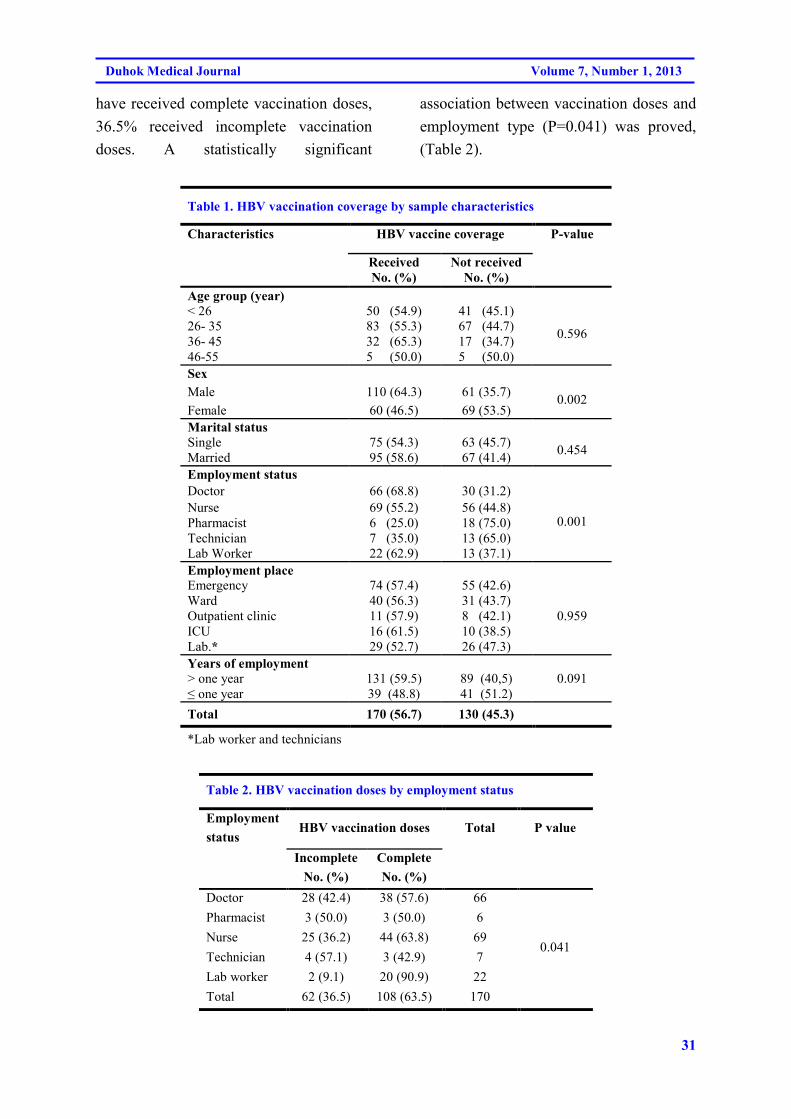
31
Duhok Medical Journal Volume 7, Number 1, 2013
have received complete vaccination doses,
36.5% received incomplete vaccination
doses. A statistically significant
association between vaccination doses and
employment type (P=0.041) was proved,
(Table 2).
Table 1. HBV vaccination coverage by sample characteristics
Characteristics HBV vaccine coverage P-value
Received
No. (%)
Not received
No. (%)
Age group (year)
< 26 50 (54.9) 41 (45.1)
0.596 26- 35 83 (55.3) 67 (44.7)
36- 45 32 (65.3) 17 (34.7)
46-55 5 (50.0) 5 (50.0)
Sex
Male 110 (64.3) 61 (35.7) 0.002
Female 60 (46.5) 69 (53.5)
Marital status
Single 75 (54.3) 63 (45.7) 0.454
Married 95 (58.6) 67 (41.4)
Employment status
Doctor 66 (68.8) 30 (31.2)
0.001 Nurse 69 (55.2) 56 (44.8)
Pharmacist 6 (25.0) 18 (75.0)
Technician 7 (35.0) 13 (65.0)
Lab Worker 22 (62.9) 13 (37.1)
Employment place
Emergency 74 (57.4) 55 (42.6)
0.959
Ward 40 (56.3) 31 (43.7)
Outpatient clinic 11 (57.9) 8 (42.1)
ICU 16 (61.5) 10 (38.5)
Lab.* 29 (52.7) 26 (47.3)
Years of employment
> one year
≤ one year
131 (59.5)
39 (48.8)
89 (40,5)
41 (51.2)
0.091
Total 170 (56.7) 130 (45.3)
*Lab worker and technicians
Table 2. HBV vaccination doses by employment status
Employment
status HBV vaccination doses Total P value
Incomplete
No. (%)
Complete
No. (%)
Doctor 28 (42.4) 38 (57.6) 66
0.041
Pharmacist 3 (50.0) 3 (50.0) 6
Nurse 25 (36.2) 44 (63.8) 69
Technician 4 (57.1) 3 (42.9) 7
Lab worker 2 (9.1) 20 (90.9) 22
Total 62 (36.5) 108 (63.5) 170
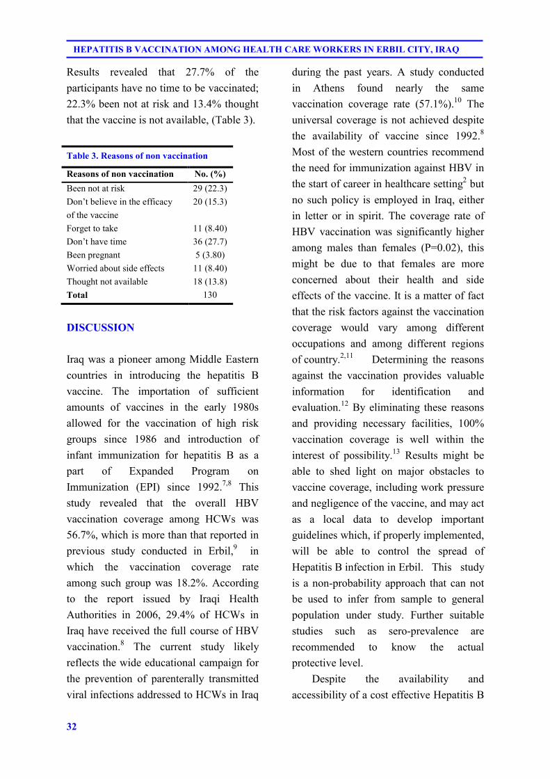
32
HEPATITIS B VACCINATION AMONG HEALTH CARE WORKERS IN ERBIL CITY, IRAQ
Results revealed that 27.7% of the
participants have no time to be vaccinated;
22.3% been not at risk and 13.4% thought
that the vaccine is not available, (Table 3).
Table 3. Reasons of non vaccination
Reasons of non vaccination No. (%)
Been not at risk 29 (22.3)
Don’t believe in the efficacy
of the vaccine
20 (15.3)
Forget to take 11 (8.40)
Don’t have time 36 (27.7)
Been pregnant 5 (3.80)
Worried about side effects 11 (8.40)
Thought not available 18 (13.8)
Total 130
DISCUSSION
Iraq was a pioneer among Middle Eastern
countries in introducing the hepatitis B
vaccine. The importation of sufficient
amounts of vaccines in the early 1980s
allowed for the vaccination of high risk
groups since 1986 and introduction of
infant immunization for hepatitis B as a
part of Expanded Program on
Immunization (EPI) since 1992.7,8
This
study revealed that the overall HBV
vaccination coverage among HCWs was
56.7%, which is more than that reported in
previous study conducted in Erbil,9 in
which the vaccination coverage rate
among such group was 18.2%. According
to the report issued by Iraqi Health
Authorities in 2006, 29.4% of HCWs in
Iraq have received the full course of HBV
vaccination.8 The current study likely
reflects the wide educational campaign for
the prevention of parenterally transmitted
viral infections addressed to HCWs in Iraq
during the past years. A study conducted
in Athens found nearly the same
vaccination coverage rate (57.1%).10
The
universal coverage is not achieved despite
the availability of vaccine since 1992.8
Most of the western countries recommend
the need for immunization against HBV in
the start of career in healthcare setting2 but
no such policy is employed in Iraq, either
in letter or in spirit. The coverage rate of
HBV vaccination was significantly higher
among males than females (P=0.02), this
might be due to that females are more
concerned about their health and side
effects of the vaccine. It is a matter of fact
that the risk factors against the vaccination
coverage would vary among different
occupations and among different regions
of country.2,11
Determining the reasons
against the vaccination provides valuable
information for identification and
evaluation.12
By eliminating these reasons
and providing necessary facilities, 100%
vaccination coverage is well within the
interest of possibility.13
Results might be
able to shed light on major obstacles to
vaccine coverage, including work pressure
and negligence of the vaccine, and may act
as a local data to develop important
guidelines which, if properly implemented,
will be able to control the spread of
Hepatitis B infection in Erbil. This study
is a non-probability approach that can not
be used to infer from sample to general
population under study. Further suitable
studies such as sero-prevalence are
recommended to know the actual
protective level.
Despite the availability and
accessibility of a cost effective Hepatitis B
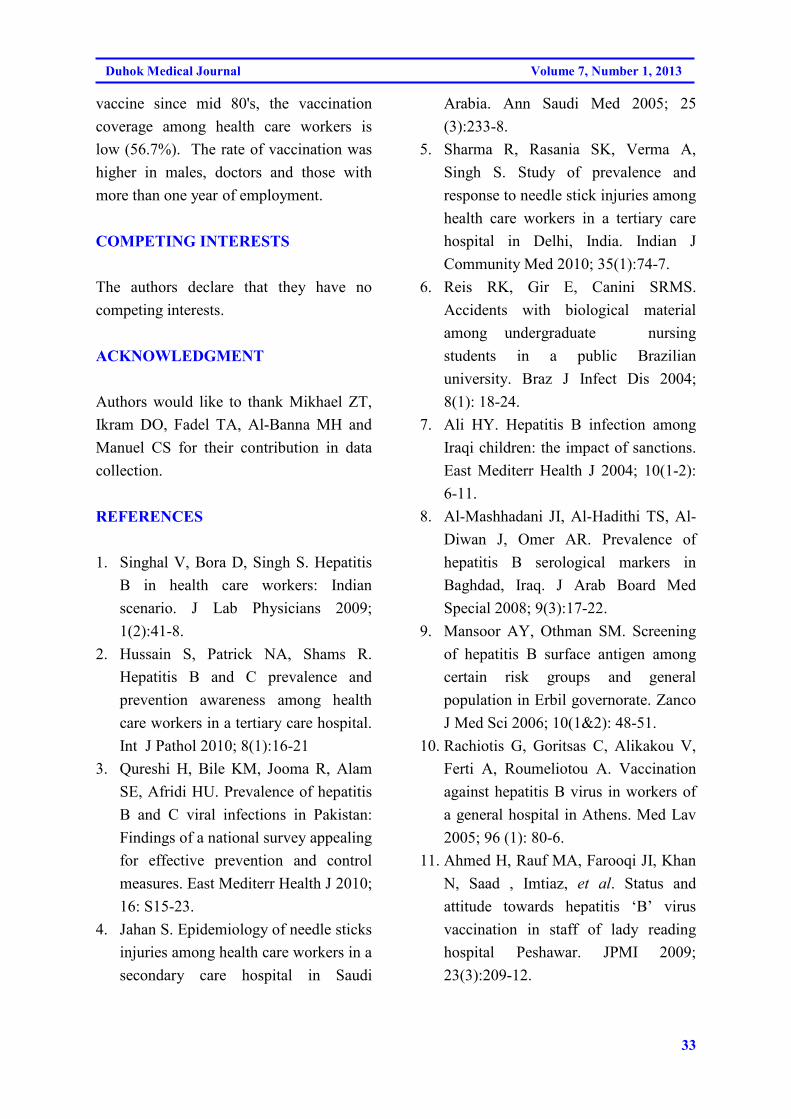
33
Duhok Medical Journal Volume 7, Number 1, 2013
vaccine since mid 80's, the vaccination
coverage among health care workers is
low (56.7%). The rate of vaccination was
higher in males, doctors and those with
more than one year of employment.
COMPETING INTERESTS
The authors declare that they have no
competing interests.
ACKNOWLEDGMENT
Authors would like to thank Mikhael ZT,
Ikram DO, Fadel TA, Al-Banna MH and
Manuel CS for their contribution in data
collection.
REFERENCES
1. Singhal V, Bora D, Singh S. Hepatitis
B in health care workers: Indian
scenario. J Lab Physicians 2009;
1(2):41-8.
2. Hussain S, Patrick NA, Shams R.
Hepatitis B and C prevalence and
prevention awareness among health
care workers in a tertiary care hospital.
Int J Pathol 2010; 8(1):16-21
3. Qureshi H, Bile KM, Jooma R, Alam
SE, Afridi HU. Prevalence of hepatitis
B and C viral infections in Pakistan:
Findings of a national survey appealing
for effective prevention and control
measures. East Mediterr Health J 2010;
16: S15-23.
4. Jahan S. Epidemiology of needle sticks
injuries among health care workers in a
secondary care hospital in Saudi
Arabia. Ann Saudi Med 2005; 25
(3):233-8.
5. Sharma R, Rasania SK, Verma A,
Singh S. Study of prevalence and
response to needle stick injuries among
health care workers in a tertiary care
hospital in Delhi, India. Indian J
Community Med 2010; 35(1):74-7.
6. Reis RK, Gir E, Canini SRMS.
Accidents with biological material
among undergraduate nursing
students in a public Brazilian
university. Braz J Infect Dis 2004;
8(1): 18-24.
7. Ali HY. Hepatitis B infection among
Iraqi children: the impact of sanctions.
East Mediterr Health J 2004; 10(1-2):
6-11.
8. Al-Mashhadani JI, Al-Hadithi TS, Al-
Diwan J, Omer AR. Prevalence of
hepatitis B serological markers in
Baghdad, Iraq. J Arab Board Med
Special 2008; 9(3):17-22.
9. Mansoor AY, Othman SM. Screening
of hepatitis B surface antigen among
certain risk groups and general
population in Erbil governorate. Zanco
J Med Sci 2006; 10(1&2): 48-51.
10. Rachiotis G, Goritsas C, Alikakou V,
Ferti A, Roumeliotou A. Vaccination
against hepatitis B virus in workers of
a general hospital in Athens. Med Lav
2005; 96 (1): 80-6.
11. Ahmed H, Rauf MA, Farooqi JI, Khan
N, Saad , Imtiaz, et al. Status and
attitude towards hepatitis ‘B’ virus
vaccination in staff of lady reading
hospital Peshawar. JPMI 2009;
23(3):209-12.
34
HEPATITIS B VACCINATION AMONG HEALTH CARE WORKERS IN ERBIL CITY, IRAQ
12. Amini-Ranjbar S, Motlagh ME.
Hepatitis B vaccination coverage
among Iranian medical students and
nursing staff. Am J Applied Sci 2008;
5(6):747-9.
13. Naz S, Ahmad M, Asghar H.
Prevalence of hepatitis ‘B’ among
hospital personnel in combined
military hospital (CMH)
Muzaffarabad. Int J Agri Biol 2002;
4(2):227-30.
35
Duhok Medical Journal Volume 7, Number 1, 2013
ón‚íqón‚íqón‚íqón‚íq@@@@@@@@‹ŽïÜìóè@ñŠb’@óÜ@��슇äóm@ðäa‡äóàŠbØ@çaíŽïä@óÜ@l@ñŠüu@Šó�u@ð�û‹îbÄ@ñómíØbà‹ŽïÜìóè@ñŠb’@óÜ@��슇äóm@ðäa‡äóàŠbØ@çaíŽïä@óÜ@l@ñŠüu@Šó�u@ð�û‹îbÄ@ñómíØbà‹ŽïÜìóè@ñŠb’@óÜ@��슇äóm@ðäa‡äóàŠbØ@çaíŽïä@óÜ@l@ñŠüu@Šó�u@ð�û‹îbÄ@ñómíØbà‹ŽïÜìóè@ñŠb’@óÜ@��슇äóm@ðäa‡äóàŠbØ@çaíŽïä@óÜ@l@ñŠüu@Šó�u@ð�û‹îbÄ@ñómíØbà@M@M@M@M×a�È×a�È×a�È×a�È@@@@@@@@
@@@@@@@@wäbàŠb÷@ì@ôØó“ŽïqwäbàŠb÷@ì@ôØó“ŽïqwäbàŠb÷@ì@ôØó“ŽïqwäbàŠb÷@ì@ôØó“ŽïqZZZZ@@@çbØòìín“îó�Žïq@ómłìì@óÜ40@@M@60@@E@ñŠüu@Êóu@ð�û‹îbÄ@ô䆋Øìóè@ô’ü‚óäB @@@õ‹Žî†ìbš@ôäa‡äóàŠbØ@óÜ
@ôîó“ïq@ôØbå�‹m@õŠbØüè@üi@òìónŽî4Šó�ò†@ôn�슇äóm@ Z÷@ìó÷@çaíŽïä@óÜ@çbmíØ@õò‰Žî4Š@ô䆋Ø@õŠbî†@üi@òìaŠ†@ãb−ó÷@óîòìóåîím@ãó@ì@ çbmíØ@ õò‰ŽîŠ@ çaíŽïä@ õ‡äòíîóq@ ôä‡äb�äó�Üóè@ üi@ bèòìóè@ L@ çóØò†ŠbØ@ ôn�슇äóm@ õ‹Žî†ìbš@ õŠaíi@ óÜ@ óØ@ õóäa‡äóàŠbØ
ôn�슇äóm@õ‹Žî†ìbš@õŠaíi@ôäa‡äóàŠbØ@óÜ@ôn“�@ôäbØóî‡äó¸ójîbm.@@åïÜíØóÄ@æŽïÙîŠåïÜíØóÄ@æŽïÙîŠåïÜíØóÄ@æŽïÙîŠåïÜíØóÄ@æŽïÙôŽôŽôŽôZZZZ@@@õòìbà@óÜ@òìaŠ‡àb−ó÷@óîòìóåîím@ãó÷1@M@@9M@2011@@@@bm1M@5@–@2012@@@ôåm‹�Šòì@ói@‹ŽïÜìóè@õŠb’@óÜ
@óäìí¹@I@@ŽÞràb�@H‹ŽïÜìóè@ôäbØóîŠbØ‹ŽïÐ@õóäbƒ’ü‚óä@óÜ@çbØòŒaìbïu@ó’ói@óÜ@ôn�슇äóm@õ‹ŽîŠ†ìbš@õaŠíi@ôäa‡äóàŠbØ@óÜ.@@ãb−ó÷ãb−ó÷ãb−ó÷ãb−ó÷ZZZZ@@óîòìóåîím@ãó÷@I300@@HäóàŠbØ@‹Žïä@õò‰Žî4Š@óØ@õü‚@ónŽî‹�ò†@ôn�슇äóm@õ‹Žî†ìbš@õ‡@I57@@H@E@Žôà@õò‰Žî4Š@òì@I43@@@H@E
@óÜ@çbïäóàóm@óØ@I22M55@@ Žßb�@Hìíi@@N@ðîa4‹ÙŽïm@aìóØ@a†@ðäb“ïq@óîòìóåîím@ãó÷@ðàb−ó÷56,7@@E@óÜ@òì@çìa‹míØ@çaìíiŠa‡’ói@óÜ@@a†@çbïäaíŽïä63,3@@@E@@çìa‹míØ@õìaìómóiI3@Šbu@Hi@@ãłó6,5@@E@óÜ@�àóØ3@@çìa‹míØ@Šbu@N@óîòíŽï’@�@Žôà@ì‹Žïä@çaíŽïä@óÜ@çbmíØ@õò‰Žî4Š
@@ìíi64,3@@@E@@ì46@@@E@Ûóî@õaì†@óÜ@Ûói@N@óØ@çbmíØ@õò‰Žî4Š@ Žßó�óÜ@熋؊bØ@õŠüy@çaíŽïä@óÜ@óîóè@�Žïèói@ôØóî‡äòíîóq@bèòìŠóèòìçbÙ“î�q@çaíŽïä@óÜ@ìíiŒŠói@ŠûŒ.@@
ãb−ó÷Šò†ãb−ó÷Šò†ãb−ó÷Šò†ãb−ó÷Šò†ZZZZ@ñŠmíØ@õò‰Žî@çb56,7@@E@bèòìŠóè@òì@çbÙ“î�q@çaíŽïä@óÜ@bèòìŠóè@òì@‹Žïä@ñŒó�ò4Š@ôäaìíiŠa‡’ói@óÜ@ìíiŒŠóiŠûŒ@óØò‰Žî4Š@óØ@ìíi@óÜ@çóØò†ŠbØ@óØ@õóäaìóÜ@Ûòì@çbØó�ïÔbm@ì@æmìóØbî‹Ð@ô’óióÜ@çóØò†ŠbØ@òóØ@õóäb�óØ@ìó÷@çaíŽïä@óÜ@ò‹mŒŠói@óØò‰ŽîŠ@óØ@@pìóØŠò†
óäbƒ’ü‚óä@ñ‹m@ôäbØóåîí’N@@
36
HEPATITIS B VACCINATION AMONG HEALTH CARE WORKERS IN ERBIL CITY, IRAQ
الخلاصة
لقاح الكبد الفايروسي نمط ب بين الكوادر الصحية في مدينة اربيل، العراق
العاملين في مجال في B التهاب الكبد بفيروس من حالات العدوى ٪60-40 النامية، في البلدان :خلفية واهداف البحث
العاملين في مجال الرعاية بين يمالتطع لتحديد معدل أجريت هذه الدراسة وقد .مهنية مخاطرل الصحية تعود الرعاية
. الصحية العاملين في مجال الرعاية من العامة الخصائصالتطعيم و بين نسبة ودراسة العلاقة ،الصحية
عينة مناسبة على 2012مارس 1 و 2011سبتمبر 1 منللفترة اربيل في مدينة مقطعية أجريت دراسة :طرق البحث
.في مستشفى أربيل التعليمي مختلف الأقسام نم العاملين في الرعاية الصحية من
معدل . (٪43) الإناث و (٪57) من العاملين في الرعاية الصحية وكانت نسبة الذكور 300عينة شملت :النتائج
(٪63.5) هذه المجموعة بينالتطعيم و تلقوا من المشاركين ٪56.7 النتائج أن كشفتو قد. سنة 55الى 22 العمرما بين
(٪64.3)الإناث كانت معنويأ أعلى من ربين الذكو التطعيم نسبة .جزئيا همتم تطعيم(%36.5) و ثلاث جرعات او قد أكمل
.بين الأطباء أعلى الذي كان التطعيمالعمل و نوع علاقة معنوية بين وكان هناك. على التوالي (46.5٪)
التطعيم و تبين أيضا ان نسبة. و كذلك الأطباء ورفي جنس الذك كان أعلى و ، ٪56.7كان التطعيم معدل :اتالاستنتاج
.الأخرى مجالات العمل من المختبر قسم تليها في قسم الطوارئ بين الذين يعملون أعلى كان

37
Duhok Medical Journal Volume 7, Number 1, 2013
THE EPIDEMIOLOGICAL, CLINICAL AND LABORATORY CHARACTERISTICS
OF TYPHOID FEVER OUTBREAK IN SULAIMANI GOVERNORATE DURING
2007-2008
MOHAMMED O. MOHAMMED, MBChB-MD-FICMS- FRCP (London)*
HEMN M. MUSTAFA, MBChB- FIBMS (Med.)**
MOHAMMED A. ALSHEIKHANI, MBChB- CABM- FRCP (London)***
Submitted 16 Mar 2013; accepted 3 Nov 2013
ABSTRACT
Background and objectives Sporadic cases of typhoid are frequently reported in Kurdistan
Region, Iraq. The disease, however, has always got the potentiality to occur in epidemic.
During years 2007-2008, an outbreak of Typhoid fever (T.F) had occurred in Sulaimani-Iraqi
Kurdistan. The aim of the study is to describe the epidemiological, clinical and laboratory
characteristics of that outbreak in Sulaimani during that period.
Methods Three hundred and five patients were enrolled in the study. All were admitted in the
general teaching hospital of Sulaimani. Full history, clinical Examination, Hematological,
biochemical, abdominal ultrasound and blood culture and sensitivity were carried out.
Results Of the 305 patients, 207 of them had positive blood culture; the other 98 patients had
negative blood cultures. In blood culture positive group, 53.1% were females and 46.9% were
males. Their age ranged between 12-57 years. Blood cultures were positive in 67.1%, 17.9%,
15% for Salmonella typhi, S.paratyphi B and S.paratyphi A respectively. All were sensitive to
ceftriaxone and all were resistant to chloramphenicol.
Conclusions In this study we realized that T.F emerged as one of most common causes of
febrile illnesses in Sulaimani during the years 2007-2008 due to an outbreak that affected the
city specially the areas with poor sanitary conditions. The clinical, epidemiological &
antibiotics resistant patterns were almost similar (with minor differences) to those reported
internationally especially the developing countries.
Duhok Med J 2013;7(1): 37-48. Key words: Enteric fever, Typhoid fever, Multidrug-resistant typhoid fever
he term enteric fever was proposed as
an alternative designation to
distinguish typhoid fever from typhus.1
The introduction of chloramphenicol for
the treatment of typhoid fever in 1948
transformed a severe, debilitating, and
often fatal disease into a readily treatable
condition. Typhoid fever (T.F) is endemic
in many parts of the developing world and
tends to occur in outbreaks and epidemic
forms.2
Risk factors include contaminated
water or ice,3 flooding. Food and drinks
purchased from street vendors, raw fruits
and vegetables grown in fields fertilized
with sewage, lack of hand washing and
toilet access,4 ill household contacts
5 and
evidence of prior Helicobacter pylori
infection (an association probably related
to chronically reduced gastric acidity),
with recent use of antimicrobial drugs.6
The development of severe disease (which
occurs in ~10–15% of patients) depends
on host factors (immunosuppressant,
antacid therapy, previous exposure, and
T
* Assistant Professor / Consultant Gastroenterologist, College of Medicine / University of Sulaimani
** Specialist physician, Department of Medicine, General Teaching Hospital
*** Assistant Professor / Consultant Physician, College of Medicine / University of Sulaimani
Correspondence author: Mohammed O. Mohammed. Email: [email protected]

38
THE EPIDEMIOLOGICAL, CLINICAL AND LABORATORY CHARACTERISTICS OF TYPHOID FEVER .
OUTBREAK
vaccination), strain virulence and
inoculum, and choice of antibiotic
therapy.7
Bleeding is the most common
complication, occurring in up to 10 percent
of patients. It results from erosion of a
necrotic Peyer’s patch through the wall of
an enteric vessel, may occur at the end of
the 2nd
week or during the 3rd
week of the
illness. In the majority of cases, the
bleeding is slight and resolves without the
need for blood transfusion, but in 2 percent
of cases, bleeding is clinically significant
and can be rapidly fatal if a large vessel is
involved.7,8
Intestinal (usually Ileal)
perforation is the most serious
complication, occurring in 1 to 3 percent
of hospitalized patients. Perforation may
be manifested by an acute abdomen or,
more covertly, by simple worsening of
abdominal pain, rising pulse, and falling
blood pressure in an already sick patient.7
This study was designed to describe the
clinical, epidemiological, and laboratory
characteristics of typhoid fever outbreak in
Sulaimani during the years of 2007-2008.
METHODS
This study was conducted between Oct. -
1st -2007 and June -30
th -2008 in the
General Teaching Hospital of Sulaimani.
Three hundred and five patients were
included in the study. Those who were
referred from outside the Sulaimani city,
has no municipality, were regarded to be
from rural areas. All patients were
admitted in the General teaching hospital
of Sulaimani during an outbreak of T.F in
Sulaimani City, in which all cases of fever
were suspected to be typhoid until proving
otherwise. The Inclusion criteria were any
patient who had fever for at least for 3
days before presentation and had one or
more symptoms, signs or investigation
results that made typhoid fever more likely
e.g. pattern of fever, splenomegaly,
leucopenia, infection of other family
members….etc. We exclude other causes
of fever with similar presentation.
A special form was filled for each
patient including demographic data like
gender, age, and residence, duration of
fever, symptoms, family history, and
source of water supply, also thorough
examination was performed. Abdominal
ultrasonography, complete blood count,
biochemical, and liver function test in
addition to blood culture/ sensitivity were
done. Patients with pancytopenia were
underwent bone marrow examination,
those with mealena or bleeding per rectum
underwent colonoscopy and those with
suspected perforated viscus surgical
opinion were sought. For those with
elevated liver transaminases, hepatic viral
markers were sent. Widal test was not used
for diagnosis because of its high false
positive results. Positive blood cultures
were subjected to antimicrobial
susceptibility test using Kirby-Bauer
method9 using eight antimicrobial agents;
1- amoxicillin, 2- ampicillin, 3-
chloramphenicol, 4- co-trimoxazole, 5-
ceftriaxon, 6- cefotaxime, 7- ciprofloxacin
and 8- gentamicin. The diameter of
inhibition zone was considered to evaluate
the susceptibility according to the National
Committee for Clinical Laboratory
Standards, Subcommittee on
Antimicrobial Susceptibility Testing,
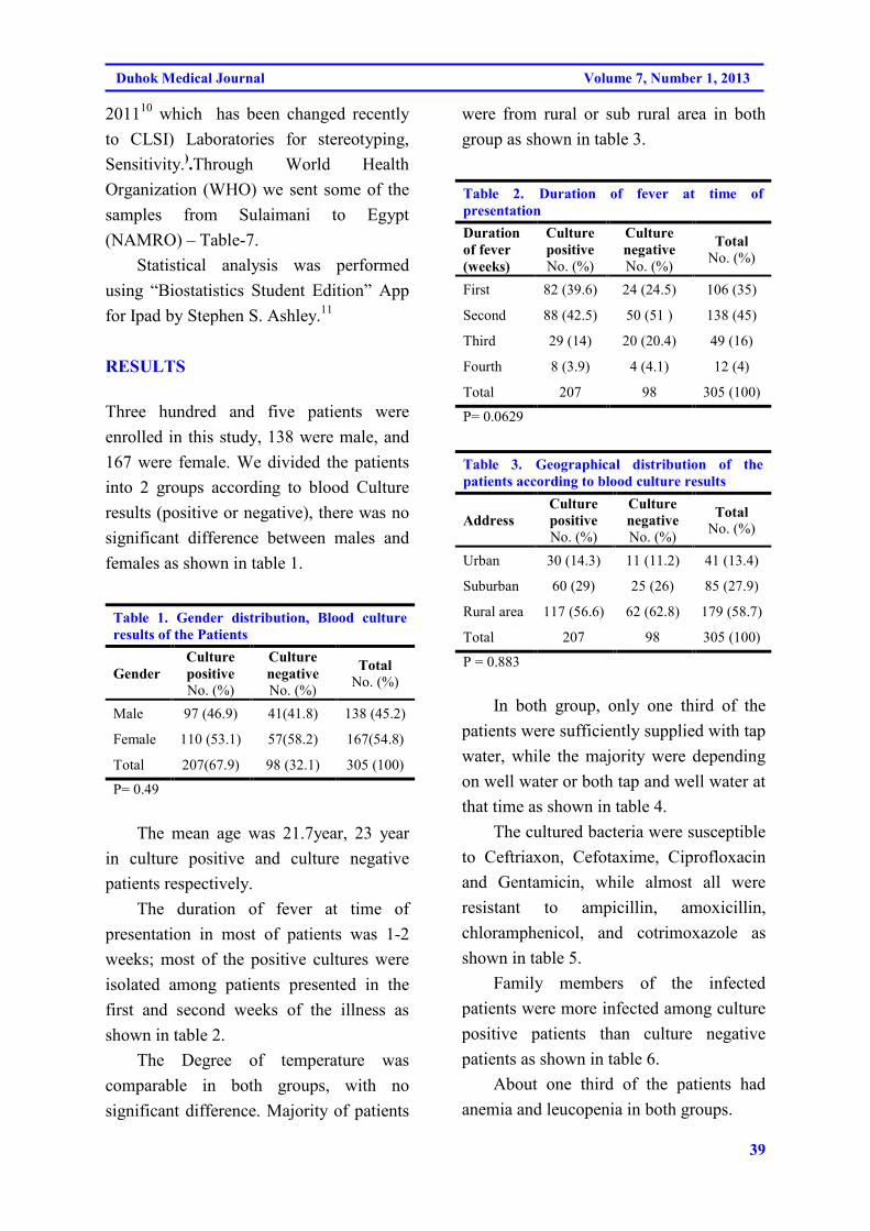
39
Duhok Medical Journal Volume 7, Number 1, 2013
201110
which has been changed recently
to CLSI) Laboratories for stereotyping,
Sensitivity.).Through World Health
Organization (WHO) we sent some of the
samples from Sulaimani to Egypt
(NAMRO) – Table-7.
Statistical analysis was performed
using “Biostatistics Student Edition” App
for Ipad by Stephen S. Ashley.11
RESULTS
Three hundred and five patients were
enrolled in this study, 138 were male, and
167 were female. We divided the patients
into 2 groups according to blood Culture
results (positive or negative), there was no
significant difference between males and
females as shown in table 1.
Table 1. Gender distribution, Blood culture
results of the Patients
Gender
Culture
positive
No. (%)
Culture
negative
No. (%)
Total
No. (%)
Male 97 (46.9) 41(41.8) 138 (45.2)
Female 110 (53.1) 57(58.2) 167(54.8)
Total 207(67.9) 98 (32.1) 305 (100)
P= 0.49
The mean age was 21.7year, 23 year
in culture positive and culture negative
patients respectively.
The duration of fever at time of
presentation in most of patients was 1-2
weeks; most of the positive cultures were
isolated among patients presented in the
first and second weeks of the illness as
shown in table 2.
The Degree of temperature was
comparable in both groups, with no
significant difference. Majority of patients
were from rural or sub rural area in both
group as shown in table 3.
Table 2. Duration of fever at time of presentation
Duration
of fever
(weeks)
Culture
positive
No. (%)
Culture
negative
No. (%)
Total
No. (%)
First 82 (39.6) 24 (24.5) 106 (35)
Second 88 (42.5) 50 (51 ) 138 (45)
Third 29 (14) 20 (20.4) 49 (16)
Fourth 8 (3.9) 4 (4.1) 12 (4)
Total 207 98 305 (100)
P= 0.0629
Table 3. Geographical distribution of the patients according to blood culture results
Address
Culture
positive
No. (%)
Culture
negative
No. (%)
Total
No. (%)
Urban 30 (14.3) 11 (11.2) 41 (13.4)
Suburban 60 (29) 25 (26) 85 (27.9)
Rural area 117 (56.6) 62 (62.8) 179 (58.7)
Total 207 98 305 (100)
P = 0.883
In both group, only one third of the
patients were sufficiently supplied with tap
water, while the majority were depending
on well water or both tap and well water at
that time as shown in table 4.
The cultured bacteria were susceptible
to Ceftriaxon, Cefotaxime, Ciprofloxacin
and Gentamicin, while almost all were
resistant to ampicillin, amoxicillin,
chloramphenicol, and cotrimoxazole as
shown in table 5.
Family members of the infected
patients were more infected among culture
positive patients than culture negative
patients as shown in table 6.
About one third of the patients had
anemia and leucopenia in both groups.
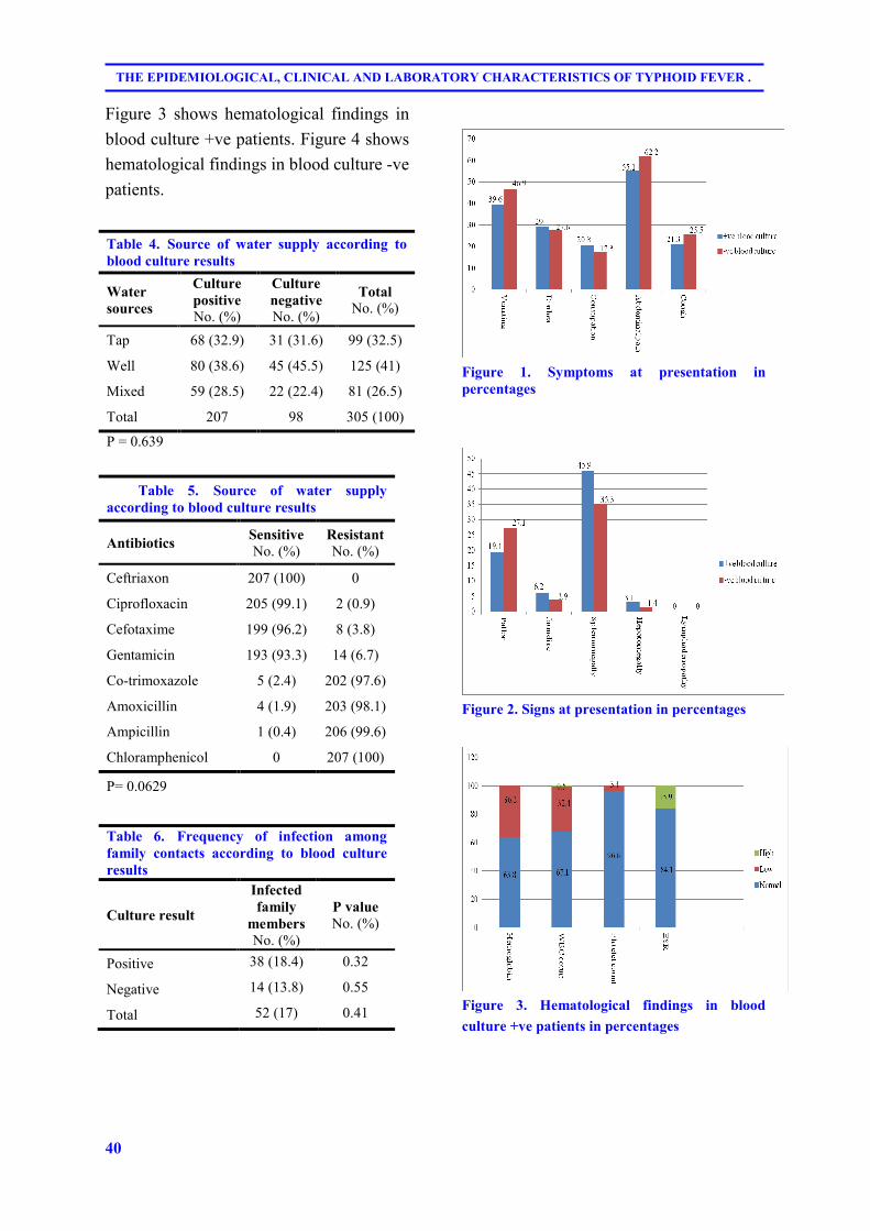
40
THE EPIDEMIOLOGICAL, CLINICAL AND LABORATORY CHARACTERISTICS OF TYPHOID FEVER .
OUTBREAK
Figure 3 shows hematological findings in
blood culture +ve patients. Figure 4 shows
hematological findings in blood culture -ve
patients.
Table 4. Source of water supply according to blood culture results
Water
sources
Culture
positive
No. (%)
Culture
negative
No. (%)
Total
No. (%)
Tap 68 (32.9) 31 (31.6) 99 (32.5)
Well 80 (38.6) 45 (45.5) 125 (41)
Mixed 59 (28.5) 22 (22.4) 81 (26.5)
Total 207 98 305 (100)
P = 0.639
Table 5. Source of water supply according to blood culture results
Antibiotics Sensitive
No. (%) Resistant
No. (%)
Ceftriaxon 207 (100) 0
Ciprofloxacin 205 (99.1) 2 (0.9)
Cefotaxime 199 (96.2) 8 (3.8)
Gentamicin 193 (93.3) 14 (6.7)
Co-trimoxazole 5 (2.4) 202 (97.6)
Amoxicillin 4 (1.9) 203 (98.1)
Ampicillin 1 (0.4) 206 (99.6)
Chloramphenicol 0 207 (100)
P= 0.0629
Table 6. Frequency of infection among family contacts according to blood culture
results
Culture result
Infected
family
members
No. (%)
P value
No. (%)
Positive 38 (18.4) 0.32
Negative 14 (13.8) 0.55
Total 52 (17) 0.41
Figure 1. Symptoms at presentation in
percentages
Figure 2. Signs at presentation in percentages
Figure 3. Hematological findings in blood
culture +ve patients in percentages
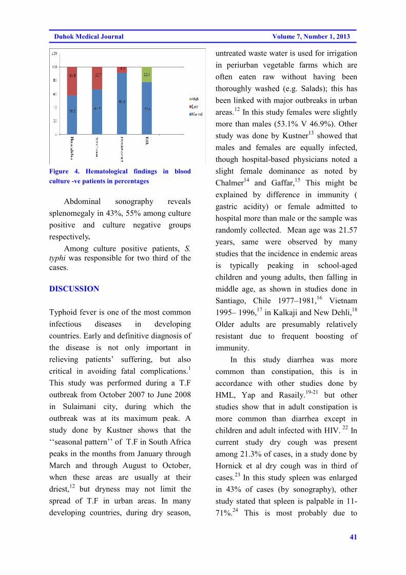
41
Duhok Medical Journal Volume 7, Number 1, 2013
Figure 4. Hematological findings in blood
culture -ve patients in percentages
Abdominal sonography reveals
splenomegaly in 43%, 55% among culture
positive and culture negative groups
respectively.
Among culture positive patients, S.
typhi was responsible for two third of the
cases.
DISCUSSION
Typhoid fever is one of the most common
infectious diseases in developing
countries. Early and definitive diagnosis of
the disease is not only important in
relieving patients’ suffering, but also
critical in avoiding fatal complications.1
This study was performed during a T.F
outbreak from October 2007 to June 2008
in Sulaimani city, during which the
outbreak was at its maximum peak. A
study done by Kustner shows that the
‘‘seasonal pattern’’ of T.F in South Africa
peaks in the months from January through
March and through August to October,
when these areas are usually at their
driest,12
but dryness may not limit the
spread of T.F in urban areas. In many
developing countries, during dry season,
untreated waste water is used for irrigation
in periurban vegetable farms which are
often eaten raw without having been
thoroughly washed (e.g. Salads); this has
been linked with major outbreaks in urban
areas.12
In this study females were slightly
more than males (53.1% V 46.9%). Other
study was done by Kustner13
showed that
males and females are equally infected,
though hospital-based physicians noted a
slight female dominance as noted by
Chalmer14
and Gaffar,15
This might be
explained by difference in immunity (
gastric acidity) or female admitted to
hospital more than male or the sample was
randomly collected. Mean age was 21.57
years, same were observed by many
studies that the incidence in endemic areas
is typically peaking in school-aged
children and young adults, then falling in
middle age, as shown in studies done in
Santiago, Chile 1977–1981,16
Vietnam
1995– 1996,17
in Kalkaji and New Dehli,18
Older adults are presumably relatively
resistant due to frequent boosting of
immunity.
In this study diarrhea was more
common than constipation, this is in
accordance with other studies done by
HML, Yap and Rasaily.19-21
but other
studies show that in adult constipation is
more common than diarrhea except in
children and adult infected with HIV. 22
In
current study dry cough was present
among 21.3% of cases, in a study done by
Hornick et al dry cough was in third of
cases.23
In this study spleen was enlarged
in 43% of cases (by sonography), other
study stated that spleen is palpable in 11-
71%.24
This is most probably due to
42
THE EPIDEMIOLOGICAL, CLINICAL AND LABORATORY CHARACTERISTICS OF TYPHOID FEVER .
OUTBREAK
variability in the time of presentation.
Hepatomegaly was present in 3.4% (by
sonography); this was less than other study
which showed hepatic enlargement in 14-
65%.25
Also this is most probably due to
variability in the time of presentation. In
this study we didn’t find any patient with
rose spots, this is in agreement with some
other observers, who didn’t see them at all,
like Chalmers,16
this might be explained
by our darker skin color. The study found
that 36.2% of patients were anemic, 32.4%
were leucopenic and 3.4% were
thrombocytopenic. These results were
comparable to a study done by Yaramis et
al in Turkey, where 38% of patients were
anemic, 18% leucopenic, 10% were
thrombocytopenic.25
In another study done
by Rasoolinejad in Iran, 72.9% were
anemic, 11.2% leucopenic and 9.1% were
thrombocytopenic.26
Also 3% of the
patients had pancytopenia that is most
probably due to extensive
hemophagocytosis known as infection-
associated hemophagocytosis syndrome
(IAHS). In this study 3.9% of patients had
jaundice, 5.8% had elevated ALT / AST,
which differs from the study done by
Rasoolinejad in Iran, that showed 1.8% of
cases had jaundice, 24.2% had elevated
ALT / AST.26
The reason behind this
different may be due to racial differences
Among the blood positive cultures 67.1%
were positive for Salmonella typhi, 15%
for Salmonella paratyphi A and 17.9% for
Salmonella paratyphi B. This is also
comparable with other study estimated that
there is one case of paratyphoid fever for
every four cases of typhoid fever, but the
incidence of infection associated with S.
Paratyphi A appears to be increasing,
especially in India.3
In our study all cases
were resistant to chloramphenicol, most of
them resistant to amoxicillin, ampicillin
and co-trimoxazole, nearly the same
results observed by Phuong et al. who
reported that, 85% of isolates of S. typhi
were resistant to chloramphenicol,
ampicillin and trimethoprim-
sulfamethoxazole.27
World-wide spread of
multi-drug resistant (MDR) strains of
S.typhi poses a serious therapeutic
challenge.28,29
In our study all cultures
were sensitive to ceftriaxon. There have
been sporadic reports of high-level
resistance to ceftriaxone (Minimal
inhibitory concentration, 64 mg/liter) in S.
enterica Serotype typhi and S. enterica
serotype paratyphi although these strains
are very rare.30,31
We record 2 cases with
ciprofloxacin resistant. In many area of the
world S.typhi strains resistant to
ciprofloxacin have already been reported.29
One of the culture positive female who
was in a visit to Sulaimani treated
successfully ,after she returned to
Germany got relapse and she had been
fully investigated and found to have
S.typhi strain that is resistant to ampicillin,
ampicillin/sulbactam, piperacillin,
cefotaxime, ceftazidime, cefepime,
chloramphenicol, streptomycin,
Trimethoprim /sulfamethoxazole ,
azithromycin, and nalidixic acid. A
reduced susceptibility to ciprofloxacin was
detected (MICCIP = 1µg/mL). The isolate
was susceptible to Imipenem, meropenem,
gentamicin, tobramycin, and amikacin.32
Also a study done by Mohammed OM
et al in Sulaimani/Iraq (2009) showed high
43
Duhok Medical Journal Volume 7, Number 1, 2013
resistant rate for chloramphenicol,
Trimethoprim, amoxicillin& streptomycin
but very low resistant rate to amikacin,
azithromycin, ciprofloxacin &ceftriaxon.33
In current study 98 cases were blood
cultures negative, Culture negative will not
totally exclude typhoid fever, depending
on type of media used, no of bacteria. In
this study previous antibiotics were used in
96% of them who were treated with
antibiotics before presentation, 75.5% of
them presented after first week, these 2
factors may be the main reason behind the
negativity of blood cultures, this is also
observed in 2 studies done by Wain J. et al
they showed that the sensitivity of blood
culture is higher in the first week of the
illness and is reduced by prior use of
antibiotics.34, 35
In the current study 53% of
those with Splenomegaly presented in the
2nd
week and 71.4% of complications had
occurred in 3rd
week of the disease of the
disease, this is similar to observation of
Stuart.36
In conclusion typhoid fever emerged
as one of the most common causes of
febrile illnesses in Sulaimani during the
years 2007-2008. The clinical,
epidemiological and antibiotics resistant
patterns were mostly similar (with minor
differences) to those reported
internationally specially the developing
countries. Infection in urban areas is not
uncommon in relation to rural areas with
more poor sanitation and low levels of
education. Young adults were mostly
affected. After fever, abdominal pain and
vomiting were the main presenting
features. Diarrhea was more common than
constipation among those infected with
typhoid fever. Ceftriaxon and
ciprofloxacin were most sensitive
antibiotics. Resistance developed to almost
all previously sensitive antibiotics such as
amoxicillin, ampicillin, chloramphenicol
and others, and our results were approved
by WHO laboratories (NAMRO) in Egypt.
Negativity of blood cultures most probably
related to delay in presentation and
previous antibiotics use.
REFERENCES
1. Levine MM. Typhoid fever. In: Evans
AS, Brachman PS, (Eds). Bacterial
Infections of Humans. Epidemiology
and Control 3rd
ed. New York,
Plenum Medical BookCompany, 1998,
p.839-58.
2. Woodward TE, Smadel JE, Ley HL.
Preliminary report on the beneficial
effect of Chloromycetin in the
treatment of typhoid fever. Ann Intern
Med. 1948;29:131-4.
3. Black RE, Cisneros L, Levine MM, ,
Banfi A, Lobos H, Rodriguez H. Case-
control study to identify risk factors for
pediatric endemic typhoid fever in
Santiago, Chile. Bull World Health
Organ. 1985; 63(5):899-904.
4. Luby SP, Faizan MK, Fisher-Hoch SP,
Syed A, Mintz ED, Bhutta ZA. Risk
factors for typhoid fever in an endemic
setting, Karachi, Pakistan.Epidemiol
Infect. 1998;120(2):129-38.
5. Luxemburger C, Chau MC, Mai NL,
Wain J, Tran TH, Simpson JA. Risk
factors for typhoid fever in the
Mekong delta, southern Viet Nam: a
case-control study.Trans R Soc Trop
Med Hyg .2001; 95(1):19-23.
44
THE EPIDEMIOLOGICAL, CLINICAL AND LABORATORY CHARACTERISTICS OF TYPHOID FEVER .
OUTBREAK
6. Gasem MH, Dolmans WM, Keuter
MM,Djokomoeljanto RR.Poor food
hygiene and housing as RFs for
typhoid fever in Semarang, Indonesia.
Trop Med Int Health. 2001; 6(6):484-
90.
7. Van Basten JP, Stocken, brugger R.
Typhoid perforation: a review of the
literature since 1960. Trop Geogr
Med .1994; 46(6):336-9.
8. Dockrell DH, Sundar S, Angus BJ.
Infectious disease, In Davidson’s
Principles& Practice of Medicine,21st
ed. Churchill Livingstone, 2011.p.334-
5.
9. Bauer AW, Kirby WM, Sherris JC,
Turck M. Antibiotic susceptibility
testing by a standardized single disk
method. Am J Clin Pathol.
1966;45(4):493-6.
10. CLSI. Performance Standards for
Antimicrobial Susceptibility Testing;
Twenty-First Informational
Supplement. CLSI document M100-
S21 Wayne, PA: Clinical and
Laboratory Standards Institute;. 2011.
11. Ashley SS. Biostatistics Student
Edition. Apple store; 2011 [cited 2012
10/4/2012]; Available from:
http://itunes.apple.com/us/app/biostatis
tics-student-edition/id415378283?m.t
12. Hohmann EL, Oletta CA, Killeen KP,
Miller SI.phoP/phoQ-deleted
Salmonella typhi (Ty800) is a safe and
immunogenic single-dose typhoid
fever vaccine in volunteers. J Infect
Dis.1996;173(6): 1408-14.
13. Kustner HGV. Trends in four major
communicable diseases. S Afr Med J
.1979; 55(12):460-73.
14. Chalmers IM. Typhoid fever in an
endemic area: a 'great imitator'. S A fr
Med J. 1971; 45(17): 470-2.
15. Abdool Gaffar MS, Seedat YK,
Coovadia YM, Khan Q. The white cell
counts in typhoid fever. Trop Geogr
Med .1992; 44(1-2): 23-7.
16. Ferreccio C, Morris JG Jr, Valdivieso
C, Prenzel I, Sotomayor V, Drusano
GL. Efficacy of ciprofloxacin in the
treatment of chronic typhoid carriers. J
Infect Dis. 1988; 157(6):1235-9.
17. Lin FY, Ho VA, Khiem HB, Trach
DD, Bay PV, Thanh TC. The efficacy
of a Salmonella typhi Vi conjugate
vaccine in two to five years old
children. N Engl J M. 2001; 344(17):
1263-9.
18. Sinha A, Sazawal S, Kumar R, Sood S,
Reddaiah VP, Singh B. Typhoid fever
in children aged less than 5 years.
Lancet. 1999; 354: 734-7.
19. Oh HML, Masayu Z, Chew SK.
Typhoid fever in hospitalized children
in Singapore. J Infect. 1997;
34(4):237-42.
20. Yap YF, Puthucheary SD. Typhoid
fever in children—a retrospective
study of 54 cases from Malaysia.
Singapore Med J. 1998; 39(6):260-2.
21. Rasaily R, Dutta P, Saha MR, Mitra U,
Lahiri M, Pal SC. Multi-drug resistant
typhoid fever in hospitalized
children.Clinical, bacteriological and
epidemiological profiles.Eur J
Epidemiol. 1994; 10(1):41-6.
22. Gotuzzo E, Frisancho O, Sanchez J,
Liendo G, Carrillo C, Black RE.
Association between the acquired
immunodeficiency syndrome and
45
Duhok Medical Journal Volume 7, Number 1, 2013
infection with Salmonella typhi or
Salmonella paratyphi in an endemic
typhoid area. Arch Intern Med. 1991;
151(1): 381-2.
23. Hornick RB. Typhoid fever. In:
Hoeprich PD, Jordan MC, Ronald AR,
(eds). Infectious Diseases.A Treatise of
Infectious Processes.5th
ed.
Philadelphia, J B Lippincott Company,
1994: 747 –53.
24. Seebaran AR, Coovadia YM, Bhana
RH, Rajput MC, Naidoo BT, Haffejee
IE. Typhoid fever in the adult and
pediatric Indian population of Durban.
S Afr Med J .1990; 77(6):14-7.
25. Ahmet Y, Idris Y; Selahattin K.
Clinical and laboratory presentation of
typhoid fever. International
Pediatrics.2001;16(4) : 227-30.
26. Rasoolinejad M, Esmailpoor N,
Mogbel B. Salmonella Hepatitis
(analysis of hepatic involvement in 107
patients with typhoid fever). Medical
Journal of Islamic World Academy of
Science. 2006;16(2): 93-4.
27. Cao XT, Kneen R, Nguyen TA,
Truong DL, White NJ, Parry CM.A
comparative study of ofloxacin and
cefixime for treatment of Typhoid
fever in children.The Dong Nai
Pediatric Center Typhoid Study Group.
Pediatric Infect Dis J. 1999; 18(3):245-
8.
28. Bhutta ZA, Farooqui BJ, Sturm
AW.Eradication of a multiple drug
resistant Salmonella paratyphi A
causing meningitis with ciprofloxacin.
J Infect .1992; 25(2):215-9.
29. Pokharel BM, Koirala J, Dahal RK,
Mishra SK, Khadga PK, Tuladhar
NR.Multidrug-resistant and extended-
spectrum β-lactamase (ESBL)–
producing Salmonella enterica
(serotypes Typhi and Paratyphi A)
from blood isolates in Nepal:
surveillance of resistance and a search
for newer alternatives. Int J Infect Dis.
2006;10:434–8.
30. Saha SK, Talukder SY, Islam M, Saha
S. A highly ceftriaxon-resistant
Salmonella typhi in
Bangladesh.Pediatr Infect Dis J .1999;
18(4):387.
31. Mandal BK. Salmonella typhi and
other salmonellas. Gut .1994;
35(6):726-8.
32. Abrescia FF, Gobbi F, Angheben A,
Gobbo M, Bisoffi Z, Falda A,et al.
Salmonella enterica Serovar Typhi
with CTX-M β-Lactamase,Germany.
Emerging Infectious Diseases.
2009;15(9):1535.
33. Mohammed O, Srwa A, Ibrahim S.
Bacteriological and Molecular Genetic
Study of Salmonella typhi Isolated
From Patients with Enteric Fever.
Journal of Koya University. 2009;12:
57-76.
34. Wain J, Diep TS, Ho VA, Walsh AM,
Nguyen TT, Parry CM, et al.
Quantitation of bacteria in blood of
typhoid fever patients and relationship
between counts and clinical features,
transmissibility, and antibiotic
resistance. J Clin Microbiol .1998;
36(6):1683-7.
35. Wain J, Pham VB, Ha V, Nguyen NM,
To SD, Walsh AL, et al. Quantization
of bacteria in bone marrow from
patients with typhoid fever:

46
THE EPIDEMIOLOGICAL, CLINICAL AND LABORATORY CHARACTERISTICS OF TYPHOID FEVER .
OUTBREAK
relationship between counts and
clinical features. J Clin Microbiol.
2001; 39(4):1571-6.
36. Hosoglu S, Aldemir M, Akalin S,
Geyik MF, Tacyildiz IH, Loeb M. Risk
Factors for Enteric Perforation in
Patients with Typhoid Fever. Am J
Epidemiol.2004; 160 (1): 46-50.
47
Duhok Medical Journal Volume 7, Number 1, 2013
ón‚íqón‚íqón‚íqón‚íq@@@@@@@@@ô�Üb�@óÜ@ôäbáŽïÝ�@õb��ŽîŠbq@óÜ@bmóäa‹�@õbmóq@ôäbØóïîó�ïÔbmì@ôÙåïÝØ@ì@ôïàŠ†@báï�@ô�Üb�@óÜ@ôäbáŽïÝ�@õb��ŽîŠbq@óÜ@bmóäa‹�@õbmóq@ôäbØóïîó�ïÔbmì@ôÙåïÝØ@ì@ôïàŠ†@báï�@ô�Üb�@óÜ@ôäbáŽïÝ�@õb��ŽîŠbq@óÜ@bmóäa‹�@õbmóq@ôäbØóïîó�ïÔbmì@ôÙåïÝØ@ì@ôïàŠ†@báï�@ô�Üb�@óÜ@ôäbáŽïÝ�@õb��ŽîŠbq@óÜ@bmóäa‹�@õbmóq@ôäbØóïîó�ïÔbmì@ôÙåïÝØ@ì@ôïàŠ†@báï�2007@@@@@M@M@M@M2008@@@@ŒŒŒŒ@@@@@@@@
��
wäbàŠb÷@ ì@ ôØó“ŽïqwäbàŠb÷@ ì@ ôØó“ŽïqwäbàŠb÷@ ì@ ôØó“ŽïqwäbàŠb÷@ ì@ ôØó“ŽïqZZZZ@Šbu@Šbu@ ói@a‡Ôa�È@ ôäbn�†ŠíØ@ óÜ@ pa†ò†@ ìì.Š@ bmóäa‹�@ ô’ü‚óäL@@ôäa†ìì.Š@ õŠó�ó÷@ ã�ýói@óîóè@Šóè@bmóq@õòíŽï’óióØóïï’ü‚óä@N@ôä�ýb�@óÜ2007@@M2008@@õb��ŽîŠbq@óÜ@óØóïï’ü‚óä@õòŠìó�@ôØóîbmóq@Œ
@a‡îìì.Š@ôäbáŽïÝ�@NôÙåïÝØì@ðïàŠ†@báï�@ôån�‚Šò†@òìóåîím@ãó÷@ô−bàb÷@ì@ìíi@óîbmóq@ãó÷@ôäbØóîó�ïÔbm.@@ŽôåïÜíØóÄ@æŽïÙôåïÜíØóÄ@æŽïÙôåïÜíØóÄ@æŽïÙôåïÜíØóÄ@æŽïÙîŠZZZZ@@õŠbØ‹ŽïÐ@õóäbƒ’ü‚óä@óÜ@çbîìíàóè@óØ@çìíia‹Ø@Šó�@óÜ@çbîòìóåï�ÜüÙŽïÜ@•ü‚óä@wåŽïq@ì†ó�@ Žð�
õìbäóè@ôåïåÙ“q@ ì@ çìíia‹Ø@ ŽðÜ@ çbîìaìóm@ õŠbï�‹q@ çbîìíàóè@ L@ çìíia‹åŽîìó‚@ ôäbáŽïÝ�@ óÜ@@@@ôÙåïÝØôÙåïÝØôÙåïÝØôÙåïÝØ@@@@ìíàóè@ òì@ôåïåÙ“q@@a‹Ø@üi@çbïäbØón�îíŽïq@óîó�ïÔbm@N@@
ãb−ó÷ãb−ó÷ãb−ó÷ãb−ó÷ZZZZ@@óÜ305@@L@•ü‚óä207@@@ìíjn�‚Šò†@õóØóïï’ü‚óä@õŠbØüè@óØóïï’ü‚óä@üi@æŽîí‚@ôä‡äbš@ôàb−ó÷@çbîòì@óÜ@æŽîí‚@ôä‡äbš@ôàb−ó÷@98@çbî@ôåŽîŠóä@@çìíi@N@óÜ@L@ìíjn�‚Šò†@õóØóïï’ü‚óä@æŽîí‚@ôä‡äbš@óØ@õóäaìó÷
53.1@ E@ òì@ çìíi@ Žðà46.9@@ çìíi@ ‹Žïä@ çbî@ N@ óÜ@ çbØó’ü‚óä@ ôäóàóm12@M@57@@ ìíi@ Žßb�@ N@çbØóiû‹Ùïà@ ìíàóèüi@ çìíi‹�Šói@ õbmóäa‹�@@ @ ßüÙïåïÑàaŠüÝØ@@ çí�Øbî�Ñï�@ ŠójàaŠói@ çìíióä‹�Šói@ òì@ Ni@ çbØóiû‹Ùïà@óÜ@ çìíi@�î‹
S.typhi @LS.paratyphi B @LS. paratyphi A @@@õò‰Žî.Š@ói67.1@[email protected]@E@ì15@EÛóî@õaì†óÜ@Ûóî.@@ãb−ó÷Šò†ãb−ó÷Šò†ãb−ó÷Šò†ãb−ó÷Šò†ZZZZ@@ôäbáŽïÝ�@óÜ@óØ@a†óîbmóq@ìóÜ@ìíi@bm@ôØòŠó�@õüè@a†óîòìbà@ìóÜ@bmóäa‹�@óØ@pìóØŠò†@a†óîòìóåîím@ãóÜ@óÜ@a‡îìì.Š20007@@M2008@@ìíi@ãóØ@bïm@çbïåŽîìb‚@ì@Ûbq@óØ@õóäbåŽîí’@ìóÜ@pójîbm@ói@L@N@ôÙåïÝØ@ì@ôïàŠ†@báï�
Úî�ä@ óàŠ†@ ãó÷@ ôäbØóîó�ïÔbm@ìíi@õóäbàŠ†@ ìóÜ@óÜ@ óØ@ôäbm�ýì@@ôØóîŒaìbïu@ ‡äóš@ Žßó�@ óÜ@ pa†ó÷@ ìì.Š@ Žð�@ôäbéïua‡àóØN@@
48
THE EPIDEMIOLOGICAL, CLINICAL AND LABORATORY CHARACTERISTICS OF TYPHOID FEVER .
OUTBREAK
الخلاصة
الخصائص الوبائية و السريرية والمختبرية لوباء الحمى التيفوئيدية في محافظة السليمانية للفترة ما
م 2008- 2007بين
راق ولكن امكانية حدوث بين الحين والاخر فى كوردستان الع" ان مرض التيفوئيد يحدث كثيرا :خلفية واهداف البحث
م حدث وباء كبير لمرض التيفوئيد في محافظة السليمانية )2008 - 2007(خلال سنة . المرض كوباء تضل قائمة . وكان من اهداف هذا البحث اظهار الخصائص السريرية والوبائية والمختبرية لهذا الوباء
وا مسثشفى التعليمى الباطنى العام في السليمانية و جميعم مريض خلال الوباء قد أُدخل 305ضمن دراسة :طرق البحث .سُألوا عن مرضهم و أجري لهم الفحص السريرى والفحوص المختبرية المطلوبة
مريض كانت نتيجة 98مقابل 207مريض ، كانت نتيجة زرع الدم لمكروب التيفوئيد موجب عند 305ضمن :النتائج. سنة 57-12كان اعمار المرضى بين . أنثى والبقية ذكر % 53.1وجبة كانت وضمن المجموعة الم. زرع الدم سالب
% 15، % 17.9، % 67.1بنسب A باراتايفى، B، باراتايفى عبارة عن نوع تايفى وكانت بكتريا التيفوئيد المزروعة .على التوالي
مى فى تلك الفترة خاصة في المناطق قليلى أظهرت الدراسة بأن حمى التيفوئيد كانت السبب الرئيسى للح :اتالاستنتاجكانت خصائص الوباء قريبة مع بعض الاأختلافات عن خصائص وباء التيفوئيد التى تحدث في مناطق العالم . النظافة
.الثالث

49
Duhok Medical Journal Volume 7, Number 1, 2013
VITAMIN D STATUS IN PREGNANT AND NON-PREGNANT WOMEN IN A
KURDISTAN REGION-NORTH IRAQ
DHIA J. AL-TIMIMI, Mphil, PhD*
FERWERDIN SH. BARZINGI, MSc, PhD*
NARIN A. MOSSA, MSc**
Submitted 4 Sep 2013; accepted 3 Nov 2013
ABSTRACT
Background and objectives Maternal vitamin D deficiency is a common finding during
pregnancy and is a widespread public health problem in many populations. The aim of the
study was to determine the prevalence of vitamin D and the current vitamin D status among a
sample selected from the healthy women in Kurdistan Region, Iraq.
Methods A cross-sectional study was made of 25-hydroxy vitamin D levels of 300
apparently healthy women (200 pregnant and 100 non-pregnant). They were selected from
women attending the Antenatal care unit, Obstetrics and Gynecology Department , Azadi
Teaching Hospital, Duhok, during the period from May 2012 to November 2012.
Results The study confirms that vitamin D status is low among pregnant and non- pregnant
women in a Kurdistan Region population. It is thus indicates the need for screening serum
vitamin D levels in healthy women, particularly during the course of pregnancy.
Conclusions The study confirms that vitamin D status is low among pregnant and non-
pregnant women in a Kurdistan Region population. It is thus indicates the need for screening
serum vitamin D levels in healthy women, particularly during the course of pregnancy.
Duhok Med J 2013;7(1): 49-56. Key words: Vitamin D, Pregnancy, Healthy women
Vitamin D is an essential fat-soluble
vitamin, which is required for the
maintenance of good health. A high
prevalence of vitamin D deficiency is now
recognized in pregnant women1 Vitamin D
is important in pregnancy because it has
implications for both maternal and child
health. It has been reported that vitamin D
deficiency during pregnancy is associated
with multiple adverse health outcomes in
mothers such as gestational diabetes,1 pre-
eclampsia, preterm birth and low birth
weight.2 Cesarean deliveries are four times
more common among those displaying
lower 25(OH) D levels.3 Moreover,
neonatal respiratory infections, and
children low bone mineral density, type-1
diabetes, and eczema are more common in
those with vitamin D deficiency.4 In Iraq,
reports on this issue are limited and the
prevalence of vitamin D deficiency has not
been established among Kurdish women .
Hence the present study was conducted to
determine the current vitamin D status in
pregnant and non-pregnant women
selected from a healthy population in
Kurdistan Region, and to identify
population group for whom vitamin D
supplement may be a concern.
*Department of Clinical Biochemistry, College of Medicine, University of Duhok, Iraq. **Directorate of Health-Duhok.
Corresponding Author: Dr. Dhia J. Al-Timimi, Professor, Department of clinical Biochemistry, College of
Medicine, University of Duhok, Duhok, Kurdistan Region, Iraq,Email: [email protected], Phone:
+4967504228908
50
VITAMIN D STATUS IN PREGNANT AND NON-PREGNANT WOMEN IN …….
METHODS
This study was conducted at the antenatal
care unit of Azadi teaching hospital,
Duhok city, Kurdistan Region, Iraq. Three
hundred women were included in this
study. Of these, 200 were pregnant women
at different trimesters (50 in the first
trimester, 50 in the second trimester and
100 in the third trimester). Classification
of pregnant women in regard to weeks of
gestation was based on the record of last
menstrual period (LMP) and the results of
ultrasound (U.S).Gestational age was
determined by LMP and by the imaging
technique of ultrasonography.5
The
remainders (100) were age matched
apparently healthy non-pregnant women
selected from the medical staff and
relatives of women attending the same
hospital.
The non-probability consecutive
sampling technique was used. It was a
cross -sectional study, in which a group of
apparently healthy pregnant women was
compared with a control group of healthy
non-pregnant women to find out the status
of serum vitamin D levels during
pregnancy. Verbal consents were obtained
from each woman after explaining the
nature of the study. Ethical approval to
carry out this study was given by the local
ethics committee of Duhok Heath
Directorate.
Pregnant women were fit with
inclusion criteria: age 20-35 years, parity
1-4, no history of (DM, hypertension,
renal disease, liver disease, thyroid and
parathyroid disease or taking vitamin D or
calcium supplementation).
A pre-tests questionnaire was used to
obtain information from the participants
about age, parity, past medical history,
past obstetric history, and drug history.
Assessment of vitamin D status was done
according to the following criteria (6).
Vitamin D deficiency is defined as a < 10
ng/ml, Vitamin D insufficiency 10 – 29.9
ng/ml, sufficient 30 – 150 ng/ml and toxic
levels ≥ 150 ng/ml.
Serum concentration of 25(OH) D
was measured by ELISA technique (Accu-
Bind ELISA micro wells). Statistical
analysis was performed using SPSS
software, vision 21.0. Independent t-test
was used to assess differences in serum
analyte among groups. The Chi-square test
was used to compare the tallies or counts
of categorical between two or more
independent groups.
RESULTS
The mean (SD) values of serum 25(OH) D
level of pregnant women was 27.0 (15.6)
ng/ml and the non-pregnant women was
31.7(13.3) ng/ml, p<0.01. Of the three
hundred studied women 9.7 %( n=29)
were vitamin D deficiency. The prevalence
of vitamin D deficiency among pregnant
and non-pregnant women was 14% and
1%, respectively. The prevalence of
vitamin D insufficiency was 43.3 %(
n=130) in pregnant women and 17.7 %(
n=52) in non-pregnant women (Table 1).
Vitamin D deficiency was more prominent
in women in the third trimester (8.5%) as
compared to the first and second trimesters
(2.5%, 3.0%) respectively (Table 2).
Pregnant women with age ≥30 years had a
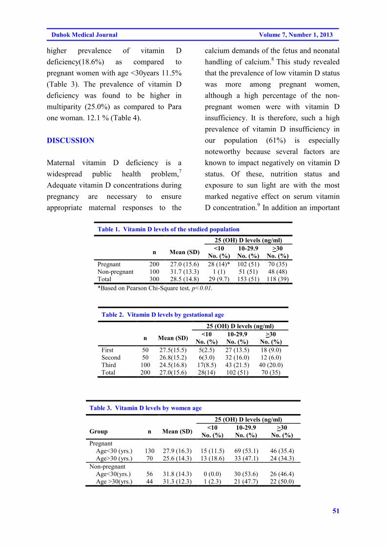
51
Duhok Medical Journal Volume 7, Number 1, 2013
higher prevalence of vitamin D
deficiency(18.6%) as compared to
pregnant women with age <30years 11.5%
(Table 3). The prevalence of vitamin D
deficiency was found to be higher in
multiparity (25.0%) as compared to Para
one woman. 12.1 % (Table 4).
DISCUSSION
Maternal vitamin D deficiency is a
widespread public health problem,7
Adequate vitamin D concentrations during
pregnancy are necessary to ensure
appropriate maternal responses to the
calcium demands of the fetus and neonatal
handling of calcium.8
This study revealed
that the prevalence of low vitamin D status
was more among pregnant women,
although a high percentage of the non-
pregnant women were with vitamin D
insufficiency. It is therefore, such a high
prevalence of vitamin D insufficiency in
our population (61%) is especially
noteworthy because several factors are
known to impact negatively on vitamin D
status. Of these, nutrition status and
exposure to sun light are with the most
marked negative effect on serum vitamin
D concentration.9 In addition an important
Table 1. Vitamin D levels of the studied population
25 (OH) D levels (ng/ml)
n Mean (SD) <10
No. (%)
10-29.9
No. (%)
>30
No. (%)
Pregnant 200 27.0 (15.6) 28 (14)* 102 (51) 70 (35)
Non-pregnant 100 31.7 (13.3) 1 (1) 51 (51) 48 (48)
Total 300 28.5 (14.8) 29 (9.7) 153 (51) 118 (39)
*Based on Pearson Chi-Square test, p<0.01.
Table 2. Vitamin D levels by gestational age
25 (OH) D levels (ng/ml)
n Mean (SD) <10
No. (%)
10-29.9
No. (%)
>30
No. (%)
First 50 27.5(15.5) 5(2.5) 27 (13.5) 18 (9.0)
Second 50 26.8(15.2) 6(3.0) 32 (16.0) 12 (6.0)
Third 100 24.5(16.8) 17(8.5) 43 (21.5) 40 (20.0)
Total 200 27.0(15.6) 28(14) 102 (51) 70 (35)
Table 3. Vitamin D levels by women age
25 (OH) D levels (ng/ml)
Group n Mean (SD) <10
No. (%)
10-29.9
No. (%)
>30
No. (%)
Pregnant
Age<30 (yrs.) 130 27.9 (16.3) 15 (11.5) 69 (53.1) 46 (35.4)
Age>30 (yrs.) 70 25.6 (14.3) 13 (18.6) 33 (47.1) 24 (34.3)
Non-pregnant
Age<30(yrs.) 56 31.8 (14.3) 0 (0.0) 30 (53.6) 26 (46.4)
Age >30(yrs.) 44 31.3 (12.3) 1 (2.3) 21 (47.7) 22 (50.0)
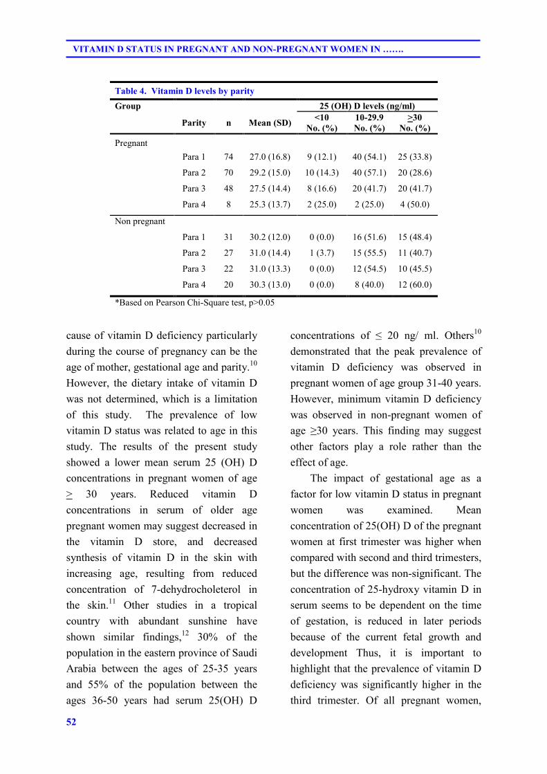
52
VITAMIN D STATUS IN PREGNANT AND NON-PREGNANT WOMEN IN …….
Table 4. Vitamin D levels by parity
Group 25 (OH) D levels (ng/ml)
Parity n Mean (SD) <10
No. (%)
10-29.9
No. (%)
>30
No. (%)
Pregnant
Para 1 74 27.0 (16.8) 9 (12.1) 40 (54.1) 25 (33.8)
Para 2 70 29.2 (15.0) 10 (14.3) 40 (57.1) 20 (28.6)
Para 3 48 27.5 (14.4) 8 (16.6) 20 (41.7) 20 (41.7)
Para 4 8 25.3 (13.7) 2 (25.0) 2 (25.0) 4 (50.0)
Non pregnant
Para 1 31 30.2 (12.0) 0 (0.0) 16 (51.6) 15 (48.4)
Para 2 27 31.0 (14.4) 1 (3.7) 15 (55.5) 11 (40.7)
Para 3 22 31.0 (13.3) 0 (0.0) 12 (54.5) 10 (45.5)
Para 4 20 30.3 (13.0) 0 (0.0) 8 (40.0) 12 (60.0)
*Based on Pearson Chi-Square test, p>0.05
cause of vitamin D deficiency particularly
during the course of pregnancy can be the
age of mother, gestational age and parity.10
However, the dietary intake of vitamin D
was not determined, which is a limitation
of this study. The prevalence of low
vitamin D status was related to age in this
study. The results of the present study
showed a lower mean serum 25 (OH) D
concentrations in pregnant women of age
> 30 years. Reduced vitamin D
concentrations in serum of older age
pregnant women may suggest decreased in
the vitamin D store, and decreased
synthesis of vitamin D in the skin with
increasing age, resulting from reduced
concentration of 7-dehydrocholeterol in
the skin.11
Other studies in a tropical
country with abundant sunshine have
shown similar findings,12
30% of the
population in the eastern province of Saudi
Arabia between the ages of 25-35 years
and 55% of the population between the
ages 36-50 years had serum 25(OH) D
concentrations of ≤ 20 ng/ ml. Others10
demonstrated that the peak prevalence of
vitamin D deficiency was observed in
pregnant women of age group 31-40 years.
However, minimum vitamin D deficiency
was observed in non-pregnant women of
age ≥30 years. This finding may suggest
other factors play a role rather than the
effect of age.
The impact of gestational age as a
factor for low vitamin D status in pregnant
women was examined. Mean
concentration of 25(OH) D of the pregnant
women at first trimester was higher when
compared with second and third trimesters,
but the difference was non-significant. The
concentration of 25-hydroxy vitamin D in
serum seems to be dependent on the time
of gestation, is reduced in later periods
because of the current fetal growth and
development Thus, it is important to
highlight that the prevalence of vitamin D
deficiency was significantly higher in the
third trimester. Of all pregnant women,
53
Duhok Medical Journal Volume 7, Number 1, 2013
65% were vitamin D insufficiency, 30%
were in the third trimester Vs 16% in the
first trimester. These data are consistent
with finding reported by others. Reported
data from Pakistan shows pregnant women
whose clothes that partly or completely
covered their body had serum 25(OH) D
levels below 30ng/ml at delivery.13
Vitamin D is also called the sunshine
vitamin because the major source (80%) of
vitamin D is Ultraviolet light. Kurdistan
Region is rich in abundant sunshine
throughout the year. However, the present
study showed vitamin D deficiency is
common in our locality, particularly in
pregnant women at third trimester. This
paradox may partly be explained by the
prevalent social and cultural practice in
Kurdistan that precludes adequate
exposure of pregnant women to sunshine.
Increasing urbanization is also one of the
reasons that results in poor outdoor
activity. Any reductions in sun exposure
attributable to the limited mobility during
later stages of pregnancy can cause also
vitamin D deficiency.
To rule out the impact of parity on
vitamin D status; It was found that the risk
of severe vitamin D deficiency was
significantly higher for pregnant women
with multi parity than in women with one
parity (25% Vs 12.1%), while the risk of
vitamin D insufficiency was nearly similar
for pregnant and non-pregnant women
with multipara (50% and 40%)
respectively. It appears that the present
results are comparable with other studies
done elsewhere reports14
that having given
birth two or more times, giving birth to
more than one child at a time (e.g. twins),
are significant risk factors for vitamin D
deficiency during pregnancy. However,
equal number of women from different age
groups and parity was not included in
study, which is another limitation of this
study
The current study confirms that
vitamin D status is low among pregnant
and non- pregnant women. Although low
vitamin D status may be caused by a
number of factors, including insufficient
synthesis in the skin and inadequate intake
or absorption of vitamin D, age of the
mother, gestational age and parity may
impact negatively on vitamin D status. It is
thus indicates the need for screening serum
vitamin D levels in healthy women,
particularly during the course of pregnancy
.Vitamin D supplementation may be an
effective public health intervention means
to improve the vitamin D status of the
population. A large scale study is
recommended on vitamin D status and its
relation with outcome of pregnancy in
Kurdistan Region-Iraq population.
REFERENCES
1. Zhang C, Qiu C, Hu FB, David RM,
van Dam RM, Bralley A et.al .
Maternal plasma 25-hydroxyvitamin D
concentrations and the risk for
gestational diabetes mellitus. PloS one
2008; 3(11:e 3753.
2. Bodnar LM, Catov JM, Simhan HN,
Holick MF, Powers RW, Roberts JM..
Maternal vitamin D deficiency
increases the risk of preeclampsia.J
Clin Endocrinol Metab 2007;
92(9):3517-22.
54
VITAMIN D STATUS IN PREGNANT AND NON-PREGNANT WOMEN IN …….
3. Merewood A, Mehta SD, Chen TC,
Bauchner H, Holick MF. Association
between vitamin D deficiency and
primary cesarean section. J Clin
Endocrinol Metab 2009;94(3):940-5..
4. Hensel KJ, Randis TM, Gelber SE,
Ratner AJ. Pregnancy-specific
association of vitamin D deficiency
and bacterial vaginosis . Am J Obstet
Gynecol 2011; 204(1): e41-49.
5. Hacker NF. Hacker and Moore's
Essential of Obstetrics and
Gynocology 2010. 5th
ed:Linda Van
Pelt.
6. Holick MF. Vitamin D staus:
measurement, interpretation, and
clinical application. Ann. Epidemiol
2009; 19(2): 73-8..
7. Holick MF, Chen TC. Vitamin D
deficiency: a worldwide problem with
health consequences. Am J Clin Nutr
2008; 87(4): 1080s-86s.
8. Holick MF. High prevalence of
vitamin D inadequacy and implications
for health. Mayo Clin Proc,
2006;81(3):353-73.
9. Aslam M, Masood Z, Sattar A, Qudsia
M. Vitamin D deficiency; prevalence
in pregnant women. Professional Med
J Apr 2012;19(2):208-13.
10. Holick MF. Vitamin D deficiency. N
Engl J Med 2007;357(3):266-281.
11. Holmes VA, Barnes MS, Alexander
HD, McFaul P, Wallace JM. Vitamin
D deficiency and insufficiency in
pregnant women; a longitudinal study.
Br J Nutr 2009;102(6):876-81.
12. Al Turki HA, Sadat Ali M, Elg AH,
Al Mulhim FA, Al Ali AK. 25-
Hydroxyvitamin D levels among
healthy Saudi Arabian women. Saudi
Med J 2008; 29(12): 1765-8.
13. Hossain N, Khanani R, Hussain-
Kanani F, Shah T, Arif S Pal L. High
prevalence of vitamin D deficiency in
Pakistani mothers and thier newborns.
Int J Gynecol Obstet 2011;112(3):229-
33.
14. Andersen LB,Abrahamsen B, Dalgard
C, Kyhl HB, Beck-Nielsen SS, Frost-
Nielsen M, et al. Parity and tanned
white skin as noval predictors of
vitamin D status in early pregnancy: a
population-based cohort study. Clin
Endocrinol 2013; 79(3):333-341.
55
Duhok Medical Journal Volume 7, Number 1, 2013
ón‚íqón‚íqón‚íqón‚íq@@@@@@@@Žo�b÷Žo�b÷Žo�b÷Žo�b÷@@@@ß@çbï�ìì†@óä@µî@ì@çbï�ìì†@µäˆ@Óò‡Ü@†@µàbnïÄß@çbï�ìì†@óä@µî@ì@çbï�ìì†@µäˆ@Óò‡Ü@†@µàbnïÄß@çbï�ìì†@óä@µî@ì@çbï�ìì†@µäˆ@Óò‡Ü@†@µàbnïÄß@çbï�ìì†@óä@µî@ì@çbï�ìì†@µäˆ@Óò‡Ü@†@µàbnïÄ@@@@@Žôäbn�†ŠíØ@bºŠóè@Žôäbn�†ŠíØ@bºŠóè@Žôäbn�†ŠíØ@bºŠóè@Žôäbn�†ŠíØ@bºŠóè@M@M@M@M@@@@ŽôÔa�È@ŽŠíØbiŽôÔa�È@ŽŠíØbiŽôÔa�È@ŽŠíØbiŽôÔa�È@ŽŠíØbi@@@@@@@@
��
wäbàŠb÷@ì@ôØó“ŽïqwäbàŠb÷@ì@ôØó“ŽïqwäbàŠb÷@ì@ôØó“ŽïqwäbàŠb÷@ì@ôØó“ŽïqZZZZ@@bïàóÝ‚b�@µ“îŠb÷ˆ@Úï÷@æmŠbà‰è@ónïè†ì@a‡øïäbï�ìì†@ùàò†@ß@óÄýóiŠói@bØò†Šbî†@çbÙîa†@Óò‡Ü@†@µàbnïÄ@bäíjáïØýóiŠói@ ÛóÜó�@µî@�“�@ Ä@ N@µäˆˆ@ Ša†Šíå�@ bØóàíØ@Óò‡Ü@ a†óØíä@ùàò††@ †@µàbnïÄ@ ùn�b÷@ bä‹ØŠíå�@ íiˆ@ ç‹Ø@ ómbè@ óåïÜíØóÄ@ Äó÷
@ß@ãóÝ‚b�@µî@çbï�ìì†@óä@µîì@çbï�ìì†@ùäbn�†ŠíØ@bºŠóèM@×a�ÈN@@ŽôåïÜíØóÄ@æŽïÙôåïÜíØóÄ@æŽïÙôåïÜíØóÄ@æŽïÙôåïÜíØóÄ@æŽïÙîŠZZZZ@@aŠbîc@aŠói@Äbä†@úìb¾2012@@bî@úìì†@båî‹“m@ì2012@bØóåïÜíØóÄ(cross section)@Øómbè@ì@ç‹300@@óÄí£@çˆ
æm‹�@ ~@ bäaìˆ200@@ ì@ çìíi@ ãóÝ‚b�ì@ çbï�ìì†@ çˆ100@çìíi@ ãóÝ‚b�@ Šóè@ ùÜói@ çbï�ìì†óä@ N@ì@ çbmò‹Ðb÷@ ù’ójÜ@ æmŠa‰jÜóè@ óäíjmbèùØíè†@ß@�“�@bî@ñ†aŒb÷@bäbƒ’í‚óåÜ@ùäíjØìŠaŒN@@
ãb−ó÷ãb−ó÷ãb−ó÷ãb−ó÷ZZZZ@çbï�ìì†@µäˆ@Óò‡Ü@ a‡øåîí‚@Óbä†@†@µàbnïÄ@ùn�b÷@ùîa‹Ùøm @}27.0 (15.6) ng/ml{@@çbï�ìì†@óä@µäˆ@ ì}31.7
(13.3) ng/ml{@ ~P<0.01@ Nçbï�ìì†@ µäˆ@ Óò‡Ü@ Ša숆@ bî@ †@ µàbnïÄ@ bäíjáïØ@ bäíjÄýóiŠói(14.0%)@@çbï�ìì†@ óä@ µäˆ@ ì(1.0%)N@@@†@µàbnïÄ@bäíjáïØ(<30 ng/ml)@@ìíióè}130(43.3%){@@ì@çbï�ìì†@µäˆ@Óò‡Ü}52 (17.7%){@@µäˆ@Óò‡Üçbï�ìì†óäN@i@æî‹m‡åÝi@Ša숆@bî@†@µàbnïÄ@bäíjáïØ@íi@çíjÄýóiŠóI8.5@HE@µî@ùïèbàì†@µÅîóè@ù�@ß@µäˆ@çaì@Óò‡Ü@ç‹Ùïåïjøm@ómbè
ì@ùïäbï�ìì†@@æîíia‡îóq@ÛìŠaŒ@ÛóÜó�@µäˆ@çaì@Óò‡ÜI25HEN
ãb−ó÷Šò†ãb−ó÷Šò†ãb−ó÷Šò†ãb−ó÷Šò†ZZZZ@µîì@çbï�ìì†@µäˆ@Óbä†@ÄýóiŠói@µî@ùïàóÝ‚b�@µ“îŠb÷ˆ@Úï÷@æmŠbà‰è@ónïè†@†@µàbnïÄ@bäíjáïØ@@ãóÝ‚b�@µî@çbï�ìì†@óäùäbn�†ŠíØ@bºŠóè@ß@~a‡øïäbï�ìì†@úìbà†@ç‹Ø@ónïéi@a‡ïåîí‚@Óbä†@†@µàbnïÄ@íi@æî�åøÜ@ò�’bi@bvÐó÷N@@
56
VITAMIN D STATUS IN PREGNANT AND NON-PREGNANT WOMEN IN …….
الخلاصة
حالة الفيتامين د لدى النساء الحوامل وغير الحوامل في إقليم كردستان، شمال العراق
هرة شائعة خلال الحمل وتعد من مشاكل الصحة العامة الواسعة نقص فيتامين د عند الأمهات ظا :خلفية واهداف البحث
أجريت الدراسة لتحديد حالة فيتامين د الحالية لدى مجموعة محددة من النساء الحوامل وغير الحوامل الأصحاء . الانتشار
. في إقليم كردستان، عراق
, امرأة 300م إجراء دراسة مقطعية شملت ت, 2012حتى تشرين الثاني و 2012خلال الفترة ما بين أيار :طرق البحث
مستشفى , تم اختيارهن من قسم النسائية والتوليد. نساء غير حوامل أصحاء 100منهن نساء حوامل أصحاء و 200
.أزادي التعليمي العام في دهوك
31.7[امل وغير الحو ] ng/ml (15.6) 27.0[معدل مستوى فيتامين د في مصل الدم عند النساء الحوامل :النتائج
(13.3) ng/ml[ ,P<0.01 . (%1.0)وغير الحوامل (%14.0)انتشار نقص فيتامين د الشديد عند النساء الحوامل .
من ] (%17.7) 52[من النساء الحوامل وفي ] (%43.3)130[كان موجود في (ng/ml 30>)نقص مستوى فيتامين د
لوحظ في مجموعة الثلث الثالث من الحمل وعند %) 8.5(شديد أعلى انتشار لنقص فيتامين د ال .النساء الغير الحوامل
.%)25(النساء اللاتي لديهن ولادات كثيرة
إقليم , نقص فيتامين د يشكل مشكلة صحية شائعة بين النساء الحوامل وغير الحوامل الأصحاء في أن :اتالاستنتاج
.الحمللذا يفضل فحص مستوى فيتامين د في مصل الدم خلال فترة , كردستان
57
Duhok Medical Journal Volume 7, Number 1, 2013
HYPERURICEMIA AMONG PATIENTS WITH METABOLIC SYNDROME
ATTENDING DUHOK DIABETES CENTER
IDRIS H. AHMED, MSc*
DHIA J. AL-TIMIMI , MPhil, PhD**
MOHAMMED T. RASOOL, FRCP-G, FRCP, DRMR***
Submitted 27 Jan 2013; accepted 3 Nov 2013
ABSTRACT
Background and objectives Hyperuricemia is known to be associated with type2 Diabetes
Mellitus (type 2 DM) and metabolic syndrome. However, this association still needs to be
delineated in our population .The aims of this study were to evaluate the prevalence of
hyperuricemia in Kurdish type 2 DM population and to explore the metabolic factors
clustering with hyperuricemia for patients with metabolic syndrome.
Methods Across-sectional study was conducted on 400 patients with metabolic syndrome
.They were selected from patients diagnosed as type 2 DM or being treated with antidiabetic
drugs who visited Duhok Diabetes Center during the period of the study(n=3678).The
patients were divided into quartile according to their uric acid levels, lowest(first) quartile to
the highest(fourth) quartile. The metabolic syndrome was defined according to the ATP-111
criteria; we exclude patients treated for hyperuricemia.
Results The prevalence of hyperuricemia was 8.0% .The age prevalence of hyperuricemia
was 5.8% in the patients aged 40-60 years and 2.2% in those aged more than 60 years .The
mean serum uric acid was 4.98 mg/dl(95%Cl 4.79-5.15) for males and 4.18 mg/dl (95%Cl
4.01-4.35) for females. In ANOVA analysis, patients with first quartile (uric acid< 4.0 mg/dl)
were associated with lower mean values of waist circumference, blood pressure, triglycerides,
blood glucose, insulin and HOMA-IR; but higher values of HDL-cholesterol than those with
this comorbidity. A significant correlation between serum uric acid and insulin resistance was
observed(r=0.344, p=0.01), and a less significant value with waist circumference(r=0.125,
p<0.05) and triglycerides (r=0.206, p<0.05).
Conclusions In patients with metabolic syndrome, about half have uric acid levels (>4.0 to
<7.0 mg/dl) and one out of 10 has hyperuricemia. The most determinant of hyperuricemia is
waist circumference and insulin resistance.
Duhok Med J 2013;7(1): 57-64. Key words: Hyperuricemia, Metabolic syndrome, Diabetes mellitus
yperuricemia is an increasingly
common medical problem not only
in the developed countries but also in the
developing countries.1 It has been
described that hyperuricemia is associated
with features of the metabolic syndrome
(MS) such as obesity, dyslipidemia,
hyperglycemia and hypertension.2,3 This
association, however, still needs to be
delineated in our population. Hence, the
present study was conducted to determine
the prevalence of hyperuricemia and the
metabolic factors clustering with
hyperuricemia among randomly selected
patients with metabolic syndrome of
Duhok city population.
H
*Specialist in Diabetology, Duhok Diabetes Center, Duhok, Iraq
**Professor of Clinical Biochemistry, Head of the Department of Clinical Biochemistry, College of Medicine,
University of Duhok, Duhok, Iraq.
***Assistant Prof. Head of the Department of Medicine, College of Medicine, University of Duhok, Duhok, Iraq
Correspondence author: Dhia J. Al-Timimi, Phone +9647504228908. Email:[email protected]
58
HYPERURICEMIA AMONG PATIENTS WITH METABOLIC SYNDROME ……………
METHODS
Collection of data was carried out during
the period from May 2011 to October 2011
at the Duhok Diabetes Center, Duhok,
Kurdistan Region, and Iraq. A cross-
sectional study design was conducted on
400 patients with MS (161 males and 239
females). The protocol was designed that
all patients with MS visited the center
were selected randomly (every third
patient). Patients interviewed and informed
about the nature of the study and instructed
to report in fasting state. At the beginning
a total of 481 patients were enrolled in the
study. Then 81 Patients with acute
infection, pregnancy, chronic liver disease,
renal disease, and with hypouricemia
treatment were excluded from the study.
The protocol was approved by the ethical
committee of the General Directorate of
Health in Duhok Governorate. A verbal
consent of all the participants was obtained
at the start of the study. The diagnosis of
metabolic syndrome was based on the
criteria by NCEP-ATPIII.4 The
participants were classified as having
metabolic syndrome if they had at least 3
components of an NCEP-ATPIII definition
of metabolic syndrome(1) fasting plasma
glucose level of 110 mg/dl or greater,(2) a
fasting triglycerides level of 150 mg/dl or
greater, (3) an HDL-Cholesterol level<40
mg/dl for men and <50mg/dl for women,
(4) a blood pressure of 130/85 mmHg or
greater or on pharmacological treatment
for hypertension, and(5) waist
circumference>102 cm for men and
>88cm for women. A pre-tested
questionnaire designed to obtain
information on gender, birth date; waist
circumference measured for each subject
using tape measure, and blood pressure
using mercuric sphygmomanometer and
the use of medication was recorded.
Fasting blood specimens were collected
for measurement of serum uric acid,
glucose, triglycerides and HDL-
Cholesterol by Clinical Chemistry
Analyzer, Lisa Xs(open, automated,
discreet, random access).Serum Insulin
level was measured using ELISA
technique(Accu-Bind ELISA microwells),
monobind Inc. lake forest, CA92630,
UASA. Insulin resistance was calculated
using homeostasis model assessment of
insulin resistance (HOMA-IR) by the
equation:
HOMA-IR= [(FBS mg/dl) * 0.05*
insulin level uIU / ml] / 22.5. All data were
analyzed using the Statistical Package for
Social Science SPSS version 18.0. One
way ANOVA test was used to obtain
different serum uric acid quartile among
diabetic patients.
RESULTS
The prevalence of hyperuricemia
(men>7.0 mg/dl, women >6.0 mg/dl) in
patients with MS was 8.0%, (in those aged
40-60 years 5.8% verses 2.2% aged >60
years). The general characteristics of the
study patients are described in Table 1.
The mean serum uric acid was 4.98 mg/dl
(95%Cl 4.79-5.15) for men and 4.18 mg/dl
(95%Cl 4.01-4.35) for women (p
<0.05).Uricemia (>4.0 to <7.0mg/dl), 54.5
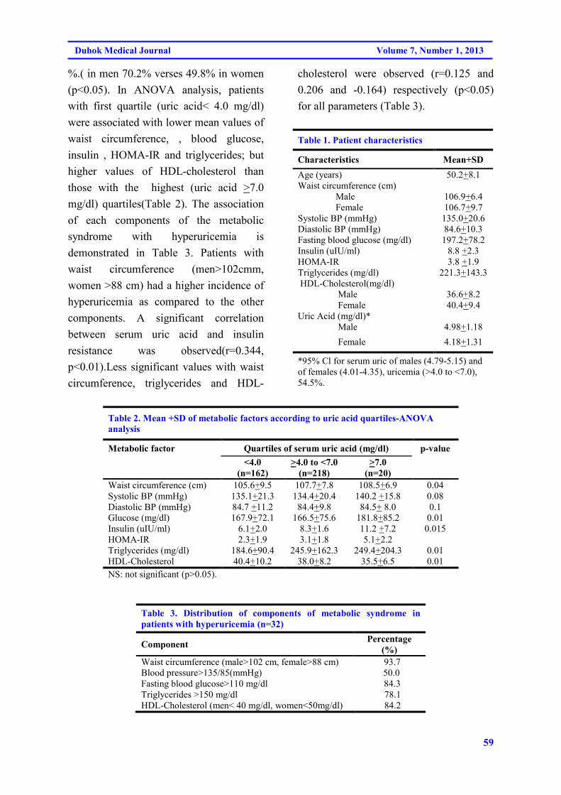
59
Duhok Medical Journal Volume 7, Number 1, 2013
%.( in men 70.2% verses 49.8% in women
(p<0.05). In ANOVA analysis, patients
with first quartile (uric acid< 4.0 mg/dl)
were associated with lower mean values of
waist circumference, , blood glucose,
insulin , HOMA-IR and triglycerides; but
higher values of HDL-cholesterol than
those with the highest (uric acid >7.0
mg/dl) quartiles(Table 2). The association
of each components of the metabolic
syndrome with hyperuricemia is
demonstrated in Table 3. Patients with
waist circumference (men>102cmm,
women >88 cm) had a higher incidence of
hyperuricemia as compared to the other
components. A significant correlation
between serum uric acid and insulin
resistance was observed(r=0.344,
p<0.01).Less significant values with waist
circumference, triglycerides and HDL-
cholesterol were observed (r=0.125 and
0.206 and -0.164) respectively (p<0.05)
for all parameters (Table 3).
Table 1. Patient characteristics
Characteristics Mean+SD
Age (years) 50.2+8.1
Waist circumference (cm)
Male 106.9+6.4
Female 106.7+9.7
Systolic BP (mmHg) 135.0+20.6
Diastolic BP (mmHg) 84.6+10.3
Fasting blood glucose (mg/dl) 197.2+78.2
Insulin (uIU/ml) 8.8 +2.3
HOMA-IR 3.8 +1.9
Triglycerides (mg/dl) 221.3+143.3
HDL-Cholesterol(mg/dl)
Male 36.6+8.2
Female 40.4+9.4
Uric Acid (mg/dl)*
Male 4.98+1.18
Female 4.18+1.31
*95% Cl for serum uric of males (4.79-5.15) and
of females (4.01-4.35), uricemia (>4.0 to <7.0),
54.5%.
Table 2. Mean +SD of metabolic factors according to uric acid quartiles-ANOVA
analysis
Metabolic factor Quartiles of serum uric acid (mg/dl) p-value
<4.0
(n=162) >4.0 to <7.0
(n=218) >7.0
(n=20)
Waist circumference (cm) 105.6+9.5 107.7+7.8 108.5+6.9 0.04
Systolic BP (mmHg) 135.1+21.3 134.4+20.4 140.2 +15.8 0.08
Diastolic BP (mmHg) 84.7 +11.2 84.4+9.8 84.5+ 8.0 0.1
Glucose (mg/dl) 167.9+72.1 166.5+75.6 181.8+85.2 0.01
Insulin (uIU/ml) 6.1+2.0 8.3+1.6 11.2 +7.2 0.015
HOMA-IR 2.3+1.9 3.1+1.8 5.1+2.2
Triglycerides (mg/dl) 184.6+90.4 245.9+162.3 249.4+204.3 0.01
HDL-Cholesterol 40.4+10.2 38.0+8.2 35.5+6.5 0.01
NS: not significant (p>0.05).
Table 3. Distribution of components of metabolic syndrome in
patients with hyperuricemia (n=32)
Component Percentage
(%) Waist circumference (male>102 cm, female>88 cm) 93.7
Blood pressure>135/85(mmHg) 50.0
Fasting blood glucose>110 mg/dl 84.3
Triglycerides >150 mg/dl 78.1
HDL-Cholesterol (men< 40 mg/dl, women<50mg/dl) 84.2
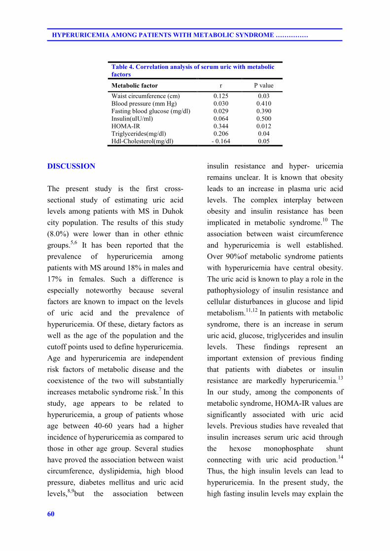
60
HYPERURICEMIA AMONG PATIENTS WITH METABOLIC SYNDROME ……………
Table 4. Correlation analysis of serum uric with metabolic
factors
Metabolic factor r P value
Waist circumference (cm) 0.125 0.03
Blood pressure (mm Hg) 0.030 0.410
Fasting blood glucose (mg/dl) 0.029 0.390
Insulin(uIU/ml) 0.064 0.500
HOMA-IR 0.344 0.012
Triglycerides(mg/dl) 0.206 0.04
Hdl-Cholesterol(mg/dl) - 0.164 0.05
DISCUSSION
The present study is the first cross-
sectional study of estimating uric acid
levels among patients with MS in Duhok
city population. The results of this study
(8.0%) were lower than in other ethnic
groups.5,6 It has been reported that the
prevalence of hyperuricemia among
patients with MS around 18% in males and
17% in females. Such a difference is
especially noteworthy because several
factors are known to impact on the levels
of uric acid and the prevalence of
hyperuricemia. Of these, dietary factors as
well as the age of the population and the
cutoff points used to define hyperuricemia.
Age and hyperuricemia are independent
risk factors of metabolic disease and the
coexistence of the two will substantially
increases metabolic syndrome risk.7 In this
study, age appears to be related to
hyperuricemia, a group of patients whose
age between 40-60 years had a higher
incidence of hyperuricemia as compared to
those in other age group. Several studies
have proved the association between waist
circumference, dyslipidemia, high blood
pressure, diabetes mellitus and uric acid
levels,8,9but the association between
insulin resistance and hyper- uricemia
remains unclear. It is known that obesity
leads to an increase in plasma uric acid
levels. The complex interplay between
obesity and insulin resistance has been
implicated in metabolic syndrome.10 The
association between waist circumference
and hyperuricemia is well established.
Over 90%of metabolic syndrome patients
with hyperuricemia have central obesity.
The uric acid is known to play a role in the
pathophysiology of insulin resistance and
cellular disturbances in glucose and lipid
metabolism.11,12
In patients with metabolic
syndrome, there is an increase in serum
uric acid, glucose, triglycerides and insulin
levels. These findings represent an
important extension of previous finding
that patients with diabetes or insulin
resistance are markedly hyperuricemia.13
In our study, among the components of
metabolic syndrome, HOMA-IR values are
significantly associated with uric acid
levels. Previous studies have revealed that
insulin increases serum uric acid through
the hexose monophosphate shunt
connecting with uric acid production.14
Thus, the high insulin levels can lead to
hyperuricemia. In the present study, the
high fasting insulin levels may explain the
61
Duhok Medical Journal Volume 7, Number 1, 2013
high levels of uric acid in patients with
metabolic syndrome.
It can be concluded that patients with
metabolic syndrome have a prevalence of
hyperuricemia of about (8%), and about
half of them have uric acid between( >4.0 -
<7.0 mg/dl).The measured uric acid status
is associated with metabolic syndrome
components. The most determinant of
hyperuricemia is waist circumference and
insulin résistance. Thus screening of
hyperuricemia in metabolic syndrome
patients is recommended and the treatment
of associated problems such as
hypertension, dyslipidemia, diabetes
mellitus and obesity is mandatory.
REFERENCES
1. Alberti KG, Zimmet P, Shaw
J.Metabolic syndrome--a new world-
wide definition. A Consensus
Statement from the International
Diabetes Federation. Diabet Med 2006;
23:469-80.
2. Conen D, Wietlisbach V, Bovet P,
Shamlaye C, Riesen W, Paccaud F,
Burnier M.'Prevalence of
hyperuricemia and relation of serum
uric acid with cardiovascular risk
factors in a developing country'. BMC
Public Health 2004; 4:9.
3. Schachter M. Uric Acid and
Hypertension'.Curr Pharm Des.
2005;11:4139-43.
4. Grund SM, Brewer Jr HB,Cleeman JI,
Smith SC Jr, Lenfant C .'Definition of
metabolic syndrome; report of the
National Heart, Lung and blood
Institute/ American Heart Association
conference on scientific issues related
to definition'. Circulation
2004;109:433-8.
5. Ishizaka N, Ishizaka Y, Toda E, Nagai
R, Yamakado M.'Association between
serum uric acid, metabolic syndrome,
and carotid atherosclerosis in Japanese
individuals'. Arterioscler Thromb Vasc
Biol 2005; 25:1038-44.
6. Nakanishi N, Okamoto M, Yoshida H,
Matsuo Y, Suzuki K, Tatara K . 'Serum
uric acid and risk for development of
hypertension and impaired fasting
glucose or Type II diabetes in Japanese
male office workers'. Eur J Epidemiol
2003;18:523-30.
7. Li Y, Stamler J, Xiao Z, Folsom A,
Tao S, Zhang H.'Serum uric acid and
its correlate in Chinese adult
population, urban and rural, of Beijing.
The PRC-USA Collaborative Study in
Cardiovascular and Cardiopulmonary
Epidemiology'. Int J Epidemiol 1997;
26:288-96.
8. Krishnan E, Kwoh CK, Schumacher
HR, Kuller L.'Hyperuricemia and
incidence of hypertension among men
without metabolic syndrome'.
Hypertension 2008; 49:298-303.
9. Coutinho TA, Turner ST, Peyser PA,
Bielak LF, Sheedy PF, 2nd, Kullo
IJ.'Associations of serum uric Acid
with markers of inflammation,
metabolic syndrome, and subclinical
coronary atherosclerosis'. Am J
Hypertens 2007; 20:83-9.
10. Matsuura F, Yamashita S, Nakamura
T, Nishida M, Nozaki S, Funahashi T,
Matsuzawa Y.'Effect of visceral fat
accumulation on uric acid metabolism
62
HYPERURICEMIA AMONG PATIENTS WITH METABOLIC SYNDROME ……………
in male obese subjects: visceral fat
obesity is linked more closely to
overproduction of uric acid than
subcutaneous fat obesity'. Metabolism
1998; 47:929-33.
11. Muscelli E, Natali A, Bianchi S,
Bigazzi R, Quinones Galvan A, Sironi
AM, Frascerra S, Ciociaro D,
Ferrannini E.'Effect of insulin on renal
sodium and uric acid handling in
essential hypertension'. Am J
Hypertens 1996; 9:746-52.
12. Quinones Galvan A, Natali A, Baldi S,
Frascerra S, Sanna G, Ciociaro D,
Ferrannini E.'Effect of insulin on uric
acid excretion in humans'. Am J
Physiol 1995; 268:E1-5.
13. GrundySM.'Hypertriglyceridemia,
insulin resistance, and the metabolic
syndrome'. Am J Cardiol 1999;
83:25F-9F.
14. Modan M, Halkin H, Karasik A, Lusky
A.'Elevated serum uric acid--a facet of
hyperinsulinaemia'. Diabetologia 1987;
30:713-8.
63
Duhok Medical Journal Volume 7, Number 1, 2013
ón‚íqón‚íqón‚íqón‚íq@@@@@@@@ðØíè†@ß@ç@ê؆@ñ‹Ø@ê’@bï’í‚@êä@ñŠ@ênä@ê�@bäa†@ëŠ@ê�@æîì@ê÷@ðà�ïÜíibnïà@µ’í‚@êä@båîí‚@Óbä†@‡ï�c@ÚîŠíîðØíè†@ß@ç@ê؆@ñ‹Ø@ê’@bï’í‚@êä@ñŠ@ênä@ê�@bäa†@ëŠ@ê�@æîì@ê÷@ðà�ïÜíibnïà@µ’í‚@êä@båîí‚@Óbä†@‡ï�c@ÚîŠíîðØíè†@ß@ç@ê؆@ñ‹Ø@ê’@bï’í‚@êä@ñŠ@ênä@ê�@bäa†@ëŠ@ê�@æîì@ê÷@ðà�ïÜíibnïà@µ’í‚@êä@båîí‚@Óbä†@‡ï�c@ÚîŠíîðØíè†@ß@ç@ê؆@ñ‹Ø@ê’@bï’í‚@êä@ñŠ@ênä@ê�@bäa†@ëŠ@ê�@æîì@ê÷@ðà�ïÜíibnïà@µ’í‚@êä@båîí‚@Óbä†@‡ï�c@ÚîŠíî@@@@@@@@
��
wäbàŠb÷@ì@ôØó“ŽïqwäbàŠb÷@ì@ôØó“ŽïqwäbàŠb÷@ì@ôØó“ŽïqwäbàŠb÷@ì@ôØó“ŽïqZZZZ@@ñŠíu@l@êîa‡î‹Ø@bî@‡ï�c@ÚîŠíî@a�îŠ@bäíi‡åÝi@líØ@ëŠbî†@bî2@ðà�Üíibnïà@bï’í‚@êä@ì@ñ‹Ø@ê’@bï’í‚@êäŒ@N@ß@ê؆@ê÷@íØ@ëŠ@êÐ@bî@ñ‡ä@êu@Àa†@êà@ðØbÑu@bä†@ç‹ØŠbî†@ênïéi@‹m@ë‡îŒ@êäa‡î‹Ø@Ó@N@êä@†@‡ï�c@ÚîŠíî@a�îŠ@bä‹ØŠbî†@�ïÜíØ@êÐ@À@b−bàŠb÷
@ñŠíu@µ’í‚2@a‡î‹Ø@ê’@µ’í‚@êä†@‡ï�c@ÚîŠíî@bäíi@‡åÝi@ña‡î‹Ø@µî@ñ†@æîŠ@ênØbÐ@bä‹ØŠbî†ì@ñ‹Ø@ê’@µî.@@ŽôåïÜíØóÄ@æŽïÙôåïÜíØóÄ@æŽïÙôåïÜíØóÄ@æŽïÙôåïÜíØóÄ@æŽïÙîŠZZZZ@@Š@ê�Ü@ça†@ãb−@ê÷@êmbè@êåïÜíØ@êÐ@Ó@ê÷400@@Û@ëì@ç‹Ùäb“ïä@o�@ë†@@êåïmbè@ñì@ê÷@íØ@ðà�ïÜíibnïà@µ’í‚@êä
@ñíu2@@ðàŒíØ@Œ@ñ‹Ø@ê’@bï’í‚@êä3678@ðØíè†@bè@êØ�î@Šbi@ß@ña†@ñ‹Ø@ê’@µï’í‚@êä@ñŠ@ênä@ê�@bäa†@ëŠ@ê�@æîì@ê÷@çb’í‚@êä@N@µ’í‚@êä@@@ì@ê÷@ì@‡ï�c@Úî‹î@bî‡åÝi@Òî†@ß@bÙîŠbu@Šbu@íi@ç‹Ø@•@êi@a‡è@æmbè@•í‚@êäça†ý@êåmbè@ç‹Ø†Š@ë@ì@@ñ‹Ø@ê’@µäbàŠ@ë†.@@
ãb−ó÷ãb−ó÷ãb−ó÷ãb−ó÷ZZZZ@@@@@‡ï�c@Úî‹î@bäíi@‡åÝi@a�îŠ@a†@�ïÜíØ@êÐ@@À†8@Eíi@N@ðä@êà@êm@†@‡ï�c@Úî‹î@bäíi@‡åÝi@a�îŠ40M60@@a†@ðÜb�5,8@Eì@íi@2,2@E@ñ@ë‡îŒ@çaì@ðîŒ@µ’í‚@êä†60@@ßb�@N@@a†@ðà@ë��†@‡ï�c@ÚîŠíî@a�îŠ4,98@âÍÝàO@íi@߆I95@ECl @4,79@–@5,15@H@ì@�ä@µî
4,18@âÍÝàO@ðà@µî@@ß@†I95@ECI @4,01@–@4,35@NH@ðÙï÷@bÙîŠbu@µ’í‚@êä@íi@bÐíäc@bä‹Ø@êÐíÝ’I@Œ@�áïØ@‡ï�c@ÚîŠíî4@âÍÝàO@†ß@@H@ì@�ïÜí�äc@ì@ñ‹Ø@ê’@a�îŠ@ì@ðä@bï�@�îŠ@ì@�îí‚@aŠb“Ð@ì@ðÙä@êm@Óbä@a�îŠ@æî�áïØíØ@íi@Šbî†HOMAIR@@Œ@‡åÝi@a�îŠì@íi@êè
�îŠ@íi@êè@�m@@@N@�ïÜí�äc@bîŠbÙåè@Š@êi@ì@‡ï�c@ÚîŠíî@aŠ@êi@Óbä†@íi@Šbî†@ñ‡ä@êî@êiIr@@]0N344L@p@@]>0,01@@H@Ó@bä†@�áïØ@a�îŠì@a‡ïÙä@êmIr@@]0N125@p@@]>0.01@H@ðäbï�@�îŠìIr@@]0N206@p@@]>0N05H.@@ãb−ó÷Šò†ãb−ó÷Šò†ãb−ó÷Šò†ãb−ó÷Šò†ZZZZ@@a�îŠ@ðà�ïÜíibnà@µ’í‚@êä@bÑïä@íØ@a‡î@µÜíØ@êÐ@À†@íi@Šbî†@a‡ïåîí‚@Óbä†@ñ‡ï�c@ÚîŠíîI> 4,0@@bm>@7,0@âÍÝàOß@†@@H
íi@êè@a‡ïåï‚@Óbä†@‡ï�c@ÚîŠíî@bäíi@‡åÝi@b’í‚@êä@Œ@@bè@ë†@Š@êè@Œ@Úï÷ì@N@ÚîŠíî@bäíi@‡åÝi@bä‹Ùäb“ïä@o�@ë†@ðî@ðØ@ëŠ@ê�@ñŠ@ênØbÐ�ïÜí�äc@bî‹ØŠ@êi@ì@êî@ðÙä@êm@Óbä@ñíïu@a‡ïåîí‚@Óbä†@ñ‡ï�aN@@
64
HYPERURICEMIA AMONG PATIENTS WITH METABOLIC SYNDROME ……………
الخلاصة
لدموي لدى مرضى المتلازمه الأيضيه الوافدين الى مركز علاج السكري في دهوكحامض البوليك ا
من مرض السكري و 2من المعروف بأن أرتفاع نسبة حامض البوليك يكون مصاحبا ل النوع :خلفية واهداف البحث
من هذه الدراسه هو لتقييم ألهدف. مع ذلك فأن هذا الأرتباط يحتاج ألى مزيد من التحديد في مجتمعنا. المتلازمه الأيضية
مرض السكري لمجتمع كردستان و لكشف العوامل الأيضيه المصاحبه 2نسبة أرتفاع حامض البوليك لدى مرضى النوع
. لأرتفاع حامض البوليك لمرضى السكري
يار العينه الذين تم تم أخت. مريض من مرضى المتلازمه الأيضيه 400أجريت هذه الدراسه المقطعيه على :طرق البحث
من مرض السكري أو من الذين يتم معالجتهم بالأدويه المضاده لمرض السكري 2تشخيصهم على أنهم مرضى من النوع
تم تقسيم المرضى حسب نسبة ). 3678العدد (والذين يترددون على مركز علاج أمرض السكري في محافظة دهوك
تم تعريف ). أعلى نسبة حامض البوليك(الى الربع الرابع ) امض البوليكأدنى نسبة ح(حامض البوليك من الربع الأول
.وقد تم أستثناء المرضى الذين يعالجون بأدوية أرتفاع نسبة حامض البوليك ATP-111المتلازمة الأيضيه حسب معيار
ذوو معدل العمر نسبة أرتفاع حامض البوليك لدى المرضى%. 8كانت نسبة أنتشار أرتفاع حامض البوليك :النتائج
كان معدل حامض . سنة 60لدى المرضى الذين كانت أعمارهم أكثر من % 2,2وكانت % 5.8سنة كانت 40-60
CI 4.01% 95(د ل للأناث /ملغم 4.18للذكور و ) Cl 4.79 – 5.15% 95(دل /ملغم 4.98البوليك في المصل
كانت لديه ) د ل/ملغم 4حامض البوليك أقل من (الأول عند أجراء تحيلي الأنوفا المرضى من ضمن الربع). 4.35 –
ومستوى أعلى HOMAIRأقل نسة من محيط الخصر وضغط الدم والدهون الثلاثيه ونسبة السكر في الدم والأنسولين و
= r(تم ملاحضة علاقه معنوية بين مستوى حامض البوليك ومقاومة الأنسولين . من الكوليستيرول ذو التركيز العالي
0.344 p =>0.01 ( ونسبة أقل من قيمة محيط الخصر)r =0.125 p =>0.01 ( ودهون ثلاثيه)r =0.206 p
=>0.05(.
<(تبين من الدراسه بأن حوالي نصف المرضى ذوو المتلازمه الأيضيه كانت لديهم حامض البوليك الدموي :اتالاستنتاج
العامل المحدد الأساسي . كانت لديهم أرتفاع في حامض البوليك الدمويوأن واحدا من عشرة ) د ل/ملغم 7,0< الى 4,0
.لأرتفاع حامض البوليك الدموي هو محيط الخصر و المقاومه للأنسولين
65
Duhok Medical Journal Volume 7, Number 1, 2013
PHYTOSTEROLS AND PHYTOSTANOLS IN PALM OILS
KAMAL A. KETULY, B.Sc., M.Appl.Sc., Ph.D.* Submitted 4 Jun 2013; accepted 3 Nov 2013
ABSTRACT
Background and objectives Phytosterols (plant sterols) are triterpenes that are important structural components of plant membranes. They serve to stabilize phospholipid bilayers in plant cell membranes just as cholesterol does in animal cell membranes. Phytostanols occur in trace levels in many plant species and they occur in high levels in tissues of only in a few cereal species. In this study the search was mainly for sitostanol which reduces cholesterol absorption in the bowel. In addition to other unidentified plant sterols in the crude palm oil (CPO), crude palm kernel oil (CPKO) and crude palm fiber oil (CPFO) extracts. Methods The unsaponifiable sterols extracts from CPO, CPKO and CPFO were isolated by preparative thin layer chromatography. The isolated sterols were analysed as free and trimethylsilylated (TMS) derivatives by GC correlations and GC-MS mass spectral data in comparison with their standard sterols. Results Qualitative and quantitative separations of the sterols are illustrated. Tables are presented showing the sterols composition determined in the oils. There were no major differences between the above three oils with respect to their main sterols composition, but the total amount of the sterols in CPFO were much higher than CPKO and CPO respectively. In addition to the well identified sterols in the palm oils (i.e cholesterol, campesterol, stigmasterol and β-sitosterol). Additional sterols were isolated and identified in CPO: stellasterol and cycloartenol. CPKO: fucosterol, 5α-4,4-dimethylcholest-8,24-dien-3β-ol and cycloartenol, CPFO: brassicasterol, sitostanol and 4,4-dimethylcholest-5-en-3β-ol. Conclusions Sitostanol was only found in CPFO (16 ppm) but was not found in CPO or CPKO. Duhok Med J 2013;7(1): 65-80. Key words: Palm oils, Phytosterols, Sitostanol
hytosterols and phytostanols are from
plants origin, with the highest
concentrations occurring in vegetable oils.
Chemically, these sterols have the same
basic structure as cholesterol but
differences arise from the side chain which
is modified by the addition of one or two
supernumerary carbon atoms at C-24 with
either α or β chirality. The 24-alkyl group
is characteristic of all phytosterols and is
preserved during subsequent steroid
metabolism in both fungi and plants to
give hormones that regulate growth and
reproduction in a manner similar to
animals.
Most phytosterols are compounds
having 28 to 30 carbon atoms and one or
two or three carbon-carbon double bonds,
typically one or two in the sterol nucleus
and sometimes a second in the alkyl side
chain. All phytosterols were shown to
derive in plants from cycloartenol and in
fungi (including yeasts), as in vertebrates,
from lanosterol, both direct products of the
cyclisation of squalene.1
More than 250 different types of
P
* Department of Biochemistry, Faculty of Medicine, University of Duhok, Duhok, Kurdistan Region – Iraq.
E-mail: [email protected]
66
PHYTOSTEROLS AND PHYTOSTANOLS IN PALM OILS
phytosterols have been reported in plant
species1. Representatives of these sterols
are campesterol, β-sitosterol
and stigmasterol (in soybean oil). β-
Sitosterol is present in all plant lipids and
is used for steroid synthesis. Stigmasterol,
which is used for the synthesis of
progesterone and vitamin D3, is known
as"Wulzen factor", a potential anti-
inflammatory compound. Its action is
mediated by the inhibition of several pro-
inflammatory and matrix degradation
mediators involved in osteoarthritis-
induced cartilage degradation.2
The non-glyceride components
content varies from oil to oil. It may range
between 2-8% in some oils, but the
constitution in most oils is 1% or below.3
Plant sterols in vegetables, fruits and
vegetable oils were extensively studied.4-9
Considerable variability in the
concentration of free sterols was observed
among different oils. Concentrations
between 200 and 400 mg/100 g are found
in oils from sesame, canola, rapeseed,
corn, and evening primrose.10 Phytosterols
produce a wide spectrum of biological
activities in animals and humans. They are
considered efficient cholesterol-lowering
agents.11,12 The lowering of serum
cholesterol by plant sterols is believed to
be the result of an inhibition of cholesterol
absorption in the small bowel.13,14 Plasma
total and LDL-cholesterol concentrations,
were significantly reduced by taking
spreads enriched with plant sterols.15 Also
it has been found that plants sterols intake
was associated with the protection of colon
carcinogenesis and with a reduction in the
risk of lung cancer and preventing
coronary heart disease.16,17 The focus of
research has shifted from plant sterols to
plant stanols because of their greater
ability to reduce intestinal cholesterol
absorption and virtually unabsorbed; thus,
plant stanol ester foods are clinically
proven, highly effective new dietary tools
for managing blood cholesterol levels.18 In
addition, they produce a wide spectrum of
therapeutic effects including anti-tumor
properties. Further data on their
metabolism and potential therapeutic
action can be found in a review article.19 A
review of physiologic and metabolic
aspects related to these cholesterol-
lowering properties may be consulted.20
The interest of adding sterols and stanols
to human food to improve health has been
discussed.21 Clinical experiments have
shown that only high amounts of stanols
(about 9 g/day) can decrease serum α-
carotene concentrations, without altering
those of vitamins A, D and E.22 As
cholesterol, phytosterols may undergo
oxidative processes. These
oxyphytosterols have been shown to have
beneficial biological properties which
deserve further investigation.23
Phytostanols are plant sterols, referred
to as stanols. They are fully-saturated
subgroup of phytosterols, have no double
bond in the ring B structure8. They are in
general produced by hydrogenation of
phytosterols. Sitostanol and campestanol
exist in quantifiable amounts in cereals,
fruits and vegetables, but generally of less
concentration than the unsaturated plant
sterols.24 To improve their solubility, plant
stanols are often combined with a fatty
acid ester to produce plant stanol esters.
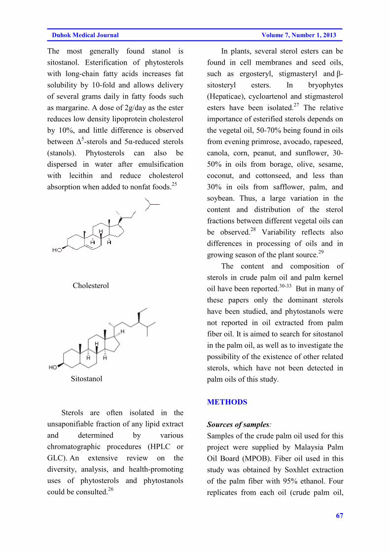
67
Duhok Medical Journal Volume 7, Number 1, 2013
The most generally found stanol is
sitostanol. Esterification of phytosterols
with long-chain fatty acids increases fat
solubility by 10-fold and allows delivery
of several grams daily in fatty foods such
as margarine. A dose of 2g/day as the ester
reduces low density lipoprotein cholesterol
by 10%, and little difference is observed
between ∆5-sterols and 5α-reduced sterols
(stanols). Phytosterols can also be
dispersed in water after emulsification
with lecithin and reduce cholesterol
absorption when added to nonfat foods.25
Cholesterol
Sitostanol
Sterols are often isolated in the
unsaponifiable fraction of any lipid extract
and determined by various
chromatographic procedures (HPLC or
GLC). An extensive review on the
diversity, analysis, and health-promoting
uses of phytosterols and phytostanols
could be consulted.26
In plants, several sterol esters can be
found in cell membranes and seed oils,
such as ergosteryl, stigmasteryl and β-
sitosteryl esters. In bryophytes
(Hepaticae), cycloartenol and stigmasterol
esters have been isolated.27 The relative
importance of esterified sterols depends on
the vegetal oil, 50-70% being found in oils
from evening primrose, avocado, rapeseed,
canola, corn, peanut, and sunflower, 30-
50% in oils from borage, olive, sesame,
coconut, and cottonseed, and less than
30% in oils from safflower, palm, and
soybean. Thus, a large variation in the
content and distribution of the sterol
fractions between different vegetal oils can
be observed.28 Variability reflects also
differences in processing of oils and in
growing season of the plant source.29
The content and composition of
sterols in crude palm oil and palm kernel
oil have been reported.30-33 But in many of
these papers only the dominant sterols
have been studied, and phytostanols were
not reported in oil extracted from palm
fiber oil. It is aimed to search for sitostanol
in the palm oil, as well as to investigate the
possibility of the existence of other related
sterols, which have not been detected in
palm oils of this study.
METHODS
Sources of samples:
Samples of the crude palm oil used for this
project were supplied by Malaysia Palm
Oil Board (MPOB). Fiber oil used in this
study was obtained by Soxhlet extraction
of the palm fiber with 95% ethanol. Four
replicates from each oil (crude palm oil,
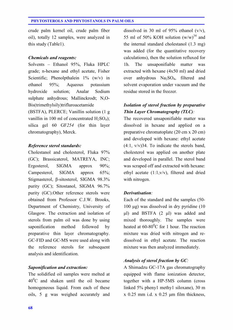
68
PHYTOSTEROLS AND PHYTOSTANOLS IN PALM OILS
crude palm kernel oil, crude palm fiber
oil), totally 12 samples, were analyzed in
this study (Table1).
Chemicals and reagents:
Solvents – Ethanol 95%, Fluka HPLC
grade; n-hexane and ethyl acetate, Fisher
Scientific; Phenolpthalein 1% (w/v) in
ethanol 95%; Aqueous potassium
hydroxide solution; Analar Sodium
sulphate anhydrous; Mallinckrodt; N,O-
Bis(trimethylsily)trifluroacetamide
(BSTFA), PLERCE; Vanillin solution (1 g
vanillin in 100 ml of concentrated H2SO4);
silica gel 60 GF254 (for thin layer
chromatography), Merck.
Reference sterol standards:
Cholestanol and cholesterol, Fluka 97%
(GC); Brassicaterol, MATREYA, INC;
Ergosterol, SIGMA approx 90%;
Campesterol, SIGMA approx 65%;
Stigmasterol, β-sitosterol, SIGMA 98.3%
purity (GC); Sitostanol, SIGMA 96.7%
purity (GC).Other reference sterols were
obtained from Professor C.J.W. Brooks,
Department of Chemistry, University of
Glasgow. The extraction and isolation of
sterols from palm oil was done by using
saponification method followed by
preparative thin layer chromatography.
GC-FID and GC-MS were used along with
the reference sterols for subsequent
analysis and identification.
Saponification and extraction:
The solidified oil samples were melted at
400C and shaken until the oil became
homogeneous liquid. From each of these
oils, 5 g was weighed accurately and
dissolved in 30 ml of 95% ethanol (v/v),
55 ml of 50% KOH solution (w/w)34 and
the internal standard cholestanol (1.3 mg)
was added (for the quantitative recovery
calculations), then the solution refluxed for
1h. The unsaponifiable matter was
extracted with hexane (4x50 ml) and dried
over anhydrous Na2SO4, filtered and
solvent evaporation under vacuum and the
residue stored in the freezer.
Isolation of sterol fraction by preparative
Thin Layer Chromatography (TLC):
The recovered unsaponifiable matter was
dissolved in hexane and applied on a
preparative chromatoplate (20 cm x 20 cm)
and developed with hexane: ethyl acetate
(4:1, v/v)34. To indicate the sterols band,
cholesterol was applied on another plate
and developed in parallel. The sterol band
was scraped off and extracted with hexane:
ethyl acetate (1:1,v/v), filtered and dried
with nitrogen.
Derivatisation:
Each of the standard and the samples (50-
100 µg) was dissolved in dry pyridine (10
µl) and BSTFA (2 µl) was added and
mixed thoroughly. The samples were
heated at 60-800C for 1 hour. The reaction
mixture was dried with nitrogen and re-
dissolved in ethyl acetate. The reaction
mixture was then analyzed immediately.
Analysis of sterol fraction by GC:
A Shimadzu GC-17A gas chromatography
equipped with flame ionization detector,
together with a HP-5MS column (cross
linked 5% pheny1 methy1 siloxane), 30 m
x 0.25 mm i.d. x 0.25 µm film thickness,
69
Duhok Medical Journal Volume 7, Number 1, 2013
was used for these analyses. The GC
conditions: Carrier gas: Nitrogen, injection
port: 2800C, Detector: 2850C, oven: 2800C,
Auxiliary: 2800C; Column pressure: 19
psi; Total flow (0.88 ml/min): Linear
velocity (29.83 cm/s) and Control mode:
Split less.
Identification and Confirmation by Using
GC-MS:
A Shimadzu GCMS QP5050A series gas
chromatograph coupled with a quadrupole
mass spectrometer was used and the
Column: SGE, forte BPX5-5% Phenyl
Polycarborane-siloxane (cross linked 5%
pheny1 methy1 siloxane), 30 m x 0.25 mm
i.d. x 0.25 µm film thickness. The GC-MS
conditions: Carrier gas: Helium; injection
port: 2800C; isothermal: oven: 2800C;
interface temp.: 2800C, column inlet
pressure: 98.6 kPa, column flow: 0.7
ml/min, linear velocity :32.8 cm/sec,
control mode: splitless, split Ratio :18,
total flow :14.3 ml/min. Temprature
program: injection port: 280.00 °C, oven
:280.00 °C (5min.), 5 °C/min to 280 °C
(40min.), Interface Temp.: 280.00 °C,
control mode :splitless, column inlet
pressure: 98.6 kPa, column flow :0.9
mL/min, linear velocity :36.5 cm/sec, split
ratio :14, total flow :14.3 mL/min .
Qualitative data were acquired in the
Electron Impact mode (70 ev) and
scanning from 50 to 550 a.m.u. at 1.53
sec/scan. The isolated sterols were
identified by GC-MS as free and their
TMS derivatives by GC correlations and
GC-MS mass spectral data in comparison
with the standard sterols.
Selected Ion Monitoring (SIM):
Mass-spectrometric sensitivity and
selectivity are greatly aided by a data
system capable of monitoring selected ions
from the mass spectra, i.e. selected ion
monitoring (SIM). From the mass spectra
of sitostanol, 3 characteristic ions (m/z:
215, 398 and 488) were chosen for
monitoring. To determine the existence of
sitostanol in the oil samples under SIM
mode, the oil samples spiked with a small
amount of sitostanol were injected into the
GC-MS.
RESULTS
The recovered phytosterols in CPO, CPKO
and CPFO and their percentage in
unsaponifiable matter are shown in (Table
1). It has been found, that the amount of
the total phytosterols in CPFO (6942
mg/kg) to be higher than, the palm oils:
CPKO (1483 mg/kg) and CPO (1122
mg/kg), respectively. The internal standard
cholestanol was added to these three oils
(Table 1) before the saponification, 62%
recoveries of the total sterols were
calculated and this has been taken in to
account. The phytosterols composition
(ppm) in these three oils and their retention
times are detailed in (Table 2).
The identification of all the isolated
sterols were made by GC and GC-MS.
These analysis data and in comparison
with their corresponding standard sterols
as free and TMS-ethers, led to the
confirmation of the isolated sterols
structures (Table 3, 4, and 5).
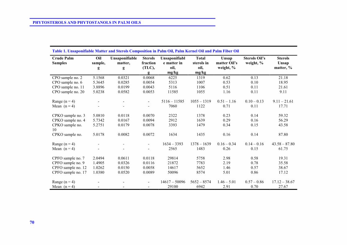
70
PHYTOSTEROLS AND PHYTOSTANOLS IN PALM OILS
Table 1. Unsaponifiable Matter and Sterols Composition in Palm Oil, Palm Kernel Oil and Palm Fiber Oil
Crude Palm
Samples
Oil
sample,
g
Unsaponifiable
matter,
g
Sterols
fraction
(TLC),
g
Unsaponifiabl
e matter in
oil,
mg/kg
Total
sterols in
oil,
mg/kg
Unsap
matter Oil’s
weight, %
Sterols Oil’s
weight, %
Sterols
Unsap
matter, %
CPO sample no. 2 5.1568 0.0321 0.0068 6225 1319 0.62 0.13 21.18 CPO sample no. 6 5.3645 0.0285 0.0054 5313 1007 0.53 0.10 18.95 CPO sample no. 11 3.8896 0.0199 0.0043 5116 1106 0.51 0.11 21.61 CPO sample no. 20 5.0238 0.0582 0.0053 11585 1055 1.16 0.11 9.11 Range (n = 4)
-
-
-
5116 – 11585
1055 – 1319
0.51 – 1.16
0.10 – 0.13
9.11 – 21.61
Mean (n = 4)
- - - 7060 1122 0.71 0.11 17.71
CPKO sample no. 3 5.0810 0.0118 0.0070 2322 1378 0.23 0.14 59.32 CPKO sample no. 4 5.7342 0.0167 0.0094 2912 1639 0.29 0.16 56.29 CPKO sample no. 10
5.2751 0.0179 0.0078 3393 1479 0.34 0.15 43.58
CPKO sample no. 5.0178 0.0082 0.0072 1634 1435 0.16 0.14 87.80 Range (n = 4)
-
-
-
1634 – 3393
1378 – 1639
0.16 – 0.34
0.14 – 0.16
43.58 – 87.80
Mean (n = 4)
- - - 2565 1483 0.26 0.15 61.75
CPFO sample no. 7 2.0494 0.0611 0.0118 29814 5758 2.98 0.58 19.31 CPFO sample no. 9 1.4905 0.0326 0.0116 21872 7783 2.19 0.78 35.58 CPFO sample no. 12 1.0262 0.0150 0.0058 14617 5652 1.46 0.57 38.67 CPFO sample no. 17 1.0380 0.0520 0.0089 50096 8574 5.01 0.86 17.12 Range (n = 4)
-
-
-
14617 – 50096
5652 – 8574
1.46 – 5.01
0.57 – 0.86
17.12 – 38.67
Mean (n = 4) - - - 29100 6942 2.91 0.70 27.67
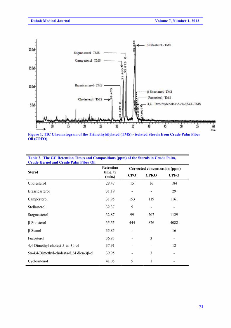
71
Duhok Medical Journal Volume 7, Number 1, 2013
Figure 1. TIC Chromatogram of the Trimethylsilylated (TMS) - isolated Sterols from Crude Palm Fiber
Oil (CPFO)
Table 2. The GC Retention Times and Compositions (ppm) of the Sterols in Crude Palm,
Crude Kernel and Crude Palm Fiber Oil
Sterol
Retention
time, tr
(min.)
Corrected concentration (ppm)
CPO CPKO CPFO
Cholesterol 28.47 15 16 184
Brassicasterol 31.19 - - 29
Campesterol 31.95 153 119 1161
Stellasterol 32.37 5 - -
Stegmasterol 32.87 99 207 1129
β-Sitosterol 35.55 444 876 4082
β-Stanol 35.85 - - 16
Fucosterol 36.83 - 3 -
4,4-Dimethyl-cholest-5-en-3β-ol 37.91 - - 12
5α-4,4-Dimethyl-cholesta-8,24 dien-3β-ol 39.95 - 3 -
Cycloartenol 41.05 5 1 -
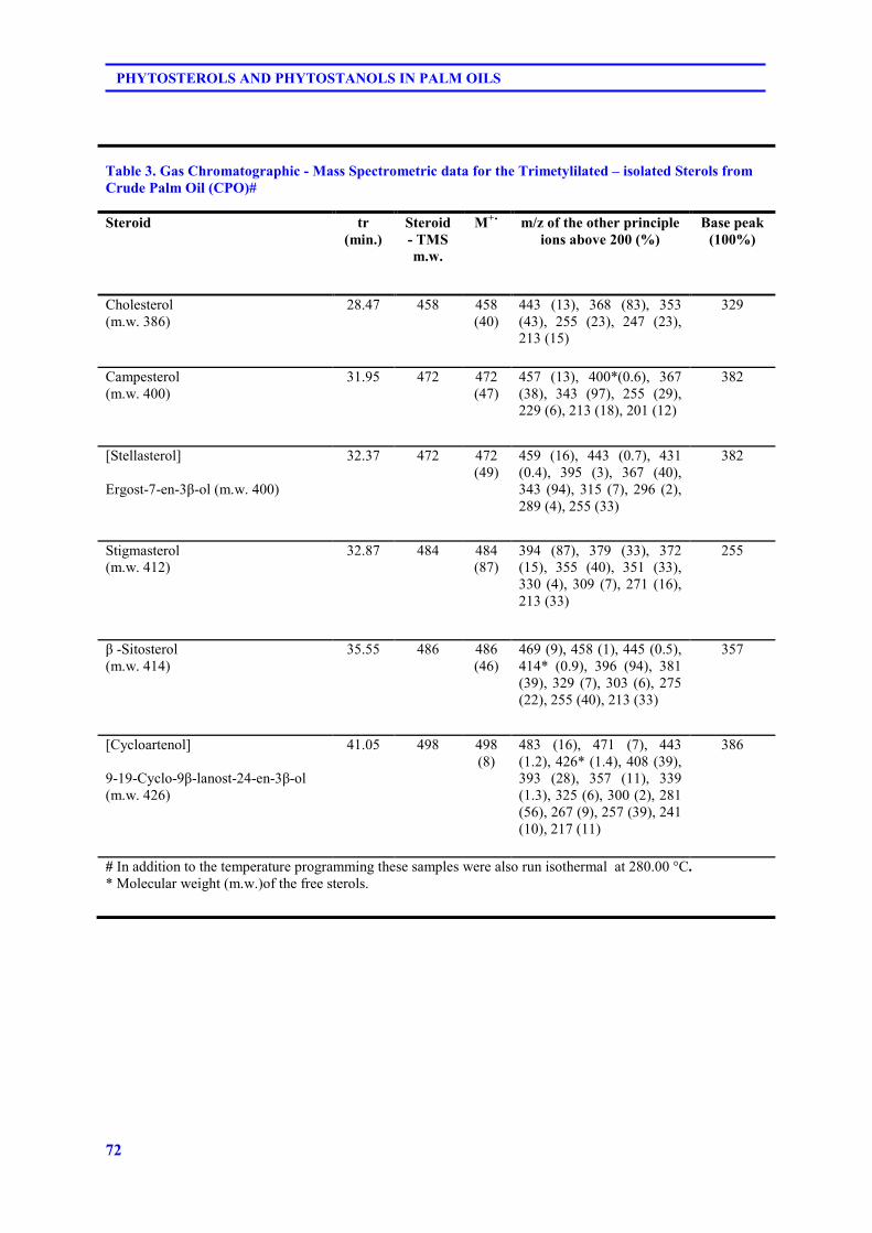
72
PHYTOSTEROLS AND PHYTOSTANOLS IN PALM OILS
Table 3. Gas Chromatographic - Mass Spectrometric data for the Trimetylilated – isolated Sterols from
Crude Palm Oil (CPO)#
Steroid tr
(min.)
Steroid
- TMS
m.w.
M+˙ m/z of the other principle
ions above 200 (%)
Base peak
(100%)
Cholesterol (m.w. 386)
28.47 458 458 (40)
443 (13), 368 (83), 353 (43), 255 (23), 247 (23), 213 (15)
329
Campesterol (m.w. 400)
31.95 472 472 (47)
457 (13), 400*(0.6), 367 (38), 343 (97), 255 (29), 229 (6), 213 (18), 201 (12)
382
[Stellasterol] Ergost-7-en-3β-ol (m.w. 400)
32.37 472 472 (49)
459 (16), 443 (0.7), 431 (0.4), 395 (3), 367 (40), 343 (94), 315 (7), 296 (2), 289 (4), 255 (33)
382
Stigmasterol (m.w. 412)
32.87 484 484 (87)
394 (87), 379 (33), 372 (15), 355 (40), 351 (33), 330 (4), 309 (7), 271 (16), 213 (33)
255
β -Sitosterol (m.w. 414)
35.55
486 486 (46)
469 (9), 458 (1), 445 (0.5), 414* (0.9), 396 (94), 381 (39), 329 (7), 303 (6), 275 (22), 255 (40), 213 (33)
357
[Cycloartenol] 9-19-Cyclo-9β-lanost-24-en-3β-ol (m.w. 426)
41.05 498 498 (8)
483 (16), 471 (7), 443 (1.2), 426* (1.4), 408 (39), 393 (28), 357 (11), 339 (1.3), 325 (6), 300 (2), 281 (56), 267 (9), 257 (39), 241 (10), 217 (11)
386
# In addition to the temperature programming these samples were also run isothermal at 280.00 °C. * Molecular weight (m.w.)of the free sterols.
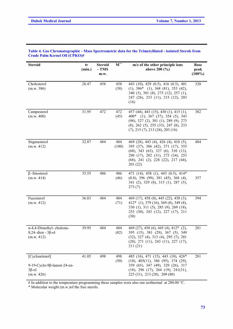
73
Duhok Medical Journal Volume 7, Number 1, 2013
Table 4. Gas Chromatographic - Mass Spectrometric data for the Trimetylilated - isolated Sterols from
Crude Palm Kernel Oil (CPKO)#
Steroid tr
(min.)
Steroid
- TMS
m.w.
M+˙ m/z of the other principle ions
above 200 (%)
Base
peak
(100%)
Cholesterol (m.w. 386)
28.47 458 458 (38)
443 (10), 429 (0.5), 416 (0.5), 401 (1), 386* (1), 368 (81), 353 (42), 340 (3), 301 (8), 275 (12), 257 (1), 247 (26), 233 (11), 215 (12), 203 (16)
320
Campesterol (m.w. 400)
31.95 472 472 (45)
457 (44), 443 (15), 430 (1), 415 (1), 400* (1), 367 (37), 354 (5), 343 (98), 327 (2), 301 (1), 289 (9), 273 (8), 262 (5), 255 (33), 247 (8), 233 (7), 215 (7), 213 (24), 203 (16)
382
Stigmasterol (m.w. 412)
32.87 484 484 (100)
469 (28), 443 (4), 426 (4), 410 (5), 395 (37), 386 (42), 371 (17), 355 (68), 343 (63), 327 (6), 310 (13), 296 (17), 282 (31), 273 (34), 253 (68), 241 (2), 228 (22), 217 (44), 201 (22)
484
β -Sitosterol (m.w. 414)
35.55
486 486 (46)
471 (14), 458 (1), 443 (0.5), 414* (0.8), 396 (99), 381 (45), 368 (4), 341 (2), 329 (8), 315 (1), 287 (5), 273 (7)
357
Fucosterol (m.w. 412)
36.83 484 484 (71)
469 (17), 458 (8), 445 (22), 430 (3), 412* (1), 379 (16), 369 (8), 349 (8), 330 (1), 311 (5), 285 (9), 269 (18), 253 (30), 243 (12), 227 (17), 211 (30)
394
α-4,4-Dimethyl- cholesta-8,24–dien - 3β-ol (m.w. 412)
39.95 484
484 (82)
469 (27), 458 (6), 445 (4), 412* (2), 395 (15), 381 (29), 367 (5), 349 (32), 327 (4), 313 (4), 295 (7), 281 (28), 271 (11), 243 (11), 227 (17), 211 (21)
281
[Cycloartenol] 9-19-Cyclo-9β-lanost-24-en-3β-ol (m.w. 426)
41.05 498 498 (50)
485 (16), 471 (15), 443 (10), 426* (10), 405(11), 386 (95), 374 (29), 359 (65), 347 (49), 329 (26), 317 (18), 296 (17), 264 (19), 241(31), 225 (31), 213 (28), 209 (80)
281
# In addition to the temperature programming these samples were also run isothermal at 280.00 °C. * Molecular weight (m.w.)of the free sterols.
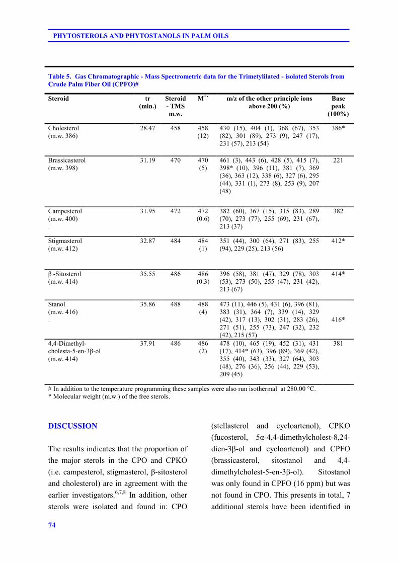
74
PHYTOSTEROLS AND PHYTOSTANOLS IN PALM OILS
DISCUSSION
The results indicates that the proportion of
the major sterols in the CPO and CPKO
(i.e. campesterol, stigmasterol, β-sitosterol
and cholesterol) are in agreement with the
earlier investigators.6,7,8 In addition, other
sterols were isolated and found in: CPO
(stellasterol and cycloartenol), CPKO
(fucosterol, 5α-4,4-dimethylcholest-8,24-
dien-3β-ol and cycloartenol) and CPFO
(brassicasterol, sitostanol and 4,4-
dimethylcholest-5-en-3β-ol). Sitostanol
was only found in CPFO (16 ppm) but was
not found in CPO. This presents in total, 7
additional sterols have been identified in
Table 5. Gas Chromatographic - Mass Spectrometric data for the Trimetylilated - isolated Sterols from Crude Palm Fiber Oil (CPFO)#
Steroid tr
(min.)
Steroid
- TMS
m.w.
M+˙ m/z of the other principle ions
above 200 (%)
Base
peak
(100%)
Cholesterol (m.w. 386)
28.47 458 458 (12)
430 (15), 404 (1), 368 (67), 353 (82), 301 (89), 273 (9), 247 (17), 231 (57), 213 (54)
386*
Brassicasterol (m.w. 398)
31.19 470 470 (5)
461 (3), 443 (6), 428 (5), 415 (7), 398* (10), 396 (11), 381 (7), 369 (36), 363 (12), 338 (6), 327 (6), 295 (44), 331 (1), 273 (8), 253 (9), 207 (48)
221
Campesterol (m.w. 400) .
31.95 472 472 (0.6)
382 (60), 367 (15), 315 (83), 289 (70), 273 (77), 255 (69), 231 (67), 213 (37)
382
Stigmasterol (m.w. 412)
32.87 484 484 (1)
351 (44), 300 (64), 271 (83), 255 (94), 229 (25), 213 (56)
412*
β -Sitosterol (m.w. 414)
35.55
486 486 (0.3)
396 (58), 381 (47), 329 (78), 303 (53), 273 (50), 255 (47), 231 (42), 213 (67)
414*
Stanol (m.w. 416) .
35.86 488 488 (4)
473 (11), 446 (5), 431 (6), 396 (81), 383 (31), 364 (7), 339 (14), 329 (42), 317 (13), 302 (31), 283 (26), 271 (51), 255 (73), 247 (32), 232 (42), 215 (57)
416*
4,4-Dimethyl- cholesta-5-en-3β-ol (m.w. 414)
37.91 486
486 (2)
478 (10), 465 (19), 452 (31), 431 (17), 414* (63), 396 (89), 369 (42), 355 (40), 343 (33), 327 (64), 303 (48), 276 (36), 256 (44), 229 (53), 209 (45)
381
# In addition to the temperature programming these samples were also run isothermal at 280.00 °C. * Molecular weight (m.w.) of the free sterols.
75
Duhok Medical Journal Volume 7, Number 1, 2013
these three oils. But brassicasterol was
only found in CPFO, although it has been
reported in CPO in trace amounts.8, 31 The
several common points of EI–MS
fragmentation can be used to predict
fragments in phytosterols with the
structures of their corresponding standards.
The mass spectra of the common
phytosterols in the isolated three oils
which were each analyzed by GC–MS as
the free form (M+) or as TMS ethers
(M++72). These were corresponded to
campesterol (m/z 400 and 472 ),
stigmasterol (m/z 412 and 484)) and
sitosterol (m/z 414 and 486). Their
common fragment ions (m/z ) were: 255m
273, 329 and 343.35 The identified sterols
as TMS-ethers from the three oils were all
presented their EI-MS molecular ions
which it corresponds to their molecular
weight. These also contains fragment ions
(m/z ) which corresponds to the molecular
weights of these free sterols. The
focosterol was distinguished from its
epimer ∆5-avenasterol by its abundant EI
mass spectra base peak, m/z 394. While the
m/z 296 is much more abundant in ∆5-
avenasterol.36 Sterols extracted from the
oils of CPO, CPKO and CPFO were
separated by the GC-capillary column and
identified, GC-MS-TIC. Their retention
time correlation and compositions.
Sitostanol (as TMS) was separated from β-
sitosterol (Figure 1) with EI-MS molecular
ion (m/z 488) which it corresponds to its
molecular weight. Also it contains a base
peak fragment ion (m/z 416, 100%) as its
free stanol. For further confirmation,
sitostanol in CPFO was detected as the
trimethylsilyl derivative by using the GC-
MS single ion monitoring (SIM) mode at
characteristic ions (m/z 215, 396, 488) and
this was confirmed by spiking with the
standard sitostanol-TMS. The retention
times of the free sterols and their TMS
derivatives on the GC column showed
approximately to be the same, but the
derivatised sterols had less adsorption on
the column and more symmetrical peaks.
The GC and GC-MS results of the isolated
sterols from CPO and CPKO were in the
agreement with the published data.6,37,38
There were no major differences
between the three palm oils extracts, with
respect to their main sterols composition,
but the total amount of the sterols in CPFO
were much higher than CPKO and CPO
respectively. In addition to the well
identified sterols in the palm oils (i.e
cholesterol, campesterol, stigmasterol and
β-sitosterol). Additional sterols were
isolated and identified in CPO: stellasterol
and cycloartenol, CPKO: fucosterol, 5α-
4,4-dimethylcholest-8,24-dien-3β-ol and
cycloartenol, CPFO: brassicasterol,
sitostanol and 4,4-dimethylcholest-5-en-
3β-ol. Sitostanol was only found in CPFO
(16 ppm) but was not found in CPO or
CPKO.
ACKNOWLEDGMENTS
I thank Dr. Choo Yuen May of the
Malaysia Palm Oil Board (MPOB) for
providing the crude palm oils and some of
the standard sterols and the GC column. I
am indebted to the late Professor C. J. W.
Brooks of Chemistry department, Glasgow
University - UK for his gifts of the
reference sterols. I am greatfull to Miss

76
PHYTOSTEROLS AND PHYTOSTANOLS IN PALM OILS
Emmy Dayana Ahmad, SUCXeS
Laboratory, Pharmacology Department,
Faculty of Medicine, University of Malaya
for the GC-MS analysis. Also thanks to
the research assistant, Han Ai Chen for the
preparation and extraction of the sterol
samples and Professor A. Hamid A. Hadi,
Chemistry Department University of
Malaya for the research collaboration and
for providing the facilities and the
required reagents for this project. I also
extend my thanks for the translation of the
Abstract to Kurdish by Mr. Sherwan Taha
Ameen (Duhok) and to Arabic, by Mr.
Dhia K. Soramli (Duhok University).
REFERENCES
1- Moreau RA, Whitker BD, Hicks
KB. Phytosterols, Phytostanols, and
their conjugates in foods. Prog. Lip.
Res. 2002; 41: 457-500.
2- Gabay O, Sanchez C, Salvat C,
Chevy F, Breton M, Nourissat G,
Wolf C, Jacques C, et al.
Stigmasterol: A phytosterol with
potential anti-osteoarthritic
properties. Osteoathr Cartil. 2010;
18(1): 106-116.
3- Rao Narasinga BS. Non-Glyderide
Components of Oil and Fats and
Their Nutritional and Health
Significance. Malaysian Oil Sci.
Technol. 1998; 7(1): 21-28.
4- Normen L, Johnsson M, Andersson
H, Van Gameren Y, Dutta P.
Phytosterol glycosides reduce
cholesterol absorption in humans.
Eur. J. Nutr. 1999; 38:84-89.
5- Jeong TM, Itoh T, Tamura T,
Analysis of Sterol Fractions from
Twenty Vegetable Oils. Lipids.
1974; 9 (11): 912-927.
6- Itoh T, Tamura T, Matsumoto T.
Sterol Composition of 19 Vegetable
Oils. J. Am. Oil Chem. Soc. 1973;
50: 122-125.
7- Itoh T, Tamura T, Matsumoto T.
Methylsterol Compositions of 19
Vegetable Oils. , J. Am. Oil Chem.,
Soc. 1973; 50: 300-303.
8- Itoh T, Tamura T, Matsumoto T.
Sterols and Methylsterols in some
Tropical and subtropical Vegetable
oils. Oleagineux. 1974; 29 (5): 253-
256.
9- Weihrauch JL, Gardner JM. Sterol
Content of Foods of Plant Origin. J.
America. Dietic Assoc. 1978; 73:
39-46.
10- Phillips KM, Ruggio DM, Toivo JI,
Swank MA, Simpkins AH. Free and
esterified sterol composition of
edible oils and fats. J Food. Comp
Anal. 2002; 15: 123–142.
11- Trautwein EA, Duchateau GSMJE,
Lin Y, Mel’nikov SM,
Molhuizen HOF, Ntanios FY.
Proposed mechanisms of
Cholesterol-lowering action of Plant
Sterols. Eur J Lipid Sci Technol.
2003; 105: 171-185.
12- Ostlund RE. Phytosterols,
cholesterol absorption and healthy
diets. Lipids. 2007; 42(1): 41-45.
13- Normen L, Dutta P, Lia A,
Andersson H. Soy strerol esters and
betasitostanol esters as inhibitors of
77
Duhok Medical Journal Volume 7, Number 1, 2013
cholesterol. Am. J. Clin. Nutr. 2000;
71(4): 908-913.
14- Zhao J, Gershkovich P, Wasan KM.
Evaluation of the effect of plant
sterols on the intestinal processing
of cholesterol. Int. J. Pharm. 2012;
436(1-2): 707-710.
15- Sierksma A, Weststrate JA, Meiher
GW. Spreads enriched with plant
sterols Brit. J. Nutr. 1999; 82 (4):
273-282.
16- Mendilaharsu M, De Stefani E,
Deneo- pellegrini H, Carzoglio J,
Ronco A. Phytosterols and the risk
of lung cancer. Lung Cancer. 1998,
21(1): 37-45.
17- Genser B, Silbernagel G, De Backer
G, Bruckert E, Carmena R,
Chapman M.J, et al. Plant sterols
and cardiovascular disease.. Eur
Heart J. 2012; 33(4): 444–451.
18- Cater NB. Historical and scientific
basis for the development of plant
stanol ester foods as cholesterol-
lowering agents. Eur Heart
J. 1999;1(suppl):S36-S44.
19- Ling WH, Jones PJ. Dietary
phytosterols: a review of
metabolism. Life Sci 1995; 57(3):
195-206.
20- Brufau G, Canelab MA, Rafecas M.
Phytosterols: physiologic and
metabolic aspects. Nutr. Res. 2008;
28: 217-225.
21- Caswell H, Denny A & Lunn J.
Plant sterol and stanol esters. Nutr.
Bull 2008; 33: 368–373.
22- Gylling H, Hallikainen M, Nissinen
MJ, Miettinen TA. The effect of a
very high daily plant stanol ester
intake. Clin. Nutr. 2010; 29(1):
112–118.
23- Hovenkamp E, Demonty I, Plat J,
Lütjohann D, Mensink RP,
Trautwein EA. Biological effects of
oxidized phytosterols. Prog. Lip.
Res. 2008; 47(1): 37-49.
24- Dutta PC, Applelqvist L-A.
Saturated Sterols (Stanols) in
Unhydrogenated and Hydrogenate.
J. Sci. Food and Agric. 1996; 71(3):
383-391.
25- Ostlund, RE Jr. Phytosteros in
human nutrition. Ann. Rev. Nutr.
2002; 22: 533-549.
26- Moreau RA, Whitaker BD, Hicks,
KB. Phytosterols, phytostanols, and
their conjugates in foods. Prog. Lip.
Res. 2002; 41(6): 457-500.
27- Toyota M, Omatsu I, Braggins J,
Asakawa Y. Lipid constituents of
the New Zealand Liverwort
Plagiochila circinalis. J. Oleo Sci.
2006; 55: 579-582.
28- Verleyen T, Forcadesa M, Verhea
R, Dewettinckb K, Huyghebaertb A
and De Greyt W. Analysis of free
and esterified sterols in vegetable
oils. J. Am. Oil Chem. Soc. 2002;
79(2) 117-122.
29- Phillips KM, Ruggio DM and
Khorassani MS. Analysis of steryl
glucosides in food. J. Food Comp.
Anal. 2002; 15: 123-140.
30- Downes MJ. Determination of sterol
composition of crude vegetable oils.
Part II. Palm Oil and Palm oil
Fractions. Leatherhead Fd. R. A.
Tech. Circ, 1982; No. 781.
31- Downes MJ. Determination of sterol
78
PHYTOSTEROLS AND PHYTOSTANOLS IN PALM OILS
32- composition of crude vegetable oils.
Part IX. Palm Kernel Oil.
Leatherhead Fd. R. A. Research
Reports. 1985; No. 519.
33- Choo, Yuen-May; Yap, Soon-Chee;
Ooi, Cheng-Keat; Ma, Ah-Ngan;
Goh, Swee-Hock; Ong, Augustine
Soon-Hock. Recovered oil from
palm-pressed fiber. J. Am. Oil
Chem. Soc. 1996; 73(5): 599-602.
34- Lau Harrison LN, Puah CW, Choo
YM, Ma Ah Ngan, Chuah CH.
Simultaneous quantification of free
fatty acids, free sterols. Lipids;.
2005; 40(5): 523-528.
35- Del Riao JC and Ana Gutiea R.
Chemical composition of Abaca. J.
Agric. Food Chem. 2006; 54(13):
4600-4610.
36- Rahier A, Benveniste P. In: Nes
WD, Parish EJ. Analysis of sterols
and other biologically significant
steroids. San Diego: Academic
Press; 1989; 223–250.
37- Knights BA, Brooks CJW. Isomers
of 24-ethylidenecholesterol: GC-MS
characterization. Phytochemistry
1969; 8:463–467.
38- Knights BA. Gas~chromatographic
analysis of plant sterols. J. Gas
Chromatogr. 1967; 273-282.
39- Slover HT, Thompson RH Jr. and
Merola GV. Determination of
tocopherols and sterols by capillary
gas chromatography J. Am. Oil
Chem. Soc. 1983; 60(8): 1524-
1528.
79
Duhok Medical Journal Volume 7, Number 1, 2013
ón‚íqón‚íqón‚íqón‚íq@@@@@@@@÷Üì�n�ínîbÐ÷Üì�n�ínîbÐ÷Üì�n�ínîbÐ÷Üì�n�ínîbÐ@@@@@Äbä†@Äbä†@Äbä†@Äbä†æîììŠæîììŠæîììŠæîììŠ@@@@æŽïîæŽïîæŽïîæŽïî@@@@àŠí‚àŠí‚àŠí‚àŠí‚a‡Žïa‡Žïa‡Žïa‡Žï ÷Üíäbn�ínîbÐ÷Üíäbn�ínîbÐ÷Üíäbn�ínîbÐ÷Üíäbn�ínîbÐ
��
wäbàŠb÷@ì@ôØó“ŽïqwäbàŠb÷@ì@ôØó“ŽïqwäbàŠb÷@ì@ôØó“ŽïqwäbàŠb÷@ì@ôØó“ŽïqZZZZ@@ßì�n�ínîbÐ@Ißì�nێ�@ëììŠ@@ôØ@Hژ@�îa‹mپ@÷jî@~úێêØ@پێ@ژ@æmbéØگçŠگ@bä†ڤ@çb‚ێëììŠ@ç@a‡ïØ@Nê÷@ڤ@@êmbéØ@ê؆ŠbØ@lپێ çۆ@xێگ@ bä‹Ø‹îچðÜíÑ�íÐ@ bî@ ôäaìì†@ båîپbä†@ ‘‡îڤ@Šaíî†ێëììŠ@ bäb‚@ ç@è@ ôØê@ëìŠ@@ bØ@ bچ�@ßì��ïÜíØ@ çaìa
ê؆ŠbØ@bä†@pچ@ڤæîێŠaíî†@çێëììŠ@bäb‚@ç@ôØN@l@oïi†Šbî†@ßì�n�ínîbÐڕێژێÛ@çێêiŠìŒ@†@ã@Šíu@õێëììŠ@ç@êi@ì@ a‡äbØ@ßێ@@Šb@l@oïi†ێژێŒ@çۆæm@l@Šێ@çb’@ßێë‡åè@ç@Šíu@Ûێím@çڤa‡äaN@ßڤێ@ßێµÜíØێ@@~ßێگë@ê�@bî@çbîŠ@ëŠ@l@ôØۆ@ê�@ß@@ô�Üíäbn�@Š
ê÷@aì@ã@íØژÛ@¶ì��ïÜíØ@bäbîێê؆@ã@çýí²Š@ß@p@~@ìپێñ@ô�Üì�n�@æmbéØێê÷@õ†@ç@ڤêä@æî@@‘bïä@~ê÷@ìێµmbè@íØ@ç@ë†@@çbåï÷Šژ@ëììŠ@Ûێ@æîììŠێ@àŠí‚b@@ãb‚@(CPO@æîììŠ@ìگ@ێ�îa@ãŠí‚ێ@I(CPKO@ì @æîììŠژ@ێ@ßb“îŠێç@æîììŠێ@ãŠí‚ێ@(CPFO)N@@
@æŽïÙîŠ@æŽïÙîŠ@æŽïÙîŠ@æŽïÙôåïÜíØóÄŽôåïÜíØóÄŽôåïÜíØóÄŽôåïÜíØóÄZZZZ@ێپê÷@ô�Üì�n�@µmbéØ@ìێ@çë†@µmbè@íØ@@çbåï÷Šڤ@ژ@çbåîììŠ@çaێپ@ژpbéØێ‹i@ç‹Øa†íu@µmbè@õ†@çێŠ@bØژێë@@ôî–@ímbàì‹Øگ@ì@ôÐaŠێپ@bîbÙîŠbè@l@ç‹Øa†íu@µmbè@µmbéØ(GC & GC-MS@ëì@@Ûێپoi@µmbéØێ@ëì@ë†@Û@@ômbèŠژ@I(TMSN @@
ãb−ó÷ãb−ó÷ãb−ó÷ãb−ó÷ZZZZ@@ bî@ì@õŠíu@bä‹Øa†íuڕێژë@@íi@ôîڤ@çaپێpbéØێê÷@¶ì�n�@ç@ìێ†@µmbè@íØ@çêi@µn“‚@ß@çbäa@p@ì@‘bïäŠێê؆‹î†@ a†@@p’ôn@گ@ێÜì�n�@ç÷ێpbéØپێ@ێêîý@ç@ژêà@ç@êä@çŒ@@çìíiێðèa†íu@چa‡äbåîììŠ@ça@Nڤß@ç‹ØŠbà@ژ@êè@µmbè@íØ@çێê÷@ç@ìێpbéØپێ
êè@ ß@‘@ Šێ@Šíuێê�@ ß@ æåîììŠ@ ç@êà@ õŠ@bàb÷ژë@پێõ‹Ø@ Nêi@ßێ@Šێژ@ aگë†@ ômbè@ ñ@ôn’@@ çbåï÷Šژ@÷Üì�nێ�@I(CPFO@bîپ@‹mêi@ãŠí‚@µåîììŠ@ß@ê�bïäŠێ@@~ëì@@Û@Zì��ïÜíØڵ@~ãbØپì�n�îێگ‘~@@ڵì�n�@bàڵ@bnïi@ì@Mì�n�ínîbڵ�@~÷Üì�nێ�@Œێë†@@ômbèêî@êî@ ômbè@ ì@ ça†ý@bä†@ ç‹ØŠbî†ڤ@I(CPO@@Zì�n�þïnڵ�@@ ~íåïmŠb÷ìþÙîb�@ ìڵ@@ ~I(CPKO@Zì�n�íØíÐڵ@@ ~5@ê÷@@ bÑÜM4,4@M@
ñb�ïºa†ڵ@o�ÜíØM24,8M@µîa†@~3MbnïiM@@ßí÷(CPFO@@Zì�n�bÙï�a‹iڵ@@~íäbn�ínîbڵ�@@ì4@~4@– ñb�ïºa†ڵo�ÜíØM5@MæîM3bnïi@M@í÷ڵ@@N@@
ãb−ó÷Šò†ãb−ó÷Šò†ãb−ó÷Šò†ãb−ó÷Šò†ZZZZ@@@@íäbnڵ�@oiێ@êmbè@@´î†ß (16 ppm) @oiێ@i‹ێژ@Ia (CPKOì@@@I (CPFO)@@liê@ڵێ@êä@@ß@´î†@pbèICPONH@@@@
80
PHYTOSTEROLS AND PHYTOSTANOLS IN PALM OILS
الخلاصة
فايتوستيرولس وفايتوستانولس في زيوت النخيل
من الترايتربيينس، هو من المركبات المهمة في خلايا النباتات، ) السترولس النباتي(فايتوستيرول :ية واهداف البحثخلف تعمل هذه المركبات على استقرار الطبقة المزدوجة من الفوسفوليبدس في جدار خلية النبات كما تعمل الكوليسترول في
بكميات ضئيلة في معظم أنواع النباتات، لكن وجودها يكون بكميات عالية وتوجد هذه المركبات .الخلية الحيوانية اغشيةفي هذه الدراسة، سلط البحث الضوء على مواد الستانولس التي تقلل من امتصاص . في أنسجة بعض أنواع الحبوب
، )CPO(ل الخام الكوليسترول في الأمعاء، ومركبات الستيرولس الأخرى غير المُعروفة والمستخلصة من نبات زيت النخي .)CPFO(والزيت المستخلص من ألياف زيت النخيل ) CPKO(وزيت جوز النخيل
بطريقة (CPFO) و (CPKO) و(CPO)فصلت مستخلصات الستيرولات الغير قايلة للتصوبن من :طرق البحثبات الأخرى بطريقة فصلت مركبات الستيرولس المستخلصة من هذه الزيوت عن المرككذلك . الكروماتوكرافي التحضيري
كذلك بواسطة ) TMS(بوصفها مركبات منفصلة، وبوصفها مشتقات ) GC & GC-MS(كروماتوكرافي بواسطة -الكميGC وGC-MS وكتلة البيانات الطيفية؛ إذ تمت مقارنة هذه البيانات مع ما يطابقها من الستيرولس القياسي.
مركبات الستيرولس في الجداول المعروضة وتبين من خلالها مكونات تم توضيح وشرح الفصل النوعي والكمي ل :النتائجالستيرولس المقاسة في هذه الزيوت، ومن الجداول تبن عدم وجود فروقات كبيرة من ناحية مكونات الستيرولس الرئيسة في
) CPFO(لس في فضلا عما توصل اليه البحث من كون الكمية الكلية المستخلصة من الستيرو . الزيوت الثلاثة أعلاه: بالإضافة الى الستيرولس المعروفة في زيوت النخيل، مثل. على التوالي) CPO(و ) CPKO(أكثر من كل من
عُزلت مركبات ستيرولس إضافية وشخصت في . سايتوستيرول –الكوليستيرول، والكامبيستيرول، والستيكما ستيرول، والبيتا )CPO(كلاوأرتينول، الستيلاستيرول والساي: وهذه المركبات)CPKO:( دايمثايل كولست- 4.4-ألفا 5فوكوستيرول، و-بيتا -3 ين - 5 -دايميثايل كولست - 4.4و السايتوستانول و البراسيكاستيرول ،): CPFO(و أول-بيتا- 3-دايين- 8،24 .أول –
د في كل من و لم يوج) ppm 16(فقط وبكمية ) CPFO(توصل البحث إلى وجود مركب الستانول في :اتالاستنتاج)CPO (و)CPKO(.
81
Duhok Medical Journal Volume 7, Number 1, 2013
CARDIAC ANGIOSARCOMA WITH HAEMOPERICARDIUM
HATEM L. AL-FARHAN, MBBS, MRCP (UK)*
AFTAB A. SIDDIQUI, MBBS, FCPS, MRCP(UK),FRCP(Edin), FRCP(Ireland)*
YASSER W. SHAREF, MBChB, MRCP(Ireland), MRCP(UK)*
ADIL F. AL-LAWATI, MD, MRCP (UK)*
Submitted 23 Apr 2012; accepted 1 Jun 2013
SUMMARY
Cardiac angiosarcoma is a rare condition. However, it remains the most common malignant tumor of the heart. The right atrium is the most common primary site and the pericardium is the second common site. In adults, about 25% of primary tumors of the heart are malignant; angiosarcoma accounts for 35% to 40% of them with male preponderance, as reported from autopsy series. The diagnosis of cardiac angiosarcoma may be easily missed because of its non-specific presentation. In this report, we have presented a young gentleman who presented with significnat haemopericardium and who was diagnosed to have angiosarcoma of the pericardium. Duhok Med J 2013;7(1): 81-85. Key words: Shortness of breath, Haemopericardium, Cardiac angiosarcoma
ngiosarcoma is a tumor of
mesenchymal origin; however, the
etiology of these tumors remains largely
unclear.1 Cardiac angiosarcomas occur
most commonly in the right atrium, in
contrast to benign tumors of heart, which
primarily occur in the left atrium. The
incidence of cardiac angiosarcoma has
been reported to be around 35% to 40% of
primary malignant tumors of the heart with
more occurrence in males as revealed from
autopsy data.2 Cardiac angiosarcoma
develops primarily in the third to fifth
decade of life and it is more common in
men than in women. The clinical diagnosis
is often difficult and may be missed
because of the nonspecific presentation.
Symptoms can be either cardiac or
systemic. Metastases occur in
approximately 66–89% of cases at the time
of diagnosis and are mostly found in the
lungs, liver, bone, lymph nodes, and
central nervous system. As cardiac
angiosarcoma is rarely reported, we are
presenting this case which was admitted at
Sultan Qaboos University Hospital, Oman.
CASE PRESENTATION
A 28-year-old Omani gentleman was
presented with worsening shortness of
breath, and productive cough with whitish
sputum for more than three months.
Initially his symptoms were mostly related
to exertion that increased before his
admission. He denied any chest pain,
constitutional symptoms, fever or wheeze.
He has never smoked and there is no
family history of asthma or any chest
diseases.
A
* Department of Medicine, Sultan Qaboos University Hospital, Muscat, Sultanate of Oman
Correspondence author: Dr. Aftab A. Siddiqui, Consultant in Internal Medicine, Department of Medicne, Sultan
Qaboos University Hospital, POBOX 35, AlKhodh 123, Muscat, Sultanate of Oman; E-mail:
[email protected], Fax:+968 24141198
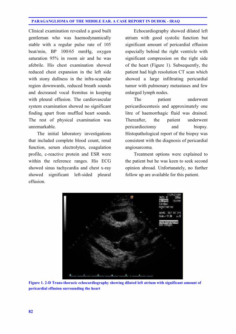
82
PARAGANGLIOMA OF THE MIDDLE EAR. A CASE REPORT IN DUHOK - IRAQ
Clinical examination revealed a good built
gentleman who was haemodynamically
stable with a regular pulse rate of 105
beat/min, BP 100/65 mmHg, oxygen
saturation 95% in room air and he was
afebrile. His chest examination showed
reduced chest expansion in the left side
with stony dullness in the infra-scapular
region downwards, reduced breath sounds
and decreased vocal fremitus in keeping
with pleural effusion. The cardiovascular
system examination showed no significant
finding apart from muffled heart sounds.
The rest of physical examination was
unremarkable.
The initial laboratory investigations
that included complete blood count, renal
function, serum electrolytes, coagulation
profile, c-reactive protein and ESR were
within the reference ranges. His ECG
showed sinus tachycardia and chest x-ray
showed significant left-sided pleural
effusion.
Echocardiography showed dilated left
atrium with good systolic function but
significant amount of pericardial effusion
especially behind the right ventricle with
significant compression on the right side
of the heart (Figure 1). Subsequently, the
patient had high resolution CT scan which
showed a large infiltrating pericardial
tumor with pulmonary metastases and few
enlarged lymph nodes.
The patient underwent
pericardiocentesis and approximately one
litre of haemorrhagic fluid was drained.
Thereafter, the patient underwent
pericardiectomy and biopsy.
Histopathological report of the biopsy was
consistent with the diagnosis of pericardial
angiosarcoma.
Treatment options were explained to
the patient but he was keen to seek second
opinion abroad. Unfortunately, no further
follow up are available for this patient.
Figure 1. 2-D Trans-thoracic echocardiography showing dilated left atrium with significant amount of
pericardial effusion surrounding the heart

83
Duhok Medical Journal Volume 7, Number 1, 2013
DISCUSSION
The diagnosis of cardiac angiosarcoma
may be easily missed because of its non-
specific presentation. Typically the
patients are presented with dyspnea, low
grade fever, signs of caval obstruction,
pericardial pain and cough,1 The dyspnea
is thought to be due to obstruction of the
caval veins, right ventricle inflow or due to
pericardial tamponade.2,3 Investigations of
this disease might also be not specific. The
ECG may show non specific changes with
low QRS voltage. Chest x-ray usually
shows globular hear or massive
cardiomegaly.1 Echocardiography and
especially, transesophageal
echocardiography, can precisely locate the
tumor, define its extent, and may
accurately predict tumor type. Most cases
are found to have haemopericordium and
in two different necropsy review of
patients with angiosarcoma, the
pericardium was obliterated by the tumor.3
MRI currently appears to be the imaging
modality of choice in the assessment of a
patient with known cardiac mass.4
The main treatment strategy in cardiac
angiosarcoma is surgical resection with or
without chemotherapy and radiation.
However, regardless of treatment, the
prognosis is very poor with a mean
survival of several months after the initial
presentation.2 This is thought to be due to
the locally invasive nature of the leision.
In addition, surgical removal is limited by
the amount of cardiac tissue that can be
resected. The high rate of haematogenous
spread with the poor response to both
chemotherapy and radiotherapy, also
contribute to the poor prognosis of this
condition.5 Novel approaches, such as the
use of interleukin-2, have been reported to
be effective. In a case of cardiac
angiosarcoma treated with a combination
of chemotherapy and immunotherapy,
survival of 30 months after surgery has
been reported.6 Cardiac transplant has
been performed in some cases with early
diagnosis and incomplete resection of the
tumor with worsened results because the
immunosuppressive therapy can increase
the risk of progression of cancer disease.7
In conclusion, Cardiac angiosarcoma
is a rare condition and its diagnosis is
usually difficult to make because of the
nonspecific presentation. Initial diagnosis
is usually suggested at echocardiography,
but identification of mediastinal invasion
and extracardiac metastases is best
detected with CT and MR imaging. The
overall prognosis remains short despite the
advances in novel approaches. There is,
however, an encouraging trend toward
more accurate and earlier diagnosis, in
large part because of improved imaging
techniques.
REFERENCES
1. Lin TK, Stech JM, Eckert WG, Lin JJ,
Farha SJ, Hagan CT. Pericardial
angiosarcoma simulating pericardial
effusion by echocardiography. Chest.
1978; 73(6): 881-3.
2. Brandt RR, Arnold R, Bohle RM, Dill
T, Hamm CW. Cardiac angiosarcoma:
case report and review of the literature.
Z Kardiol. 2005; 94(12): 824-8.
84
PARAGANGLIOMA OF THE MIDDLE EAR. A CASE REPORT IN DUHOK - IRAQ
3. Cheesman MG, Webester J, Rowlands
D. Pericardial obliteration by
angiosarcoma. Br Heart J. 1988; 60(3):
262-3.
4. Mader MT, Poulton TB, White RD.
Malignant tumors of the heart and
great vessels: MR imaging appearance.
Radiographics. 1997; 17(1): 145-53.
5. Yao H, Miyamoto T, Mukai S,
Yamamura M, Nakagawa T, Ryomoto
M. Angiosarcoma of the pericardium.
Review of 9 reports from Japan.
Japanese J Thorac Cardiovas Surg.
2003; 51(2): 65-7.
6. Kakizaki S, Tagari H, Hosaka Y.
Cardiac angiosarcoma responding to
multidisciplinary treatment. Int J
Cardiol. 1997; 62(3): 273-5.
7. Überfuhr P, Meiser B, Fuchs A,
Schulze C, Rechenspurner H, Falk M,
et al. Heart transplantation: an
approach to treating primary cardiac
sarcoma. J Heart Lung Transplant.
2002; 21(10): 1135-9.
85
Duhok Medical Journal Volume 7, Number 1, 2013
ón‚íqón‚íqón‚íqón‚íq@@@@@@@@a†@¶†@a†Šóq@†@æîí‚óÄb÷@ßó�†@¶†@bî@aŠbàò†@bÙŽî‹�a†@¶†@a†Šóq@†@æîí‚óÄb÷@ßó�†@¶†@bî@aŠbàò†@bÙŽî‹�a†@¶†@a†Šóq@†@æîí‚óÄb÷@ßó�†@¶†@bî@aŠbàò†@bÙŽî‹�a†@¶†@a†Šóq@†@æîí‚óÄb÷@ßó�†@¶†@bî@aŠbàò†@bÙŽî‹�@@@@–@@@@ŽôØómóÜby@bä‹ÙmŠíqaŠŽôØómóÜby@bä‹ÙmŠíqaŠŽôØómóÜby@bä‹ÙmŠíqaŠŽôØómóÜby@bä‹ÙmŠíqaŠ@@@@@@@@
��
¶†@æŽïî@Žõ‹Žï’ó−óq@æŽïÙŽî‹�@ŽõŠíu@æî‹mó“à@oïåïà†@Ž¶ói@óáŽïØ@ÛóÜó�@æŽïmóÜby@ˆ@¶†@bî@aŠbàò†@bÙŽî‹�@íØ@ò‡äóšŠóè@NaŠìˆ<@Žôn�aŠ@bî@¶††Šóq@ì@Žõ‹Žï’ó−óq@ŽôÄ@íi@óéu@æî‹mó“à@õŠó�ÜoŽïè†@Žõìì†@a�Žî‹Ü@¶†@a@N@a‰Žî‹i@õ‹Žï’ó−óq@ŽõŠíu@a†@¶†@æŽïÙŽî‹�@ôàóè@Äbä†25@E@oŽïè†
@çaìˆ@ì35@–@40@EŽôä‹à@Žôàò‡Ü@a†@b‚óm@bä‹ØóÄì‹’@æŽïmŠíqaŠ@†@Šbî†@ôØòì@ò‹Žïä@ŽõŒó�òŠ@Óò‡Ü@�q@ì@óîaŠbàò†@bÙŽî‹�@ŽõŠíu@ˆ@N@ÛóÜó�iˆ@ç‹Ùäb“ïån�ò†@ónŽïèbä@óÙŽî‹�@òŠíu@Äó÷@çaŠbuòŒŠói@æŽïî@ Žõì@æŽïäb“ïä@Šó@N@íØ@óïî‹Ù“ŽïÙ“Žïq@ôØóàýòŒ@ ŽômóÜby@óà@a†@ ŽômŠíqaŠ@ ŽŽôĆ
ìíióè@¶†@a†ŠórÜ@õ‹Žï’ó−ói@bî@aŠbàò†@bÙŽî‹�@íØ@ç‹Ùäb“ïån�ò†@ómbèì@¶†@a†Šóq@Šó�Ü@ìíióè@ÄbšŠói@bØóåîí‚óÄb÷.@@@@@@
الخلاصة
قـــــرير حالـــــةت - الورم الوعائي القلبي مع نضوح دموي في شغاف القلب
.على الرغم من كون الورم الوعائي القلبي من الحالات النادرة، لكنه يبقى الأكثر شيوعاً من بين أورام القلب السرطانيةمن % 25تشكل الأورام السرطانية نسبة . يعتبر الاذين الأيمن المنطقة الأكثر شيوعاً للورم الأولي و يليه شغاف القلب
هي من نوع الورم الوعائي السرطاني وبأكثرية لدى الذكور كما % 40 - 35مجمل أورام القلب الأولية لدى البالغين ومنها في هذا التقرير . كثيراً ما يصعب تشخيص هذا الورم وذلك لأعراضه الغير محددة. بينته تقارير التحليل النسيجي للجثث
.وي واضح في شغاف القلب و تم تشخيصه كحالة ورم وعائي للشغافقمنا بعرض حالة شخص بالغ لديه نضوح دم
86
PARAGANGLIOMA OF THE MIDDLE EAR. A CASE REPORT IN DUHOK - IRAQ
PARAGANGLIOMA OF THE MIDDLE EAR. A CASE REPORT IN DUHOK - IRAQ
ALAA H. RAZAK, M.B.Ch.B., M.Sc (pathology), M.I.B.M.S*
JENA WARDA, M.B.Ch.B**
Submitted 11 Nov 2013; accepted 5 Sep 2013
SUMMARY
Paragangliomas are rare tumors of the paraganglia. Here, we report the clinical,
histopathological and immunohistochemical findings of a rare case of paragnglioma in a 20
year old lady who presented as an external ear polyp extending from the middle ear.
Histopathological features raised the diagnosis of paraganglioma, and immunohistochemical
stains confirmed the diagnosis.
Duhok Med J 2013;7(1): 86-92. Key words: Paraganglioma, Ear polyp
araganglioma is the generic term
applied to tumors of paraganglia
regardless of location. The only exception,
largely on the basis of tradition, is the
paraganglioma of the adrenal medulla,
which is rarely designated as such but
universally known as pheochromocytoma.
By extension, paragangliomas located
outside the adrenal gland that are
obviously chromaffin and associated with
clinical evidence of norepinephrine and/or
epinephrine secretion also have been
designated as extra-adrenal
pheochromocytomas. Most of these arise
from orthosympathetic-related
paraganglia, whereas most of the
nonchromaffin, nonfunctioning
paragangliomas originate from
parasympathetic-related organs. It is not
possible on morphologic grounds to
distinguish between these two types or to
predict whether a tumor is functioning at
the clinical level or not.1
Extra-adrenal paragangliomas are
very uncommon tumors arising from
neuroectodermal-derived paraganglion
tissue. These tumors are usually located
along the vascular tree, especially near
the carotid body, the jugulotympanic
body, or the mediastinal vessels.2
Jugulotympanic paragangliomas (JTP)
tumors have a predilection for women.
They arise from microscopic collections of
paraganglia, which were described by
Guild in a study involving serial sectioning
of temporal bones; the first case of a
patient with JTP was reported by
Rosenwasser in 1945. These paraganglia
(average of two to three in each temporal
bone) may be found along the course of
the Jacobsen nerve (tympanic branch of
the 9th cranial nerve), the nerve of Arnold
(auricular branch of the 10th cranial
nerve), the adventitia of the jugular bulb,
the osseous canal connecting the jugular
fossa to the middle ear cavity, or within
P
* Assistant professor in pathology, Faculty of Medical Sciences, School of Medicine, Duhok University,
Kurdistan-Iraq
** Faculty of Medical Sciences, School of Medicine, Duhok University, Kurdistan-Iraq
Correspondence author: Dr. Alaa Hani Razak, 0096475048013-10, E-mail address [email protected]
87
Duhok Medical Journal Volume 7, Number 1, 2013
the middle ear (usually over the cochlear
promontory). The varied locations of these
paraganglia and the complex anatomy of
this area form the basis for the different
clinical presentations of patients with JTP.
Small tumors arising over the cochlear
promontory (tympanic paraganglioma)
may arise as an aural polyp, with filling of
the middle ear cavity or extension into the
external ear canal. JTP can involve the
temporal bone, with intracranial extension,
or appear as a mass at the base of the skull,
with erosion of the jugular foramen.
Rarely, these tumors display clinical
evidence of malignancy, primarily
metastatic disease. The most common sites
of metastasis include lymph nodes,
skeleton, lungs, and liver. The histologic
appearance of these tumors is usually not
predictive of biologic behavior.3
CASE PRESENTATION
A 20 year old lady presented to the ENT
clinic at Azady Teaching Hospital, Duhok
City suffering from severe reduction in
hearing in the left ear and intermittent
bleeding. On examination, the surgeon
noted a mass extending from the middle
ear, damaging the tympanic membrane and
extending as a polyp into the external ear.
There was a complete conductive hearing
loss in that ear. CT scan examination
confirmed that the mass was originated
from the middle ear and eroding the
mastoid bone. Surgical excision was done
and the specimen was sent for
histopathology. Grossly, the lesion
appeared as an ovoid vascular polyp-like
lesion measuring 8x5x4 mm.
Microscopically, the lesion was well-
defined sub-epithelial, formed of nests of
bland looking polygonal cells with finely
granular cytoplasm. The nests were
surrounded by spindle cells. The
intervening stroma was well vascularized
(Figure 1and 2). The diagnosis of
paragnaglioma was aroused but
serominous adenoma and adenocarcinoma
couldn’t be excluded.
Immunohistochemically, using the
automated Ventana immunotechnique, the
tumor nests cells were strongly positive for
chromogranin (Figure 3), CD56 (Figure 4)
and NSE while the surrounding spindle
(sustentacular) cells stained positive for S-
100 protein (Figure 5 and 6).
DISCUSSION
Paragangliomas are rare
neuroendocrine tumors derived from
the extra-adrenal paraganglia. Head and
neck paraganglia are associated with the
parasympathetic nervous system, the
largest being the carotid body, with
others found at the vagus nerve, the
jugular bulb, the tympanic branch of
the ascending pharyngeal artery, the
larynx, and other sites.4 These tumors are
more common in female.5 The common
presenting symptoms are usually pulsating
tinnitus and conductive hearing loss,
although large tumors may erode the
tympanic membrane and appear as a
bleeding aural polyp,6 as did our patient.
Most reported cases behaved in a benign
way, however, very rare occasions of
malignant behavior had beed reported.4
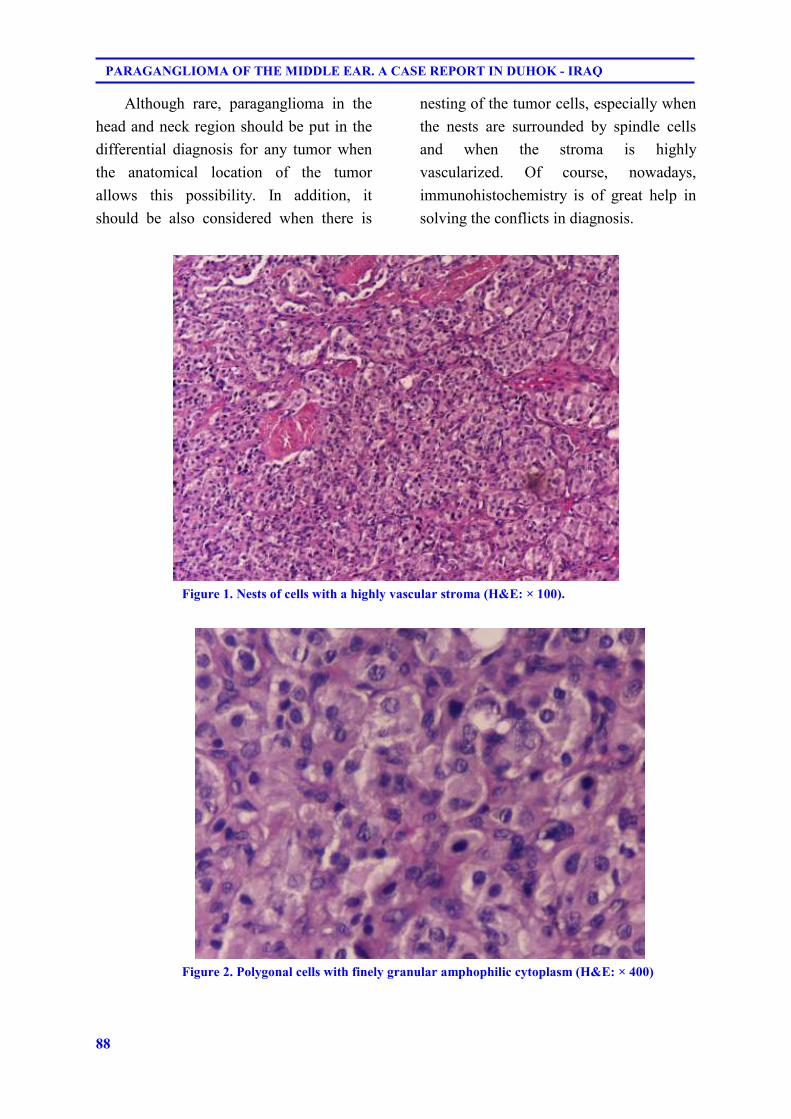
88
PARAGANGLIOMA OF THE MIDDLE EAR. A CASE REPORT IN DUHOK - IRAQ
Although rare, paraganglioma in the
head and neck region should be put in the
differential diagnosis for any tumor when
the anatomical location of the tumor
allows this possibility. In addition, it
should be also considered when there is
nesting of the tumor cells, especially when
the nests are surrounded by spindle cells
and when the stroma is highly
vascularized. Of course, nowadays,
immunohistochemistry is of great help in
solving the conflicts in diagnosis.
Figure 1. Nests of cells with a highly vascular stroma (H&E: × 100).
Figure 2. Polygonal cells with finely granular amphophilic cytoplasm (H&E: × 400)
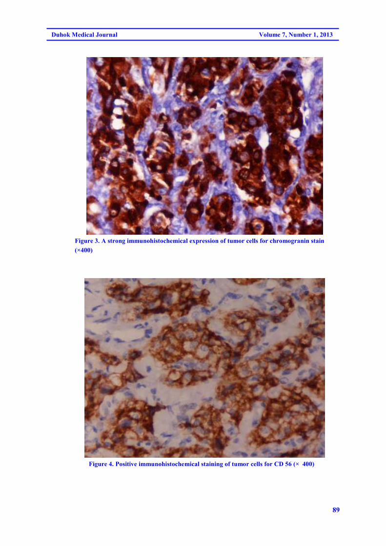
89
Duhok Medical Journal Volume 7, Number 1, 2013
Figure 3. A strong immunohistochemical expression of tumor cells for chromogranin stain
(×400)
Figure 4. Positive immunohistochemical staining of tumor cells for CD 56 (× 400)
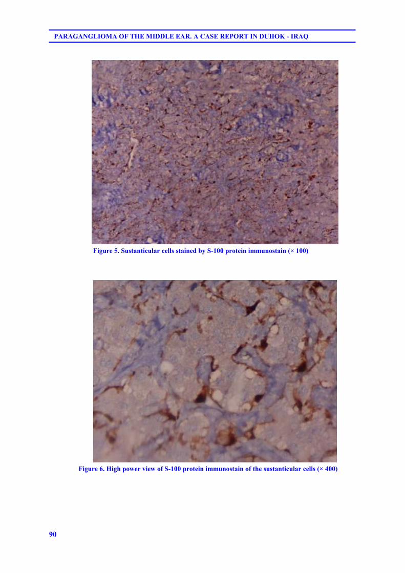
90
PARAGANGLIOMA OF THE MIDDLE EAR. A CASE REPORT IN DUHOK - IRAQ
Figure 5. Sustanticular cells stained by S-100 protein immunostain (× 100)
Figure 6. High power view of S-100 protein immunostain of the sustanticular cells (× 400)
91
Duhok Medical Journal Volume 7, Number 1, 2013
REFERENCES
1. Rosai J. Rosai and Ackerman’s
surgical pathology. 9th ed. Vol 1.
Edinburgh: Mosby, 2004.
2. da Silvaa RA, Grossa JF, Haddada FJ,
Cíntia AB, and Younesa RN. Primary
pulmonary paraganglioma: case report
and literature review. Clinics.
2006;61(1):83-6.
3. Mills SE. Sternberg's diagnostic
surgical pathology. 5th ed. Lippincott
Williams & Wilkins, 2010.
4. Nedev PI. Paraganglioma of the
middle ear – case report. Trakia
Journal of Sciences 2008; 6(2): 36-39.
5. Kuczkowski J, Przewoźny T, Biernat
W, Dubaniewicz-Wybieralska M.
Nonchromaffin paragangliomas
(chemodectoma) of the middle ear in
the material of the ENT Department
Medical University of Gdańsk.
Otolaryngol Pol. 2011; 65(1):40-6.
6. Neto M, Isabela M. de Vuono, Souza
LR, Testa JR, Pizarro GU, Barros F.
Tympanic Paragangliomas: case
reports. Rev Bras Otorrinolaringol.
2005; 71(1): 97-100.

92
PARAGANGLIOMA OF THE MIDDLE EAR. A CASE REPORT IN DUHOK - IRAQ
ón‚íqón‚íqón‚íqón‚íq@@@@@@@@ä@Žôèí�@ß@bàíï�äb�Šbqä@Žôèí�@ß@bàíï�äb�Šbqä@Žôèí�@ß@bàíï�äb�Šbqä@Žôèí�@ß@bàíï�äb�Šbq@o�aŠóÄb@o�aŠóÄb@o�aŠóÄb@o�aŠóÄb–@@@@ŽôØómóÜby@bä‹ÙmŠíqaŠŽôØómóÜby@bä‹ÙmŠíqaŠŽôØómóÜby@bä‹ÙmŠíqaŠŽôØómóÜby@bä‹ÙmŠíqaŠ@@@@@@@@
��
a†bïÝ�äó�aŠbq@Óbä†@pò††@õìŠ@çaŠbu@âïØ@óØóàòŠòì@bàíïÝ�äó�aŠbq@~@bØó¹b‚@óà20@@õóè@bî@óàòŠòì@Äó÷@íØ@ôØíè†@õ‹îˆbjÜ@oî†@çýb�íjnÐóØŠò†@õì@ôèí�ˆ@íØ@ôn’í�@bØò†bîŒ@õòíï’@Šó�Ü@~@ì@ô’í‚óä@bïîóäb’@båïåÙ“q@l@õì@õŠíuˆ@µi@o�aŠ@o“q@æîbï’@ãó÷@ì
@bïîóäb’@bî‹�ó�Šói@båïåÙ“qõìbïáïØ.@@
الخلاصة
ـــــــــــي الاذن الوســـــــــــطى ـــــــــــا ف ــــــة- اورام الباراكانكليوم ــــــرير حال تق
اورام الباراكانكليوما هي من الاورام النادره التي تصيب الانسجه المجاوره للعقد العصبيه و هنا نوثق الحاله السريريه و
هذه الاورام لدى سيده في العشرين من نتائج الفحص النسيجي و الفحص المناعي النسيجي الكيميائي لحاله نادره من
عمرها و التي ظهرت على شكل زائده لحميه في الاذن الخارجيه ممتده من الاذن الوسطى
الصفات النسيجيه للورم اثارت الريبه في تشخيص الباراكانكليوما و لكن الصبغات المناعيه النسيجيه الكيميائيه اثبتت
. التشخيص بصوره قطعيه
93
Duhok Medical Journal Volume 7, Number 1, 2013
ERRATUM
Duhok Med J 2013;7(1): 93-94.
Article: A CYTOPATHOLOGICAL STUDY OF THE EFFECT OF SMOKING ON
THE ORAL EPITHELIAL CELLS IN RELATION TO ORAL HEALTH STATUS BY
THE MICRONUCLEUS ASSAY
Authors: SAEED H. SAEED, WASEN H. YOUNIS
Journal: Duhok Med J 2012;6 Suppl 3:170-177.
In this article, we regret to inform the authors and our readers that there was a printing error
in page 172. The following correction is to be made:
Substitute page 172 (table 1 through 4) with the corrected version (table 1 though 6)
presented in the back of this erratum page.
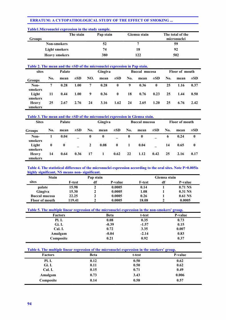
94
ERRATUM: A CYTOPATHOLOGICAL STUDY OF THE EFFECT OF SMOKING ...
Table1.Micronuclei expression in the study sample.
The total of the
micronuclei
Giemsa stain Pap stain The stain
Groups
59 7 52 Non-smokers
92 18 74 Light smokers
502 122 380 Heavy smokers
Table 2. The mean and the ±SD of the micronuclei expression in Pap stain.
sites
Groups
Palate Gingiva Buccal mucosa Floor of mouth
No. mean ±SD NO. mean ±SD No. mean ±SD No. mean ±SD
Non-
smokers
7 0.28 1.00 7 0.28 0 9 0.36 0 25 1.16 0.37
Light
smokers
11 0.44 1.00 9 0.36 0 18 0.76 0.23 25 1.44 0.50
Heavy
smokers
25 2.67 2.76 24 3.16 1.62 24 2.65 1.20 25 6.76 2.42
Table 3. The mean and the ±SD of the micronuclei expression in Giemsa stain.
Sites
Groups
Palate Gingiva Buccal mucosa Floor of mouth
No. mean ±SD No. mean ±SD No. mean ±SD No. mean ±SD
Non-
smokers
1 0.04 _ 0 0 _ 0 0 _ 6 0.24 0
Light
smokers
0 0 _ 2 0.08 0 1 0.04 _ 14 0.65 0
Heavy
smokers
14 0.64 0.36 17 1 0.62 22 1.12 0.42 25 2.16 0.17
Table 4. The statistical difference of the micronuclei expression according to the oral sites. Note P˃0.005is
highly significant, NS means non- significant.
Stain
sites
Pap stain Giemsa stain
F-test df P-value F-test df P-value
palate 15.98 2 0.0005 0.14 1 0.71 NS
Gingiva 15.30 2 0.0005 1.08 1 0.31 NS
Buccal mucosa 22.25 2 0.0005 0.26 1 0.61 NS
Floor of mouth 119.41 2 0.0005 18.08 2 0.0005
Table 5. The multiple linear regression of the micronuclei expression in the non-smokers' group.
P-value t-test Beta Factors
0.73 0.35 0.08 Pl. I.
0.15 -1.57 -0.39 Gi. I.
0.007 3.35 0.72 Cal. I.
0.83 -2.14 -0.04 Amalgam
0.37 0.92 0.21 Composite
Table 6. The multiple linear regression of the micronuclei expression in the smokers' group.
P-value t-test Beta Factors
0.62 0.50 0.12 Pl. I.
0.62 0.50 0.11 Gi. I.
0.49 0.71 0.15 Cal. I.
0.006 3.43 0.73 Amalgam
0.57 0.58 0.14 Composite