how killer cells kill
TRANSCRIPT
How Killer Cells Kill
These immune-system cells recognize a target, close in on it and bind tightly to it. Then they secrete onto its surface a lethal pore-forming protein that causes the target cell to leak and die
by John Ding-E Young and Zanvil A. Cohn
The immune system is commonly likened to an army and its various cells to soldiers.
The analogy is nowhere more appropriate than in the case of the cells called killer cells. Their primary duty is to seek out and destroy the body's own cells when they go wrong: to kill tumor cells and cells that have been infected by viruses (and perhaps by other foreign agents). For some years it has been clear that killer cells do their job with great efficiency, first seeking out a miscreant target cell, then binding tightly to it and finally doing something to it that causes its death, while at the same time sparing innocent bystander cells. But what exactly do they do? Just how do the killer cells kill?
The answer is beginning to come clear. Having bound to an appropriate victim, the killer cell takes aim at the target's surface and shoots it full of holes. More specifically, it fires molecules of a lethal protein. The molecules bore into the target cell's surface membrane and form porelike channels. The target cell leaks, and soon it dies.
Work in a number of laboratories, including our own at Rockefeller University, has shown that the poreforming protein is part of the armamentarium of the two kinds of killer cell, the cytotoxic T cell and the so-called natural killer cell. We have found a protein with a similar function in another immune cell, the eosinophil. Moreover, the same protein, or one very like it, appears to be responsible for attacks on human cells by an amoeba that causes severe dysentery.
We begin to think pore-forming proteins may be a major weapon in a wide range of cell-mediated killing. Knowing more about the process should have important medical payoffs. It may eventually be pOSSible,
38
for example, to treat amebic dysentery and some other parasitic, fungal and bacterial diseases by blocking a pore-forming protein. Finding a way to enhance the pore-forming process in immune-system cells might be even more important: in principle it should be helpful in the treatment of both cancer and such intractable viral diseases as AIDS.
The killer cells are elements of the cellular immune system, but
their precise and lethal functioning can be studied and understood only
in the context of the immune system as a whole. It has a humoral component as well as a cellular one. The humoral system defends the body primarily against bacteria and toxic molecules. Its weapons are antibodies, or immunoglobulins, which are synthesized and secreted by the cells called B lymphocytes. Each of millions of B cells synthesizes a particular antibody, which recognizes a particular antigen (a molecular recognition pattern); the cell displays the antibody on its surface. When the lymphocyte encounters a bacterial
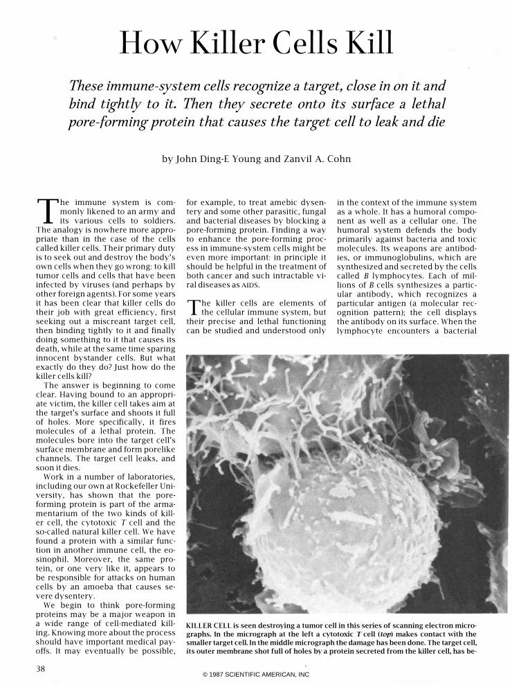
KILLER CELL is seen destroying a tumor cell in this series of scanning electron micro
graphs. In the micrograph at the left a cytotoxic T cell (top) makes contact with the
smaller target cell. In the middle micrograph the damage has been done. The target cell,
its outer membrane shot full of holes by a protein secreted from the killer cell, has be-
© 1987 SCIENTIFIC AMERICAN, INC

cell or a toxin bearing that antigen, the lymphocyte proliferates. Some of its progeny become memory cells that will respond to the same antigen faster the next time; most of the progeny become plasma cells that manufacture a large amount of the antibody and secrete it. The antibody binds to the antigen. Toxins are precipitated or otherwise neutralized by the binding itself. In the case of an invading cell the binding sets off a cascade of reactions at the cell surface involving a group of blood-serum proteins collectively known as complement. The end result of the cascade is the death of the cell.
The same precursor cells that give rise to B lymphocytes are the ancestors of a varied family of T lymphocytes that are the basis of the cellular immune system. Some T cells, called helper cells and suppressor cells, modulate both the humoral and the cellular system, chiefly by secreting chemical messengers called lymphokines. The major effector cell of the cellular system is the cytotoxic T lymphocyte, or killer T cell, we have mentioned. Its main targets are virusinfected cells. The other type of killer cell introduced above, the natural killer cell, is also a lymphocyte. Its
precise lineage is not clear, but it seems to be closely related to the cytotoxic T cell. Its main targets are thought to be tumor cells, and perhaps also cells infected by agents other than viruses.
As in the case of B lymphocytes, the function of T lymphocytes depends in the first instance on proper recognition of an appropriate target. T cells display on their surface specific receptors, very similar to the B cell's antibodies, that recognize and bind to particular cell-surface antigens. T cells are more selective than B cells, however. They recognize and bind to an antigen only when it is "presented" by one of a group of surface molecules known collectively as the major histocompatibility complex (MHC). Natural killer cells, as their name implies, are less choosy about what they attack. Their receptors are less discriminating, and they are not restricted by the MHC.
Once either the killer T cell or the natural killer cell has identified
its target, the killer cell binds tightly to the target cell. This close contact triggers the lethal process and also ensures that neighboring cells are not indiscriminately destroyed.
That much was known a decade ago, but the nature of the killing process continued to be a mystery. The first clues to the mystery came in the early 1970's from the laboratories of a number of investigators, notably Eric Martz of the University of Massachusetts at Amherst, Christopher S. Henney of the Immunex Corporation in Seattle, William R. Clark of the University of California at Los Angeles, Pierre Golstein of the Center for Immunology in Marseilles and Gideon Berke of the Weizmann Institute of Science in Israel. Their work dissected the killing process into a sequence of discrete stages.
First, it became clear, a "conjugate" is formed as the lymphocyte and its target come in close contact. Then some kind of lethal hit is delivered, injuring the target cell. The hit initiates what seems to be a programmed death of the injured target: death proceeds in a predetermined manner (provided only that calcium ions are present) even as the conjugate breaks up and the lymphocyte goes on to initiate a new killing cycle.
Henney and Martz were among the first to suggest that a lymphocyte appeared to kill by somehow damaging the plasma (outer) membrane of
come leaky and an influx of water has made it balloon; it has lost
many of its projecting villi and there is a large cavity in the mem
brane. In the micrograph at the right there is nothing left of the
tumor cell but its nucleus and some debris <the other nucleus in
the micrograph is that of a second target cell). The cells were
prepared by Chau-Ching Liu and the scanning microscopy was
done by Gilla Kaplan. The cells are enlarged 5,700 diameters in
the first two micrographs and 4,200 diameters in the third one.
39 © 1987 SCIENTIFIC AMERICAN, INC

CULTURED CYTOTOXIC T CELL is enlarged 1,400 diameters in an electron micro
graph (left) made by the authors with Hans Hengartner and Ecl<hard R Podack The dark
storage granules in its cytoplasm, enlarged 7,600 diameters (right), have an electron
dense outer membrane; their amorphous matrix contains perforin, the lethal protein_
its target. Their proposal was based on their observation that radioactive molecules introduced into a target cell as markers leaked out rapidly when the target was damaged by a lymphocyte. The membrane became permeable only to markers of a certain maximum size, suggesting that the damage to the membrane might take the form of holes, or pores.
That possibility became a likelihood in 1980. Robert R. Dourmashkin and Pierre Henkart of the National Cancer Institute and their colleagues examined the surface of damaged target cells, enlarged many thousands of diameters in electron micrographs, and detected ringlike structures that appeared to be holes in the membrane. Their finding was extended three years later by Eckhard R. Podack of the New York Medical College and Gunther Dennert of the University of Southern California School of Medicine, who studied the effect of cultured killer cells on tumor cells. They established that the surface of the target cell was pocked with holes whose internal diameter ranged from five to 20 nanometers (millionths of a millimeter).
It was still not at all clear whether the pores were actually inflicted by
the lymphocytes or simply reflected a terminal stage of cell death caused by some other form of injury. The search for an answer was long hampered by the lack of a ready source of killer cells. Then in 1977 Steven Gillis of Immunex, Kendall A. Smith of the Dartmouth Medical School and Robert C. Gallo's group at the National Cancer Institute succeeded in maintaining mouse lymphocytes-
40
both cytotoxic T cells and natural killer cells-in the laboratory. They were able to do so by identifying particular nutrients and growth factors required for the survival of these cells in culture; one key factor turned out to be the lymphokine called interleukin-2. Culturing made it possible to grow lymphocyte clones derived from known parent cells, and so to have at hand an abundant and homogeneous source of cytotoxic T cells and natural killer cells with known characteristics. The way was cleared for detailed analysis of such cells with the tools of cell biology and biochemistry.
An obvious feature of the lymphocytes clearly called for investigation. Micrographs had revealed numerous small, dark organelles (subcellular elements) in the cytoplasm. They appeared to be storage granules, which are common in secretory cells. Such granules provide an efficient means of accumulating and packaging a substance synthesized by the cell so that large quantities of it can be released quickly at the right time. The release is accomplished by exocytosis: the granules move to the periphery of the cell, fuse with the plasma membrane and disgorge their contents.
Several investigators had seen that early in the cell-killing process the granules in a killer lymphocyte become concentrated in the part of the cell that is in close contact with the target. Then Abraham Kupfer and S. Jonathan Singer of the University of California at San Diego and Dennert noted that the granule-packaging Golgi apparatus seems to be directed toward that contact region
and that a number of proteins of the cell's cytoskeleton (its fibrous internal framework) "reorient" toward the target cell soon after contact is eStablished, apparently providing the motive force that redirects the Golgi and the granules.
The reorientation of the cytoskeleton and the movement of the Golgi stacks and granules take place only when and where the lymphocyte binds to an appropriate target. Roger Y. Tsien of the University of California at Berkeley showed that the binding induces an explosive increase of calcium ions in the cell; the increase triggers exocytosis. John H. Yanelli and Victor H. Engelhard of the University of Virginia have made cinemicrographs showing granules reorienting in the cytoplasm and then fusing with the plasma membrane.
All in all, the evidence suggested that contact with an appropriate target causes the killer cell to aim its secretory apparatus at the target and fire a lethal agent contained in its granules. It was necessary first to establish that the granules are in fact the shell casings and then to identify the projectile itself.
The first task was to isolate the granules and see if they alone
could kill. That was done independently in 1984 by Henkart and Podack and by our group. We exploited the various techniques of subcellular fractionation, the objective of which is to break up a cell into its components and find what component contains a particular enzyme or carries out a particular function. Killer lymphocytes are broken into bits and pieces by subjecting them to pressure in nitrogen gas. The cellular debris is layered onto a density gradient of inert particles and then spun in a high-speed centrifuge. The various organelles come to rest, depending on their density, in discrete bands. We examined each of the fractions in the electron microscope and assayed them for enzymatic activity and ability to kill cells.
One fraction, which electron micrography showed consisted almost entirely of granules, was enriched in certain enzyme activities and was a potent killer: when the isolated granules were mixed with red blood cells or tumor cells in a medium containing calcium ions, the cells died within minutes. Electron micrographs revealed that the cells' surface carried ringlike lesions virtually indistinguishable from those produced by intact killer cells. And so the gran-
© 1987 SCIENTIFIC AMERICAN, INC
ules were shown indeed to contain the killer cells' lethal secretion. . The secreted agent itself was soon identified. In 1985, collaborating with Podack and Hans Hengartner of University Hospital in Zurich, we found a protein that all by itself (in the presence of calcium) reproduces the observed membrane lesions and the killing inflicted by intact killer cells and by granules. We isolated and purified the protein by passing extracts of granules through chromatographic columns that separated proteins on the basis of electric charge and of molecular mass, and screening the separated proteins for their efficiency in lysing red blood cells: breaking down their membrane and causing them to burst. The killer protein was isolated independently by Danielle Masson and Jiirg Tschopp of the University of Lausanne.
So far only this one pore-forming protein, which is often called perforin (because it perforates membranes), has been found in the granules of cytotoxic T lymphocytes or natural killer cells. Its molecular mass is 70,000 daltons. Once cells are exposed to perforin in the presence of calcium ions they are lysed within a few minutes. On the other hand, if calcium ions are added to perforin before it makes contact with cells, the protein's killing activity is abolished. The effect sounds paradoxical, but actually it leads to an important insight into just how perforin kills.
The 70-kilodalton molecules secreted by a killer cell insert into
the target-cell membrane. There (in the presence of calcium ions) the individual molecules (monomers) polymerize, or link up with one another. The polymer they form can assume various shapes, but under optimal conditions the final product resembles a cylinder. In the electron microscope it looks like a ring when seen in cross section and like two parallel lines when seen in longitudinal section. A fully formed ring, as Podack and Dennert first noted, ranges from five to 20 nanometers in internal diameter.
For perforin to damage a target cell the calcium-mediated polymerization must take place entirely within the cell's membrane. The reason is that only the perforin monomer can insert into a membrane; if polymerization takes place in solution, in the absence of nearby membrane, the resultant polymer cannot enter the membrane and cannot kill. The protective effect is easy to understand.
Any secreted perforin that spills into the extracellular space or the bloodstream, where calcium is abundant, must immediately undergo polymerization and thereby be rendered inactive, virtually eliminating the possibility of "accidental" injury to nontarget cells.
On an appropriate target cell, however, the tubular pores shaped by polymerization bring about rapid, measurable changes in the target cell. The plasma membrane of a living cell retains proteins and other large molecules within the cell (except when they are to be secreted) and segregates different ions, keeping some inside and some outside the cell. The segregation of positive and negative ions establishes a transmembrane electric potential. When the membrane leaks, ions and water tend to flow down their electrochemical gradients toward equilibration, and so there is a drop in membrane potential. If the holes in the membrane are of limited size, there is an additional effect, known as the Donnan effect. The large molecules inside the membrane cannot pass through it; water and salts from the extracellular fluid pour through the
membrane toward the side that has the proteins-into the cell. The cell swells, and eventually it bursts.
By impaling target cells with microelectrodes, we were able to measure a significant drop in membrane potential soon after perforin was administered. With sensitive electronic equipment we were even able to measure the ion current flowing through individual pores. Our results were consistent with the predictions of the Donnan effect. The measurements showed, moreover, that the perforin holes are stable structures that persist as open pathways in the membrane. By determining just which ions and small molecules got through the damaged membrane, we were able to estimate that the functional internal diameter of the pores ranges from one to 10 nanometers (as opposed to the observed diameter in micrographs of from five to 20 nanometers).
G'ven the apparently undiscriminating efficiency of perforin as
a killer, how can one account for the specificity of killing by lymphocytes? We have mentioned that "accidental" killing by perforin that gets
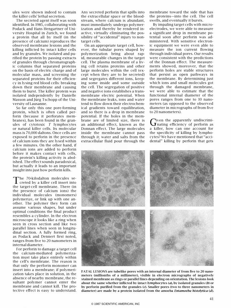
FATAL LESIONS are tubelike pores with an internal diameter of from five to 20 nano
meters (millionths of a millimeter), visible in electron micrographs of negatively
stained membrane as rings or parallel lines depending on orientation. The lesions look
about the same whether inflicted by intact lymphocytes (a), by isolated granules (b) or
by perforin purified from the granules (c). Smaller pores (two to three nanometers in
diameter) are made by a protein isolated from the amoeba Entamoeba hisro/ylica (d).
4 1
© 1987 SCIENTIFIC AMERICAN, INC

2
GOLGI STACK
GRANULE TARGET CELL
STAGES IN KILLING PROCESS were identified some time ago.
The killer lymphocyte (1) recognizes the target cell and makes
close contact with it (2). On contact the cell's granules and the
Golgi complex that forms them are reoriented toward the target
DETAILS OF KILLING PROCESS have now been elucidated. A
rise in the lymphocyte's calcium-ion level, apparently triggered
by the receptor-mediated binding of the killer cell to its target
(1), brings about exocytosis, in which the granules fuse with the
cell membrane (2) and disgorge their perforin (3) into the small
42
cell; perforin is secreted and forms pores in the target-cell mem
brane (see illustration below). Having launched its lethal missiles,
the killer cell withdraws and goes on to kill again (3); the dam
aged target cell dies a programmed death within minutes (4).
TARGET CELL
intercellular space abutting the target. Calcium there changes
the conformation of the individual perforin molecules, or mono
mers (4), which then bind to the target-cell membrane (5) and in·
sert into it (6). The monomers polymerize like staves of a barrel
(7) to form pores (8) that admit water and salts and kill the cell.
© 1987 SCIENTIFIC AMERICAN, INC

away from the lymphocyte-target interface is prevented by calcium-induced polymerization of the protein. What one might term "purposeful" killing of nontarget cells (as a result of contact with a killer lymphocyte) is prevented, on the other hand, by the lymphocyte's ability to recognize an appropriate cell-one displaying, for example, either viral antigens or tumor antigens. In other words, the specificity of killing resides solely in the requirement for close contact, which depends in turn on the binding of killer cell to target cell through the interaction of antigens on the target cell with receptors on the killer cell.
But what keeps the killer cell from killing itself? It cannot be the closecontact requirement, since the killer cell's membrane is continually exposed directly to secreted perforin. Chau-Ching Uu, a graduate student in our laboratory, and one of us (Young) collaborated with Clark recently to show that even purified perforin fails to kill either cytotoxic T cells or natural killer cells. The selfprotective mechanism is not known, but we have a hypothesis. We think the killer-cell membrane may incorporate a special protein, which we call protectin, that is very similar to perforin. The close homology would promote a kind of "faulty polymerization": the protectin would rapidly combine with any perforin monomer that gets to the killer-cell membrane, thereby preventing either the insertion of perforin into the membrane or the normal perforin-toperforin polymerization that would form a pore. We are now engaged in an intensive search for our hypothetical protein.
All the recent studies we have described here were done with mouselymphocyte cultures maintained in the laboratory. It was conceivable that perforin was just a laboratory curiosity and not truly the lymphocytes' weapon in vivo. Collaborating with Bice Perussia of the Wistar Institute in Philadelphia and Uu, we looked for perforin in lymphocytes freshly obtained from human blood. We could find none. When the lymphocytes were stimulated with interleukin-2, however, they proliferated in vitro and began to synthesize perforin. We found this to be the case for both cytotoxic T cells and natural killer cells; similar results have been reported by Leora S. Zalman and Hans J. Muller-Eberhard of the Research Institute of Scripps Clinic and their colleagues. The in vitro effect of interleukin-2 presumably reflects its
TARGET CELL
PROTECTIVE MECHANISM that prevents self-inflicted death of the killer cell has been
postulated by the authors. They think a protein, protectin, that is very similar to perfo
rin is concentrated in the lymphocyte membrane; perforin monomers bind to the pro
tectin and are prevented from polymerizing to form pores in the killer-cell membrane.
effect in the body, where it is produced by helper T cells and promotes a range of immune responses.
Indeed, the effect we noted in the laboratory may explain an apparent clinical effect of interleukin-2 first reported in 1984 by Steven A. Ro-
senberg of the National Cancer Institute. He devised a novel therapy for certain intractable cancers in which lymphocytes extracted from the blood of a patient are stimulated with interleukin-2 outside the body and then infused back into the patient.
HUMORAL IMMUNE SYSTEM forms pores much like those inflicted by the cellular sys
tem's killer lymphocytes. Binding of antibodies to a target cell triggers a cascade in
which successive proteins of the complement system are activated. Eventually the pro·
tein C 5b·6 binds to the target's surface membrane, after which C7, C8 and a number of
C9 proteins aggregate to form a pore (left). In contrast, the pores made by killer cells are
formed by the self-aggregation of one kind of subunit: the perforin monomer (right).
43 © 1987 SCIENTIFIC AMERICAN, INC
The lymphocytes are presumably thereby activated to kill more efficiently; some tumor regression has been observed in some patients. The procedure is highly toxic and therefore still in an experimental stage, but a better understanding of how to induce the expression of perforin by killer lymphocytes will certainly have a role in designing immunotherapies for cancer.
t\ lthough it seems clear that killer .t-\.cells kill by punching holes in target-cell membranes, the lymphocytes may have other weapons too. John H. Russell of Washington University in St. Louis has put forward an "internal disintegration" model to explain the killing event. The model is based on the observation that early in the course of target-cell injury the membrane enclosing the nucleus of the cell ruptures and the DNA in the nucleus breaks up into small fragments. Russell and others argue that the death of the target cell results from the cleaving of the DNA, which is presumably triggered by some signal emitted by the killer cell. The model has not been elaborated in detail, but it cannot be excluded by our
findings. Perhaps there are several different killing mechanisms.
Indeed, we have found that certain cytotoxic T-cell lines maintained in culture for some time continue to exert killing activity even though they do not secrete perforin. Do they secrete something else? We have preliminary evidence that a molecule we callieukalexin, which kills cells over the course of several hours rather than minutes, may be present in killer cells. It is just possible that even if perforin pores first admit only water and salts and are too small to pass most proteins, they eventually enlarge enough (through additional polymerization) to admit one chain of a protein-perhaps leukalexin-that cleaves DNA. Alternatively, pore formation might lead eventually to the admission of a DNA-cleaving protein by somehow promoting endocytosis, the process by which large molecules are generally taken into cells.
Pore formation may not be the only killing mechanism for killer lymphocytes, but it is clearly an efficient one. It figures, for example, in humoral immunity as well as in cellular immunity: it is the end result of the complement cascade that kills bacteria
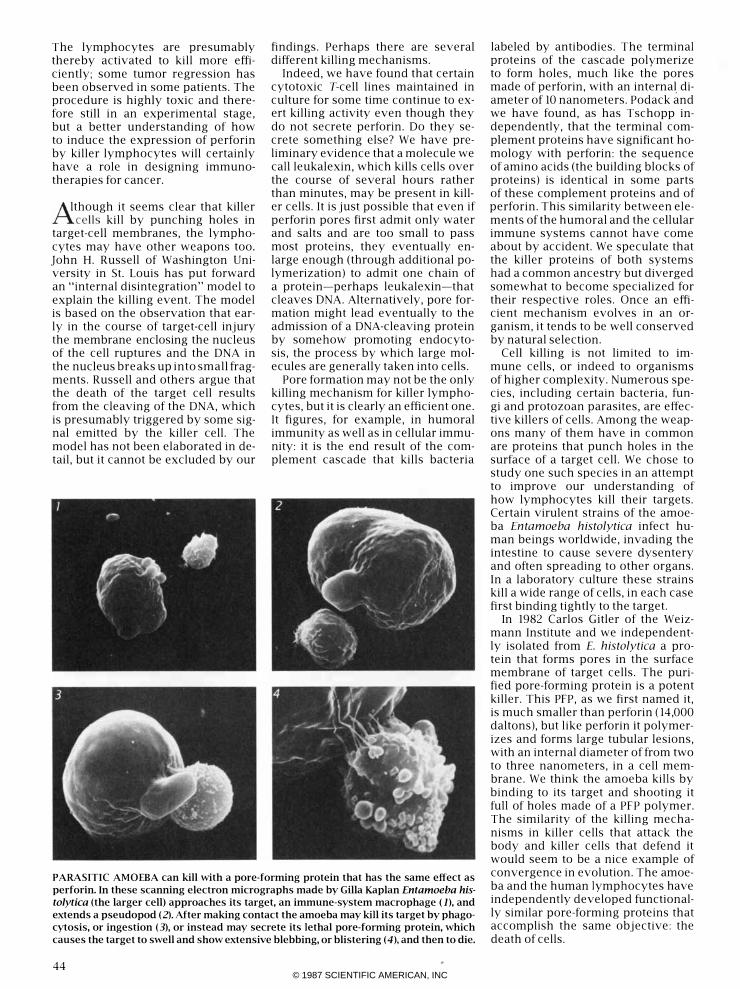
PARASITIC AMOEBA can kill with a pore-forming protein that has the same effect as
perforin. In these scanning electron micrographs made by Gilla Kaplan Entamoeba his
tolytica (the larger cell) approaches its target, an immune-system macrophage (1), and
extends a pseudopod (2). After making contact the amoeba may kill its target by phago
cytosis, or ingestion (3), or instead may secrete its lethal pore-forming protein, which
causes the target to swell and show extensive blebbing, or blistering (4), and then to die.
44
labeled by antibodies. The terminal proteins of the cascade polymerize to form holes, much like the pores made of perforin, with an internal diameter of 10 nanometers. Podack and we have found, as has Tschopp independently, that the terminal complement proteins have significant homology with perforin: the sequence of amino acids (the building blocks of proteins) is identical in some parts of these complement proteins and of perforin. This similarity between elements of the humoral and the cellular immune systems cannot have come about by accident. We speculate that the killer proteins of both systems had a common ancestry but diverged somewhat to become specialized for their respective roles. Once an efficient mechanism evolves in an organism, it tends to be well conserved by natural selection.
Cell killing is not limited to immune cells, or indeed to organisms of higher complexity. Numerous species, including certain bacteria, fungi and protozoan parasites, are effective killers of cells. Among the weapons many of them have in common are proteins that punch holes in the surface of a target cell. We chose to study one such species in an attempt to improve our understanding of how lymphocytes kill their targets. Certain virulent strains of the amoeba Entamoeba histolytica infect human beings worldwide, invading the intestine to cause severe dysentery and often spreading to other organs. In a laboratory culture these strains kill a wide range of cells, in each case first binding tightly to the target.
In 1982 Carlos Gitler of the Weizmann Institute and we independently isolated from E. histolytica a protein that forms pores in the surface membrane of target cells. The purified pore-forming protein is a potent killer. This PFP, as we first named it, is much smaller than perforin (14,000 daltons), but like perforin it polymerizes and forms large tubular lesions, with an internal diameter of from two to three nanometers, in a cell membrane. We think the amoeba kills by binding to its target and shooting it full of holes made of a PFP polymer. The similarity of the killing mechanisms in killer cells that attack the body and killer cells that defend it would seem to be a nice example of convergence in evolution. The amoeba and the human lymphocytes have independently developed functionally similar pore-forming proteins that accomplish the same objective: the death of cells.
© 1987 SCIENTIFIC AMERICAN, INC

© 1987 SCIENTIFIC AMERICAN, INC