kingdom plantae - f://one design · pdf filekingdom plantae objectives - be able to recognize...
TRANSCRIPT

122Ramp. Copyright © 2012 by F.one Design. All rights reserved.
Lab Exercise 22
Kingdom Plantae
Objectives
- Be able to recognize and name the plant phyla studied
- Understand the life cycle of these phyla
- Know what alternation of generations means
- Be able to distinguish between sporophyte and
gametophyte
- Be able to identify and name important reproductive
structures in the phyla studied
Introduction
The Kingdom Plantae is composed of multicellular, photo-
synthetic organisms which differ from the animal kingdom
not only in their ability to manufacture their own food, but
also in the number of phases they pass through in their life
cycle.
When diploid animals reproduce, specialized cells undergo
meiosis and produce haploid cells that mature into gam-
etes (sperm or eggs). These gametes join at fertilization,
producing a new diploid animal and completing the life
cycle. When diploid plants (called sporophytes) reproduce,
specialized cells undergo meiosis and produce haploid
cells called spores. These do not become gametes but
germinate and grow into multicellular haploid plants called
gametophytes. Specialized cells in the gametophyte then
produce gametes by mitosis.
These gametes will join at fertilization and produce a new
sporophyte plant, completing the life cycle.
Because a plant’s life cycle has these two distinct multicel-
lular phases, this life cycle is referred to as Alternation of
Generations. A generalized plant’s life cycle is shown in
the diagram.
Plant groups differ largely by the appearance of structures
associated with their life cycle, the relative amount of time
the sporophyte and gametophyte persist, and the ability of
the sporophyte and gametophyte to survive independently.
To begin examining these, go to the Diversity section of
the BiologyOne DVD and select ‘Kingdom Plantae’. The
four phyla covered in this lab are introduced. Click on the
forward arrow in the lower right to begin your investigation
of the Phylum Bryophyta.

222Ramp. Copyright © 2012 by F.one Design. All rights reserved.
Activity 22.1Division Bryophyta
The Bryophyta include the mosses, hornworts, and liv-
erworts. These are relatively small plants found in habi-
tats that are wet at least once during the year but most
Bryophytes can withstand drought periods by becoming
dormant. The gametophyte generation is dominant in this
group.
In the Kingdom Plantae simulation of the BiologyOne
DVD, examine the gametophyte plant of bryophytes. Note
the relatively small size. Usually these grow no taller than
20 cm. Along the axis of the plant are flat, leaflike struc-
tures called microphylls. If you examine the anatomy of
the gametophyte, you would find an absence of tissue to
conduct water or nutrients. Answer the questions in the
Results Section.
The structures which produce gametes are located at the
top of the gametophyte plant among the top microphylls.
The structure that produces sperm cells is termed an an-
theridium (pl. antheridia) and the structure that produces
an egg cell is termed an archegonium (pl. archegonia).
The flagellated sperm swim through a film of water to
reach the archegonia and then swim down the central
canal to fertilize the egg.
After fertilization, the newly formed zygote grows into
the sporophyte plant. However, the sporophyte is never
released from the gametophyte; it just grows from the top
of the gametophyte. The part of the sporophyte called the
foot anchors the sporophyte in the gametophyte and ab-
sorbs food and water from the gametophyte. At the top of
the sporophyte, a capsule is formed, within which meiosis
occurs and the spores are produced. When the spores are
mature, the top of the capsule, the operculum, pops off
and the spores are then dispersed by the wind. Frequently,
an immature capsule is covered by a thin tissue layer
called the calyptra. This is actually derived from the top of
the archegonium and consequently is haploid tissue.
After the spores are released they will begin to grow when
environmental conditions are favorable. The first structure
that grows from a spore is an alga-like filament of cells.
This is called the protonema and will eventually develop
into the mature gametophyte.
Structures of Moss Life Cycle
gametophyteantheridia archegonia
calyptera
sporophytecapsule
spore
protonema
322Ramp. Copyright © 2012 by F.one Design. All rights reserved.
Other members of the Phylum Bryophyta include the liv-
erworts and the hornworts (some place these in separate
phyla).
After studying the structures of moss, label the illustration
of the moss life cycle in the Results Section.
Activity 22.2Division Pterophyta
The Pterophyta are the ferns. This is a very diverse group
with members found growing from the tropical forests to
above the Arctic Circle. The majority are found in warm,
moist habitats. Like the Bryophytes, for completion of the
life cycle, the ferns require a wet period in the growing
season. Unlike the Bryophytes, the sporophytic phase is
the dominant generation.
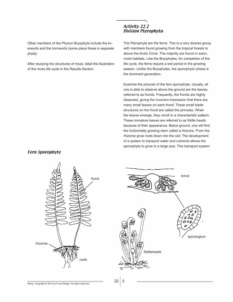
Examine the pictures of the fern sporophyte. Usually, all
one is able to observe above the ground are the leaves,
referred to as fronds. Frequently, the fronds are highly
dissected, giving the incorrect impression that there are
many small leaves on each frond. These small blade
structures on the frond are called the pinnules. When
the leaves emerge, they unroll in a characteristic pattern.
These immature leaves are referred to as fiddle heads
because of their appearance. Below ground, one will find
the horizontally growing stem called a rhizome. From the
rhizome grow roots down into the soil. The development
of a system to transport water and nutrients allows the
sporophyte to grow to a large size. This transport system
Fern Sporophyte
frond
rhizome
roots
fiddleheads
sorus
sporangium

422Ramp. Copyright © 2012 by F.one Design. All rights reserved.
is referred to as the vascular system and is composed of
two principle tissues: xylem to transport water and miner-
als from the roots and phloem to transport sugar nutrients
from the leaves. Vascular tissues also provide structural
support to the plant.
On the under side of the fronds, you frequently find
structures that appear as dark spots or, in some species,
as rows or mats. These are the locations where spore-pro-
ducing structures are located. These structures are called
the sori (singular = sorus). These are clusters of spore
producing sacs called sporangia. Around the outside of
each sporangium is a layer of cells with thick walls called
the annulus. These cells help to open the sporangium and
disperse the mature spores. The sporangia are connected
to the center of the sorus by stalks. In many species, a
flap of tissue covers the sporangia of a sorus. This flap is
called the indusium and helps protect the sporangia as
they mature.
After the spores are released, when favorable conditions
occur they will germinate and grow into the gametophyte
plant. Initially the gametophyte is filamentous but soon
grows into a somewhat heart shaped plant, but remains
only a few cell layers thick. The gametophyte is green and
photosynthetic. On the bottom side of the gametophyte
are hairlike cells called rhizoids which extend down into
the substrate to anchor the gametophyte and help absorb
water and nutrients. Examine the pictures of fern gameto-
phytes and answer the questions in the Results Section.
On the lower surface of the gametophyte, specialized
structures are formed to produce the sperm and the eggs.
The sperm-producing structure is called the antheridium;
the egg producing structure is called the archegonium.
Depending on the species, the gametophyte may produce
both antheridia and archegonia or may only produce one
type. When both are produced on a single gametophyte,
the archegonia are usually found near the notch of the
‘heart’. The antheridia are usually found among the rhi-
zoids, closer to the bottom of the heart shape .
When the flagellated sperm are mature, they are released
and swim through a film of water to an archegonium. There
they will swim down the central canal to fertilize the egg.
The newly formed zygote will grow to form a new sporo-
phyte. Initially, the sporophyte remains attached to the
gametophyte, using it for nutrition. However, very shortly,
the sporophyte develops roots and grows independently of
the gametophyte. The gametophyte will eventually wither
and die. Examine the micrographs of young sporophytes.
Any young sporophytes will grow out from the underside of
the gametophyte, usually emerging somewhere around its
‘notch.’
Divisions with similar life cycles include the Lycophyta and
Equisetophyta.
Fern Gametophyte
antheridium
sperm
archegonium
gametophyte
young
sporophyte
protonema
spore
gametophyte
522Ramp. Copyright © 2012 by F.one Design. All rights reserved.
Activity 22.3Division Coniferophyta
The Coniferophyta includes the pines, firs, and spruces.
These plants are found throughout the world, but are
especially abundant in cooler climates. The sporophyte
phase is dominant in the life cycle, producing plants which
are the largest in the world (the California Redwoods,
Sequoia sempervirens, reach heights of 340 feet).
Look at the diagram of the stem cross section shown here.
This is part of the sporophyte generation. The axis of the
branch and stem is mostly vascular tissue (secondary
xylem and secondary phloem). These tissues have been
produced by a layer of dividing cells called the vascu-
lar cambium, located between the bark and the wood.
Secondary xylem is produced toward the center, creating
the wood, while secondary phloem is produced toward the
outside, creating part of the bark. Seasonal variations in
growth create the annual rings seen in wood.
Examine one of the pictures that shows a branch with
needles. At the base of the needles is a small scale
called the scale leaf. From the axis of this scale leaf is the
structure called the spur shoot which actually produces
the needles. Most conifer needles are evergreen (retain
their leaves year round) rather than deciduous (lose their
leaves annually). Can you think of advantages of an ever-
green habit? Disadvantages? Place your thoughts in the
Results Section.
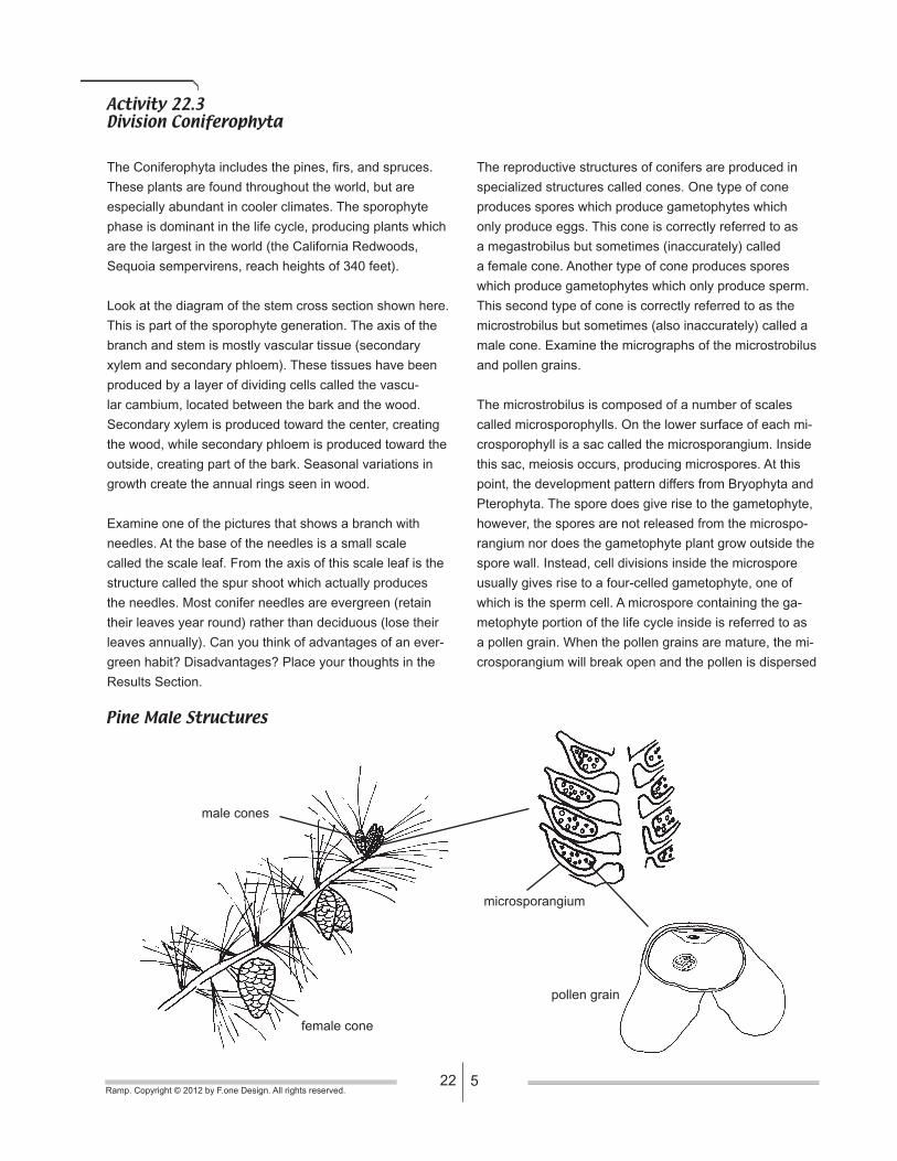
The reproductive structures of conifers are produced in
specialized structures called cones. One type of cone
produces spores which produce gametophytes which
only produce eggs. This cone is correctly referred to as
a megastrobilus but sometimes (inaccurately) called
a female cone. Another type of cone produces spores
which produce gametophytes which only produce sperm.
This second type of cone is correctly referred to as the
microstrobilus but sometimes (also inaccurately) called a
male cone. Examine the micrographs of the microstrobilus
and pollen grains.
The microstrobilus is composed of a number of scales
called microsporophylls. On the lower surface of each mi-
crosporophyll is a sac called the microsporangium. Inside
this sac, meiosis occurs, producing microspores. At this
point, the development pattern differs from Bryophyta and
Pterophyta. The spore does give rise to the gametophyte,
however, the spores are not released from the microspo-
rangium nor does the gametophyte plant grow outside the
spore wall. Instead, cell divisions inside the microspore
usually gives rise to a four-celled gametophyte, one of
which is the sperm cell. A microspore containing the ga-
metophyte portion of the life cycle inside is referred to as
a pollen grain. When the pollen grains are mature, the mi-
crosporangium will break open and the pollen is dispersed
Pine Male Structures
male cones
female cone
microsporangium
pollen grain

622Ramp. Copyright © 2012 by F.one Design. All rights reserved.
by wind. Make your own drawing of these structures in the
Results Section.
Next examine the pictures and the whole specimen of
the megastrobilus. Like the microstrobilus, the megastro-
bilus is composed of a number of scales. Here they are
referred to as ovuliferous scales. On the upper surface
of each ovuliferous scale is a sac, which at its center, will
produce a single megaspore by meiosis. Surrounding
and protecting this spore are several layers of tissue; the
outermost layers make up the integument. This structure,
a megaspore surrounded by tissue layers including the
integument, is called an ovule. When an ovule matures, it
is referred to as a seed.
Inside the spore, the gametophyte portion of the life
cycle will develop. As with the microspores, the gameto-
phyte will not be released from inside the spore wall. The
gametophyte will grow and eventually produce two egg
containing structures, the archegonia. Generally, only one
of these archegonia will be fertilized to produce a new
sporophyte generation. Make your own diagram of an
ovule in the Results Section.
When pollen, dispersed by the wind, is deposited near an
ovule, a specialized cell of the pollen’s gametophyte will
grow out of the pollen grain and produce a pollen tube
which will eventually reach the gametophyte inside the
ovule. Here, the sperm nucleus, which is located near the
end of the pollen tube, will be released and fuse with the
egg cell. In conifers, the maturation of a fertilized ovule,
now the seed, is a prolonged process taking as long as
two years. Examine the megastrobilus cones available
and try to determine its stage of development using the
diagram of a conifer’s life cycle.
Other divisions with a life cycles similar to the pines are
the Cycaophyta, Ginkgophyta and Gnetophyta.
Pine Female Structures
male cones
female cone
ovuliferous
scale
female cone
bract
ovule / seed
embryo
ovule

722Ramp. Copyright © 2012 by F.one Design. All rights reserved.
Activity 22.4Division Magnoliophyta
The Magnoliophyta include all the flowering plants. This
group is the most recently evolved group of plants as
well as the most successful. Flowering plants are found
throughout the world. The flower of flowering plants is a
reproductive structure which is much more efficient than
the cone structures of conifers however the basic struc-
tures are very similar.
The sporophyte is the dominant phase of the flowering
plant’s life cycle. All of these sporophytes will produce
xylem and phloem but only some will produce secondary
xylem (wood) and secondary phloem (part of the bark).
Those plants which do not produce wood are referred to
as herbaceous. The leaves of flowering plants are usually
flattened and blade-like, unlike the needles of conifers.
Frequently these leaves are deciduous, shed each year.
The reproductive structures of these plants are located
within the flower. The flower also contains some non-re-
productive structures, the sepals and petals.
In flowering plants, the development of pollen from micro-
spores is much the same as in the conifers. This occurs
in sacs called anthers. Just as with conifers, ovules are
produced but here in the flowering plants, the ovules are
enclosed within another structure called the ovary. After
fertilization, the ovules mature into the seeds and the
ovary matures into the fruit.
A unique characteristic of the flowering plants is that when
fertilization occurs, two sperm are actually delivered by the
pollen tube. One of these sperm will fertilize the egg cell in
the megagametophyte, the other will fuse with two other
cells in the gametophyte to produce a tissue with three
sets of chromosomes, said to be triploid or 3N. This 3N
tissue is called the endosperm and will initially serve as a
nutrient source for the developing embryo. This process is
referred to as double fertilization.
A more detailed examination of reproduction in flowering
plants is found in other lab modules.
Sporophyte of Flowering Plants
flower

822Ramp. Copyright © 2012 by F.one Design. All rights reserved.
Results SectionName _______________________Lab Exercise 22
Activity 22.1Division Bryophyta
1. _____________________
5. _____________________4. _____________________
2. _____________________
3. _____________________
gametophyte
operculum
(chamber)sporophyte

922Ramp. Copyright © 2012 by F.one Design. All rights reserved.
Activity 22.2Division Pterophyta
1. _____________________
5. _____________________
6. _____________________
3. _____________________
sperm
2. _____________________
4. _____________________

1022Ramp. Copyright © 2012 by F.one Design. All rights reserved.
Activity 22.3Division Coniferophyta
3. _____________________
5. _____________________
2. _____________________
1. _____________________
4. _____________________
(type of cone)
(type of cone)
(center structure)
(entire round structure)