lysophosphatidylcholine potentiates phenylephrine...
TRANSCRIPT

JPET # 97964
1
Lysophosphatidylcholine Potentiates Phenylephrine Responses in Rat
Mesenteric Arterial Bed through Modulation of Thromboxane A2
Rui Zhang, Brian Rodrigues and Kathleen M. MacLeod
Division of Pharmacology and Toxicology
Faculty of Pharmaceutical Sciences
University of British Columbia, Vancouver, Canada (RZ, BR, KMM)
JPET Fast Forward. Published on January 4, 2006 as DOI:10.1124/jpet.105.097964
Copyright 2006 by the American Society for Pharmacology and Experimental Therapeutics.
This article has not been copyedited and formatted. The final version may differ from this version.JPET Fast Forward. Published on January 4, 2006 as DOI: 10.1124/jpet.105.097964
at ASPE
T Journals on July 30, 2018
jpet.aspetjournals.orgD
ownloaded from

JPET # 97964
2
Running title – Vasoactive effects of lysophosphatidylcholine
Address for correspondence:
Kathleen M. MacLeod, PhD
Faculty of Pharmaceutical Sciences, University of British Columbia
2146 East Mall, Vancouver, BC, Canada V6T 1Z3
Tel. (604) 822-3830, Fax (604) 822-3035, e-mail: [email protected]
Number of Text pages (including abstract): 15
Number of Tables: 0
Number of Figures: 7
Number of References: 39
Number of words in the “Abstract”: 249
Number of words in the “Introduction”: 435
Number of words in the “Discussion”: 943
Abbreviations:
AA, arachidonic acid; Ach, acetylcholine; COX, cyclooxygenase; DRC, dose response
curve; EIA, enzyme-immunoassay; EDR, endothelium-dependent relaxation; EDHF,
endothelium-dependent relaxing factor; LDL, low density lipoprotein; L-NMMA, NG-
monomethyl-L-arginine methyl ester; LPC, lysophosphatidylcholine; NO, nitric oxide;
NOS, nitric oxide synthase; MAB, mesenteric arterial bed; PC, phosphatidylcholine; PE,
phenylephrine; PLA2, phospholipase A2; PLC, phospholipase C; PLD, phospholipase D;
PGI2, prostacyclin; TxA2, thromboxane A2; TxB2, thromboxane B2
Recommended section assignment: Cardiovascular
This article has not been copyedited and formatted. The final version may differ from this version.JPET Fast Forward. Published on January 4, 2006 as DOI: 10.1124/jpet.105.097964
at ASPE
T Journals on July 30, 2018
jpet.aspetjournals.orgD
ownloaded from

JPET # 97964
3
Abstract
Lysophosphatidylcholine (LPC) plays important physiological and pathophysiological
roles in the cardiovascular system. Despite this, there is little information about its effects
on vasoreactivity of resistance vessels. The present study was designed to characterize the
effects of LPC in the isolated perfused rat mesenteric arterial bed (MAB) and to investigate
the underlying mechanisms of the changes it produced. Perfusion with 10 µM LPC for 40
min did not significantly affect basal perfusion pressure or reactivity of MAB to the alpha1-
adrenoceptor agonist phenylephrine (PE), but almost completely abolished the maximum
endothelium-dependent relaxation (EDR) to acetylcholine (Ach), reducing it from 93±5%
to 7±4% (p<0.001). Following washout of LPC for 60 min, the vasodilator response to Ach
partially recovered, while the vasoconstrictor response to PE was markedly enhanced, the
pD2 value increasing from 7.50±0.04 to 8.13±0.15, and maximum response to 199±24% of
control (p<0.001). Pretreatment with either indomethacin, a non-selective inhibitor of
cyclooxygenase, or SQ-29548, a selective thromboxane receptor antagonist, completely
prevented the potentiation of the PE response following washout of LPC. In untreated
MABs, only the highest concentration of PE produced a significant increase in
thromboxane A2 (TxA2) production (assessed by enzyme-immunoassay of TxB2 levels).
This was prevented by perfusion with LPC, but was significantly increased after LPC
washout. The basal release of TxA2 was not modified by LPC. These results demonstrate
that LPC exerts both immediate and residual effects on the reactivity of the rat MAB, and
that these effects are at least partially due to modification of PE-induced TxA2 production.
This article has not been copyedited and formatted. The final version may differ from this version.JPET Fast Forward. Published on January 4, 2006 as DOI: 10.1124/jpet.105.097964
at ASPE
T Journals on July 30, 2018
jpet.aspetjournals.orgD
ownloaded from

JPET # 97964
4
Introduction
Lysophosphatidylcholine (1-acyl-sn-glycero-3-phosphocholine, LPC) is an important
lysophospholipid that is generated as a breakdown product of phosphatidylcholine by
phospholipase A2 (PLA2). Present in cell membranes and oxidized lipoproteins, it plays
important physiological and pathophysiological roles in both humans and animals. For
instance, LPC is responsible for the delivery of fatty acids and choline to tissues (Xu,
2002). On the other hand, elevated levels of LPC have been linked to the cardiovascular
complications associated with diabetes, atherosclerosis and ischemia (Takahara et al., 1997;
Shi et al., 1999; Sonoki et al., 2003).
In the cardiovascular system, LPC adversely modifies vasoreactivity in conduit arteries.
Hence, in large artery ring preparations, LPC can impair endothelium-dependent relaxation
(EDR) (Fukao et al., 1995; Froese et al., 1999; Vuong et al., 2001), and enhance agonist-
induced contractions. The latter effect includes potentiation of contractile responses
induced by high-K+, UK14,304 (Suenaga et al., 2003) and angiotensin II (Galle et al., 2003;
Suenaga and Kamata, 2003). These effects of LPC may be partially explained by the
results of studies in cultured endothelial and vascular smooth muscle cells, where LPC was
shown to activate protein kinase C (Kohno et al., 2001), increase intracellular Ca2+
(Terasawa et al., 2002) and activate or inhibit MAP kinase (Bassa et al., 1999; Rikitake et
al., 2000). In addition to its role in modulating vascular contractility, it is generally
accepted that LPC is the major pathological component of oxidized low density lipoprotein
(LDL) in promoting atherosclerosis (Takahara et al., 1997; Sonoki et al., 2003).
This article has not been copyedited and formatted. The final version may differ from this version.JPET Fast Forward. Published on January 4, 2006 as DOI: 10.1124/jpet.105.097964
at ASPE
T Journals on July 30, 2018
jpet.aspetjournals.orgD
ownloaded from

JPET # 97964
5
Several lines of evidence also suggest a vaso-protective role for LPC. In rabbits, LPC
produces potent endothelium-dependent vascular smooth muscle relaxation, both in vivo
and in vitro (Menon and Bing, 1991; Wolf et al., 1991). In umbilical vein endothelial cells,
LPC has been shown to enhance endothelial expression of eNOS and cyclooxygenase-2
(COX-2), leading to increased synthesis of nitric oxide (NO) and prostacyclin (PGI2)
(Zembowicz et al., 1995a; Zembowicz et al., 1995b). NO and PGI2 act synergistically to
block monocyte adhesion, smooth muscle cell proliferation, platelet activation, and
vasoconstriction (Vane and Botting, 1995; Naseem, 2005).
Although it has been extensively studied, the contribution of LPC in regulating vascular
resistance has not been completely elucidated, as the majority of previous studies have used
either large blood vessels or isolated cells. The mesenteric arterial bed (MAB) is an
important effector organ regulating blood pressure, with small structural and functional
changes eliciting significant alterations in peripheral resistance (Caveney et al., 1998). The
objective of the present study was to determine the vasoactive effects of LPC in the isolated
perfused MAB, and to investigate the underlying mechanisms of the changes it produced.
This article has not been copyedited and formatted. The final version may differ from this version.JPET Fast Forward. Published on January 4, 2006 as DOI: 10.1124/jpet.105.097964
at ASPE
T Journals on July 30, 2018
jpet.aspetjournals.orgD
ownloaded from

JPET # 97964
6
Methods
Animals. The investigation conforms to the guidelines of the Canadian Council on
Animal Care. Male Wistar rats were obtained from the Animal Care Center, University of
British Columbia. Animals were housed under a 12 h light: 12 h dark regime, and given
free access to standard rat chow (PMI Feeds, Richmond, VA), and tap water.
Isolated perfused rat mesenteric arterial bed. Male Wistar rats (300-400 g) were
anesthetized with sodium pentobarbital (60mg/kg, i.p.), and the mesenteric arterial bed
(MAB) was isolated as described previously (He and MacLeod, 2002). Briefly, the
abdominal cavity was opened, and the superior mesenteric artery was cannulated through
an incision at its confluence with the dorsal aorta. The whole MAB was then separated by
cutting close to the intestinal border. The MAB was flushed with heparinized warm Krebs–
bicarbonate buffer with the following composition: 113 mM NaCl, 4.7 mM KCl, 11.5 mM
glucose, 1.2 mM MgSO4, 2.5 mM CaCl2, 1.2 mM KH2PO4, and 25.0 mM NaHCO3. The
pH of the buffer following saturation with a 95% O2-5% CO2 gas mixture was 7.4.
Subsequent to flushing, the MAB was transferred into a jacketed organ chamber, and
perfused through the cannula with Krebs buffer maintained at 37°C, and gassed with 95%
O2-5% CO2. Perfusion rate was kept constant at 3 ml/min using a peristaltic pump (Buchler
Instruments, Buchler Fort Lee, N.J.). The perfusate flowed out through the cut ends of the
mesenteric arterial bed. Vascular responses were detected as changes in perfusion pressure,
and this was continuously measured and recorded using a pressure transducer (PD23ID,
Gould, Statham, Calif.) placed between the perfusate and the cannula, and connected to a
Grass polygraph (model 79D, Grass Instruments, Quincy, Mass.). The perfused MAB was
This article has not been copyedited and formatted. The final version may differ from this version.JPET Fast Forward. Published on January 4, 2006 as DOI: 10.1124/jpet.105.097964
at ASPE
T Journals on July 30, 2018
jpet.aspetjournals.orgD
ownloaded from

JPET # 97964
7
allowed to stabilize for 40 min before four bolus injections of KCl (0.4 mmol). Perfusion
pressure was allowed to return to baseline following each injection of KCl. After further
equilibration for 40 min, dose response curves (DRCs) to various agonists were performed.
Vasoconstrictor responses. To determine the effects of LPC on vasoconstrictor
responses of the MAB, bolus injections of phenylephrine (PE) (0.9-300 nmol) or KCl (50-
800 µmol) were given. These first DRCs to PE or KCl served as controls. Once perfusion
pressure returned to baseline, palmitoyl-LPC (0.1-10 µM) was added to the perfusion
buffer. In preliminary experiments, none of these concentrations of LPC per se produced
any observable change in contractility. In all subsequent experiments, 10 µM LPC was
used. After 40 min of perfusion with LPC, the second PE/KCl DRC was constructed in the
presence of LPC. The tissues were then switched back to normal Krebs buffer, and
perfused for one hour, followed by construction of a third PE/KCl DRC. The 60-min
washout was designed to test whether the expected effect of LPC was reversible or not.
Perfusion pressure was allowed to return to baseline between each PE/KCl injection. In
order to test if the vascular responses are affected by time, a control experiment consisting
of three consecutive DRCs to PE and KCl were done without the addition of LPC.
To determine the potential contribution of NO and thromboxane A2 (TxA2) to the
observed modulatory effects of LPC on PE responses, the LPC perfusion and washout
procedures described above were performed in the presence of a NOS inhibitor (NG-
monomethyl-L-arginine methyl ester; L-NMMA, 300 µM), a COX inhibitor (indomethacin;
20 µM), or a TxA2 receptor antagonist (SQ-29548; 0.3 µM). All inhibitors were added to
This article has not been copyedited and formatted. The final version may differ from this version.JPET Fast Forward. Published on January 4, 2006 as DOI: 10.1124/jpet.105.097964
at ASPE
T Journals on July 30, 2018
jpet.aspetjournals.orgD
ownloaded from

JPET # 97964
8
the perfusate following the first DRC, and kept present throughout the second and third
DRCs.
Vasodilatory response. To assess the effects of LPC on endothelium-dependent
relaxation (EDR), MABs were precontracted with submaximal concentrations of PE (1-3
µM). Subsequently, increasing concentrations of acetylcholine (Ach; 3 nmol-0.3 µM) were
administered until maximal relaxation was attained. MABs were then perfused for 40 min
with 10 µM LPC, and the response to PE and Ach repeated. A third Ach response was
obtained after washout of LPC for 1 hour. Control experiments consisted of three
consecutive Ach-induced responses in the absence of LPC.
Effluent collection. To measure the content of thromboxane B2 (TxB2), a stable
metabolite of TxA2, in the MAB effluent, samples were collected for 2 min on six different
occasions as follows: before the first application of PE; during the first PE-DRC at each PE
dose; following the 40 min perfusion with LPC; during the second PE-DRC at each PE
dose; after the 60 min washout of LPC; during the third PE-DRC at each PE dose. Samples
were stored at -70ºC, until assayed.
Enzyme-immunoassay (EIA) of TxB2. The collected perfusates were extracted using
Solid Phase Extraction C-18 Cartridges (Cayman Chemical, Michigan, USA) following
cartridge activation by 5 ml of methanol and 5 ml of ultra-pure water. A 0.5 ml aliquot of
each sample was acidified to ~ pH 4.0, and loaded onto the cartridge. The cartridge was
washed with 5 ml of ultra-pure water, followed by 5 ml hexane. The TxB2 fraction was
eluted with 5 ml ethyl acetate containing 1% methanol. The eluate was dried under a
stream of nitrogen, and reconstituted with 0.5 ml EIA buffer. The concentration of TxB2 in
This article has not been copyedited and formatted. The final version may differ from this version.JPET Fast Forward. Published on January 4, 2006 as DOI: 10.1124/jpet.105.097964
at ASPE
T Journals on July 30, 2018
jpet.aspetjournals.orgD
ownloaded from

JPET # 97964
9
the eluate was determined by EIA, using a commercially available kit (Cayman Chemical,
Michigan, USA).
Chemicals. L-phenylephrine hydrochloride, 1-palmitoyl-sn-glycero-3-phosphocholine
and indomethacin were obtained from Sigma Chemical Co. (St. Louis, Mo.). L-NMMA
(NG-monomethyl-L-arginine methyl ester) and SQ-29548 ([1S-[1a,2a(Z),3a,4a]]-7-[3-[[2-
[(phenylamino) carbonyl]hydrazino] methyl]-7-oxabicyclo [2.2.1]hept-2-yl]-5-heptanoic
acid) were purchased from BIOMOL International, L.P. (Plymouth Meeting, PA). Stock
solutions of PE (0.01 M), LPC (0.01 M) and L-NMMA (0.1 M) were prepared in distilled
water. Indomethacin and SQ-29548 were dissolved in 100% ethanol, and prepared as stock
solutions of 0.1 M and 0.1 mM, respectively. Solutions of PE and indomethacin were made
fresh. LPC, and all inhibitors were further diluted to the required concentration in the
perfusate reservoir. The final ethanol concentrations (0.03-0.3%, v/v) were without effect
on contractile responses.
Data analysis. KCl-induced vasoconstrictor responses were expressed as the absolute
increase in perfusion pressure. PE responses were expressed as a percentage of the
maximum response of the first PE-DRC. The negative log of the PE concentration
producing 50% of maximum response (pD2) was obtained by nonlinear regression analysis
of individual DRCs using Graphpad Prism version 4.0 (Graphpad Software, San Diego,
Calif).
All data are presented as mean ± S.E.M. Data were analyzed for significant differences
using NCSS. Student’s unpaired t test was used for comparisons between two means.
One-way ANOVA followed by Newman-Keul’s test was used for comparison of more than
two means. Two-way ANOVA, using the general linear model approach (repeated
This article has not been copyedited and formatted. The final version may differ from this version.JPET Fast Forward. Published on January 4, 2006 as DOI: 10.1124/jpet.105.097964
at ASPE
T Journals on July 30, 2018
jpet.aspetjournals.orgD
ownloaded from

JPET # 97964
10
measures) followed by Newman-Keul’s test was used for comparisons between PE-DRCs
and KCl-DRCs. P<0.05 was considered statistically significant.
This article has not been copyedited and formatted. The final version may differ from this version.JPET Fast Forward. Published on January 4, 2006 as DOI: 10.1124/jpet.105.097964
at ASPE
T Journals on July 30, 2018
jpet.aspetjournals.orgD
ownloaded from

JPET # 97964
11
Results
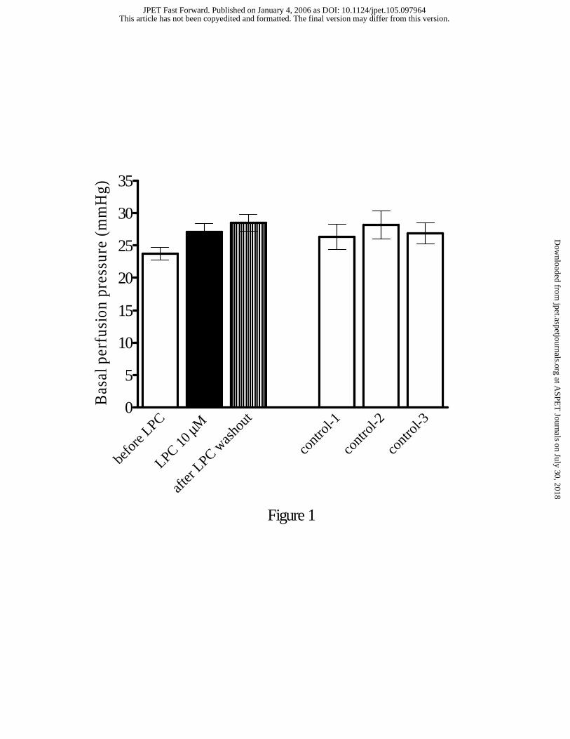
Effect of LPC on basal perfusion pressure. To examine the effect of LPC on basal
perfusion pressure, isolated MABs were incubated with varying concentrations of LPC
(0.1-10 µM). Perfusion pressure was measured during a 40 min perfusion with LPC, or 1
hour after washout. In Fig. 1 (left panel), the effects of 10 µM LPC on basal perfusion
pressure are shown. LPC had no significant effect on perfusion pressure, either during
incubation or following washout, suggesting that this lysophospholipid has no direct effect
on the contractility of MAB. Basal MAB perfusion pressure in untreated control tissues
remained unchanged throughout the experimental period (Fig. 1, right panel).
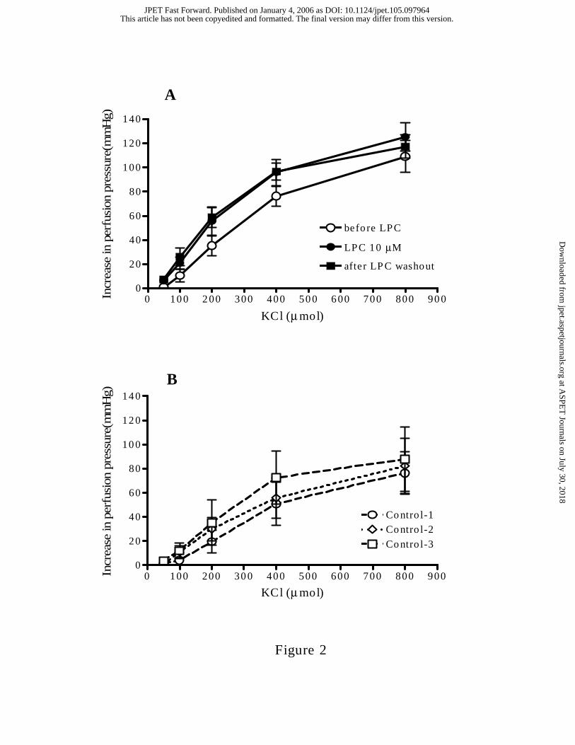
KCL-induced contractile responses following LPC. Pressor responses of the MAB to
bolus injections of KCl (50-800 µmol) were not significantly altered, either during
perfusion with 10 µM LPC, or after LPC washout (Fig. 2A). In untreated control tissues,
KCl-DRCs remained constant over time (Fig. 2B).
Effects of LPC on PE-induced contractile responses. Bolus injection of PE (0.9-300
nmol) produced a concentration-dependent increase in perfusion pressure that did not
significantly change over time (Fig. 3A). The response to PE was unaffected by prior
perfusion with LPC for either 40 (Fig. 3B) or 150 (data not shown) min. Interestingly,
following washout of LPC for 1 hour, the PE response was markedly enhanced (Fig. 3B).
The maximum response to PE increased to 199±24% of control (P<0.001) following LPC
washout, while the PE pD2 value increased from 7.50±0.04 to 8.13±0.15 (P<0.001).
Further increasing the dose of PE above 90 nmol resulted in a decline in the contractile
response.
This article has not been copyedited and formatted. The final version may differ from this version.JPET Fast Forward. Published on January 4, 2006 as DOI: 10.1124/jpet.105.097964
at ASPE
T Journals on July 30, 2018
jpet.aspetjournals.orgD
ownloaded from

JPET # 97964
12
Involvement of NO and TxA2 in mediating the direct effects of LPC perfusion. Prior
to LPC perfusion, Ach (0.1-0.3 µmol) produced a maximum relaxation of 93±5% of the
response to PE (Fig. 4, left panel). The Ach response remained unchanged over time (Fig.
4, right panel). LPC perfusion almost completely abrogated the response to Ach, reducing
the maximum relaxation to 7±4% (Fig. 4, left panel), suggesting that LPC is diminishing
the actions of both NO and EDHF. In a separate experiment, we tested the effects of a
NOS inhibitor on PE responses. As expected, pretreatment with L-NMMA significantly
enhanced the PE response (Fig. 5A), with both an increase in the maximum response and
pD2 value (control, 7.03±0.45; L-NMMA, 7.49±0.17; P<0.001) being apparent. However,
in the presence of LPC, the enhancement of the PE response by L-NMMA was prevented
(Fig. 5A). These data suggest that in addition to suppressing NO, LPC may be exerting
other actions to limit vasoconstrictor release or responsiveness to PE.
TxA2 is a major vasoconstrictor prostanoid implicated in regulation of vascular tone in
response to α1-adrenoceptor stimulation in the MAB. To investigate the involvement of
TxA2 in mediating PE responses, we measured levels of TxB2, the stable metabolite of
TxA2. Basal release of TxB2 was stable over time. In untreated MAB, only the highest
dose of PE tested (300 nmol) produced an increase in TxA2 production (Fig. 6). LPC
perfusion for 40 min had no effect on basal levels of TxB2 but suppressed its enhancement
by PE (Fig. 6), suggesting that LPC prevents the ability of PE to increase TxA2 production.
Involvement of NO and TxA2 in mediating the indirect effects (after washout) of
LPC. Washout of LPC induced partial recovery of the Ach response (Fig. 4). Despite this
fractional recovery, contractile responses to PE were augmented (Fig. 3B), suggesting that
This article has not been copyedited and formatted. The final version may differ from this version.JPET Fast Forward. Published on January 4, 2006 as DOI: 10.1124/jpet.105.097964
at ASPE
T Journals on July 30, 2018
jpet.aspetjournals.orgD
ownloaded from

JPET # 97964
13
amplification of vasoconstrictor pathways, in addition to reduction of vasodilators such as
NO, may be responsible for this effect. To investigate this possibility, following washout
of LPC, a DRC to PE was performed in the presence of L-NMMA (Fig. 5B). Interestingly,
even in the presence of the NOS inhibitor, the response to PE was shifted to the left
following LPC washout (pD2 value in the presence of L-NMMA alone, 7.54±0.07;
following LPC washout and in the presence of L-NMMA, 8.16±2.05; P<0.05). These data
suggest that the enhancement of PE response following LPC washout is mediated by
mechanisms in addition to NO inhibition.
To further investigate mechanisms facilitating the enhanced PE response after LPC
washout, TxB2 levels in the perfusion medium were measured. Interestingly, TxB2 levels
only increased following LPC washout (Fig. 6), suggesting a role for this vasoconstrictor in
the magnified response to PE. MABs were then pretreated with either a COX inhibitor or a
TxA2 receptor antagonist. Both indomethacin (pD2 value after LPC washout, 8.24±0.07;
LPC washout plus indomethacin, 7.74±0.25; P<0.05) and SQ-29548 (pD2 value after LPC
washout, 8.24±0.07; LPC washout plus SQ-29548, 7.73±0.10; P<0.05) completely
prevented the enhancement of the PE DRC following LPC washout (Fig. 7). Neither
indomethacin nor SQ-29548 significantly changed PE-induced vasoconstriction by
themselves (data not shown).
This article has not been copyedited and formatted. The final version may differ from this version.JPET Fast Forward. Published on January 4, 2006 as DOI: 10.1124/jpet.105.097964
at ASPE
T Journals on July 30, 2018
jpet.aspetjournals.orgD
ownloaded from

JPET # 97964
14
Discussion
To our knowledge, the present study is the first to investigate the effects of LPC on the
reactivity of a perfused resistance artery bed. As reported previously in conduit blood
vessels, LPC perfusion of MAB caused impairment in Ach-induced maximum relaxation.
The novel finding in the present study was the potentiation of PE-induced contractile
responses following washout of LPC, an effect likely related to increased production of
TxA2.
The balance between vasodilators (NO; endothelium-dependent hyperpolarizing factor,
EDHF; PGI2) and vasoconstrictors (TxA2; endothelin-1) is pivotal in the regulation of
vascular tone. As most of these vasoactive factors originate from vascular endothelium, we
examined the effects of LPC on endothelial function. Measurement of EDR supported the
established view that LPC inhibits EDR, likely through inactivation of NO and EDHF
(Cowan and Steffen, 1995; Froese et al., 1999; Rikitake et al., 2000). Impaired EDR is
normally linked to potentiated vasoconstrictor responses, an observation we confirmed in
preliminary studies, in which various methods were used to denude MAB of endothelial
cells, including perfusion with air (Tatchum-Talom and Atkinson, 1997), distilled water,
sodium deoxycholate (Cusma-Pelogia et al., 1993), and CHAPS (McCulloch and Randall,
1996). Mechanical denudation of endothelium, which was associated with loss of the
relaxant response to Ach, caused an enhancement of PE responses (data not shown).
However, the impaired Ach-induced EDR caused by LPC perfusion was not accompanied
by an increased response to PE. Other studies using aortic rings from normal rats have also
demonstrated similar effects of LPC, that is, reduced EDR with no significant change in PE
This article has not been copyedited and formatted. The final version may differ from this version.JPET Fast Forward. Published on January 4, 2006 as DOI: 10.1124/jpet.105.097964
at ASPE
T Journals on July 30, 2018
jpet.aspetjournals.orgD
ownloaded from

JPET # 97964
15
responses (Ceylan et al., 2004). These data suggest that even in the presence of NO/EDHF
inhibition by LPC, vasoconstrictor responsiveness or release may also be inhibited by LPC,
thus preventing augmentation in responses to PE. This possibility is supported by the
observation that LPC prevented amplification of PE responses in the presence of L-
NMMA, and reduced the PE-induced production of TxA2.
Unexpectedly, the response to PE was dramatically potentiated only following removal
of LPC. A time-dependent mechanism can be ruled out, as prolonged perfusion with LPC
(150 min) did not produce any potentiation of PE responses. The enhancement of the PE
response could be blocked by both indomethacin and SQ-29548, suggesting an important
contribution of vasoconstrictor prostanoids, which was confirmed by the finding of
potentiation of PE-induced TxA2 production. At present, the mechanism for this rebound
production of excessive TxA2 is not known, nor is it clear whether this effect is global, or
localized to the MAB. The contribution of time in producing the potentiated contractile
responses and TxA2 production in response to PE can be ruled out, as these were stable
over time in the absence of LPC. These effects were unlikely to be due solely to continued
endothelial impairment, as Ach-induced relaxation had partially recovered. Furthermore,
the enhancement of PE responses produced by mechanical removal of the endothelium
could not be abolished by pretreatment with SQ-29548 (data not shown).
As a potent inducer of platelet aggregation, vasoconstriction and bronchoconstriction,
TxA2 has been implicated in vascular pathogenesis (Dogne et al., 2004). Although the
major source of TxA2 is platelets, this vasoconstrictor can also be produced within the
vascular walls, by both endothelial (Ally and Horrobin, 1980) and smooth muscle cells
(Shiokoshi et al., 2002). The mechanism of TxA2 synthesis includes phospholipid
This article has not been copyedited and formatted. The final version may differ from this version.JPET Fast Forward. Published on January 4, 2006 as DOI: 10.1124/jpet.105.097964
at ASPE
T Journals on July 30, 2018
jpet.aspetjournals.orgD
ownloaded from
JPET # 97964
16
hydrolysis by PLA2, release of AA, and metabolism to TxA2 by the COX-TxA2 synthase
pathway. Alpha1-receptor activation initiates phospholipid hydrolysis, and release of TxA2
(Terzic et al., 1993; Nishio et al., 1996; Ruan et al., 1998; Bolla et al., 2002; Parmentier et
al., 2004). To our knowledge, an effect of LPC on α1-adrenoceptor mediated TxA2
production in the vasculature has not previously been reported. However, our observation
that LPC abrogated the increase in TxA2 production produced by PE in the MAB is
consistent with a previous study in platelets, in which an inhibitory effect of LPC on
agonist-induced TxA2 production was observed (Yuan et al., 1996). Although we are not
aware of any studies showing potentiation of TxA2 production by LPC, an interaction
between these two agents has previously been suggested. For instance, LPC was found to
enhance the ability of TxA2 to increase vascular smooth muscle cell proliferation (Koba et
al., 2000). Taken together, these findings strongly suggest that LPC is able to alter the
biosynthesis of TxA2, as well as its downstream signaling. Details of the mechanism of
these modulatory effects need to be further investigated. Irrespective of the mechanism(s),
the increased release of TxA2 could be a major contributor towards hypertensive (Seeger et
al., 1989), thrombotic and ischemic diseases (Ally and Horrobin, 1980; Muller, 1991).
It is tempting to speculate that conditions that evoke an increase in LPC levels initially
trigger compensatory mechanisms to neutralize its injurious effects. Thus, the suppression
of PE-induced TxA2 production could compensate for the endothelium dysfunction. Wu
and coworkers have also suggested that LPC is a double-faced molecule, which can
produce both vasoprotective and proatherogenic mechanisms (Zembowicz et al., 1995a;
Zembowicz et al., 1995b). An additional caveat is the suggestion that residual effects of
This article has not been copyedited and formatted. The final version may differ from this version.JPET Fast Forward. Published on January 4, 2006 as DOI: 10.1124/jpet.105.097964
at ASPE
T Journals on July 30, 2018
jpet.aspetjournals.orgD
ownloaded from

JPET # 97964
17
LPC after washout could arise from its metabolism to lysophosphatidic acid (Tokumura,
2004). Given this complex biology, investigation of the in vivo temporal effects of LPC
under normal and pathological conditions would be beneficial.
In conclusion, our results for the first time demonstrate effects of LPC in the rat
resistance arterial bed, even following washout. These included potentiated PE responses,
and enhanced TxA2 production. Thus, when evaluating the effects of LPC in the
vasculature, both its immediate and residual effects need to be considered. Although
unclear, the mechanism for this residual effect, especially the relationship between LPC and
LPA, is currently being evaluated.
This article has not been copyedited and formatted. The final version may differ from this version.JPET Fast Forward. Published on January 4, 2006 as DOI: 10.1124/jpet.105.097964
at ASPE
T Journals on July 30, 2018
jpet.aspetjournals.orgD
ownloaded from
JPET # 97964
18
References
Ally AI and Horrobin DF (1980) Thromboxane A2 in blood vessel walls and its
physiological significance: relevance to thrombosis and hypertension.
Prostaglandins Med 4:431-438.
Bassa BV, Roh DD, Vaziri ND, Kirschenbaum MA and Kamanna VS (1999)
Lysophosphatidylcholine activates mesangial cell PKC and MAP kinase by
PLCgamma-1 and tyrosine kinase-Ras pathways. Am J Physiol 277:F328-337.
Bolla M, Matrougui K, Loufrani L, Maclouf J, Levy B, Levy-Toledano S, Habib A and
Henrion D (2002) p38 mitogen-activated protein kinase activation is required for
thromboxane- induced contraction in perfused and pressurized rat mesenteric
resistance arteries. J Vasc Res 39:353-360.
Caveney SW, Culhane JM, Taylor DA and Fleming WW (1998) Preparation and
application of an isolated superior mesenteric arterial vascular preparation. J
Cardiovasc Pharmacol 32:721-727.
Ceylan A, Karasu C, Aktan F and Ozansoy G (2004) Simvastatin treatment restores
vasoconstriction and the inhibitory effect of LPC on endothelial relaxation via
affecting oxidizing metabolism in diabetic rats. Diabetes Nutr Metab 17:203-210.
Cowan CL and Steffen RP (1995) Lysophosphatidylcholine inhibits relaxation of rabbit
abdominal aorta mediated by endothelium-derived nitric oxide and endothelium-
derived hyperpolarizing factor independent of protein kinase C activation.
Arterioscler Thromb Vasc Biol 15:2290-2297.
This article has not been copyedited and formatted. The final version may differ from this version.JPET Fast Forward. Published on January 4, 2006 as DOI: 10.1124/jpet.105.097964
at ASPE
T Journals on July 30, 2018
jpet.aspetjournals.orgD
ownloaded from
JPET # 97964
19
Cusma-Pelogia N, Oliveira SF, Nigro D, de Carvalho MH, Scivoletto R and Fortes ZB
(1993) Endothelium inactivation in in vitro perfused vascular beds. Comparison of
methods. J Pharmacol Toxicol Methods 29:157-163.
Dogne JM, Hanson J, de Leval X, Masereel B, Kolh P and Pirotte B (2004) New
developments on thromboxane modulators. Mini Rev Med Chem 4:649-657.
Froese DE, McMaster J, Man RY, Choy PC and Kroeger EA (1999) Inhibition of
endothelium-dependent vascular relaxation by lysophosphatidylcholine: impact of
lysophosphatidylcholine on mechanisms involving endothelium-derived nitric oxide
and endothelium derived hyperpolarizing factor. Mol Cell Biochem 197:1-6.
Fukao M, Hattori Y, Kanno M, Sakuma I and Kitabatake A (1995) Evidence for selective
inhibition by lysophosphatidylcholine of acetylcholine-induced endothelium-
dependent hyperpolarization and relaxation in rat mesenteric artery. Br J Pharmacol
116:1541-1543.
Galle J, Mameghani A, Bolz SS, Gambaryan S, Gorg M, Quaschning T, Raff U, Barth H,
Seibold S, Wanner C and Pohl U (2003) Oxidized LDL and its compound
lysophosphatidylcholine potentiate AngII-induced vasoconstriction by stimulation
of RhoA. J Am Soc Nephrol 14:1471-1479.
He Y and MacLeod KM (2002) Modulation of noradrenaline-induced vasoconstriction in
isolated perfused mesenteric arterial beds from obese Zucker rats in the presence
and absence of insulin. Can J Physiol Pharmacol 80:171-179.
Koba S, Pakala R, Watanabe T, Katagiri T and Benedict CR (2000) Synergistic interaction
between thromboxane A2 and mildly oxidized low density lipoproteins on vascular
This article has not been copyedited and formatted. The final version may differ from this version.JPET Fast Forward. Published on January 4, 2006 as DOI: 10.1124/jpet.105.097964
at ASPE
T Journals on July 30, 2018
jpet.aspetjournals.orgD
ownloaded from
JPET # 97964
20
smooth muscle cell proliferation. Prostaglandins Leukot Essent Fatty Acids 63:329-
335.
Kohno M, Ohmori K, Wada Y, Kondo I, Noma T, Fujita N, Mizushige K and Mandal AK
(2001) Inhibition by eicosapentaenoic acid of oxidized-LDL- and
lysophosphatidylcholine-induced human coronary artery smooth muscle cell
production of endothelin. J Vasc Res 38:379-388.
McCulloch AI and Randall MD (1996) Modulation of vasorelaxant responses to potassium
channel openers by basal nitric oxide in the rat isolated superior mesenteric arterial
bed. Br J Pharmacol 117:859-866.
Menon NK and Bing RJ (1991) Nitroarginine does not inhibit lysophosphatidylcholine
(LPC)-induced vascular relaxation and accumulation of cyclic GMP. Proc Soc Exp
Biol Med 196:461-463.
Muller B (1991) Pharmacology of thromboxane A2, prostacyclin and other eicosanoids in
the cardiovascular system. Therapie 46:217-221.
Naseem KM (2005) The role of nitric oxide in cardiovascular diseases. Mol Aspects Med
26:33-65.
Nishio E, Nakata H, Arimura S and Watanabe Y (1996) alpha-1-Adrenergic receptor
stimulation causes arachidonic acid release through pertussis toxin-sensitive GTP-
binding protein and JNK activation in rabbit aortic smooth muscle cells. Biochem
Biophys Res Commun 219:277-282.
Parmentier JH, Gandhi GK, Wiggins MT, Saeed AE, Bourgoin SG and Malik KU (2004)
Protein kinase Czeta regulates phospholipase D activity in rat-1 fibroblasts
expressing the alpha1A adrenergic receptor. BMC Cell Biol 5:4.
This article has not been copyedited and formatted. The final version may differ from this version.JPET Fast Forward. Published on January 4, 2006 as DOI: 10.1124/jpet.105.097964
at ASPE
T Journals on July 30, 2018
jpet.aspetjournals.orgD
ownloaded from
JPET # 97964
21
Rikitake Y, Hirata K, Kawashima S, Inoue N, Akita H, Kawai Y, Nakagawa Y and
Yokoyama M (2000) Inhibition of endothelium-dependent arterial relaxation by
oxidized phosphatidylcholine. Atherosclerosis 152:79-87.
Ruan Y, Kan H, Parmentier JH, Fatima S, Allen LF and Malik KU (1998) Alpha-1A
adrenergic receptor stimulation with phenylephrine promotes arachidonic acid
release by activation of phospholipase D in rat-1 fibroblasts: inhibition by protein
kinase A. J Pharmacol Exp Ther 284:576-585.
Seeger W, Walter H, Suttorp N, Muhly M and Bhakdi S (1989) Thromboxane-mediated
hypertension and vascular leakage evoked by low doses of Escherichia coli
hemolysin in rabbit lungs. J Clin Invest 84:220-227.
Shi AH, Yoshinari M, Wakisaka M, Iwase M and Fujishima M (1999)
Lysophosphatidylcholine molecular species in low density lipoprotein of type 2
diabetes. Horm Metab Res 31:283-286.
Shiokoshi T, Ohsaki Y, Kawabe J, Fujino T and Kikuchi K (2002) Downregulation of nitric
oxide accumulation by cyclooxygenase-2 induction and thromboxane A2
production in interleukin-1beta-stimulated rat aortic smooth muscle cells. J
Hypertens 20:455-461.
Sonoki K, Iwase M, Iino K, Ichikawa K, Ohdo S, Higuchi S, Yoshinari M and Iida M
(2003) Atherogenic role of lysophosphatidylcholine in low-density lipoprotein
modified by phospholipase A2 and in diabetic patients: protection by nitric oxide
donor. Metabolism 52:308-314.
This article has not been copyedited and formatted. The final version may differ from this version.JPET Fast Forward. Published on January 4, 2006 as DOI: 10.1124/jpet.105.097964
at ASPE
T Journals on July 30, 2018
jpet.aspetjournals.orgD
ownloaded from

JPET # 97964
22
Suenaga H and Kamata K (2003) Lysophosphatidylcholine activates extracellular-signal-
regulated protein kinase and potentiates vascular contractile responses in rat aorta. J
Pharmacol Sci 92:348-358.
Takahara N, Kashiwagi A, Nishio Y, Harada N, Kojima H, Maegawa H, Hidaka H and
Kikkawa R (1997) Oxidized lipoproteins found in patients with NIDDM stimulate
radical-induced monocyte chemoattractant protein-1 mRNA expression in cultured
human endothelial cells. Diabetologia 40:662-670.
Tatchum-Talom R and Atkinson J (1997) Disruption of the rat mesenteric arterial bed
endothelial function by air perfusion. Life Sci 60:2407-2416.
Terasawa K, Nakajima T, Iida H, Iwasawa K, Oonuma H, Jo T, Morita T, Nakamura F,
Fujimori Y, Toyo-oka T and Nagai R (2002) Nonselective cation currents regulate
membrane potential of rabbit coronary arterial cell: modulation by
lysophosphatidylcholine. Circulation 106:3111-3119.
Terzic A, Puceat M, Vassort G and Vogel SM (1993) Cardiac alpha 1-adrenoceptors: an
overview. Pharmacol Rev 45:147-175.
Tokumura A (2004) Metabolic pathways and physiological and pathological significances
of lysolipid phosphate mediators. J Cell Biochem 92:869-881.
Vane JR and Botting RM (1995) Pharmacodynamic profile of prostacyclin. Am J Cardiol
75:3A-10A.
Vuong TD, de Kimpe S, de Roos R, Rabelink TJ, Koomans HA and Joles JA (2001)
Albumin restores lysophosphatidylcholine-induced inhibition of vasodilation in rat
aorta. Kidney Int 60:1088-1096.
This article has not been copyedited and formatted. The final version may differ from this version.JPET Fast Forward. Published on January 4, 2006 as DOI: 10.1124/jpet.105.097964
at ASPE
T Journals on July 30, 2018
jpet.aspetjournals.orgD
ownloaded from
JPET # 97964
23
Wolf A, Saito T, Dudek R and Bing RJ (1991) The effect of lysophosphatidylcholine on
coronary and renal circulation in the rabbit. Lipids 26:223-226.
Xu Y (2002) Sphingosylphosphorylcholine and lysophosphatidylcholine: G protein-
coupled receptors and receptor-mediated signal transduction. Biochim Biophys Acta
1582:81-88.
Yuan Y, Schoenwaelder SM, Salem HH and Jackson SP (1996) The bioactive
phospholipid, lysophosphatidylcholine, induces cellular effects via G-protein-
dependent activation of adenylyl cyclase. J Biol Chem 271:27090-27098.
Zembowicz A, Jones SL and Wu KK (1995a) Induction of cyclooxygenase-2 in human
umbilical vein endothelial cells by lysophosphatidylcholine. J Clin Invest 96:1688-
1692.
Zembowicz A, Tang JL and Wu KK (1995b) Transcriptional induction of endothelial nitric
oxide synthase type III by lysophosphatidylcholine. J Biol Chem 270:17006-17010.
This article has not been copyedited and formatted. The final version may differ from this version.JPET Fast Forward. Published on January 4, 2006 as DOI: 10.1124/jpet.105.097964
at ASPE
T Journals on July 30, 2018
jpet.aspetjournals.orgD
ownloaded from

JPET # 97964
24
Footnotes
This project was supported by a program grant from the Heart and Stroke Foundation of
BC & Yukon.
Reprint request should be addressed to:
Kathleen M. MacLeod, PhD, 2146 East Mall, Vancouver, BC, Canada, V6T 1Z3
This article has not been copyedited and formatted. The final version may differ from this version.JPET Fast Forward. Published on January 4, 2006 as DOI: 10.1124/jpet.105.097964
at ASPE
T Journals on July 30, 2018
jpet.aspetjournals.orgD
ownloaded from

JPET # 97964
25
Figure legends
Figure 1: Basal perfusion pressure before, following 40 min perfusion with 10 µM LPC,
and after washout of LPC for 60 min (n=25, left panel). The effects of LPC at lower
concentrations were not shown. The right panel depicts basal perfusion pressure in
untreated control tissues (n=7) at the same fixed time intervals. Data represent the Mean ±
S.E.M.
Figure 2: A. Dose response curves of MABs to KCl before, following 40 min perfusion
with 10 µM LPC, and after washout of LPC for 60 min (n=6). B. Dose response curves to
KCl in untreated control MABs (n=4) at the same fixed time intervals. Data represent the
Mean ± S.E.M.
Figure 3: A. Dose response curves to PE in untreated control MABs (n=11) at the same
fixed time intervals. B. Dose response curves of MABs to PE before, following 40 min
perfusion with 10 µM LPC, and after washout of LPC for 60 min (n=20). **P<0.01 versus
all other responses at the same dose (Two-way ANOVA followed by Newman-Keuls test).
All data represent the Mean ± S.E.M.
Figure 4: Maximal Ach-induced relaxation of perfused MABs precontracted with PE (1-3
µM) before, following 40 min perfusion with 10 µM LPC, and after washout of LPC for 60
min (n=5, left panel). The right panel depicts maximal Ach-induced relaxation in untreated
control tissues (n=5) at the same fixed time intervals. Data represent the Mean ± S.E.M.
This article has not been copyedited and formatted. The final version may differ from this version.JPET Fast Forward. Published on January 4, 2006 as DOI: 10.1124/jpet.105.097964
at ASPE
T Journals on July 30, 2018
jpet.aspetjournals.orgD
ownloaded from

JPET # 97964
26
*P<0.05 versus “before LPC” and “after LPC washout”. #P<0.05 versus “before LPC” and
“LPC 10 µM” (One-way ANOVA followed by Newman-Keuls test).
Figure 5: A. Dose response curves to PE in untreated MAB, in MAB treated with 300 µM
L-NMMA alone for 40 min, and in MAB treated with 10 µM LPC plus 300 µM L-NMMA
for 40 min. Data represent the Mean ± S.E.M. n represents the number of the experiments.
*P<0.05 versus all other responses at the same dose (Two-way ANOVA followed by
Newman-Keuls test). B. Dose response curves to PE in untreated MAB, and in MAB
pretreated with 300 µM L-NMMA alone for 140 min, after the washout of LPC for 60 min,
and after the washout of LPC in the presence of 300 µM L-NMMA. Data represent the
Mean ± S.E.M. n represents the number of the experiments. #P<0.05 versus all other
responses at the same dose except “after LPC washout + L-NMMA 300 µM”. @P<0.05
versus all other responses at the same dose (Two-way ANOVA followed by Newman-
Keuls test).
Figure 6: TxB2 release from MAB in response to PE before, following 40 min perfusion
with 10 µM LPC, and after washout of LPC for 60 min (n=8). Data represent the Mean ±
S.E.M. #P<0.05 versus all other PE doses in “before LPC” (One-way ANOVA followed by
Newman-Keuls test). *P<0.05 versus all the other groups at the same dose (Two-way
ANOVA followed by Newman-Keuls test).
This article has not been copyedited and formatted. The final version may differ from this version.JPET Fast Forward. Published on January 4, 2006 as DOI: 10.1124/jpet.105.097964
at ASPE
T Journals on July 30, 2018
jpet.aspetjournals.orgD
ownloaded from

JPET # 97964
27
Figure 7: Dose response curves to PE in untreated MABs, and in MAB following the
washout of LPC for 60 min, in the absence and presence of inhibitors (indomethacin 20 µM
or SQ-29548 0.3 µM). Data represent the Mean ± S.E.M. n represents the number of the
experiments. *P<0.05 versus all other responses at the same dose (Two-way ANOVA
followed by Newman-Keuls test).
This article has not been copyedited and formatted. The final version may differ from this version.JPET Fast Forward. Published on January 4, 2006 as DOI: 10.1124/jpet.105.097964
at ASPE
T Journals on July 30, 2018
jpet.aspetjournals.orgD
ownloaded from
befo
re LPC M
µ
LPC 10
after
LPC was
hout
contr
ol-1
contr
ol-2
contr
ol-3
0
5
10
15
20
25
30
35
Figure 1
Bas
al p
erfu
sion
pre
ssur
e (m
mH
g)
This article has not been copyedited and formatted. The final version may differ from this version.JPET Fast Forward. Published on January 4, 2006 as DOI: 10.1124/jpet.105.097964
at ASPE
T Journals on July 30, 2018
jpet.aspetjournals.orgD
ownloaded from
0 10 0 2 00 300 400 50 0 6 00 700 800 90 00
20
40
60
80
100
120
140
before LPC
LPC 10 µM
after LP C washout
A
KCl (µ mol)
Incr
ease
in p
erfu
sion
pre
ssur
e(m
mH
g)
0 100 200 30 0 400 500 600 700 800 9000
20
40
60
80
10 0
12 0
14 0
Co ntro l-1Co ntro l-2
Co ntro l-3
B
KCl (µ mol)
Incr
ease
in p
erfu
sion
pre
ssur
e(m
mH
g)
Figure 2
This article has not been copyedited and formatted. The final version may differ from this version.JPET Fast Forward. Published on January 4, 2006 as DOI: 10.1124/jpet.105.097964
at ASPE
T Journals on July 30, 2018
jpet.aspetjournals.orgD
ownloaded from

0.1 1 10 100 10000
2 5
5 0
7 5
1 0 0
1 2 5
1 5 0
1 7 5
2 0 0
2 2 5
c ontrol-2
c ontrol-1
A
c ontrol-3
P E(nmo l)
% Ini
tial m
axim
um res
pons
e
0 .1 1 1 0 1 0 0 1 0 0 00
2 5
5 0
7 5
1 0 0
1 2 5
1 5 0
1 7 5
2 0 0
2 2 5 **
*
LP C 10 µM
afte r LP C w ashout
before LP C
B
*
**
P E(nmo l)
% Ini
tial m
axim
um r
espo
nse
F igure 3
This article has not been copyedited and formatted. The final version may differ from this version.JPET Fast Forward. Published on January 4, 2006 as DOI: 10.1124/jpet.105.097964
at ASPE
T Journals on July 30, 2018
jpet.aspetjournals.orgD
ownloaded from

befo
re LPC Mµ
LPC 10
after
LPC was
hout
cont
rol-1
cont
rol-2
cont
rol-3
0
25
50
75
100
*
#
Figure 4
Max
imum
Ach
-ind
uced
rel
axat
ion
(%)
This article has not been copyedited and formatted. The final version may differ from this version.JPET Fast Forward. Published on January 4, 2006 as DOI: 10.1124/jpet.105.097964
at ASPE
T Journals on July 30, 2018
jpet.aspetjournals.orgD
ownloaded from

0.1 1 10 100 10000
50
100
150
200
250
L-NMMA 300 µM (n=8)
LPC10 µM+L-NMMA 300 µM (n=7)
control (n=11)
A
*
PE(nmol)
% I
nitia
l max
imum
res
pons
e
0 .1 1 10 100 10000
50
100
150
200
250
after LPC washout (n=20)
after LPC washout+L-NM M A 300 µM (n=7)
L-NM M A 300 µM (n=8)
control (n=11)
B
#
#
#
Figure 5
@
@
PE(nmol)
% I
nitia
l max
imum
res
pons
e
This article has not been copyedited and formatted. The final version may differ from this version.JPET Fast Forward. Published on January 4, 2006 as DOI: 10.1124/jpet.105.097964
at ASPE
T Journals on July 30, 2018
jpet.aspetjournals.orgD
ownloaded from

0 10 100 10000
100
200
300
400
500before LPC
LPC 10 µM
after LPC washout *
*
*
*
#
Figure 6
PE(nmol)
TxB
2 (p
g/m
l)
This article has not been copyedited and formatted. The final version may differ from this version.JPET Fast Forward. Published on January 4, 2006 as DOI: 10.1124/jpet.105.097964
at ASPE
T Journals on July 30, 2018
jpet.aspetjournals.orgD
ownloaded from

0.1 1 10 100 10000
25
50
75
100
125
150
175
200
225
after LPC washout (n=20)
after LPC washout+Indo 20 µM (n=12)
after LPC washout+ SQ 0.3 µM (n=4)
control (n=11)
*
*
*
Figure 7
PE(nmol)
% I
nitia
l max
imum
res
pons
e
This article has not been copyedited and formatted. The final version may differ from this version.JPET Fast Forward. Published on January 4, 2006 as DOI: 10.1124/jpet.105.097964
at ASPE
T Journals on July 30, 2018
jpet.aspetjournals.orgD
ownloaded from