modeling glucocorticoid-mediated fetal lung maturation: i....
TRANSCRIPT

JPET #95851
1
Modeling Glucocorticoid-Mediated Fetal Lung Maturation: I.
Temporal Patterns of Corticosteroids in Rat Pregnancy
Mahesh N. Samtani, Nancy A. Pyszczynski, Debra C. DuBois, Richard R. Almon,
and William J. Jusko
Department of Pharmaceutical Sciences, School of Pharmacy and Pharmaceutical
Sciences (M.N.S., N.A.P., D.C.D., R.R.A., W.J.J.), and Department of Biological Sciences
(D.C.D., R.R.A.), State University of New York at Buffalo, Buffalo, New York
JPET Fast Forward. Published on December 21, 2005 as DOI:10.1124/jpet.105.095851
Copyright 2005 by the American Society for Pharmacology and Experimental Therapeutics.
This article has not been copyedited and formatted. The final version may differ from this version.JPET Fast Forward. Published on December 21, 2005 as DOI: 10.1124/jpet.105.095851
at ASPE
T Journals on June 5, 2018
jpet.aspetjournals.orgD
ownloaded from

JPET #95851
2
Running Title:
Corticosteroid Temporal Patterns in Rat Pregnancy
Corresponding Author:
William J. Jusko
Department of Pharmaceutical Sciences
School of Pharmacy and Pharmaceutical Sciences
565 Hochstetter Hall
State University of New York at Buffalo
Buffalo, NY 14260
Phone: (716) 645-2842 ext 225
FAX: (716) 645-3693
E-mail: [email protected]
Number of text pages: 35
Number of tables: 3
Number of figures: 10
Number of references: 31
Number of words in the Abstract: 247
Number of words in the Introduction: 505
Number of words in the Discussion: 1460
ABBREVIATIONS: DEX, dexamethasone; CBG, corticosteroid binding globulin;
PK/PD, pharmacokinetic/pharmacodynamic; RDS, respiratory distress syndrome; GA,
This article has not been copyedited and formatted. The final version may differ from this version.JPET Fast Forward. Published on December 21, 2005 as DOI: 10.1124/jpet.105.095851
at ASPE
T Journals on June 5, 2018
jpet.aspetjournals.orgD
ownloaded from

JPET #95851
3
gestational age; IM, intramuscular; A0, body weight of a single fetus at time zero set at
GA 17; TD, doubling time
Recommended Section: Absorption, Distribution, Metabolism, and Excretion
This article has not been copyedited and formatted. The final version may differ from this version.JPET Fast Forward. Published on December 21, 2005 as DOI: 10.1124/jpet.105.095851
at ASPE
T Journals on June 5, 2018
jpet.aspetjournals.orgD
ownloaded from

JPET #95851
4
ABSTRACT
Preterm birth produces neonatal respiratory distress syndrome and dexamethasone (DEX)
is administered maternally to induce fetal lung maturation in women at risk of preterm
delivery. Antenatal DEX therapy is largely empirical and administering multiple doses of
DEX produces undesirable metabolic and developmental effects in the fetus. It is
hypothesized that pharmacokinetic/pharmacodynamic (PK/PD) assessment of the
maternal/fetal disposition and selected effects of corticosteroids will allow insights into
optimal dosing methods. An optimal regimen was defined as a dosing schedule that
would reproduce the endogenous prenatal steroid exposure and up-regulation of fetal
lung maturational markers precociously. This report focuses on designing such a regimen
from a PK standpoint in rats. The temporal profile of endogenous corticosterone in
control rats was captured using a radioimmunoassay and showed that maternal and fetal
corticosterone increased significantly during the last days of gestation. Six 1 µmol kg-1
intramuscular DEX doses separated by 12 hr intervals were administered maternally
starting on gestational age (GA) 18 and PK was captured using a liquid chromatography-
mass spectrometry assay. Unbound DEX exhibited a fetal to maternal concentration ratio
of 0.55, had a free fraction of 0.2 in maternal and 0.4 in fetal plasma, and declined with a
half-life of approximately 3 hr. DEX PK and plasma protein binding were linear during
the study and DEX exposure caused severe adrenosuppression. These temporal steroid
profiles in the fetal circulation will be used to drive the PD effects reported in a
companion paper and an optimal steroid regimen will be proposed.
This article has not been copyedited and formatted. The final version may differ from this version.JPET Fast Forward. Published on December 21, 2005 as DOI: 10.1124/jpet.105.095851
at ASPE
T Journals on June 5, 2018
jpet.aspetjournals.orgD
ownloaded from

JPET #95851
5
INTRODUCTION
Endogenous glucocorticoids play a pivotal role in programming the development of the
fetus and preparing it for life outside the womb (Liggins, 1994). During most of its
gestational life the fetus is exposed to very low levels of corticosteroids. A surge of
corticosteroids in late pregnancy turns on fetal lung surfactant production that is
necessary for lung maturation. Preterm birth occurs in about 10% of pregnancies where
the neonate does not experience the late gestational steroid surge. These prematurely born
infants experience respiratory distress syndrome (RDS) and require mechanical lung
ventilation, which itself can cause additional lung diseases. RDS is a major cause of
infant mortality and is among the most common and costly medical problems afflicting
prematurely born infants (NIH Consensus Panel, 1995).
Synthetic fluorinated corticosteroids exhibit transplacental passage when administered
to pregnant women because of low affinity for maternal corticosteroid binding globulin
(CBG) and poor breakdown by placental steroid metabolizing enzymes (Diederich et al.,
1998; Pugeat et al., 1981). DEX, in the form of its soluble phosphate ester prodrug, is
administered prenatally to induce fetal lung maturation in women at risk of preterm
delivery. This exogenous steroid administration has been shown to reduce the incidence
of neonatal RDS and is considered a rare example of medical intervention that improves
health care and produces considerable cost saving (NIH Consensus Panel, 1995).
Reports have appeared in the literature that the current empirically chosen dosing
regimen of four 6 mg doses of DEX administered every 12 hr reduces the incidence of
RDS by only 50% (Newnham, 2001). This has led to the practice of administering
multiple doses of corticosteroids, which are administered on a weekly basis for weeks
This article has not been copyedited and formatted. The final version may differ from this version.JPET Fast Forward. Published on December 21, 2005 as DOI: 10.1124/jpet.105.095851
at ASPE
T Journals on June 5, 2018
jpet.aspetjournals.orgD
ownloaded from

JPET #95851
6
and even months to women at risk for preterm delivery. Such prolonged exposure has
been found to produce adverse cardiovascular, neuronal and developmental effects in the
fetus (Newnham, 2001). There is therefore a need for dose optimization for the safe use
of these potent drugs. An assessment of glucocorticoid PK and effects in a suitable
animal model may therefore provide some insight into the optimal use of steroids in
prenatal medicine.
The first objective was to capture the glucocorticoid profile and lung maturation profile
in control fetal rats. An optimal regimen was then defined as a DEX dosing schedule that
would precociously produce the endogenous prenatal steroid exposure and up-regulation
of fetal lung maturational markers. This first report of a two part series focuses on
designing such a regimen from a pharmacokinetic standpoint in pregnant rats.
Examination of PK properties during pregnancy in rodents requires measurement of drug
concentrations in maternal and fetal plasma and total drug content in fetal tissue (Samtani
et al., 2004a). Furthermore, corticosteroid effects during pregnancy are driven by both the
endogenous and the exogenously administered corticosteroid. Finally, it is important to
assess free corticosteroids in plasma because it is the unbound drug that is accessible to
various organs for mediating steroid effects. This article reports extensive data that were
obtained to help understand the driving force behind corticosteroid PD effects in pregnant
rats.
This article has not been copyedited and formatted. The final version may differ from this version.JPET Fast Forward. Published on December 21, 2005 as DOI: 10.1124/jpet.105.095851
at ASPE
T Journals on June 5, 2018
jpet.aspetjournals.orgD
ownloaded from

JPET #95851
7
Materials and Methods
Simulation Study
The aim of this exercise was to design a dosing regimen for DEX that would mimic the
endogenous prenatal steroid exposure in the fetus. The following criteria were defined,
which are thought to be critical in obtaining optimal steroid exposure: a) The dissociation
constant for the fetal lung glucocorticoid receptor is considered to be an efficacy
threshold (Samtani et al., 2005). Maintaining fetal free plasma concentrations above this
threshold of 4.7 nM (Ballard et al., 1978) in rat might be conducive to mediating fetal
lung maturational effects. Interestingly, human and rat fetal lung glucocorticoid receptors
have very similar affinity for DEX (Ballard et al., 1978) and thus the pregnant rat is a
good animal model for studying prenatal glucocorticoid effects. b) Concentrations of
DEX above 100 nM in fetal explant cultures have been observed to have a paradoxical
effect in that these high concentrations have an inhibitory effect on the synthesis of
certain lung maturational surfactant proteins (Boggaram et al., 1989). Therefore 100 nM
was considered the upper safety limit. c) The recommended 4 doses of prenatal DEX
therapy is equivalent to the physiologic stress response experienced by premature infants,
leads to receptor occupancy in target cells of > 75%, and causes induction of
glucocorticoid-regulated genes (Ballard and Ballard, 1995). This therapy produces a peak
human fetal free plasma concentration of 30 nM (Ballard and Ballard, 1995) and we
therefore targeted this value as our concentration maximum. Surprisingly the half-life of
DEX is identical in humans and rats (Samtani and Jusko, 2005a) and by targeting a 30
nM concentration maximum we are attempting to achieve human clinical exposure in
fetal rats. d) The route of drug input for simulations was intramuscular injection and the
This article has not been copyedited and formatted. The final version may differ from this version.JPET Fast Forward. Published on December 21, 2005 as DOI: 10.1124/jpet.105.095851
at ASPE
T Journals on June 5, 2018
jpet.aspetjournals.orgD
ownloaded from

JPET #95851
8
dosing interval was fixed at 12 hr since this is the clinically used method. e) DEX dosing
was simulated to begin on gestational day 18, because sufficient fetal blood for
measurement of free and total endogenous and exogenous steroid is available only after
this stage of pregnancy. f) Free corticosteroids are elevated in the fetal circulation during
late gestation in rats starting on pregnancy day 17 up to day 21 (VanBaelen et al., 1977).
Thus DEX input into the model continued for up to six doses every 12 hr starting on
gestational day 18 and time zero was always defined as gestational day 17.
Antenatal corticosteroids are indicated for threatened preterm labor during the
gestational window of 24-34 weeks, which corresponds to 60-85% of human gestation
(Ballard and Ballard, 1995). Most studies with larger animals involving antenatal
corticosteroids are therefore conducted at 70-80% gestation. However, rats have a short
gestational life of only 22 days and the time point selected for DEX input was 18 days
(82% gestation) due to the fetal size restrictions. However, a short rodent gestational life
also translates into a very late switch on time for critical organs such as fetal lungs and
this becomes obvious in the companion paper. Expression of surfactant proteins doesn’t
start to rise until gestational day 19 and development appears to be completed by 21-22
days gestation. Thus, although 18 days gestation appears to be fairly late in development
it still represents a state where the lungs are immature and is a suitable time frame for
inducing precocious fetal lung maturation in rats.
To design a regimen that met of all the above criteria we utilized our model for DEX
pharmacokinetics in rat pregnancy (Samtani et al., 2004a). The model was developed
using some sparse literature data for total DEX concentrations in pregnant rats on
gestational day 20. The model as shown in Fig. 1 consists of a two compartment
This article has not been copyedited and formatted. The final version may differ from this version.JPET Fast Forward. Published on December 21, 2005 as DOI: 10.1124/jpet.105.095851
at ASPE
T Journals on June 5, 2018
jpet.aspetjournals.orgD
ownloaded from

JPET #95851
9
maternal/fetal exchange model with elimination only from the maternal compartment.
Such a simple system is possible only in rodents, where the fetus lacks drug metabolizing
capability (Samtani et al., 2004a). The equations used were:
,Akadt
dAIM
IM •−= Dose)0(A IM = (1a, b)
,A/FV
/FCLA)
/FV
/FCL
/FV
/FCL(Aka
dt
dAf
f
fmm
m
mf
m
mIM
m •+•+−•=
0)0(A m = (2a, b)
,A/FV
/FCLA
/FV
/FCL
dt
dAm
m
mff
f
fmf •+•−= 0)0(Af = (3a, b)
/FV
AD
f
ff = (4)
where subscript IM refers to the intramuscular drug administration compartment, A refers
to amounts including the initial amount (A(0)), D refers to DEX concentration as a
function of time (t), and the terms expressed as a function of the intramuscular
bioavailability (F) refer to apparent pharmacokinetic parameters. Subscripts m and f refer
to the mother and fetus, CL and V refer to clearance and volume of distribution, and CLmf
and CLfm represent maternal to fetal and fetal to maternal placental transfer clearances.
The parameter values for bioavailability (F) and absorption rate constant (ka) were
obtained from a recent publication on intramuscular absorption of this drug in female rats
(Samtani and Jusko, 2005a), while other parameter values were from our recent work on
PK of DEX in pregnant rats (Samtani et al., 2004a). The assumption was made that
pharmacokinetics are not affected by the advancing state of pregnancy except for the
parameter Vf, which is expected to rise markedly because of fetal growth that occurs
during the last days of gestation. Vf is expressed as:
This article has not been copyedited and formatted. The final version may differ from this version.JPET Fast Forward. Published on December 21, 2005 as DOI: 10.1124/jpet.105.095851
at ASPE
T Journals on June 5, 2018
jpet.aspetjournals.orgD
ownloaded from

JPET #95851
10
Vf =Normalized Vf • Fetal Body Weight • Litter Size (5)
Fetal Body weight = A0•eln2•t / TD (6)
where normalized Vf is the fetal volume of distribution per gm of fetal tissue from our
previous work (Samtani et al., 2004a), A0 is the body weight of a single fetus at time zero
(gestational day 17), and TD is the doubling time from the literature (Schneidereit, 1985).
Finally the total fetal concentration from equation 4 was converted to free concentration
using a free fraction of 0.14 reported for pregnant rats (Stock et al., 1980) under the
assumption that binding in fetal plasma is the same as maternal plasma.
Animals
Fifty-four time-pregnant Wistar rats were purchased from Harlan-Sprague-Dawley Inc.
(Indianapolis, IN). Animals arrived at 12 days gestation and were housed in our
University Laboratory Animal Facility maintained under constant temperature (22°C) and
humidity with a controlled 12-hour light/dark cycle. A time period of 5 days was allowed
for acclimatization. Rats had free access to rat chow and drinking water. Rats were
randomly divided into two groups. Twenty-four rats (Group I) were assigned to the
control group and thirty rats (Group 2) were assigned to the treatment group. Studies
began either on gestational day 17 (Group 1) or day 18 (Group 2) at which time these
animals weighed 330 – 440 gm. This research adheres to Principles of Laboratory Animal
Care (National Institutes of Health publication 85-23, revised 1985) and was approved by
the University at Buffalo Institutional Animal Care and Use Committee.
Experimental
Control Rats. Rats in Group I were sacrificed at 9:00 AM on GA 17, 18, 19, 20, and
21. Three additional time points at 5:00 PM on GA 18, 19, and 20 were also chosen.
This article has not been copyedited and formatted. The final version may differ from this version.JPET Fast Forward. Published on December 21, 2005 as DOI: 10.1124/jpet.105.095851
at ASPE
T Journals on June 5, 2018
jpet.aspetjournals.orgD
ownloaded from

JPET #95851
11
Three animals were sacrificed at each time point. Rats were sacrificed by exsanguination
under ketamine/xylazine anesthesia, with maternal blood drained from the abdominal
aortic artery and fetal blood collected by neck incision. Individual fetuses were weighed
and blood from all the fetuses from a single litter was pooled. Blood was collected in
EDTA-containing syringes and capillary tubes, centrifuged immediately at 4°C, and
plasma quickly harvested and aliquoted for different assays and ultrafiltration. Samples
were frozen at –20°C until analyzed. One fetus from each litter was frozen in liquid
nitrogen and stored at -80°C. Fetal lungs were excised by dissection of the chest cavity,
immediately frozen in liquid nitrogen, and stored at -80°C until used for RNA
preparation, which is described in the companion paper.
DEX Treated Rats. The dose of DEX that met the criteria described in the simulation
section was 1 µmol kg-1 (0.4 mg kg-1 DEX). Animals in Group 2 were administered up to
six doses of DEX in the form of DEX sodium phosphate (Phoenix Scientific, Inc. St.
Joseph, MO). These doses were injected intramuscularly between 8 to 9 AM and between
8 to 9 PM on GA 18, 19, and 20. Three animals were sacrificed at each of the following
time points: 1, 6, and 9 hr after the first dose on GA 18; 10 min, 2.5 and 9 hr after the
third dose on GA 19; 0.5, 4, and 9 hr after the fifth dose on GA 20; and 12 hours after the
sixth dose on GA 21. The methods for sample collection were identical to the control
group. The different sacrifice times on the four gestational days will help to illustrate the
stationarity of the PK system.
A few methodological issues need to be elucidated further. The use of corticosteroid
prodrugs in PK studies poses the risk of overestimation of corticosteroid concentrations
due to in vitro hydrolysis of prodrugs after sample collection. The ex vivo prodrug
This article has not been copyedited and formatted. The final version may differ from this version.JPET Fast Forward. Published on December 21, 2005 as DOI: 10.1124/jpet.105.095851
at ASPE
T Journals on June 5, 2018
jpet.aspetjournals.orgD
ownloaded from

JPET #95851
12
hydrolysis can be minimized by administering the prodrug via the intramuscular route
where a good fraction of the prodrug is activated at the injection site and the steroid can
be absorbed directly in the active form (Samtani and Jusko, 2005b). This is the preferable
administration route since it is used clinically and produces almost complete and rapid
DEX input (Samtani and Jusko, 2005a). To stabilize samples they should be collected in
EDTA, kept on ice during processing delays, rapidly spun down at 4°C and stored
immediately at −20 to −70 °C (Samtani and Jusko, 2005b). Prodrug hydrolysis is an
enzymatic process and is highly temperature dependant (Samtani et al., 2004b). Cooling
the samples, therefore, reduces ex vivo generation of DEX.
Non-pregnant Controls. Three non-pregnant control female rats were sacrificed
before the beginning of the dark cycle, which represents the peak of the circadian cycle
for corticosterone in rats (Hazra et al., 2004). Total and free plasma corticosterone
concentrations in plasma from these three animals was measured and compared to values
from fetal samples.
Plasma Steroid Assays
Preparation of Samples for Measurement of Unbound Steroid Concentrations.
Protein free samples for measurement of unbound corticosterone and DEX concentrations
were prepared by the method of ultrafiltration using the Amicon Centrifree Device
(Millipore, MA) with a 30 kDa molecular weight cut-off filter. 300-400 µL of fetal
plasma was spun at 1000 x g in a fixed angle rotor for 6 minutes, while 0.5 mL maternal
plasma was spun for 15 minutes at 37°C. Preliminary experiments indicated that filtration
of up to 50% of the sample did not affect binding equilibrium and binding of
corticosterone and DEX to the ultrafiltration device was negligible. The ultrafiltration
This article has not been copyedited and formatted. The final version may differ from this version.JPET Fast Forward. Published on December 21, 2005 as DOI: 10.1124/jpet.105.095851
at ASPE
T Journals on June 5, 2018
jpet.aspetjournals.orgD
ownloaded from

JPET #95851
13
conditions described above produced a filtrate volume that was 30-40% of the initial
plasma used.
DEX Analysis by Liquid Chromatography Tandem Mass Spectrometry. DEX was
measured in maternal/fetal plasma, plasma ultrafiltrate and fetal tissue. Plasma DEX
concentrations were determined by a highly sensitive and specific solid phase extraction
liquid chromatography tandem mass spectrometry method as previously described
(Samtani et al., 2005). The limit of quantitation was 0.25 nM and the inter-day and intra-
day coefficients of variation were less than 20%.
Fetuses from control rats were used for generation of pooled blank fetal tissue
homogenate for constructing standards and controls. Entire fetuses were weighed and
homogenized (4 mL/g tissue) in ice-cold phosphate-buffered saline (Gibco Invitrogen
Corp., Grand Island, NY) using a Polytron homogenizer (Brinkmann Instruments,
Westbury, NY) at speed setting 5 with 3 bursts of 10-sec duration with 30-sec intervals.
Blank plasma ultrafiltrate was obtained by spinning a 15 mL starting volume of rat
plasma in Amicon ultra-15 centrifugal filter units (Millipore, MA) with a 30 kDa
molecular weight cut-off filter in a swinging bucket rotor at 1000 x g for 45 minutes at
37°C. Standards and quality controls were prepared by spiking known quantities of
steroids into blank matrices and were run on a daily basis for assay calibration.
Sample preparation for the fetal tissue involved addition of 50 µL of the internal
standard and 1 mL of methanol to 0.5 mL of tissue homogenate. Addition of methanol
caused precipitation of proteins and extraction of steroids upon thorough vortexing. The
samples were then centrifuged at 8000g for 20 minutes and the supernatant siphoned off
into 50 mL polypropylene tubes to which 20 mL of HPLC water was added to reduce the
This article has not been copyedited and formatted. The final version may differ from this version.JPET Fast Forward. Published on December 21, 2005 as DOI: 10.1124/jpet.105.095851
at ASPE
T Journals on June 5, 2018
jpet.aspetjournals.orgD
ownloaded from

JPET #95851
14
content of methanol to < 5%. This mixture was vortexed and subjected to solid phase
extraction using Oasis HLB 20cc Vac RC 30 mg cartridges (Waters Corporation,
Milford, MA). The extraction procedure is otherwise identical to that described for
plasma, except that the funnel shaped Vac RC cartridges can accommodate a 20 mL
processed sample instead of the 1 mL processed sample described for plasma.
Sample preparation for plasma ultrafiltrates involved adding 0.75 mL HPLC water to
0.2 mL sample in polypropylene tubes. Sample volume was restricted to 200 µL because
the maximum volume of ultrafiltrate obtained during rat PK/PD studies ranges between
100-200 µL. Added was 50 µL of a methanolic stock (1 µg/mL) of prednisolone as
internal standard and thereafter the 1 mL processed ultrafiltrate sample was handled
exactly identical to the plasma samples.
Corticosterone Analysis by Radioimmunoassay. The possibility of simultaneously
monitoring corticosterone with DEX during LC/MS/MS analysis was investigated.
However, the sensitivity was not satisfactory for corticosterone during PK studies
involving DEX due to its strong adrenosuppressive effect. We therefore utilized the
commercially available ImmuChem™ Double Antibody 125I kit from MP Biomedicals
(Costa Mesa, CA) which requires only 5 µL plasma samples. Two minor modifications
have been commonly utilized in the literature to improve the sensitivity of this kit (Duffy
et al., 2000; Huot et al., 2002). This enhancement in sensitivity becomes necessary when
evaluating samples from DEX treated animals that exhibit extremely low corticosterone.
We incorporated two additional standards i.e. 36 and 18 nM into the curve by diluting the
lowest standard (72 nM) provided with the kit and decreased sample dilution from 1: 200
to 1:100. These changes improved the lower limit of quantification from 72 to 9 nM
This article has not been copyedited and formatted. The final version may differ from this version.JPET Fast Forward. Published on December 21, 2005 as DOI: 10.1124/jpet.105.095851
at ASPE
T Journals on June 5, 2018
jpet.aspetjournals.orgD
ownloaded from

JPET #95851
15
using a 5 uL plasma sample. Samples were counted in a 1272 CliniGamma counter from
LKB Wallac with a counting time of 2 minutes/tube. Finally, in our experience and in
other published reports (Pacak et al., 1995; Taymans et al., 1997) this kit has been found
to be suitable for measurement of free corticosterone in plasma ultrafiltrates. To achieve
measurable levels of free corticosterone 25 µL of ultrafiltrate samples were diluted 1:10
with assay buffer. The lower limit of quantification with plasma ultrafiltrates was 0.9 nM.
Mathematical Models
Fetal Growth. Growth curves describing the average body weight of a fetus from a
litter in control and DEX treated animals were pooled and equation 6 was fitted to the
data to obtain estimates of A0 and TD.
DEX Pharmacokinetics. Although protein binding in maternal and fetal plasma was
linear the extent of binding was not identical. Differential equations were thus allowed to
operate on free rather than total drug concentrations. The DEX PK component of the
model presented in Fig. 1 was fitted to free maternal/fetal plasma concentrations using
equations 1a to 3b. Five data sets were fitted simultaneously: a) unbound maternal plasma
DEX; b) unbound fetal plasma DEX; c) total fetal tissue DEX content expressed as pmol
per gm of fetal tissue d) total maternal plasma DEX; and e) total fetal plasma DEX; and
the output functions were:
/FmVmA
freem,D = (7)
/FfVfA
freef,D = (8)
This article has not been copyedited and formatted. The final version may differ from this version.JPET Fast Forward. Published on December 21, 2005 as DOI: 10.1124/jpet.105.095851
at ASPE
T Journals on June 5, 2018
jpet.aspetjournals.orgD
ownloaded from

JPET #95851
16
sizelitterAverageweightbodyfetalAveragefA
gmper fA
•= (9)
upfMaternalFree m,D
Totalm,D = (10)
upf FetalFree f,D
Total f,D = (11)
The additional subscripts free and total refer to unbound and total concentrations in
plasma and fup is the free fraction in plasma. The average fetal body weight in equation 9
was fixed to the fitted curve obtained from analyzing the fetal growth data in the previous
section. The average litter size during the study was 14.6. The eight fitted parameters
were: Vm/F, Normalized Vf/F (see equation 5), CLm/F, CLmf/F, CLfm/F, ka, maternal fup,
and fetal fup.
Fetal Corticosterone Temporal Patterns in Control vs. Treated Animals. Our data
demonstrated that corticosterone binding in maternal/fetal plasma was highly non-linear
and non-stationary and administration of DEX had a marked inhibitory effect on
corticosterone secretion. Thus, for the purposes of simplicity and also because the fetal
steroid concentrations are of the greatest interest since they drive fetal PD, only the fetal
corticosterone data were modeled. The corticosterone inhibition component of the model
depicted in Fig. 1 was fitted to the data using:
,Ck-kdt
dCControlFree,elo
ControlFree, •= Free,0ControlFree, C)0(C = (12)
,Ck-)ICD
D-(1k
dt
dCDEXFree,el
Free50,Freef,
Freef,o
DEXFree, •+
•=
Free,0DEXFree, C)0(C = (13)
This article has not been copyedited and formatted. The final version may differ from this version.JPET Fast Forward. Published on December 21, 2005 as DOI: 10.1124/jpet.105.095851
at ASPE
T Journals on June 5, 2018
jpet.aspetjournals.orgD
ownloaded from
JPET #95851
17
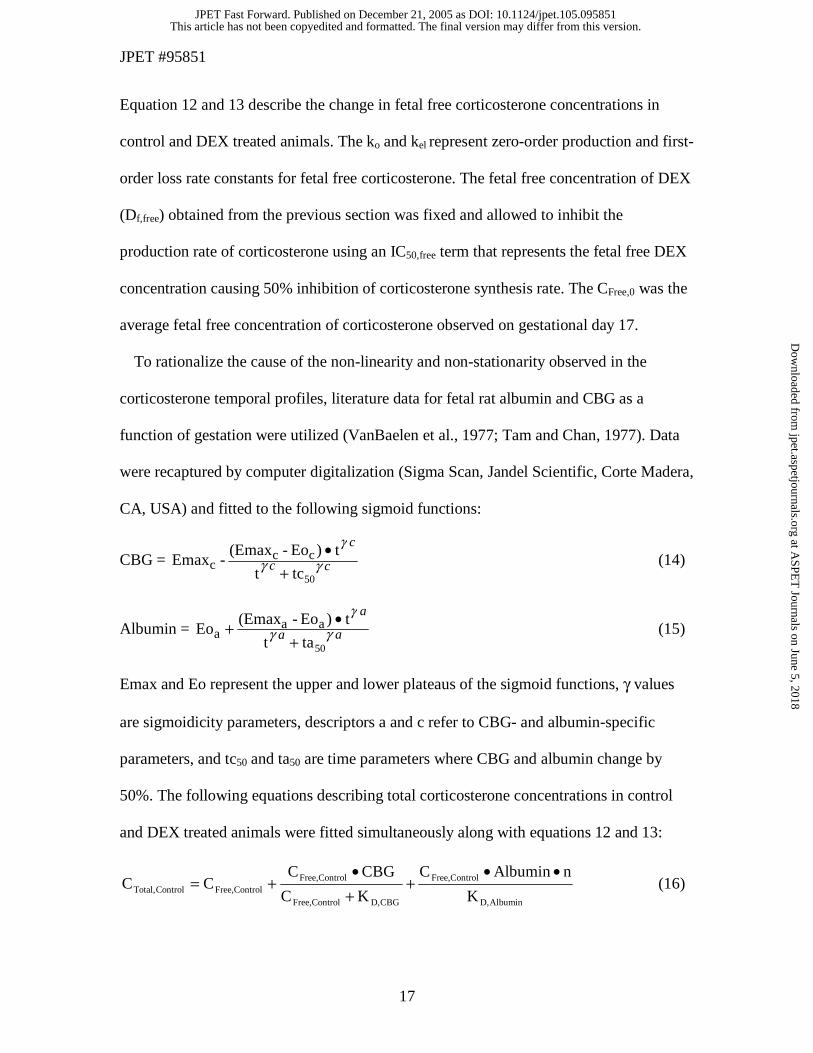
Equation 12 and 13 describe the change in fetal free corticosterone concentrations in
control and DEX treated animals. The ko and kel represent zero-order production and first-
order loss rate constants for fetal free corticosterone. The fetal free concentration of DEX
(Df,free) obtained from the previous section was fixed and allowed to inhibit the
production rate of corticosterone using an IC50,free term that represents the fetal free DEX
concentration causing 50% inhibition of corticosterone synthesis rate. The CFree,0 was the
average fetal free concentration of corticosterone observed on gestational day 17.
To rationalize the cause of the non-linearity and non-stationarity observed in the
corticosterone temporal profiles, literature data for fetal rat albumin and CBG as a
function of gestation were utilized (VanBaelen et al., 1977; Tam and Chan, 1977). Data
were recaptured by computer digitalization (Sigma Scan, Jandel Scientific, Corte Madera,
CA, USA) and fitted to the following sigmoid functions:
CBG = cc
c
γγ
γ
50tct
t)Eo-(Emax-Emax cc
c +•
(14)
Albumin = aa
a
γγ
γ
50tat
t)Eo-(EmaxEo aa
a +•+ (15)
Emax and Eo represent the upper and lower plateaus of the sigmoid functions, γ values
are sigmoidicity parameters, descriptors a and c refer to CBG- and albumin-specific
parameters, and tc50 and ta50 are time parameters where CBG and albumin change by
50%. The following equations describing total corticosterone concentrations in control
and DEX treated animals were fitted simultaneously along with equations 12 and 13:
AlbuminD,
ControlFree,
CBGD,ControlFree,
ControlFree,ControlFree,ControlTotal, K
nAlbuminC
KC
CBGCCC
••+
+•
+= (16)
This article has not been copyedited and formatted. The final version may differ from this version.JPET Fast Forward. Published on December 21, 2005 as DOI: 10.1124/jpet.105.095851
at ASPE
T Journals on June 5, 2018
jpet.aspetjournals.orgD
ownloaded from

JPET #95851
18
AlbminD,
DEXFree,
CBGD,DEXFree,
DEXFree,DEXFree,DEXTotal, K
nAlbuminC
KC
CBGCCC
••+
+•
+= (17)
KD,albumin and KD,CBG were estimated and refer to the equilibrium dissociation constants
for corticosterone binding to its two binding proteins; n is the number of binding sites per
molecule of albumin available for corticosterone binding and was fixed to 2 (Jobin and
Perrin, 1974). CBG is a high affinity, low capacity binding protein which exhibits non-
linear binding, while albumin is a high capacity, low affinity non-saturable binding
protein.
Data Analysis. Data from multiple animals were pooled and the ADAPT II program
(D'Argenio and Schumitzky, 1997) with the maximum likelihood method was applied for
all fittings. The following variance model was used:
Variance = Coefficient2•Y(t)Power (18)
where Coefficient and Power are variance parameters, and Y(t) represents the model
output function. The goodness-of-fit was assessed using correlation coefficients,
examination of residuals, visual inspection of the fitted curves, estimator criterion value,
sum of squared residuals, and coefficients of variation of the estimated parameters.
This article has not been copyedited and formatted. The final version may differ from this version.JPET Fast Forward. Published on December 21, 2005 as DOI: 10.1124/jpet.105.095851
at ASPE
T Journals on June 5, 2018
jpet.aspetjournals.orgD
ownloaded from

JPET #95851
19
Results
Simulation Study Results
Fig. 2 compares the PK results of the simulation (1 umol kg-1 dose) vs. the actual
observed fetal exposure to free DEX upon serial IM administration. The figure also
provides the desirable lower threshold, the detrimental upper threshold, and the targeted
concentration maximum. The simulation correctly predicted the time to reach
concentration maximum after each dose, but generally under predicted the fetal exposure
to free DEX.
Fetal Growth Curves
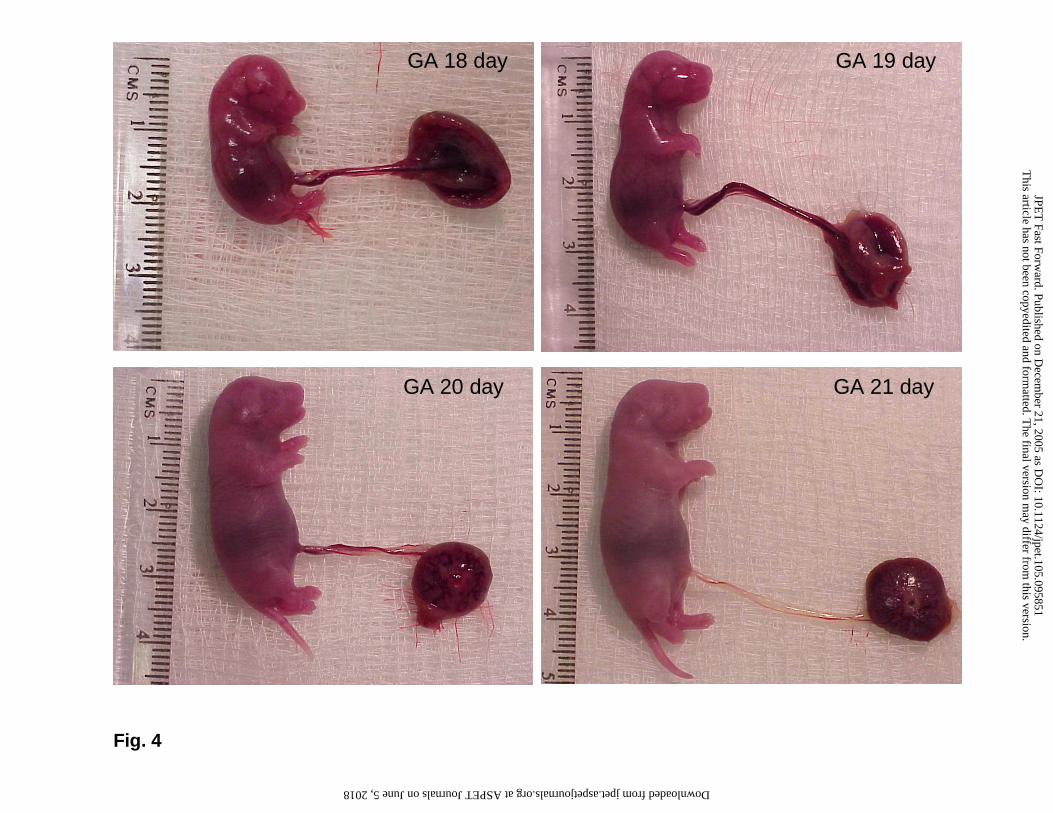
The fetal growth curves followed an exponential growth pattern (Fig. 3). Variability in
the control group on gestational day 21 (96 h) may be due to the variable litter sizes that
were obtained from 2 animals at this time point where one rat had only 8 fetuses, while
another had 19 fetuses. Parameter estimates were A0 = 1 gm (CV: 3%) and TD = 1.7 day
(CV: 3%). Fig. 4 visualizes the burst of growth that occurs in fetal rats during late
gestation.
Maternal/Fetal DEX PK
Total and unbound DEX profiles in the maternal/fetal circulation are shown in Fig. 5.
DEX appeared very rapidly in the systemic circulation after IM administration of DEX
sodium phosphate. This is evident from the maternal total DEX concentration of 500-750
nM observed at 1 hr, 10 and 30 min after the first, third and fifth doses. Fetal DEX,
although lower than maternal concentrations, exhibited rapid transfer across the placenta.
DEX could be detected in the fetal circulation at the first sacrifice time point of 1 hr after
This article has not been copyedited and formatted. The final version may differ from this version.JPET Fast Forward. Published on December 21, 2005 as DOI: 10.1124/jpet.105.095851
at ASPE
T Journals on June 5, 2018
jpet.aspetjournals.orgD
ownloaded from

JPET #95851
20
the first dose. The fetal tissue DEX contents (ng/gm fetus) were slightly higher than the
total fetal plasma concentrations, which indicate that DEX distributes extensively into
fetal tissues. On all days of the study, DEX concentrations declined with a half-life of
approximately 3 hr in maternal and fetal circulations, which is excellent agreement with
our previous results (Samtani and Jusko, 2005a; Samtani et al., 2004a). The free fraction
of DEX in the maternal circulation was about 20%, indicating that it is moderately bound
to albumin. In contrast the free fraction in the fetal circulation was approximately 40%.
DEX PK parameters listed in Table 1 were estimated with high precision, which is
evident from the < 35% CV for all parameters. Fig. 6 provides unbound maternal/fetal
data from different gestational days plotted against the time since last dose. The plot
takes the shape of a simple Bateman function and helps to verify DEX PK stationarity
during pregnancy.
Temporal Fetal Corticosterone Patterns in Control/DEX Rats
Total corticosterone in control fetal rats began to rise on GA 17, stayed high up to 19
day GA, and exhibited a shallow decline between GA 19 and 21 (Fig. 7). In contrast the
free corticosterone rose to a maximum on day 17 and stayed constant until the end of the
study. Total corticosterone in the fetus was somewhat lower than the circadian high
observed in non-pregnant controls. However, the free corticosterone in fetal rats closely
mirrored the circadian high that occurs in non-pregnant controls after GA 17. Maternal
administration of DEX caused marked adrenal suppression, which led to lowering of the
total and free corticosterone concentrations (Fig. 7). Corticosterone concentrations in
adrenosuppressed fetuses, although fairly low, were still captured by the
radioimmunoassay. Parameters describing the release and binding pattern of
This article has not been copyedited and formatted. The final version may differ from this version.JPET Fast Forward. Published on December 21, 2005 as DOI: 10.1124/jpet.105.095851
at ASPE
T Journals on June 5, 2018
jpet.aspetjournals.orgD
ownloaded from

JPET #95851
21
corticosterone in fetal rats and inhibition of corticosterone secretion by DEX are
provided in Table 2.
Plasma Protein Binding Stationarity and Linearity
Binding of DEX in maternal and fetal plasma was found to be linear (Fig. 8). However,
DEX was more extensively bound in maternal than in fetal plasma. Thus the common
assumption of equal binding in maternal/fetal plasma was found to be inappropriate.
Therefore in modeling the data the differential equations were allowed to operate on the
free rather than the total drug concentrations. Binding of corticosterone in maternal and
fetal plasma in both control and DEX treated groups was non-linear and non-stationary
(Fig. 8). The non-stationarity can be attributed to the rapidly declining CBG
concentrations in rat plasma during the last few days of gestation (Fig. 9). During this
period the albumin concentrations also change but with an increasing trend (Fig. 9).
Despite the increasing albumin concentrations, binding to this protein is expected to
remain linear due to the large size of this binding pool, which exists at 50-100 times
higher concentrations in the fetal rat as compared to CBG. The linearity of this binding
system in the fetal rat is illustrated by the binding characteristics of DEX, which binds to
albumin alone. The parameter estimates describing the change in fetal plasma binding
proteins as a function of time are shown in Table 3.
Fetal/Maternal Gradients for Corticosteroids
Fetal/maternal gradients are usually evaluated by assessing the ratio of unbound fetal to
maternal concentrations. Binding of steroids to different extents in the two plasma pools
can cause differences in total drug concentrations between the mother and the fetus. By
estimating the ratio of free drug concentrations the binding effects in the two exchanging
This article has not been copyedited and formatted. The final version may differ from this version.JPET Fast Forward. Published on December 21, 2005 as DOI: 10.1124/jpet.105.095851
at ASPE
T Journals on June 5, 2018
jpet.aspetjournals.orgD
ownloaded from

JPET #95851
22
pools can be negated. Assessment of this ratio for corticosterone in control rats shows
that this ratio is roughly distributed around one, indicating that corticosterone distributes
readily across the placental interface (Fig. 10). However, the ratio for DEX exhibits a
value of less than one in all of the thirty steroid-treated animals. This indicates that the
placenta creates a barrier for the transfer of DEX from the maternal to fetal circulation
(Fig. 10). The possible source of this placental barrier can be understood by comparing
the values of Clmf and Clfm obtained during curve fitting of the DEX PK data (Table 1). A
higher value for Clfm indicates that the placental DEX transfer occurs much more
efficiently in the fetal to maternal direction.
This article has not been copyedited and formatted. The final version may differ from this version.JPET Fast Forward. Published on December 21, 2005 as DOI: 10.1124/jpet.105.095851
at ASPE
T Journals on June 5, 2018
jpet.aspetjournals.orgD
ownloaded from

JPET #95851
23
Discussion
The simulation study underestimated the fetal free DEX exposure. This discrepancy
can be attributed to an inappropriate assumption (equal maternal/fetal plasma protein
binding) made during the simulation study. The fetal plasma protein binding of DEX was
found to be lower than that in maternal plasma. This could possibly occur because the
fetal plasma protein content is far lower than maternal plasma and because fetal rat
albumin starts to rise late in gestation (Tam and Chan, 1977). Despite the disagreement
between the simulated and observed profiles, the measured fetal DEX exposure still met
the desired criteria and thus the simulation exercise served as a suitable method for
designing a prenatal regimen.
DEX did not appear to have an effect on fetal growth and therefore growth curves from
both groups were pooled and modeled. The fitted parameters for fetal growth are in good
agreement with literature estimates of Ao = 0.7 gm and TD = 1.5 day (Schneidereit, 1985).
Binding of DEX, although different in maternal and fetal plasma, was found to be
linear in both compartments. This is somewhat unexpected considering that fetal plasma
albumin changes dramatically during the last days of gestation (Fig. 9). However the
concentration of albumin even in fetal plasma is in the high µM range. This probably
precludes saturation of albumin binding sites by DEX which circulates at nM
concentrations after administration of a 1 µmol kg-1 dose.
Binding of corticosterone in fetal plasma was found to be highly non-linear and non-
stationary. It is interesting to note that although the total concentrations of corticosterone
decline in control fetal rats after GA 19, the free concentrations stay unchanged. All these
observations can be explained by the sharp fall in CBG concentrations during late
This article has not been copyedited and formatted. The final version may differ from this version.JPET Fast Forward. Published on December 21, 2005 as DOI: 10.1124/jpet.105.095851
at ASPE
T Journals on June 5, 2018
jpet.aspetjournals.orgD
ownloaded from
JPET #95851
24
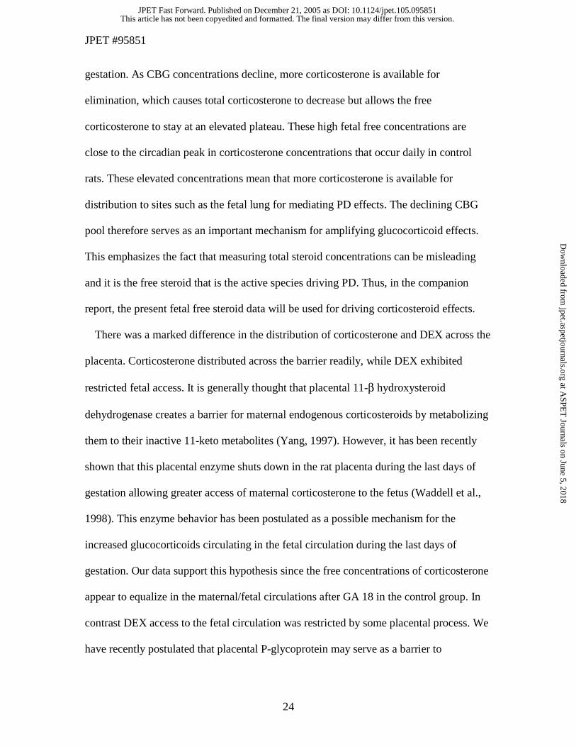
gestation. As CBG concentrations decline, more corticosterone is available for
elimination, which causes total corticosterone to decrease but allows the free
corticosterone to stay at an elevated plateau. These high fetal free concentrations are
close to the circadian peak in corticosterone concentrations that occur daily in control
rats. These elevated concentrations mean that more corticosterone is available for
distribution to sites such as the fetal lung for mediating PD effects. The declining CBG
pool therefore serves as an important mechanism for amplifying glucocorticoid effects.
This emphasizes the fact that measuring total steroid concentrations can be misleading
and it is the free steroid that is the active species driving PD. Thus, in the companion
report, the present fetal free steroid data will be used for driving corticosteroid effects.
There was a marked difference in the distribution of corticosterone and DEX across the
placenta. Corticosterone distributed across the barrier readily, while DEX exhibited
restricted fetal access. It is generally thought that placental 11-β hydroxysteroid
dehydrogenase creates a barrier for maternal endogenous corticosteroids by metabolizing
them to their inactive 11-keto metabolites (Yang, 1997). However, it has been recently
shown that this placental enzyme shuts down in the rat placenta during the last days of
gestation allowing greater access of maternal corticosterone to the fetus (Waddell et al.,
1998). This enzyme behavior has been postulated as a possible mechanism for the
increased glucocorticoids circulating in the fetal circulation during the last days of
gestation. Our data support this hypothesis since the free concentrations of corticosterone
appear to equalize in the maternal/fetal circulations after GA 18 in the control group. In
contrast DEX access to the fetal circulation was restricted by some placental process. We
have recently postulated that placental P-glycoprotein may serve as a barrier to
This article has not been copyedited and formatted. The final version may differ from this version.JPET Fast Forward. Published on December 21, 2005 as DOI: 10.1124/jpet.105.095851
at ASPE
T Journals on June 5, 2018
jpet.aspetjournals.orgD
ownloaded from

JPET #95851
25
maternally administered DEX (Samtani et al., 2004a). Most synthetic corticosteroids,
including DEX, are excellent substrates for this transporter (Yates et al., 2003) and the
placenta expresses high levels of P-glycoprotein where it serves as a pump for effluxing
xenobiotics from the fetal to the maternal circulation (Young et al., 2003). The P-
glycoprotein argument is further strengthened when corticosterone data are reconsidered.
Corticosterone, unlike synthetic corticosteroids, is not a P-glycoprotein substrate (Yates
et al., 2003; Karssen et al., 2001) and thus the lack of metabolic and efflux barriers allow
this steroid to distribute equally across the placenta. Finally, the importance of computing
free fetal/maternal concentration ratios cannot be stressed enough. Due to the lack of
available data we previously compared total fetal and maternal DEX concentrations from
the literature (Samtani et al., 2004a). This comparison revealed a fetal/maternal gradient
of 0.2. It is only now that we understand that an important reason for this gradient is the
lower binding of DEX in the fetal circulation. A higher free fraction in fetal plasma
allows greater return of DEX from the fetal to the maternal circulation. However, by
computing the fetal to maternal ratio of free DEX (average = 0.55 across the duration of
the study) it is recognized that plasma protein binding does not completely explain the
fetal to maternal gradient and some additional process restricts fetal access, which is
probably placental efflux.
It was noted that one animal sacrificed as 81 hr showed markedly lower steroid
concentrations (Fig. 5). All five measurements (maternal total and free, fetal total and
free, and fetal tissue content) from this particular animal appeared as outliers. This
observation rules out assay error since five mismatches cannot arise from the assays. By
extrapolating the curves from the previous dose it appears that this animal may not have
This article has not been copyedited and formatted. The final version may differ from this version.JPET Fast Forward. Published on December 21, 2005 as DOI: 10.1124/jpet.105.095851
at ASPE
T Journals on June 5, 2018
jpet.aspetjournals.orgD
ownloaded from

JPET #95851
26
received the fifth dose. In performing these labor intensive animal studies, the possibility
of such human error cannot be ruled out. The enormity of the study size can be judged
from the fact that the 54 pregnant animals humanely killed in this study yielded almost
800 fetuses. The possibly erroneous data points were retained during the modeling
process because the use of the maximum likelihood procedure, which is inherently a way
of weighting the data, is less susceptible to outliers during the curve fitting procedure
(Gabrielsson and Weiner, 1997). This point is emphasized by the fitted curves, which do
not appear to be influenced by the five outliers.
The estimation of DEX PK parameters made use of a simple two-compartment
maternal/fetal exchange model that assumed stationary PK across the entire duration of
the study, except for the parameter Vf. The property of stationary DEX PK is
demonstrated in Fig. 6 where unbound maternal/fetal concentrations from different
gestational days are plotted against the time since last dose. On gestational days 18, 19,
and 20 a trough kill at 9 hr was performed. The overlapping of DEX concentrations at
this time point on maternal/fetal profiles in Fig 6. shows that PK are not altered by
advancing GA. Furthermore, the data take the shape of a simple Bateman function, which
further illustrates stationarity. In constructing Fig. 6, reverse super-positioning (Bauer
and Gibaldi, 1983) to strip off contributions of the previous doses during the second to
the sixth dosing intervals was not applied. The dosing interval exceeded four elimination
half-lives and the contribution of the previous dose would be < 7%, making the
correction factor marginal. Fig. 6 also demonstrates that although only ten time points
were chosen over the course of a 72 hr study, the judicious choice of sample collection
points during this multiple-dosing study helps capture both the disposition and absorption
This article has not been copyedited and formatted. The final version may differ from this version.JPET Fast Forward. Published on December 21, 2005 as DOI: 10.1124/jpet.105.095851
at ASPE
T Journals on June 5, 2018
jpet.aspetjournals.orgD
ownloaded from

JPET #95851
27
properties of DEX with good precision (evident from the < 35% CV for all parameters in
Table 1).
Modeling the corticosterone data, although yielding parameters with high degrees of
precision, produced fitted curves that did not characterize all the data equally well.
Examples include GA 18 data for total and free corticosterone in control and DEX treated
fetuses (Fig. 7). One possible explanation is that all data in Fig. 7 were fitted
simultaneously. It is known that DEX treatment can suppress levels of CBG (VanBaelen
et al., 1977) which was not incorporated in the model. This is because quantitative
assessments of DEX effects on CBG are sparse or qualitative in nature. Given the lack of
complete information, CBG levels were assumed to be similar in control and treated
animals. Furthermore, the main purpose of capturing steroid profiles was to ascertain the
driving force behind corticosteroid effects. It is generally thought that effects are driven
by free steroid concentrations and therefore any discrepancies in fitting the total
corticosterone data are not a big concern. In addition, free corticosterone data in the DEX
treated group will have a minor contributing effect towards steroid PD because the effects
will be primarily driven by the exogenous steroid due to its higher affinity for the
glucocorticoid receptor. Thus the most important profile amongst the different curves in
Fig. 7 is the fetal free corticosterone in untreated controls and these data were captured
relatively well. Finally, the estimates of KD values in Table 2 confirm that CBG and
albumin are high and low affinity binding proteins, which is in good agreement with
corticosterone binding properties (Jobin and Perrin, 1974).
This article has not been copyedited and formatted. The final version may differ from this version.JPET Fast Forward. Published on December 21, 2005 as DOI: 10.1124/jpet.105.095851
at ASPE
T Journals on June 5, 2018
jpet.aspetjournals.orgD
ownloaded from
JPET #95851
28
Acknowledgment
We thank Donna Ruszaj for helpful discussions and technical assistance with
LC/MS/MS assay development.
This article has not been copyedited and formatted. The final version may differ from this version.JPET Fast Forward. Published on December 21, 2005 as DOI: 10.1124/jpet.105.095851
at ASPE
T Journals on June 5, 2018
jpet.aspetjournals.orgD
ownloaded from

JPET #95851
29
References
Ballard PL, Mason RJ and Douglas WH (1978) Glucocorticoid binding by isolated lung
cells. Endocrinology 102:1570-1575.
Ballard PL and Ballard RA (1995) Scientific basis and therapeutic regimens for use of
antenatal glucocorticoids. Am J Obstet Gynecol 173:254-262.
Bauer LA and Gibaldi M (1983) Computation of model-independent pharmacokinetic
parameters during multiple dosing. J Pharm Sci 72:978-979.
Boggaram V, Smith ME and Mendelson CR (1989) Regulation of expression of the gene
encoding the major surfactant protein (SP-A) in human fetal lung in vitro.
Disparate effects of glucocorticoids on transcription and on mRNA stability. J
Biol Chem 264:11421-11427.
D'Argenio DZ and Schumitzky A (1997) ADAPT II user's guide:
Pharmacokinetic/pharmacodynamic systems analysis software. Biomedical
Simulations Resource, Los Angeles.
Diederich S, Hanke B, Burkhardt P, Müller M, Schöneshöfer, Bähr V and Oelkers W
(1998) Metabolism of synthetic corticosteroids by 11β-hydroxysteroid-
dehydrogenases in man. Steroids 63:271-277.
Duffy DL, Bentley GE, Drazen DL and Ball GF (2000) Effects of testosterone on cell-
mediated and humoral immunity in non-breeding adult European starlings. Behav
Ecol 11: 654-662.
Gabrielsson J and Weiner D (1997) Pharmacokinetic and Pharmacodynamic Data
Analysis 2nd ed. Swedish Pharmaceutical Press, Stockholm, Sweden.
This article has not been copyedited and formatted. The final version may differ from this version.JPET Fast Forward. Published on December 21, 2005 as DOI: 10.1124/jpet.105.095851
at ASPE
T Journals on June 5, 2018
jpet.aspetjournals.orgD
ownloaded from

JPET #95851
30
Hazra A, Jusko WJ, Almon RA and Dubois DC (2004) Pharmacodynamics of circadian
rhythm of corticosterone effects on tyrosine aminotransferase in normal rats. The
AAPS Journal 6:Abstract T3355.
Huot RL, Plotsky PM, Lenox RH and McNamara RK (2002) Neonatal maternal
separation reduces hippocampal mossy fiber density in adult Long Evans rats.
Brain Res 950:52-63.
Jobin M and Perrin F (1974) Evaluation of three constants involved in the binding of
corticosterone to plasma proteins in the rat. Can J Biochem 52:101-105.
Karssen AM, Meijer OC, van der Sandt IC, Lucassen PJ, de Lange EC, de Boer AG and
de Kloet ER (2001) Multidrug resistance P-glycoprotein hampers the access of
cortisol but not of corticosterone to mouse and human brain. Endocrinology
142:2686-2694.
Liggins GC (1994) The role of cortisol in preparing the fetus for birth. Reprod Fertil Dev
6:141-150.
Newnham JP (2001) Is prenatal glucocorticoid administration another origin of adult
disease? Clin Exp Pharmacol Physiol 28:957-961.
NIH Consensus Panel (1995) NIH consensus development panel on the effect of
corticosteroids for fetal maturation on perinatal outcomes. JAMA 273:413-8.
Pacak K, McCarty R, Palkovits M, Cizza G, Kopin IJ, Goldstein DS and Chrousos GP
(1995) Decreased central and peripheral catecholaminergic activation in obese
Zucker rats. Endocrinology 136:4360-4367.
This article has not been copyedited and formatted. The final version may differ from this version.JPET Fast Forward. Published on December 21, 2005 as DOI: 10.1124/jpet.105.095851
at ASPE
T Journals on June 5, 2018
jpet.aspetjournals.orgD
ownloaded from

JPET #95851
31
Pugeat MM, Dunn JF and Nisula BC (1981) Transport of steroid hormones: Interaction
of 70 drugs with testosterone-binding globulin and corticosteroid-binding globulin
in human plasma. J Clin Endocrinol Metab 53:69-75.
Samtani MN, Schwab M, Nathanielsz PW and Jusko WJ (2004a) Area/Moment and
compartmental modeling of pharmacokinetics during pregnancy: applications to
maternal/fetal exposures to corticosteroids in sheep and rats. Pharm Res 21:2279-
2292.
Samtani MN, Schwab M, Nathanielsz PW and Jusko WJ (2004b) Stabilization and HPLC
analysis of betamethasone sodium phosphate in plasma. J Pharm Sci 93:726-732.
Samtani MN, Lohle M, Grant A, Nathanielsz PW and Jusko WJ (2005) Betamethasone
pharmacokinetics after two prodrug formulations in sheep: implications for
antenatal corticosteroid use. Drug Metab Dispos 33:1124-1130.
Samtani MN and Jusko WJ (2005a) Comparison of dexamethasone pharmacokinetics in
female rats after intravenous and intramuscular administration. Biopharm Drug
Dispos 26:85-91.
Samtani MN and Jusko WJ (2005b) Stability of dexamethasone sodium phosphate in rat
plasma. Int J Pharm 301:262-266.
Schneidereit M (1985) Study of fetal organ growth in Wistar rats from day 17 to 21. Lab
Anim 19:240-244.
Stock B, Dean M and Levy G (1980) Serum protein binding of drugs during and after
pregnancy in rats. J Pharmacol Exp Ther 212:264-268.
This article has not been copyedited and formatted. The final version may differ from this version.JPET Fast Forward. Published on December 21, 2005 as DOI: 10.1124/jpet.105.095851
at ASPE
T Journals on June 5, 2018
jpet.aspetjournals.orgD
ownloaded from

JPET #95851
32
Tam PP and Chan ST (1977) Changes in the composition of maternal plasma, fetal
plasma and fetal extraembryonic fluid during gestation in the rat. J Reprod Fertil
51:41-51.
Taymans SE, DeVries AC, DeVries MB, Nelson RJ, Friedman TC, Castro M,Detera-
Wadleigh S, Carter CS and Chrousos GP (1997) The hypothalamic-pituitary-
adrenal axis of prairie voles (Microtus ochrogaster): evidence for target tissue
glucocorticoid resistance. Gen Comp Endocrinol 106:48-61.
VanBaelen H, Vandoren G and DeMoor P (1977) Concentration of transcortin in the
pregnant rat and its foetuses. J Endocrinol 75:427-431.
Waddell BJ, Benediktsson R, Brown RW and Seckl JR (1998) Tissue-specific messenger
ribonucleic acid expression of 11beta-hydroxysteroid dehydrogenase types 1 and
2 and the glucocorticoid receptor within rat placenta suggests exquisite local
control of glucocorticoid action. Endocrinology 139:1517-1523.
Yang K (1997) Placental 11 beta-hydroxysteroid dehydrogenase: barrier to maternal
glucocorticoids. Rev Reprod 2:129-132.
Yates CR, Chang C, Kearbey JD, Yasuda K, Schuetz EG, Miller DD, Dalton JT and
Swaan PW (2003) Structural determinants of P-glycoprotein-mediated transport
of glucocorticoids. Pharm Res 20:1794-1803.
Young AM, Allen CE and Audus KL (2003) Efflux transporters of the human placenta.
Adv Drug Deliv Rev 55:125-132.
This article has not been copyedited and formatted. The final version may differ from this version.JPET Fast Forward. Published on December 21, 2005 as DOI: 10.1124/jpet.105.095851
at ASPE
T Journals on June 5, 2018
jpet.aspetjournals.orgD
ownloaded from
JPET #95851
33
Footnotes
a) Source of financial support: This study was supported by grant GM 24211 from the
National Institutes of Health and a predoctoral fellowship for MNS from Merck. The
liquid chromatograph/tandem mass spectrometer was obtained through a shared
instrumentation grant (S10RR14573) from the National Center for Research
Resources, National Institutes of Health.
b) Person to receive reprint requests: William J. Jusko, Ph.D., Department of
Pharmaceutical Sciences, School of Pharmacy and Pharmaceutical Sciences,
University at Buffalo, State University of New York, 565 Hochstetter Hall, Buffalo,
New York 14260. Email: [email protected]
This article has not been copyedited and formatted. The final version may differ from this version.JPET Fast Forward. Published on December 21, 2005 as DOI: 10.1124/jpet.105.095851
at ASPE
T Journals on June 5, 2018
jpet.aspetjournals.orgD
ownloaded from

JPET #95851
34
Figure legends
Fig. 1: Model of DEX pharmacokinetics and adrenosuppression mediated by free DEX in
fetal plasma. Symbols and differential equations for the model are defined in the text. The
dashed arrow and solid rectangle represent inhibition of corticosterone secretion by
unbound DEX via an indirect mechanism.
Fig. 2: PK results of the simulation (solid curve) vs. the observed fetal exposure to free
DEX (filled circles) upon intramuscular administration of 1 umol kg-1 doses. Horizontal
lines depict desirable lower and detrimental upper thresholds and the arrow indicates the
targeted concentration maximum.
Fig. 3: Fetal growth in control (open circles) and DEX treated animals (filled circles)
followed the exponential growth model depicted by the solid line.
Fig. 4: Photographs of typical rat fetuses on indicated days showing the burst of growth
that occurs during late GA.
Fig. 5: DEX PK in the mother (top) and fetus (bottom). Open and filled circles symbolize
total and free DEX concentrations, while filled triangles represent the amount of DEX in
fetal tissue. Solid, dashed, and dotted curves are model fitted results for total
concentrations, free concentrations, and fetal tissue content.
Fig. 6: Stationary DEX PK is verified by plotting the unbound maternal (open symbols)
and fetal (filled symbols) DEX concentrations on different gestational days as a function
of time since last dose. Gestational days 18, 19, 20, and 21 are symbolized by triangles,
squares, circles, and inverted triangles, respectively.
Fig. 7: Fetal corticosterone in untreated controls (top) and DEX treated animals (bottom).
Open and filled symbols represent unbound and total concentrations. Triangles represent
This article has not been copyedited and formatted. The final version may differ from this version.JPET Fast Forward. Published on December 21, 2005 as DOI: 10.1124/jpet.105.095851
at ASPE
T Journals on June 5, 2018
jpet.aspetjournals.orgD
ownloaded from

JPET #95851
35
data from non-pregnant controls sacrificed at the peak of the circadian corticosterone
cycle. The model shown in Fig. 1 was fitted to all the data simultaneously and the curves
represent model fittings.
Fig. 8: Protein binding of DEX (top) and corticosterone (bottom) in maternal/fetal
plasma. Circles represent data from DEX treated animals, while data from controls are
represented by triangles. Filled symbols represent maternal data, while open symbols are
data from fetal samples. Straight lines are regressions through the maternal/fetal data to
demonstrate linear plasma protein binding for DEX.
Fig. 9: Fetal plasma CBG (top) and albumin (bottom) as a function of advancing GA.
Solid curves represent results of fitting the sigmoid functions to the data.
Fig. 10: Unbound fetal to maternal concentration ratio for corticosterone (top) and DEX
(bottom). Horizontal lines depict the ratio of 1.
This article has not been copyedited and formatted. The final version may differ from this version.JPET Fast Forward. Published on December 21, 2005 as DOI: 10.1124/jpet.105.095851
at ASPE
T Journals on June 5, 2018
jpet.aspetjournals.orgD
ownloaded from

JPET #95851
36
Table 1. Pharmacokinetic parameters for DEX in pregnant rats
Parameter Definition Estimate CV%
Vm/F (mL) Apparent maternal volume of distribution 3097 6
Normalized Vf/F (mL/gm fetus) Apparent fetal volume of distribution 4.71 7
CLm/F (mL/hr) Apparent elimination clearance 729 5
CLmf/F (mL/hr) Apparent maternal to fetal placental transfer clearance 720 31
CLfm/F (mL/hr) Apparent fetal to maternal placental transfer clearance 1342 32
ka (hr -1) Absorption rate constant 8.52 28
Maternal fup Maternal DEX free fraction in plasma 0.18 7
Fetal fup Fetal DEX free fraction in plasma 0.39 7
This article has not been copyedited and form
atted. The final version m
ay differ from this version.
JPET
Fast Forward. Published on D
ecember 21, 2005 as D
OI: 10.1124/jpet.105.095851
at ASPET Journals on June 5, 2018 jpet.aspetjournals.org Downloaded from

JPET #95851
37
Table 2. Parameters for the release and binding of corticosterone in fetal rats and inhibition of corticosterone secretion by DEX
Parameter Definition Estimate CV%
ko (nM/hr) Zero order production rate of fetal free corticosterone 18.4 14
kel (1/hr) First order loss rate of fetal free corticosterone 0.45 9
KD,CBG (nM) Equilibrium dissociation constant for corticosterone binding to CBG 107 21
KD,Albumin (µM) Equilibrium dissociation constant for corticosterone binding to albumin 21.5 25
IC50,Free (nM) Fetal free DEX causing 50% inhibition of corticosterone synthesis rate 0.98 10
n Number of binding sites per molecule of albumin available for corticosterone binding 2 Fixed
CFree,0 (nM) Average fetal free concentration of corticosterone observed on GA 17 9.44 Fixed
This article has not been copyedited and form
atted. The final version m
ay differ from this version.
JPET
Fast Forward. Published on D
ecember 21, 2005 as D
OI: 10.1124/jpet.105.095851
at ASPET Journals on June 5, 2018 jpet.aspetjournals.org Downloaded from

JPET #95851
38
Table 3. Parameter estimates describing the change in fetal plasma binding proteins
Parameter Definition Parameter Estimates (CV %)
Albumin CBG
Emax (nM) Upper plateau of the sigmoid function 122•103 (26) 1.90•103 (1)
Eo (nM) Lower plateau of the sigmoid function 40.0•103 (Fixed) 365 (5)
t50 (hr) Time parameter where CBG and albumin change by 50% 46.3 (50) 73.0 (1)
γ Sigmoidicity parameter 1.97 (48) 7.28 (7)
This article has not been copyedited and form
atted. The final version m
ay differ from this version.
JPET
Fast Forward. Published on D
ecember 21, 2005 as D
OI: 10.1124/jpet.105.095851
at ASPET Journals on June 5, 2018 jpet.aspetjournals.org Downloaded from

Vm
Dm, Total
CL m
CL mf
CL fm
IM
ka F • Dose
Dm, Free
Df, Total
Df, Free Vf
Cfree CBG
ko
Albumin
kel
IC50,Free
Fig. 1
This article has not been copyedited and formatted. The final version may differ from this version.JPET Fast Forward. Published on December 21, 2005 as DOI: 10.1124/jpet.105.095851
at ASPE
T Journals on June 5, 2018
jpet.aspetjournals.orgD
ownloaded from

Fig. 2
Time (hr)
0 24 48 72 96 120
Dex
amet
haso
ne C
once
ntra
tion
(nM
)
0.1
1
10
100
1000
Gestational Age (day)
17 18 19 20 21 22
This article has not been copyedited and formatted. The final version may differ from this version.JPET Fast Forward. Published on December 21, 2005 as DOI: 10.1124/jpet.105.095851
at ASPE
T Journals on June 5, 2018
jpet.aspetjournals.orgD
ownloaded from

Time (hr)
0 24 48 72 96
Ave
rage
wei
ght o
f a s
ingl
e fe
tus
from
a li
tter
(gm
)
1
2
3
4
5
6
Gestational Age (day)
17 18 19 20 21
Fig. 3
This article has not been copyedited and formatted. The final version may differ from this version.JPET Fast Forward. Published on December 21, 2005 as DOI: 10.1124/jpet.105.095851
at ASPE
T Journals on June 5, 2018
jpet.aspetjournals.orgD
ownloaded from
GA 18 day GA 19 day
GA 20 day GA 21 day
Fig. 4
This article has not been copyedited and form
atted. The final version m
ay differ from this version.
JPET
Fast Forward. Published on D
ecember 21, 2005 as D
OI: 10.1124/jpet.105.095851
at ASPET Journals on June 5, 2018 jpet.aspetjournals.org Downloaded from

Fig. 5
Mat
erna
l (pm
ol p
er m
L)
1
10
100
1000
Gestational Age (day)
18.0 18.5 19.0 19.5 20.0 20.5 21.0
Time (hr)
24 36 48 60 72 84 96
Fet
al (
pmol
per
mL
or g
m o
f fet
us)
0.1
1
10
100
1000
DE
XA
ME
TH
AS
ON
E C
ON
CE
NT
RA
TIO
NS
This article has not been copyedited and formatted. The final version may differ from this version.JPET Fast Forward. Published on December 21, 2005 as DOI: 10.1124/jpet.105.095851
at ASPE
T Journals on June 5, 2018
jpet.aspetjournals.orgD
ownloaded from

Fig. 6
Time Since Last Dose (hr)
0 2 4 6 8 10 12
UN
BO
UN
D D
EX
AM
ET
HA
SO
NE
CO
NC
EN
TR
AT
ION
(nM
)
0.1
1
10
100
1000
This article has not been copyedited and formatted. The final version may differ from this version.JPET Fast Forward. Published on December 21, 2005 as DOI: 10.1124/jpet.105.095851
at ASPE
T Journals on June 5, 2018
jpet.aspetjournals.orgD
ownloaded from

Fig. 7
Fet
al U
ntre
ated
Con
trol
s
10
100
1000
Gestational Age (day)
17 18 19 20 21 22
Time (hr)
0 12 24 36 48 60 72 84 96 108 120
Fet
al in
the
DE
X G
roup
1
10
100
1000
CO
RT
ICO
ST
ER
ON
E C
ON
CE
NT
RA
TIO
NS
(nM
)
This article has not been copyedited and formatted. The final version may differ from this version.JPET Fast Forward. Published on December 21, 2005 as DOI: 10.1124/jpet.105.095851
at ASPE
T Journals on June 5, 2018
jpet.aspetjournals.orgD
ownloaded from

Fig. 8
Free DEX (nM)
0 20 40 60 80 100 120 140 160 180
Bou
nd D
EX
(nM
)
0
200
400
600
800
Free Corticosterone (nM)
0 15 30 45 60 75 90 105
Bou
nd C
ortic
oste
rone
(nM
)
0
300
600
900
1200
1500
1800
This article has not been copyedited and formatted. The final version may differ from this version.JPET Fast Forward. Published on December 21, 2005 as DOI: 10.1124/jpet.105.095851
at ASPE
T Journals on June 5, 2018
jpet.aspetjournals.orgD
ownloaded from

Fig. 9
Fet
al P
lasm
a C
BG
(nM
)
0
400
800
1200
1600
2000
2400
Gestational Age (day)
17 18 19 20 21 22
Time (hr)
0 24 48 72 96 120
Fet
al P
lasm
a A
lbum
in (
uM)
40
60
80
100
120
This article has not been copyedited and formatted. The final version may differ from this version.JPET Fast Forward. Published on December 21, 2005 as DOI: 10.1124/jpet.105.095851
at ASPE
T Journals on June 5, 2018
jpet.aspetjournals.orgD
ownloaded from
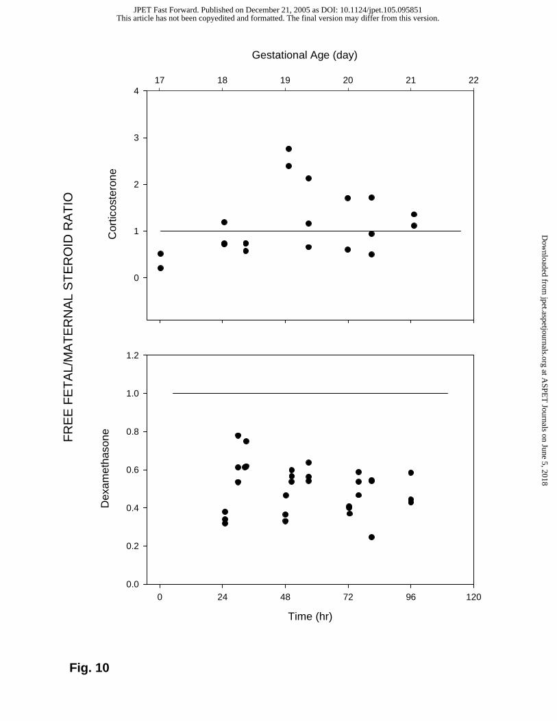
Fig. 10
Time (hr)
0 24 48 72 96 120
Dex
amet
haso
ne
0.0
0.2
0.4
0.6
0.8
1.0
1.2
FR
EE
FE
TA
L/M
AT
ER
NA
L S
TE
RO
ID R
AT
IO
Cor
ticos
tero
ne
0
1
2
3
4
Gestational Age (day)
17 18 19 20 21 22
This article has not been copyedited and formatted. The final version may differ from this version.JPET Fast Forward. Published on December 21, 2005 as DOI: 10.1124/jpet.105.095851
at ASPE
T Journals on June 5, 2018
jpet.aspetjournals.orgD
ownloaded from