regulation phosphorylation proteins i, lila, 'bproteini is aneuron-specificproteinpresentin...
TRANSCRIPT

Proc. Natl Acad. Sci. USAVol. 79, pp. 6075-6079, October 1982Neurobiology
Regulation of phosphorylation of proteins I, lila, and "'b in ratneurohypophysis in vitro by electrical stimulation and byneuroactive agents
(calcium/cAMP/dopamine/fluphenazine)
KANG Tsou* AND PAUL GREENGARDDepartment of Pharmacology, Yale University School of Medicine, 333 Cedar Street, New Haven, Connecticut 06510
Contributed by Paul Greengard, June 1, 1982
ABSTRACT The state of phosphorylation of proteins I, HI.,and Iffb-neuron-specific phosphoproteins-was studied in neu-rosecretory endings of the neurohypophysis in vitro. Briefperiods(a few seconds) of electrical stimulation caused large increases inthe state of phosphorylation of all three proteins. The three pro-teins were dephosphorylated within 1 min after termination of thestimulation. High potassium, 8-bromo-cAMP, and dopamine alsostimulated the phosphorylation of the three proteins. The effectof dopamine was blocked by the dopamine antagonist fluphen-azine. Peptide mapping of protein I revealed that electrical stim-ulation or high potassium increased the state of phosphorylationof two regions of the molecule, whereas 8-bromo-cAMP and do-pamine increased the state of phosphorylation of only one of theseregions.
Protein I is a neuron-specific protein present in most and prob-ably all presynaptic terminals, where it is at least partially as-sociated with neurotransmitter vesicles (1-5). It is a major en-dogenous substrate for both cAMP-dependent (1) and calcium/calmodulin-dependent (6, 7) protein kinases. Neurotransmit-ters (8, 9), apparently acting through cAMP, and depolarizingagents (8-10), apparently acting through calcium, have beenshown to increase the state of phosphorylation of protein I inintact preparations of the central and peripheral nervous sys-tem. Recently, it has been shown that brief periods of nerveimpulse conduction increase the state of phosphorylation ofprotein I in the rabbit superior cervical ganglion (11). These andother findings suggest that protein I may have a functional rolein the nerve terminal (12). Proteins IIIa and IIIb are also neuron-specific proteins that have cellular and subcellular distributionsand certain other properties similar to those ofprotein 1 (13, 14).
The neurohypophysis is a neuroendocrine organ of quite ho-mogeneous composition, the nervous structure ofwhich consistentirely of nerve terminals without any postsynaptic neurons.Vasopressin and oxytocin release from the neurohypophysis canbe detected in vitro, and the rate of release of these neurohor-mones can be manipulated easily. The state of phosphorylationof proteins I, IIIa, and IIIb can be determined in a single neu-rohypophysis of the rat. Therefore, we have studied the regu-lation of the state of phosphorylation of these proteins in theneurohypophysis as a step towards understanding their possiblerole in the function of nerve terminals.
METHODSIsolation, Incubation, and Stimulation of Neurohypophysis.
Sprague-Dawley rats (150-175 g) were decapitated, and theirskulls were opened through the sagittal suture. The brainstem
was lifted from behind so that the pituitary stalk could be seen,and the stalk was cut underneath the basal hypothalamus. Thebrain was then removed. After dissecting away the coveringsheath, the whole pituitary gland was removed from the baseof the skull with a moistened brush and placed in a Petri dishcontaining standard buffer (Krebs-Ringer bicarbonate buffercontaining 125 mM NaCl/5.0 mM KCI/25 mM NaHCO3/1.0mM CaCl2/1.5 mM Na2HPO4/1.5 mM MgSO4/10 mM glu-cose). The neurohypophysis with its stalk and attached frag-ments of the pars intermedia was separated from the anteriorpituitary with a brush. A half cut along the posterior midlineof the neurohypophysis was made to facilitate penetration ofdrugs into the gland.The neurohypophysis was incubated and stimulated as de-
scribed in the physiological studies of Lightman et al. (15) onvasopressin release. The pituitary stalk was drawn into the tipofa platinum wire suction electrode. The gland was then placedin a glass vial containing 15 ml of standard buffer and gassedcontinuously with 95% 02/5% CO2. The gland was preincu-bated at 370C for 45 min before stimulation or treatment withneuroactive agents. The stimulation parameters studied weresimilar to those examined in an investigation of the effect ofim-pulse conduction on the state of phosphorylation of protein Iin the rabbit superior cervical ganglion (11). Stimulation wascarried out with pulses shown in preliminary experiments to besupramaximal: a pulse amplitude of 40 V and a pulse width of2 msec.
Extraction of Proteins I, IIIH, and IIb and Assay of TheirDephosphorylated Forms. The technique of back-phosphoryl-ation (10) was used to quantitate the amounts of dephospho-proteins I, I11a, and IIIb. The principle of this technique is tohomogenize the tissue in the presence ofZn2+, which preventschanges in the state of phosphorylation of phosphoproteins byinhibiting endogenous protein kinase and protein phosphataseactivities. The proteins are then extracted, and the dephos-phorylated forms are phosphorylated with [y-32P]ATP by theexogenous catalytic subunit ofcAMP-dependent protein kinase.
After electrical stimulation or treatment with neuroactiveagents, each neurohypophysis was transferred into a glass ho-mogenizer containing 2 ml of ice-cold Zn acetate (5 mM) andhomogenized with a Teflon pestle. The homogenate was cen-trifuged at 4,000 X g for 10 min, and the pellet was resuspendedin 220 ,ul of 0.01 M citric acid (pH 3.0) at 4°C. The suspensionwas centrifuged at 23,000 x g for 15 min, and a 200-,ul aliquotof the supernatant was adjusted to pH 6 with 0.5 M Na2HPO4and centrifuged at 15,000 x g for 10 min. A 60-pul aliquot of the
Abbreviation: kDal, kilodaltons.* Present address: Department of Pharmacology III, Shanghai Instituteof Materia Medica, Chinese Academy of Sciences, Shanghai, 200031,People's Republic of China.
6075
The publication costs ofthis article were defrayed in part by page chargepayment. This article must therefore be hereby marked "advertise-ment" in accordance with 18 U. S. C. §1734 solely to indicate this fact.
Dow
nloa
ded
by g
uest
on
Janu
ary
20, 2
020
6076 Neurobiology: Tsou and Greengard
supernatant was used for protein assay. All pH 6 supernatantsfrom one experiment were then diluted to equal protein con-
centration with 10 mM Na citrate/phosphate buffer, pH 6.The standard back-phosphorylation assay mixture (final vol-
ume, 100 1l) contained 50mM Hepes (pH 7.4), 10mM MgCl2,1 mM EDTA, 3 AuM [y-32P]ATP (specific activity 50-100 cpmfmol'), 60 ,ul oftissue extract, and a low concentration (7.5 nM)of the purified catalytic subunit of cAMP-dependent proteinkinase. (This enzyme was prepared from bovine heart and sup-
plied by A. C. Nairn of this laboratory.) The assay was carriedout for 30 min at 30TC, the reaction was terminated by boilingthe assay mixture in "NaDodSO4 stop solution," and the sam-
ples were then subjected to NaDodSO4/polyacrylamide gelelectrophoresis and autoradiography as described (10). Thebands containing proteins I, IIIa, and IIIb were cut from eachlane ofthe slab gels, and radioactivity was determined by liquidscintillation spectrometry. Under the assay conditions used,phosphorylation of proteins I, IIIa, and I"b was proportionalto the amount of tissue extract over a 10-fold concentrationrange of extract.The standard assay conditions, involving a low concentration
of catalytic subunit, were chosen in order to provide selectivephosphorylation of the site in protein I that is phosphorylatedunder physiological conditions by both a cAMP-dependent anda calcium/calmodulin-dependent protein kinase (6, 16). Thissite is located in the 10-kilodalton (kDal) fragment obtainedupon digestion ofprotein I with Staphylococcus aureus protease(see below). Under standard back-phosphorylation assay con-
ditions, this site accounted for 95% of the total phosphate in-corporated into protein I (data not shown).
The total amounts ofproteins I, IIIa, and IIIb were not alteredby any of the experimental procedures used in this study.Therefore, changes in the amount of the dephosphorylatedforms of proteins I, Ina, and Ilb reflect changes in the statesof phosphorylation of those proteins.
Assay of Individual Phosphorylation Sites in Protein I. Inexperiments in which the state ofphosphorylation of individualsites in protein I was to be determined, the conditions of thestandard back-phosphorylation procedure were used except for
I 2 s 4
Ia---
44 kDaI so
32 kDaI
Elec. stim. + - +
-100
- 70un
~0
0
45 ~2
23
Ca 2- + + -
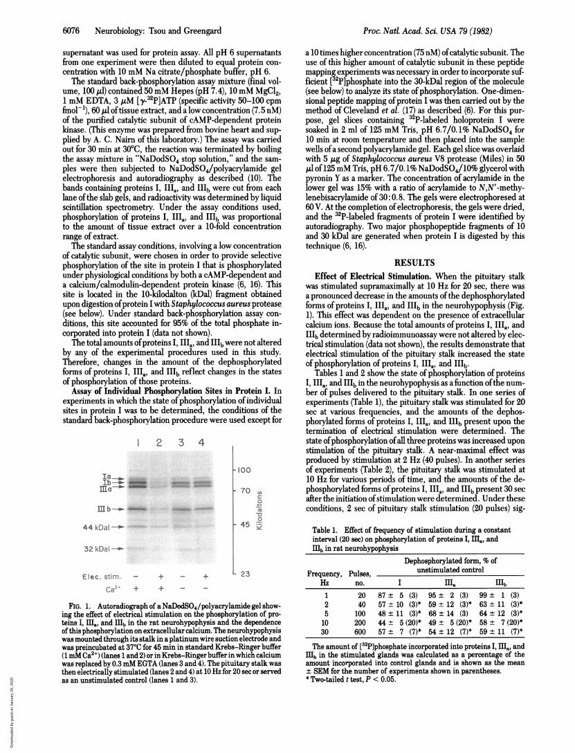
FIG. 1. Autoradiograph of a NaDodSO4/polyacrylamide gel show-ing the effect of electrical stimulation on the phosphorylation of pro-teins I, E., and IIIb in the rat neurohypophysis and the dependenceof this phosphorylation on extracellular calcium. The neurohypophysiswas mounted through its stalk in a platinum wire suction electrode andwas preincubated at 370C for 45 min in standard Krebs-Ringer buffer(1mM Ca2+) (lanes 1 and 2) or in Krebs-Ringer buffer in which calciumwas replaced by 0.3mM EGTA (lanes 3 and 4). The pituitary stalk wasthen electrically stimulated (lanes 2 and 4) at 10 Hz for 20 sec or servedas an unstimulated control (lanes 1 and 3).
a 10 times higher concentration (75 nM) ofcatalytic subunit. Theuse of this higher amount of catalytic subunit in these peptidemapping experiments was necessary in order to incorporate suf-ficient [ P]phosphate into the 30-kDal region of the molecule(see below) to analyze its state ofphosphorylation. One-dimen-sional peptide mapping ofprotein I was then carried out by themethod of Cleveland et al. (17) as described (6). For this pur-pose, gel slices containing 3 P-labeled holoprotein I weresoaked in 2 ml of 125 mM Tris, pH 6.7/0.1% NaDodSO4 for10 min at room temperature and then placed into the samplewells ofa second polyacrylamide gel. Each gel slice was overlaidwith 5 Ag of Staphylococcus aureus V8 protease (Miles) in 50A1 of 125mM Tris, pH 6.7/0.1% NaDodSO4/10% glycerol withpyronin Y as a marker. The concentration of acrylamide in thelower gel was 15% with a ratio of acrylamide to N,N'-methy-lenebisacrylamide of 30:0.8. The gels were electrophoresed at60 V. At the completion of electrophoresis, the gels were dried,and the 32P-labeled fragments of protein I were identified byautoradiography. Two major phosphopeptide fragments of 10and 30 kDal are generated when protein I is digested by thistechnique (6, 16).
RESULTSEffect of Electrical Stimulation. When the pituitary stalk
was stimulated supramaximally at 10 Hz for 20 sec, there wasa pronounced decrease in the amounts ofthe dephosphorylatedforms of proteins I, IIIa, and IIIb in the neurohypophysis (Fig.1). This effect was dependent on the presence of extracellularcalcium ions. Because the total amounts of proteins I, IIIa, andIIIb determined by radioimmunoassay were not altered by elec-trical stimulation (data not shown), the results demonstrate thatelectrical stimulation of the pituitary stalk increased the stateof phosphorylation of proteins I, IIIa, and IIIb.
Tables 1 and 2 show the state of phosphorylation of proteinsI, IIIa, and IIIb in the neurohypophysis as a function ofthe num-ber of pulses delivered to the pituitary stalk. In one series ofexperiments (Table 1), the pituitary stalk was stimulated for 20sec at various frequencies, and the amounts of the dephos-phorylated forms of proteins I, IIIa, and IIIb present upon thetermination of electrical stimulation were determined. Thestate ofphosphorylation ofall three proteins was increased uponstimulation of the pituitary stalk. A near-maximal effect wasproduced by stimulation at 2 Hz (40 pulses). In another seriesof experiments (Table 2), the pituitary stalk was stimulated at10 Hz for various periods of time, and the amounts of the de-phosphorylated forms ofproteins I, IIIa, and IIIb present 30 secafter the initiation ofstimulation were determined. Under theseconditions, 2 sec of pituitary stalk stimulation (20 pulses) sig-
Table 1. Effect of frequency of stimulation during a constantinterval (20 sec) on phosphorylation of proteins I, IIIa, andI1b in rat neurohypophysis
Dephosphorylated form, % ofFrequency, Pulses, unstimulated control
Hz no. I ma H1b1 20 87 ± 5 (3) 95 ± 2 (3) 99 ± 1 (3)2 40 57 ± 10 (3)* 59± 12 (3)* 63 ± 11 (3)*5 100 48± 11 (3)* 68± 14 (3) 64± 12 (3)*10 200 44 ± 5(20)* 49 ± 5(20)* 58 ± 7(20)*30 600 57 ± 7 (7)* 54 ± 12 (7)* 59 ± 11 (7)*
The amount of [32P]phosphate incorporated into proteins I, Ha, andI1b in the stimulated glands was calculated as a percentage of theamount incorporated into control glands and is shown as the mean+ SEM for the number of experiments shown in parentheses.* Two-tailed t test, P < 0.05.
Proc. Nad Acad. Sci. USA 79 (1982)
I L- %.Io -T
.m.p... , !W" 5*0"
A"k.,--- ......... ''L... --L- .-,-i,-.- .;---
Dow
nloa
ded
by g
uest
on
Janu
ary
20, 2
020

Proc. Natl. Acad. Sci. USA 79 (1982) 6077
Table 2. Effect of duration of stimulation at constant frequency(10 Hz) on phosphorylation of proteins I, a., and H1b inrat neurohypophysis
Duration of Dephosphorylated form, % ofstimulation, Pulses, unstimulated control
sec no. I [a ]b0 0 100 ± 9 100 ± 8 100 ± 112 20 45±8* 74±11 69±145 50 34 + 3* 63± 4* 65 ± 10*10 100 27 ±9* 57 ± 7* 44± 10*20 200 37 ± 2* 56 ± 10* 59 ± 12*
The amount of [32P]phosphate incorporated into proteins I, 11., andIb in the stimulated glands was calculated as a percentage of theamount incorporated using control glands and is shown as the mean+ SEM for four experiments.* Two-tailed t test, P < 0.05.
nificantly increased the state of phosphorylation of protein I,and 5 sec of stimulation (50 pulses) significantly increased thestate of phosphorylation of proteins III. and Ilb. Ten secondsof pituitary stalk stimulation (100 pulses) appeared to producea maximum phosphorylation of all three proteins.
Fig. 2 shows the changes in the amounts of the dephos-phorylated forms of proteins I and IIIa, observed in responseto pituitary stalk stimulation at 10 Hz for 20 sec, as a functionof the time after termination of the stimulation. A substantialdecrease in the amounts of dephosphoproteins I and IIIa wasobserved when the glands were extracted "immediately"-i. e.,within 20 sec after termination of the stimulation. The amountsof the dephosphorylated forms of proteins I and IIIa returnedto control levels within 1 min after termination of stimulation.Results obtained with protein Ilb (data not shown) in theseexperiments were erratic but similar to those shown in Fig. 2for proteins I and III.
Effect of High K+, 8-Bromo-cAMP, and Dopamine. HighK+ concentration (60mM for 30 sec) and 8-bromo-cAMP (4mMfor 10 min) reduced the amounts ofthe dephosphorylated formsof proteins I, III., and Illb in the rat neurohypophysis (Table3). The effect ofhigh K+ was abolished in calcium-free medium(not shown). Dopamine has been shown to be present in the ratneurohypophysis (18, 19) and to affect vasopressin release invitro (15). When the neurohypophysis of the rat was incubatedfor 3 min in the presence of 100 iM dopamine, a significantdecrease in the amounts of the dephosphorylated forms of pro-
100 --
C
20
tO0 3060 10 0
"a)
60M*3
20se Tiese0'~
00
to0 30 60 120 300
20-aec Time, sec
stim.
FIG. 2. Effect of electrical stimulation on the amounts of dephos-phoprotein I and dephosphoprotein ma in rat neurohypophysis as afunction of time after termination of stimulation. The pituitary stalkof each neurohypophysis was stimulated by means of a suction elec-trode at 10 Hz for 20 sec. The amount of [32P]phosphate incorporatedinto protein I and protein ma was calculated as the percentage of theamount incorporated using control glands. Each datum point repre-sents the mean ± SEM for four experiments. *, Protein I; o, proteinma.
teins I, IIi and IIIb was observed (Table 3). The effect of do-pamine was abolished in calcium-free medium (not shown). Theeffect of dopamine also was abolished by the dopamine antag-onist fluphenazine at 1 ,uM (Table 3). In contrast to the effectofdopamine, the a-agonist phenylephrine (100 ,M) and the 1-
agonist isoproterenol (10 ,uM) failed to alter substantially thestate of phosphorylation of proteins I, IIIa, and IIIb. These re-sults suggest that the effect of dopamine is not mediated by a-or ,B-adrenergic receptors but through a dopamine receptor.
Analysis of Phosphorylation Sites in Protein I. Protein I con-tains a serine residue that is phosphorylated both by a cAMP-dependent and by a calcium /calmodulin-dependent proteinkinase in the collagenase-insensitive region (10-kDal fragment)ofthe molecule and two serine residues that are phosphorylatedby a second calcium /calmodulin-dependent protein kinase inthe collagenase-sensitive region (30-kDal fragment) of the mol-ecule (6, 16). One-dimensional peptide mapping ofphosphoryl-ation sites in protein I revealed that the decrease in the amountof dephosphorylated holoprotein I seen in response either toelectrical stimulation or to high K+ concentration (Table 3) wasreflected in similar changes in both the 10-kDal fragment andthe 30-kDal fragment of the molecule (Figs. 3 and 4). In con-
Table 3. Regulation of the state of phosphorylation of proteins I, ma. and H1b inrat neurohypophysis
Duration oftreatment, Dephosphorylated form, % of unstimulated control
Agent sec I ma mb1. Electrical stimulation, 10 Hz 20 46 ± 7* 58 ± 7* 49 ± 7*2. High K+, 60 mM 30 28 ± 9* 42 ± 11* 33 ± 9*3. 8-Bromo-cAMP, 4 mM 600 28 ± 5* 25± 4* 27 ± 9*4. Dopamine, 100 AuM 180 60 ± 6* 54 ± 9* 67 ± 11*5. Dopamine, 100 ,uM, and
fluphenazine, 1 ,uM 180 104 (98-110) 102 (89-115) 89 (78-100)6. Phenylephrine, 100 pM 180 98 (88-108) 106 (101-111) 82 (77-87)7. Isoproterenol, 10 1M 180 122 (94-150) 125(122-128) 104 (99-109)
Neurohypophyses were incubated under the conditions shown. All test agents were present in standardbuffer except for dopamine, which was present in standard buffer containing 1 mM ascorbic acid to preventoxidation. The amount of [32P]phosphate incorporated into proteins I, iEa, and H1b in the experimentalglands was calculated as a percentage of the amount incorporated using control glands and is shown asthe mean ± SEM for six experiments with each agent 1-4 and as the mean and rangefor two experimentswith each agent 5-7.* Two-tailed t test, P < 0.05.
Neurobiology: Tsou and Greengard
Dow
nloa
ded
by g
uest
on
Janu
ary
20, 2
020

6078 Neurobiology: Tsou and Greengard
8
!32P]
Elec stim. - +
HighK+ - - +
-Br-cAMP - -
phosphate 100 55 40
incorp., % 100 41 18
Standardbuffer
FIG. 3. Autoradiograph showingtion and of neuroactive agents on phoin protein I. Rat neurohypophyses welation at 1 Hz for 20 sec or to high K+(4mM, 10 min), or dopamine (100 IAM,was dissolved in a modified buffer (sta1 mM ascorbic acid added to prevent itEextracted from the gland and subjectStaphylococcus aureus protease. Autosultant phosphopeptide pattern obtairkDal and 10-kDal fragments. The amrated into each of the two fragments ithis experiment was calculated as a pporated under control conditions. inco
trast, 8-bromo-cAMP and dopaminon the 10-kDal fragment.
DISCUSSIt seems likely that proteins I, IIpresent investigation, were locate(ings of the neurohypophysis ratherof this gland. The neurohypophysis
4-a0
0
C.)~0
EInCDc
0
10-
Elec.stim. High K+
system, the nervous structure ofwhich is composed entirely ofnerve terminals. Moreover, the nerve terminals are predomi-nantly secretory endings, although nonneurosecretory termi-nals containing y-aminobutyric acid or dopamine have been
.b4.r30 kDaI reported. to be present (18-20). Peptides found in the neuro-hypophysis, like enkephalins (21) and dynorphin (22), have beenreported to exist with either vasopressin or oxytocin in the neu-rosecretory terminals. Although our neurohypophyseal prepa-rations were contaminated with pars intermedia fragments, it
iii^ ^ has been demonstrated by immunocytochemistry (3) that theIO kDa intermediate lobe has very little protein I in contrast to its abun-
_ dance in the nerve terminals of the neurohypophysis. Thus, itseems likely that the phosphorylation changes observed in pro-
_ - + Dopamine tein I in this study occurred in the neurosecretory endings.+ Proteins I'Ia and IIIb are neuron-specific proteins, which have
a cellular and subcellular distribution similar to that of protein85 100 103 30 kDaI I (13, 14); therefore, it seems likely that the phosphorylation6 100 53 10 kDal changes observed in proteins I"Ia and IIIb also occurred in theneurosecretory endings of the neurohypophysis.
Modified The results of the present study indicate that the state ofbuffer phosphorylation of proteins I, IIIa, and II'b in the neurohy-
pophysis is regulated by neuroactive agents in a manner similarthe effect of electrical stimula- to that demonstrated earlier for these phosphoproteins in slicesisphorylation of individual sites of rat cerebral cortex (10), in slices of rat facial motonucleus (8),re subjected to electrical stimu- in slices of bovine superior cervical ganglion (9), and in intact(60 mM, 30 sec), 8-bromo-cAMIP rabbit superior cervical ganglion (11, 23). In addition, there are
indard Krebs-Ringer buffer with several similarities between the effects of electrical stimulationoxidation).] Then, protein I was on protein I phosphorylation in the neurohypophysis (presented to proteolytic digestion with study) and the rabbit superior cervical ganglion (11). However,)radiography illustrates the re- a significantly greater reduction (>70%) in the amount of de-ned. The arrows indicate the 30 phosphoprotein I was observed in the neurohypophysis, whichunder various test conditions in contains only nerve terminals, than in the other four prepara-urcentage of the amount incor- tions examined, where the maximal decrease in dephospho-rp., Incorporation. protein I was about 50% (8-10, 23). This may be explained by
the presence of both presynaptic and postsynaptic elements inie had significant effects only these latter preparations because postsynaptic protein I is ap-
parently not responsive to various electrical or chemical stimuli(23).
ION Proteins I, IIIa, and IIIb have been shown to be enriched inIaandstudied in the purified preparations of synaptic vesicles (2, 5, 14, 24). These[I., and IIIb, studied m the and other observations (12) suggest that these phosphoproteinsd in the neurosecretory end- may be involved in regulation of release from nerve terminals.
than in some other structure Thepresent results indicate that large changes occur in the stateis a relatively homogeneous of phosphorylation of these proteins in the nerve endings of the
neurohypophysis in response to procedures that affect the re-lease of hormones from these nerve endings (15, 25). For in-
_- -__ _ stance, the changes in state of phosphorylation of proteins I,
IIIa' and IIIb seen in the neurohypophysis in response to elec-trical stimulation and to high K+ depended on extracellular cal-cium. Vasopressin release in response to electrical stimulationand high K+ also showed a dependence on extracellular calcium
TX (26). The optimal stimulus frequency for the phosphorylationofproteins I, IIIa, and Hlb in the present work was about 10 Hz,in agreement with the optimal stimulus frequency of 10-20 Hzfor vasopressin release in vitro (25) and with the discharge fre-quency of magnocellular neurons observed in vivo in water-de-prived rats (27). Thus, the neurohypophysis should be an ex-cellent system in which to study the role of protein phosphoryl-
8-Br-cAMP Dopamine ation in general and of proteins I, IIIa, and IIIb in particular inthe regulation of release from nerve endings.
MG. 4. Effect of electrical stimulation and of neuroactive agentson phosphorylation of individual sites in protein Iin rat neurohypo-physis. Experimental conditions were asdescribed inthelegendto Fig.3. The amount of [32P]phosphate incorporated into the 30-kDal(I) and10-kDal (0 fragments under various test conditions was calculatedas a percentage of the amount incorporated under control conditionsand is shown as the mean ± SEM for four experiments.
This work was supported by U.S. Public Health Service Grants MH-17387 and NS-08440 and by a grant from the McKnight Foundation.
1. Ueda, T. & Greengard, P. (1977) J. BioL Chem. 252, 5155-5163.2. Bloom, F. E., Ueda, T., Battenberg, E. & Greengard, P. (1979)
Proc. NatL Acad. Sci. USA 76, 5982-5986.
Proc. Natl. Acad. Sci. USA 79 (1982)
a's - A_IMM- _
-A -P __--!-1_-A!
Dow
nloa
ded
by g
uest
on
Janu
ary
20, 2
020

Proc. NatL Acad. Sci. USA 79 (1982) 6079
3. DeCamilli, P., Ueda, T., Bloom, F. E., Battenberg, E. & Green-gard, P. (1979) Proc. Nati Acad. Sci. USA 76, 5977-5981.
4. Fried, G., Nestler, E. J., De Camilli, P., Stjmrne, L., Olson, L.,Lundberg, J. M., HMkfelt, T., Ouimet, C. C. & Greengard, P.(1982) Proc. Nati Acad. Sci. USA 79, 2717-2721.
5. Huttner, W. B., De Camilli, P., Schiebler, W. & Greengard, P.(1981) Soc. Neurosci. Abstr. 7, 441.
6. Huttner, W. B. & Greengard, P. (1979) Proc. NatL Acad. Sci.USA 76, 5402-5406.
7. Kennedy, M. B. & Greengard, P. (1981) Proc. NatL Acad. Sci.USA 78, 1293-1297.
8. Dolphin, A. C. & Greengard, P. (1981)J. Neurosci. 1, 191-203.9. Nestler, E. J. & Greengard, P. (1980) Proc. NatL Acad. Sci. USA
77, 7479-7483.10. Forn, J. & Greengard, P. (1978) Proc. Nate Acad. Sci. USA 75,
5195-5199.11. Nestler, E. J. & Greengard, P. (1982) Nature (London) 296,
452-454.12. Greengard, P. (1981) Harvey Lect 75, 277-331.13. Huang, C. K., Browning, M. D. & Greengard, P. (1982)J. BioL
Chem. 257, 6524-6528.14. Browning, M. D., Huang, C. K. & Greengard, P. (1982) Soc.
Neurosci. Abstr., in press.15. Lightman, S. L., Iversen, L. L. & Forsling, M. L. (1982)J. Neu-
rosci. 2, 78-81.
16. Huttner, W. B., DeGennaro, L. J. & Greengard, P. (1981) J.Biol Chem. 256, 1482-1488.
17. Cleveland, D. W., Fischer, S. G., Kirschner, M. W. & Laemmli,U. K. (1977)J. BioL Chem.-252, 1102-1106.
18. Bjorklund, A., Moore, R. Y., Nobin, A. & Stenevi, U. (1973)Brain Res. 51, 171-191.
19. Baumgarten, H. G., Bjorklund, A., Hostein, A. F. & Nobin, A.(1972) Z. Zellforsch. 126, 483-517.
20. Oertel, W. H., Mugnaini, E., Tappaz, M. L., Weise, V. K.,Dahl, A.-L., Schmechel, D. E. & Kopin, I. J. (1982) Proc. NatlAcad. Sci. USA 79, 675-679.
21. Martin, R. & Voigt, K. H. (1981) Nature (London) 289, 502-504.22. Watson, S. J., Akil, H., Fischli, W., Goldstein, A., Zimmerman,
E. & Nilaver, G. (1982) Science 216, 85-87.23. Nestler, E. J. & Greengard, P. (1982) J. Neurosci., in press.24. Ueda, T., Greengard, P., Berzins, K., Cohen, R. S., Blomberg,
F., Grab, D. J. & Siekevitz, P. (1979) J. Cell Biol 83, 308-319.25. Douglas, W. W. (1974) in Handbook of Physiology, Section 7,
Volume IV, Part 1 (American Physiology Society, Washington,DC), pp. 191-224.
26. Douglas, W. W. & Poisner, A. M. (1964)J. Physiol (London) 172,1-18.
27. Dyball, R. E. J., Wakerley, J. B., Poulain, D. A. & Brimble, M.J. (1977) Neurohypophysis, International Conference (Karger,Basel, Switzerland), pp. 78-87.
Neurobiology: Tsou and Greengard
Dow
nloa
ded
by g
uest
on
Janu
ary
20, 2
020