sa mediese tydskrif hereditary dysrhythmic congestive
TRANSCRIPT
18 Desember 1976 SA MEDIESE TYDSKRIF 2119
Hereditary Dysrhythmic Congestive CardiomyopathyA. J. BRINK,
SUMMARY
M. TORRINGTON, J. J. VA DER WALT
CASE REPORT
A 13-year-old girl was admitted to hospital 3 hours afterhaving suffered a sudden loss of consciousness associatedwith respiratory arrest and circulatory collapse. The rapidapplication of external carctiac massage and mouth-to·mouth respiration by a trained nurse enabled the patientto reach the hospital alive,
In the carctiac intensive care unit it was established thatshe had ventricular tachy-arrhythmia. The patient had astormy hospital course with repeated ventricular arrhythmias, inclucting bouts of ventricular fibrillation, and hadrepeated direct-current shock therapy. The arrhythmiaswere extremely resistant to treatment with all forms ofanti-arrhythmic drugs. An attempt to overdrive the ventricles by means of a pacemaker was also ineffective andhad to be discontinued. Partial control of the arrhythmiawas eventually achieved by a combination of digoxin, propranolol, quinidine sulphate and diphenylhydantoin. Hercondition improved to the extent that she could be discharged from hospital 2 months after admission.
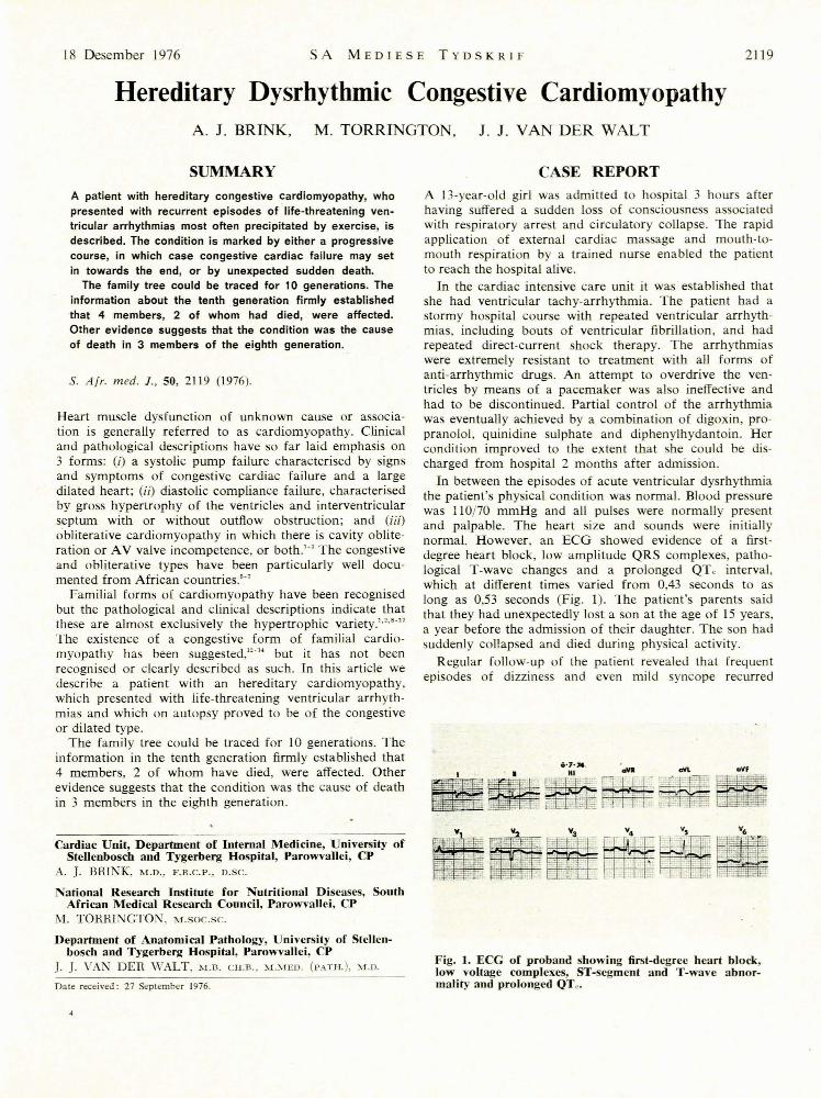
In between the episodes of acute ventricular dysrhythmiathe patient's physical condition was normal. Blood pressurewas 110/70 mmHg and all pulses were normally presentand palpable. The heart size and sounds were initiallynormal. However, an ECG showed evidence of a firstdegree heart block, low amplitude QRS complexes, pathological T-wave changes and a prolonged QTc interval,which at different times varied from 0,43 seconds to aslong as 0,53 seconds (Fig. 1). The patient's parents saidthat they had unexpectedly lost a son at the age of 15 years,a year before the admission of their daughter. The son hadsuddenly collapsed and died during physical activity.
Regular follow-up of the patient revealed that frequentepisodes of dizziness and even mild syncope recurred
A patient with hereditary congestive cardiomyopathy, whopresented with recurrent episodes of life-threatening ventricular arrhythmias most often precipitated by exercise, isdescribed. The condition is marked by either a progressivecourse, in which case congestive cardiac failure may setin towards the end, or by unexpected sudden death.
The family tree c·ould be traced for 10 generations. Theinformation about the tenth generation firmly establishedthat 4 members, 2 of whom had died, were affected.Other evidence suggests that the condition was the causeof death in 3 members of the eighth generation.
S. Afr. med. l., 50, 2119 (1976).
Heart muscle dysfunction of unknown cause or association is generally referred to as cardiomyopathy. Clinicaland pathological descriptions have so far laid emphasis on3 forms: (i) a systolic pump failure characterised by signsand symptoms of congestive cardiac failure and a largedilated heart; (ii) diastolic compliance failure, characterisedby gross hypertrophy of the ventricles and interventricularseptum with or without outflow obstruction; and (iii)obliterative cardiomyopathy in which there is cavity obliteration or A V valve incompetence, or both.'" The congestiveand obliterative types have been particularly well docu·mented from African countries.'"
Familial forms of cardiomyopathy have been recognisedbut the pathological and clinical descriptions indicate thatthese are almost exclusively the hypertrophic variety.""""The existence of a congestive form of familial cardio·myopathy has been suggested,"'" but it has not beenrecognised or clearly described as such. In this article wedescribe a patient with an hereditary cardiomyopathy,which presented with life-threatening ventricular arrhythmias and which on autopsy proved to be of the congestiveor dilated type.
The family tree could be traced for 10 generations. Theinformation in the tenth generation firmly established that4 members, 2 of whom have died, were affected. Otherevidence suggests that the condition was the cause of deathin 3 members in the eighth generation.
0-7'''. ....~~.~ ..[-.~aVf
Cardiac Unit, Department of Internal Medicine, University ofSteHenbosch and Tygerberg Hospital, Parowvallei, CP
A. J. BRINK, M.D., F.R.C.P., D.SC.
National Research Institute for Nutritional Diseases, SouthAfrican Medical Research Council, Parowvallei, CP
M. TORRINGTON, M.SOC.Sc.
Department of Anatomical Pathology, Universit)' of Stellenbosch and Tygerberg Hospital, Parowvallei, CP
]. ]. VAN DER WALT, "-LB. CH.B., ;VOIED. (PATH.), M.D.
Date received: 27 September 1976.
Fig. ]. ECG of proband showing first-degree heart block,low voltage complexes, ST-segment and T-wave abnoTmality and prolonged QTc •

2120 SA MEDICAL Jo R AL 18 December 1976
despite the therapy. These episodes were precipitated byphysical activity.
Because it was thought possible that the prolonged QTeinterval might reflect the underlying cause of the ventricular tachy-arrhythmias, the QTe interval was studied afterstellate ganglion block. tO A left cervical ganglionectomy wasdone on the strength of these studie . For a time it wasthought that mild improvement had occurred, but this ideawas oon di pelled. For some months the more seriousventricular rhythm disturbances abated and a supraventricular arrhythmia resembling an atrial flutter appearedperiodically.
Two years after the initial observations, clinical andradiological evidence showed that cardiac enlargement wastaking place (Fig. 2). A third heart sound was audible anda murmur indicating mitral incompetence appeared. Crisesof ventricular tachy-arrhythmia again supervened (Fig. 3)and she was repeatedly admitted to hospital. Progressivecardiac enlargement (Fig. 2) and an increasing degree ofimpairment of effort tolerance occurred and eventuallyleft ventricular failure supervened. The patient died 4years after first coming under observation, in a condition ofcardiac failure and with persisting crises of ventriculararrhythmia.
Fig. 2. Chest radiograph of proband showing cardiacenlargement.
AUTOPSY FINDINGS
Macroscopic ExaminationAutopsy consent was limited to an examination of the
heart only. Generalised cardiomegaly was present due to
111
4'9·73 04hOO
4·9·73
14·12·73
Fig. 3. ECG rhythm strips from proband showing ventricular tachy-arrhythmia.
hypertrophy and dilatation of all the chambers. The dilatation was more marked on the right side of the heart, butit masked hypertrophy on both sides. However, the hypertrophy was reflected in the heart weight of 482 g. Therewas no asymmetrical septal hypertrophy. The parietalendocardium of the left atrium and left ventricle showedfoci of mild thickening. Organising thrombi were presentin the right atrium as well as in both ventricles. There wereno valvular abnormalities.
Microscopic Examination
The myocardial fibres were regularly arranged and werehypertrophied but attenuated. A moderate degree of nuclearpleomorphism was present, but there were no perinuclearhaloes or glycogen pools." Occasional myofibres showedbasophilic degeneration. Small foci of active myocytolysiswere present in relation to mural thrombi or focal endocardial thickening. All chambers showed slight focal fibro-
Fig. 4. Endocardial fibro-elastosis and active myocytolysis.Note pleomorphic nuclei (Verhoeff x 200).
18 Desember 1976 SA MEDIE E TYD KRIF 2121
elastotic thickening of the parietal endocardium (Fig. 4).Small stellate areas of connective tissue increase werepresent subendocardialJy (Fig. 5). Vessels included in thesections were normal. The conduction system was dissectedaccording to the method of Hudson," but no lesions of theconducting tissues could be demonstrated.
Fig. 5. Endocardial fibro-elastosis, myofibre hypertropbyand interstitial stellate scars (Verboeff x 200).
FAMILY mSTORY
During an interview with the parents of the proband, itwas suggested by the father that 2 teenage sisters 'somehowrelated to him' were affected with a condition similar tothat of his daughter. The parents of the affected childrenwere traced. and by means of further interviews and withthe assistance of the Cape Town Dutch Reformed ChurchArchives it was possible to show that the 2 girls. aged atthis time 20 and 21 years respectively, were related throughtheir paternal branch to the proband's family. Both thesepatients had already been the subjects of a previous reportentitled 'Familial paroxysmal ventricular tachycardia'."
These girls were again examined and apart from thepreviously described episodes of syncope and vascular collapse, now admitted to dyspnoea with effort. Radiologicalexamination showed them to have cardiac enlargementand the ECGs of both girls were similar to that of theproband. Their ECGs showed low-voltage QRS complexeswith abnormal ST- and T-wave changes, and in one of thepatients abnormal left axis deviation of - 60·, suggestingleft anterior hemiblock. The echocardiogram did not revealevidence of hypertrophic cardiomyopathy. Both werereasonably well controlleo on anti-arrhythmic drugs, butwere still subject to episodes of dizziness and even syncope.Exercise was again a prominent precipitant of suchepisodes.
Genealogy
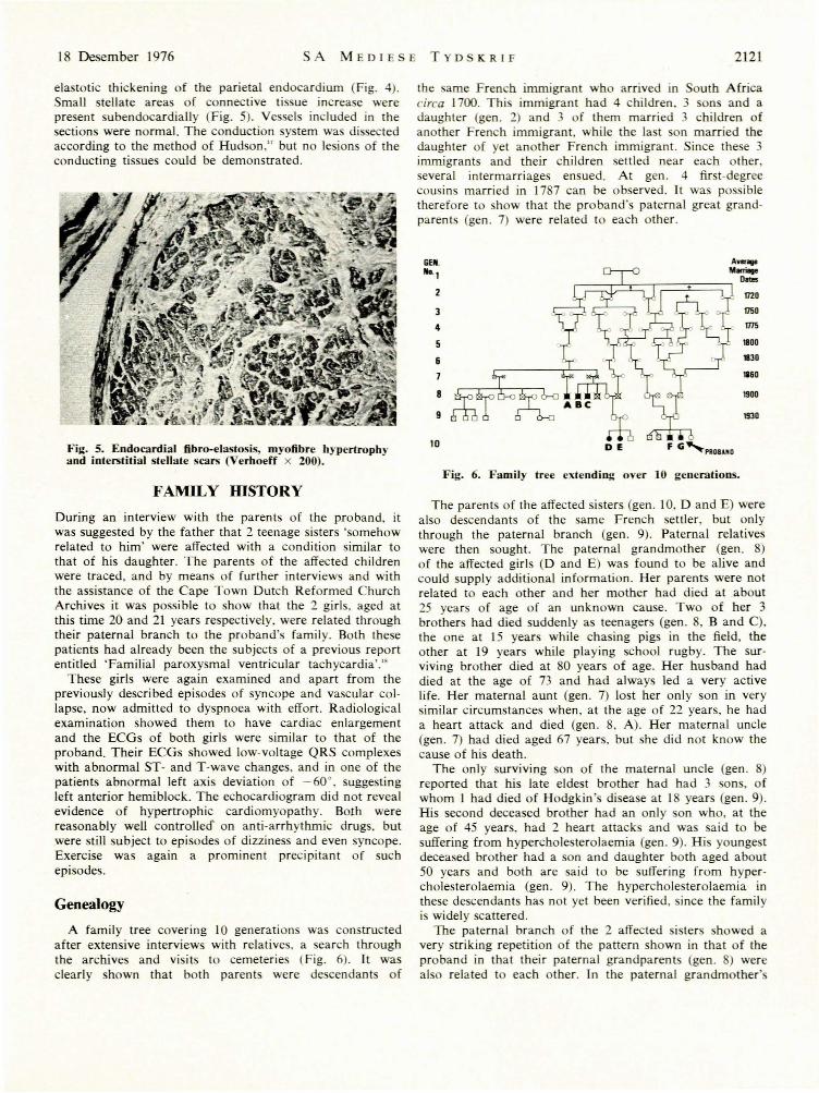
A family tree covering 10 generations was constructedafter extensive interviews with relatives. a search throughthe archives and visits to cemeteries (Fig. 6). It wasclearly shown that both parents were de cendant of
the same French immigrant who arrived in outh Africacirca 1700. Thi immigrant had 4 children, 3 sons and adaughter (gen. 2) and 3 of them married 3 children ofanother French immigrant, while the la t on married thedaughter of yet another French immigrant. Since the e 3immigrants and their children settled near each other,everal intermarriages ensued. At gen. 4 first-degree
cousins married in 1787 can be observed. It was possibletherefore to show that the proband's paternal great grandparents (gen. 7) were related to each other.
GEIl. A_"1 M......
DI1IIS
2 1720
3 1750
4 ms
5 1800
6 1830
7 1860
8 1900
9 1930
10 DE
Fig. 6. Family tree e:dending over 10 generations.
The parents of the affected sisters (gen. 10, D and E) werealso descendants of the ame French settler, but onlythrough the paternal branch (gen. 9). Paternal relativeswere then sought. The paternal grandmother (gen. 8)of the affected girls (D and E) was found to be alive andcould supply additional information. Her parents were notrelated to each other and her mother had died at about25 years of age of an unknown eau e. Two of her 3brothers had died suddenly a teenagers (gen. 8, B and C),the one at IS years while chasing pigs in the field, theother at 19 years while playing school rugby. The surviving brother died at 80 years of age. Her husband haddied at the age of 73 and had always led a very activelife. Her maternal aunt (gen. 7) lost her only son in verysimilar circumstances when, at the age of 22 years, he hada heart attack and died (gen. 8, A). Her maternal uncle(gen. 7) had died aged 67 years. but she did not know thecause of his death.
The only surviving son of the maternal uncle (gen. 8)reported that his late eldest brother had had 3 sons, ofwhom 1 had died of Hodgkin's disease at 18 years (gen. 9).His second deceased brother had an only on who, at theage of 45 years, had 2 heart attacks and was said to besuffering from hypercholesterolaemia (gen. 9). His youngestdeceased brother had a son and daughter both aged about50 years and both are said to be suffering from hypercholesterolaemia (gen. 9). The hypercholesterolaemia inthese descendants has not yet been verified, since the familyis widely scattered.
The paternal branch of the 2 affected i ters showed avery striking repetition of the pattern shown in that of theproband in that their paternal grandparents (gen. ) werealso related to each other. Tn the paternal grandmother'

2122 SA MEDICAL JOUR 'AL 18 December 1976
family (gen. 8, A, B and C) a consanguineous marriage(gen. 4) of 2 first-degree cousins had occurred,
DISCUSSIO
The clinical picture presented by the proband and the 2sisters (D and E) (Fig. 6) is unLike that of other casesof cardiomyopathy so far described, both with regardto the course of the disease and symptoms. Patients withcongestive forms of cardiomyopathy""" characteristicallydevelop evidence of congestive cardiac failure, without lifethreatening ventricular arrhythmias; so, too, do patientswith obliterative types of cardiomyopathy",7 and Laffler'sfibroplastic parietal endocarditis," Literature on the featuresand course of idiopathic non-obstructive cardiomyopathy',·,7.'" and on hypertrophic obstructive cardiomyopathy'·3,,, indicates that while sudden death and episodesof syncope are well known, this appears to be unrelated torecorded episodes of acute ventricular dysrhythmia. Theseforms of cardiomyopathy also do not tend to show a prolonged course with repeated episodes of syncope. Moreover, on closer examination relevant literature suggeststhat this type of history is to be found in the hypertrophicforms of cardiomyopathy and more frequently in thefamilial types of this particular condition.'·3,,,.,,
Another notable aspect of the symptomatology is theprecipitation of syncope by exercise, This was evident inthe history of the proband and of the related sisters (D andE) and the sudden death recorded in the deceased patients(B, C and F) was said to be related to exercise.
A prolongation of the QT interval was observed in theproband and directed the attention to the condition of theprolonged QT syndrome.""" Because of the gravity of thepatient's condition, and because of theoretical considerations on the imbalance of sympathetic tone and its influence in arrhythmias related to the prolonged QTc interval," a left cervical sympathectomy was done, No definiteimprovement was obtained by this procedure. The prolonged QTc interval was more likely an expression of thecardiomyopathy as such with enlargement of the heart.The cardiomyopathic aspect became more evident whenthe heart was observed to enlarge and when signs of congestive failure, a third heart sound and a murmur indicative of mitral incompetence appeared, The ECGs of theproband and the affected sisters (D and E) (Fig. 6) alsosuggested myocardial pathology and were unlike the ECGsof the hypertrophic types of cardiomyopathy.'
A familial and hereditary type of congestive cardiomyopathy is not well recognised. In a description of identical twins with cardiomyopathy the authors suggest that onetwin had a hypertrophic cardiomyopathy while the othermay have had a congestive form of this condition." Otherauthors do not attempt to draw a clear distinction betweenthe dillerent types when describing the familial forms, butthe pathological findings published generally support thediagnosis of hypertrophic varieties as being of the familialform."'''' An African type of congestive cardiomyopathydescribed in 2 brothers" is more likely to be coincidental inan area where this condition is extremely prevalent.
The pathological findings in the proband's heart werequite unlike those expected in classic hypertrophic cardio-
myopathy. The attenuated muscle fibres as well as the increase in the elastic and smooth muscle elements of theendocardium indicate that cardiac dilatation had beenpresent for a considerable time. Twenty-six blocks of theheart were examined, and in all but one the arrangement ofthe myofibres was very regular without any evidence of'whorl' formation as described by Olsen." The most likelydiagnosis is that this patient· has suffered from a congestivecardiomyopathy. In one block, however, two tiny foci wereencountered where the liypertrophied myocardial fibreswere irregularly arranged. However, If one applied thehistological hypertrophic cardiomyopathy index (HHI),allotting points for each of the observed histologicalchanges (fibrosis, bizarre l1uclei, disappearing myocardialfibres, perinuclear haloes, whorls and short runs of myofibres)," a value of only 40% was obtained. This is belowthe figure on which definite hypertrophic cardiomyopathycan be diagnosed. However, we considered whether thiscase did not belong to the very rare group of hypertrophic cardiomyopathies described by Olsen"', and in whichclinical obstruction had been minimal. Although asymmetrical septal hypertrophy is usually seen in these patients, itmay be absent and yet show a focal distribution of abnormal myocardial fibres. There were two reasons why itwas unlikely that our patient belonged to this category ofpatients: the HHI was low and only two tiny abnormal fociwere found in a very large number of blocks examined. Itwas therefore concluded that this patient had suffered fromcongestive cardiomyopathy.
The inheritance pattern is not clear. The father andmother of the 2 affected sisters were found to be normal."Both parents of the proband were also found to be normal.A familial relationship was, however, discovered betweenA-B-C, D-E and F-G. The grandfather of A-B and C(gen. 6) is a direct descendant via the male link of theeldest son of the initial immigrant. The paternal greatgrandfather of D and E (gen. 7) was also married to adescendant of the same immigrant but exact relationshipscould not be established because of lack of data. Thepaternal great-grandmother of F and G (gen. 7) was adirect descendant of the youngest son of the initial immigrant.
Thirty-seven marriages, between 1720 and 1860, werestudied. The surname of the initial immigrant seems todisappear entirely in the female line. Yet, in 1860 no lessthan 4 out of the 6 marriages contracted were betweencouples of whom the male or female were carriers of thesurname of the initial immigrant. It is possible, therefore,that the members of this family led very isolated livesand allowed a certain amount of inbreeding to take place.If a recessive mode of inheritance is considered for thefamily of the proband G, then the chances for a heterozygous condition in the parents can be calculated for themother as 1 : 512, for the father as 1 : 32. For the abnormality in D and E (gen. 10), intermarriage cannot beregarded as the possible mechanism, since the parents didnot have a known common ancestor. The possibility thatthe female parent of this marriage did carry the gene canonly be arrived at through knowledge of the incidence ofthe heterozygote in the general population. This is notavailable. The same applies to the family of A. Band C

I8 Desernber I976 SA MEDIESE TYDSKRIF 2123
(gen. 8). A dominant gene with variable penetration can beconsidered in individuals A, B, C, D and E, with a mutationin one of the parents (gen. 6). The fact of a commonancestor for the families of A, B, C, D, E, F and G maythen be coincidental. A more complex mode of inheritance where more than one gene is involved cannot beascertained with the present information. A multifactorialinheritance usually gives a clinical picture of different degrees of the condition in affected individuals. Perhaps thedisease is a result of an assortive mating and its expressionis regarded as a dominant new condition finding its originin an isolated community.""
To date the only other cases of comparable descriptionthat have been brought to our attention are those described as idiopathic familial myocardiopathy in 5 generations." The authors describe a non-hypertrophic cardiomyopathy with globular dilated left ventricles, endocardialfibro-elastosis and mitral regurgitation. This condition developed in several members of at least 3, and possibly 5,generations. However, symptoms in· the 4 patients who diedsuddenly were minimal during life and signs of heartfailure were present for only a brief time before death, ifat all. Precordial murmurs, moreover, were most oftendetected in the first few years of life. The inheritancepatterns of the disease suggest an autosomal dominantmode of inheritance.
The pathology described appears to resemble that of ourpatients. The clinical manifestations, however, differ: aprolonged course with recurrent frequent episodes ofventricular dysrhythmia and absence of early detection ofmurmurs occurs in our patients and not in theirs, but asimilarity exists with regard to minimal and late onset ofmyocardial failure.
The fatal forms of familial cardiac arrhythmias described by others31
-33 do not have the pathological features
of a congestive cardiomyopathy, nor could we find evidence of pathology in the conducting system such as wasthought to be present in other cases.31
,s>
The underlying genetic defect in our patients appears tohave a complex origin and to be the consequence of intermarriage and degree of inbreeding. In future it might bepossible to carry out studies to define such a defect moreaccurately, as has been done in the case of the inbred myopathy of the BlO.l4.6 strain of the Syrian hamster.34 This
myopathy, which presents most prominently as a conge tivetype of cardiomyopathy with clinical signs of cardiacfailure, has been shown to be the consequence of defectivetransfer ribonucleic acid.""'"
REFERE CES
I. Goodwin, J. F. (1970): Lancet, I, 731.2. Braunwald, E., Morrow, A. G., Cornell, W. P., Aygen, M. M. and
Hilbish, T. R. (1960): Amer. J. Med., 29, 924.3. Wigle, E. D. in Wolstenholrne, G. E. w. and O'Connor, M., eds
(1964): CIBA Foundation Symposium - Cardiomyopathies, p. 49.London: J. & A. Churchill.
4. Korb, G., Bajusz, E. and Rona, G., eds (1973): Cardiomyopathies,p. 9. Baltimore: University Park Press.
5. Bedford, D. E. and Konstam, G. L. S. (1946): Brit. Heart J., 8,236.
6. Becker, B. J. P., Chatgidakis, C. B. and Van Lingen, B. (1953):Circulation, 7, 345.
7. Brink, A. J. and Weber, H. W. (1966): S. Air. med. 1., 40, 455.Evans, W. (1949): Brit. Heart 1., 11, 68.
9. Gaunt, R. T. and Lecutier, M. A. (1956): Ibid., 18, 251.10. Battersby, E. J. and Glenner, G. G. (1961): Amer. J. Med., 30,
382.11. Barry, M. and Hall, M. (1962): Brit. Heart 1., 24, 613.12. Anselrni, A., Suarez, 1. A., Anselmi, G., Moleiro, F., Suarez, de C.
and Ruesta, V. (1975): Amer. J. Cardio!., 35, 97.13. Kariv, 1., Kreisler, B., Sherf, L., Feldman, S. and Rosenthal, T.
(1971): Amer. 1. Cardio!., 28, 693.14. Owor, R. and Rwomushana, R. J. W. (1975): E. Afr. med. 1.,
52, 372.15. Yanowitz, F., Preston, 1. B. and Abildskov, 1. A. (1966): Circulat.
Re ., 18, 416.16. Van oorden, S., Olsen, E. G. 1. and Pearse, A. G. E. (1971):
Cardiovasc. Res., 5. 118.17. Hudson, R. E. B. (1963): 1. clin. Path., 16,492.18. Sacks, H. S., Matison, R. and Kennelly, B. M. (1974): Amer. Heart
1., 87, 217.19. Brink, A. 1. and Weber, H. w. (1963): Amer. 1. Med., 34, 52.20. KUbler, W., Gleichman, U., Loogen, F. and LUck, 1. (1973): In
Op cit. 4 , p. 613.21. Loogen, F., Krelhaus, W. and KUbler, W. (1973): In Op cir. 4 , p. 629.22. lervell, A. and Lange· ielsen, F. (1957): Amer. Heart 1., 54, 59.23. Ward, O. C. (1964): 1. Irish med. Ass., 54, 103.24. Garza, L. A., Vick, R .. L., ora, 1. 1. and Mc arnara, D. G.
(1970): Circulation, 41, 39.25. lames, T. N. (1969): Mod. Conc. cardiov. Dis., 38. 35.26. Philips, 1. and lehinose, H. (1970): Chest, 58, 236.27. Emanuel, R., Withers, R. and O'Brien, K. (1971): Lancet, 2, 1065.28. Olsen, E. G. 1. (1971): CIBA Foundation Study Group No. 37,
p. 183. London: 1. & A. Churchill.29. Stern, C. (1960): Principals of Human Genetics, 2nd ed., p. 379.
San Francisco: W. H. Freeman.30. Ross, R. S., BulkJey, B. H., Hutchins, G. M., Harshey, S. 1., 10nes,
R. A., Kraus, H., Liebman, 1., Thorne, C. M., Weinberg, S. B. andWeech, A. A. (submitted for publication to Circulation).
31. Gault, 1. H., Cantwell, 1., Lev, M. and Braunwald, E. (1972):Amer. 1. Cardio!., 29. 548.
32. Green, 1. R., Korovetz, M. 1., ShankJin, D. R., De Vito, 1. 1. andTaylor, W. 1. (1969): Arch. intern. Med., 124, 359.
33. Wennevold, A., Melchior, 1. C. and Sandoe, E. (1965): Acta med.scand., 177, 557.
34. Lochner, A., Brink, A. 1. and Van der Wait, 1. 1. (1970): 1. Mo!.Cel!. Cardio!., I, 47.
35. Bester, A. 1. and Gevers, W. (1973): Biochem. 1., 132, 203.36. Idem (1975): 1. Mol. Cell. Cardio!., 7, 325.