the neocortex of cetaceans: cytoarchitecture and...
TRANSCRIPT

Ann. N.Y. Acad. Sci. ISSN 0077-8923
ANNALS OF THE NEW YORK ACADEMY OF SCIENCESIssue: New Perspectives on Neurobehavioral Evolution
The neocortex of cetaceans: cytoarchitecture andcomparison with other aquatic and terrestrial species
Camilla Butti,1 Mary Ann Raghanti,2 Chet C. Sherwood,3 and Patrick R. Hof11Department of Neuroscience, Mount Sinai School of Medicine, New York, New York. 2Department of Anthropology andSchool of Biomedical Sciences, Kent State University, Kent, Ohio. 3Department of Anthropology, The George WashingtonUniversity, Washington, DC
Address for correspondence: Camilla Butti, Department of Neuroscience, Mount Sinai School of Medicine, Box 1065, OneGustave L. Levy Place, New York, NY 10029. [email protected]
The evolutionary process of readaptation to the aquatic environment was accompanied by extreme anatomical andphysiological changes in the brain. This review discusses cortical specializations in the three major lineages of marinemammals in comparison to related terrestrial and semiaquatic species. Different groups of marine mammals adopteda wide range of strategies to cope with the challenges of aquatic living. Cetaceans and hippopotamids possess acompletely agranular neocortex in contrast to phocids and sirenians; vertical modules are observed in deep layersV and VI in manatees, cetaceans, phocids, and hippopotamids, but in different cortical areas; and clustering inlayer II appears in the insular cortex of hippopotamids, phocids, and cetaceans. Finally, von Economo neurons arepresent in cetaceans, hippopotamids, sirenians, and some phocids, with specific, yet different, cortical distributions.The interpretation of the evolutionary and functional significance of such specializations, and their relationshipswith the degrees of adaptation to the aquatic environment and phylogeny, remain difficult to trace, at least untilcomprehensive data, including representative species from all of the major mammalian families, become available.
Keywords: cetaceans; neocortex; evolution
What are marine mammals?
Marine mammals are a highly diverse group ofspecies that resulted from the reinvasion of theaquatic environment by terrestrial species. Ma-rine mammals are fully or partially dependent onthe aquatic environment for survival and includecetaceans (whales, dolphins, and porpoises), sireni-ans (manatees and dugongs), and some carnivores(sea otters, polar bears, and the pinnipeds, seals, sealions, and walruses). The degree of adaptation tothe aquatic environment is extremely variable, withcetaceans and the unrelated sirenians being the onlytwo extant groups fully dependent on an aquaticlifestyle.
Phylogenetic position, degree of adaptation tothe aquatic environment, and lifestyles all resultedin major differences in morphological and physi-ological adaptations, including those of the brain.Cetaceans evolved extremely large and convolutedbrains that set them apart from most other mam-
mals; sirenians represent a unique departure fromthe usual organization of the brain in large mam-mals, having a lissencephalic brain with only fewprominent fissures; and marine carnivores possessa brain that is comparable, at least in external mor-phology and proportions, to that of large terrestrialcarnivores (Fig. 1).
Origin and evolution of cetaceans,sirenians, and pinnipeds
According to the fossil record, cetaceans divergedfrom terrestrial mammals approximately 52 mil-lion years ago (mya).1 Early cetaceans, the Archeo-cetes, were a group of semiaquatic nonecholocat-ing and nonfilter-feeding animals inhabiting marineand fresh waters that arose from terrestrial mam-mals such as anthracotheres, raoellids, and mesony-chids.2 In the early Eocene between 45 and 53 mya,Archeocetes diversified into Pakicetidae, Ambulo-cetidae, and Remingtonocetidae, all inferred to be
doi: 10.1111/j.1749-6632.2011.05980.xAnn. N.Y. Acad. Sci. 1225 (2011) 47–58 c© 2011 New York Academy of Sciences. 47

Neocortex in marine mammals Butti et al.
Figure 1. Macroscopic views of the brains of the species dis-cussed in this review. Lateral (A, C, E, G, I), midline (B, D, F,L), dorsal (H, K), and coronal (J) views. c, caudal; d, dorsal; r,rostral; v, ventral. Scale bars = 5 cm.
semiaquatic mammals that could inhabit either landor sea.3 In the middle Eocene, a more derived groupof mammals arose, the Protocetidae, that possesseda lifestyle probably similar to modern pinnipeds and
lived in water but depended on a terrestrial lifestylefor reproduction. In the late Eocene, 38–40 mya, theBasilosauridae appeared, a group of fully aquaticmammals with morphological features and feedingand hearing capacities comparable to early odonto-cetes and mysticetes.4 The early Oligocene, about 35mya, is marked by the appearance of the Neoceti,5
the clade of modern whales including their stemtaxa. The oldest baleen-bearing mysticetes date backto the mid-Oligocene, about 28–29 mya, a period ofgreat diversification of Mysticeti.6 The oldest odon-tocetes are from the early Oligocene, about 32 mya.7
Sperm whales (Physeter macrocephalus and Kogiaspp.) are considered the most basal extant odonto-cetes.8 However, only the early Pliocene, 2.5–5 mya,witnessed the divergence of the main crown cetaceangenera.4,5
The classification and phylogenetic position ofcetaceans within Eutheria has been debated giventhe divergent conclusions drawn by different au-thors.4 Recent data provide both molecular9 andmorphological10,11 evidence for the inclusion ofcetaceans within the Artiodactyla (even-toed un-gulates) and for a sister-taxon relationship betweencetaceans and hippopotamids,10,12 thereby creatinga new clade, Cetancodonta.13 The currently acceptedclassification groups are Cetacea (dolphins, whales,and porpoises) and Artiodactyla in the unrankedtaxon Cetartiodactyla. According to this classifica-tion, the Order Cetacea includes the two subor-ders, Mysticeti (baleen whales, with 14 species in4 families) and Odontoceti (toothed whales, with74 species in 10 families).14
Sirenians probably originated from large earlyherbivores, an ancestry that they share with ele-phants and hyraxes.15 They also most likely adaptedto aquatic life at approximately the same time ascetaceans, 50–60 mya during the Eocene.16 The ex-istence of the oldest sirenians, Prorastomus and Pro-tosiren, is known from Eocene fossil records of theWest Indies, Pakistan, North Africa, and Europe.17
Sirenians are the closest phylogenetic relatives toelephants,18 and the order includes only two extantfamilies: Dugongidae (dugongs) and Trichechidae(manatees).
Pinnipeds (and otters) originated from ursids,19
mustelids,20 and possibly an unresolved ances-tor, as supported by recent evidence.21 The oldestpinnipeds, Elianarctos and Pteronarctos, are fromthe Oligocene, 25–27 mya, and Miocene, 19–15
48 Ann. N.Y. Acad. Sci. 1225 (2011) 47–58 c© 2011 New York Academy of Sciences.

Butti et al. Neocortex in marine mammals
mya, respectively, which supports a later adapta-tion to the aquatic environment than sirenians andcetaceans.22
The brain of fully aquatic marine mammals
Structure and function of the neocortexof cetaceansFossil evidence shows that modifications in cranialmorphology occurred during cetacean evolutionand that the process of telescoping and migrationof the narial apertures onto the dorsal apex of theskull23 led to the modern cetacean skull anatomy.The morphology of the cetacean brain reflectsthese anatomical changes in cranial morphology,with structural modifications such as foreshorten-ing along the beak-fluke axis and lateral widen-ing.24 Modern cetaceans possess the largest brainsin absolute size and relative to body sizes, and theirstructural complexity is increasingly recognized tobe related to sociality and cognition rather than toadaptation to the aquatic environment.25
One of the most fascinating characteristics of thebrain of cetaceans is the size and the extreme fold-ing of the neocortex26–28 (Fig. 1A–D). The limbiclobe is extensive and includes well-developed cingu-late, insular, and parahippocampal cortices.26,27,29
In contrast to the elaboration of the neocortex, thepaleocortex (rhinencephalon) and archicortex (hip-pocampal formation) are very reduced, although thehippocampal formation contains all its subregions(dentate gyrus, hippocampus proper, and subicu-lum), while the entorhinal cortex is large.26,29–31
This suggests that, in cetaceans, the pathways in-volved in learning, memory, and spatial navigationlikely are organized very differently than in terres-trial mammals. The cetacean cortex is agranularowing to the lack or underdevelopment of layerIV.26,29,31–33 The general layering pattern is char-acterized by a thick layer I that is far more cellularthan in most terrestrial species, a densely popu-lated layer II that contains extraverted neurons withdendrites extending into layer I, a wide pyramidallayer III, a layer V containing very large and clus-tering pyramidal neurons, and a multiform layerVI26,34–36 (Figs. 2A–C and 3A, B). Specific corticalpatterns are observed in the auditory and visual cor-tices where striking columns of neurons in layers Vand VI (Fig. 3A and B) are proposed to be associatedto specific thalamic afferents.26
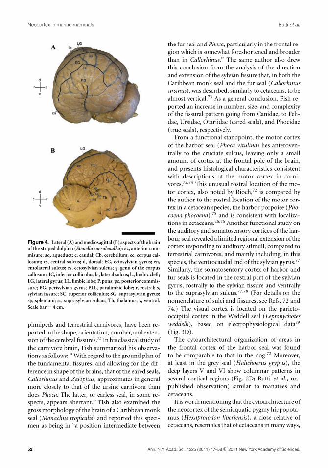
The lack or underdevelopment of layer IV, givenits major role as input for thalamocortical affer-ents, has been related to a possible different strategyin cortical wiring in cetaceans.26,37 The remarkabledevelopment of the cetacean neocortex results in acomplex pattern of gyrification characterized by aprominent and almost vertical sylvian fissure (tech-nically a pseudosylvian fissure) that is surroundedconcentrically toward the vertex of the hemisphereby the ectosylvian, suprasylvian, lateral, and ento-lateral sulci, respectively26,28,29,38 (Fig. 4).
A few physiological mapping studies provide evi-dence about the functional organization of the neo-cortex of cetaceans. The caudal cortical domain sit-uated between the ectosylvian and the suprasylviansulci, the ectosylvian gyrus, corresponds to the sec-ondary auditory field; almost the entire rostrocaudalextent of the cortex positioned between the supra-sylvian and entolateral sulci, the suprasylvian gyrus,forms a belt along the vertex of the hemisphere thatcorresponds to the primary auditory field;39–44 thecortex located between the lateral and the entolateralsulci, at the vertex of the hemisphere, in the lateralgyrus, corresponds to the primary visual field.37,40,44
On the rostroventral extent of the cortex originatesthe cruciate sulcus that extends rostrocaudally, de-lineating the boundaries between the primary mo-tor and primary somatosensory fields40,45 (Fig. 5).The remainder of the lateral surface of the hemi-sphere is likely occupied by “association cortices”connecting the auditory, somatosensory, and motorfields.28
Studies of the neocortical distribution andmorphology of neurons expressing neurochemi-cal markers such as calcium-binding proteins showthat the molecular organization of the neocortex ofcetaceans is similar to that of ungulates but sets themapart from other groups, reflecting phylogeneticrelationships among evolutionary distinct mam-malian branches.35,46–48 Moreover, the percent ofGABAergic neurons in the visual cortex of cetaceansis comparable to that in terrestrial mammals suchas the cat and macaque monkey.49
In the past, quantitative studies of neocorticalorganization that used a variety of methodologiespointed to a high glial cell-to-neuron number ra-tio as a peculiar characteristic of cetaceans.38,50–52
This subsequently gave rise to a controversial hy-pothesis about the functional significance of thisratio,53 which was challenged on several grounds in
Ann. N.Y. Acad. Sci. 1225 (2011) 47–58 c© 2011 New York Academy of Sciences. 49

Neocortex in marine mammals Butti et al.
Figure 2. Examples of neocortical cytoarchitecture of somatosensory and insular cortices in some of the species discussedin this review. Somatosensory cortex (A–E and G); anterior insular cortex (F). Arrowheads (F) point to a neuronal cluster,or “Rindenkerne,” in layer VI, a specialization unique to sirenians. Cortical layers are indicated by Roman numerals. Scalebar = 400 �m.
a recent review.54 Marino et al. have stressed fur-ther the need for standardization of methods usedin acquiring quantitative neuroanatomical data incomparative studies.54
Structure and function of the neocortexof sireniansIn contrast to the large size and the dramatic sulca-tion and gyrification of the cetacean brain, sirenians
50 Ann. N.Y. Acad. Sci. 1225 (2011) 47–58 c© 2011 New York Academy of Sciences.

Butti et al. Neocortex in marine mammals
Figure 3. Examples of cytoarchitecture of the visual cortex in some of the species discussed in this review. Cortical layers areindicated by Roman numerals. Scale bar = 400 �m.
present an extreme and unusual case of lissencephalywith the presence of only a few pronounced fissures,which are accompanied by a relatively small size24,55
(Fig. 1E and F). These differences are even moreenhanced at a cytoarchitectural level, with the neo-cortex of sirenians being thicker and including botha well-organized internal granular layer IV56–58—unlike the situation in cetaceans—and columnarpatterns of layers V and VI in most cortical areas56,57
(Fig. 2E). In sirenians, seven cytoarchitectural areas,which represent 25% of the total neocortical surfacearea, are devoted to somatosensory functions,56–58
and a possible overlapping of the entire primary au-ditory cortex with somatosensory function has beenproposed in manatees.58 The somatosensory nucleiof the thalamus and brainstem are much larger thanthose devoted to other functions,59 which supportsthe primary role of the somatosensory perceptionof the environment for this species.56,57,60–62 Theformation of clusters of neurons in deep layer VI(Rindenkerne; Fig. 2F) is a unique specialization ofsirenians that has been compared to the barrels ofthe somatosensory cortex of rodents, and has beensuggested to be a functional representation of theirunique tactile hairs.58,62–64
Comparison with large semiaquatic andterrestrial mammals
From a comparative viewpoint, the gross anatomyof the brains of both African and Indian elephants(Loxodonta africana and Elephas maximus, respec-
tively) is more comparable in size, level of sulcationand gyrification, and cytoarchitectural specializa-tion to that of cetaceans rather than to that of its clos-est phylogenetic relatives, the sirenians18 (Fig. 1E, Fand K, L).
The few studies available on the gross anatomyand structure of the cerebral cortex of elephants65
highlight a gyral complexity that is second only tocetaceans and includes an expanded neocortex withenlarged temporal, frontal, insular, parietal, cingu-late, and hippocampal cortices, but with a poor de-velopment of the occipital cortex.66 A recent studythat examined the neuronal morphology of pyra-midal neurons in the superficial layers of frontaland occipital cortices of the African elephant high-lighted differences between this species, primates,and rodents, including dendritic length, branchingpatterns, and orientation.67,68 The neocortex of theelephant, like that of cetaceans, contains superficialpyramidal neurons that possess bifurcating apicaldendrites,33,67,69,70 and has been suggested to have ahigh glia-to-neuron ratio.71
Only a few reports are available on the exter-nal morphology of the brain and cytoarchitectureof the pinniped neocortex, and they are mostly fo-cused on a particular species and a restricted corti-cal domain. In terms of gross morphology, the brainof pinnipeds is comparable in shape, proportions,and cortical folding to that of large terrestrial carni-vores72 (Fig. 1G–J). However, minor species-specificdifferences among pinnipeds, as well as between
Ann. N.Y. Acad. Sci. 1225 (2011) 47–58 c© 2011 New York Academy of Sciences. 51
Neocortex in marine mammals Butti et al.
Figure 4. Lateral (A) and mediosagittal (B) aspects of the brainof the striped dolphin (Stenella coeruleoalba): ac, anterior com-misure; aq, aqueduct; c, caudal; Cb, cerebellum; cc, corpus cal-losum; cs, central sulcus; d, dorsal; EG, ectosylvian gyrus; en,entolateral sulcus; es, ectosylvian sulcus; g, genu of the corpuscallosum; IC, inferior colliculus; la, lateral sulcus; lc, limbic cleft;LG, lateral gyrus; LL, limbic lobe; P, pons; pc, posterior commis-sure; PG, perisylvian gyrus; PLL, paralimbic lobe; r, rostral; s,sylvian fissure; SC, superior colliculus; SG, suprasylvian gyrus;sp, splenium; ss, suprasylvian sulcus; Th, thalamus; v, ventral.Scale bar = 4 cm.
pinnipeds and terrestrial carnivores, have been re-ported in the shape, orientation, number, and exten-sion of the cerebral fissures.73 In his classical study ofthe carnivore brain, Fish summarized his observa-tions as follows: “ With regard to the ground plan ofthe fundamental fissures, and allowing for the dif-ference in shape of the brains, that of the eared seals,Callorhinus and Zalophus, approximates in generalmore closely to that of the ursine carnivora thandoes Phoca. The latter, or earless seal, in some re-spects, appears aberrant.” Fish also examined thegross morphology of the brain of a Caribbean monkseal (Monachus tropicalis) and reported this speci-men as being in “a position intermediate between
the fur seal and Phoca, particularly in the frontal re-gion which is somewhat foreshortened and broaderthan in Callorhinus.” The same author also drewthis conclusion from the analysis of the directionand extension of the sylvian fissure that, in both theCaribbean monk seal and the fur seal (Callorhinusursinus), was described, similarly to cetaceans, to bealmost vertical.73 As a general conclusion, Fish re-ported an increase in number, size, and complexityof the fissural pattern going from Canidae, to Feli-dae, Ursidae, Otariidae (eared seals), and Phocidae(true seals), respectively.
From a functional standpoint, the motor cortexof the harbor seal (Phoca vitulina) lies anteroven-trally to the cruciate sulcus, leaving only a smallamount of cortex at the frontal pole of the brain,and presents histological characteristics consistentwith descriptions of the motor cortex in carni-vores.72,74 This unusual rostral location of the mo-tor cortex, also noted by Rioch,72 is compared bythe author to the rostral location of the motor cor-tex in a cetacean species, the harbor porpoise (Pho-coena phocoena),75 and is consistent with localiza-tions in cetaceans.26,76 Another functional study onthe auditory and somatosensory cortices of the har-bour seal revealed a limited regional extension of thecortex responding to auditory stimuli, compared toterrestrial carnivores, and mainly including, in thisspecies, the ventrocaudal end of the sylvian gyrus.77
Similarly, the somatosensory cortex of harbor andfur seals is located in the rostral part of the sylviangyrus, rostrally to the sylvian fissure and ventrallyto the suprasylvian sulcus.77,78 (For details on thenomenclature of sulci and fissures, see Refs. 72 and74.) The visual cortex is located on the parieto-occipital cortex in the Weddell seal (Leptonychotesweddelli), based on electrophysiological data79
(Fig. 3D).The cytoarchitectural organization of areas in
the frontal cortex of the harbor seal was foundto be comparable to that in the dog.72 Moreover,at least in the grey seal (Halichoerus grypus), thedeep layers V and VI show columnar patterns inseveral cortical regions (Fig. 2D; Butti et al., un-published observation) similar to manatees andcetaceans.
It is worth mentioning that the cytoarchitecture ofthe neocortex of the semiaquatic pygmy hippopota-mus (Hexaprotodon liberiensis), a close relative ofcetaceans, resembles that of cetaceans in many ways,
52 Ann. N.Y. Acad. Sci. 1225 (2011) 47–58 c© 2011 New York Academy of Sciences.

Butti et al. Neocortex in marine mammals
Figure 5. Dorsal (A) and rostral (B) aspects, and dorsalschematic view (C) of the brain of the bottlenose dolphin (Tur-siops truncatus) showing the localization of primary corticalareas. A1, primary auditory cortex; c, caudal; cs, cruciate sul-cus; crs, coronary sulcus; d, dorsal; en, entolateral sulcus; l,lateral; la, lateral sulcus; M1, primary motor cortex; r, rostral;S1, primary somatosensory cortex; v, ventral; V1, primary visualcortex. Scale bar = 5 cm.
including the absence of layer IV throughout theneocortex (Butti et al., unpublished observations),a densely packed and clustered layer II in the in-sular cortex,80 and in the putative somatosensorycortex, as well as the presence of vertical modulesof neurons in layer VI of somatosensory and visualputative cortices (Figs. 2G and 3C; Butti et al., un-published observations). In our comparative studyof the organization of the insular cortex, we ob-served a complete agranularity of the anterior sec-tor of the insula in the Atlantic walrus (Odobenusrosmarus rosmarus),80 which is similar to what hasbeen observed in the cat81 but contrasts with thesituation in the dog, where a “dysgranular” cortex ispresent.82
Cortical specializations: von Economoneurons and laminar clusters
In most of the cetacean species studied, layer V ofthe anterior cingulate (ACC), anterior and fron-toinsular (AI and FI), and frontopolar (FP) corticescontains von Economo neurons (VENs), 83 a pop-ulation of projection neurons originally describedin humans, great apes, and later elephants, whichare suggested to play a role in interoception, social-ity, and cognition.26,80,84–91 Recent evidence showsa rapid increase in the number of VENs during thefirst eight months after birth in humans as well asan hemispheric asymmetry in their distribution inhominids, which is possibly related to asymmetriesin the sympathetic and parasympathetic divisionsof the autonomic nervous system.89 VENs are largerthan neighboring pyramidal neurons in cetaceansand hominoids,84,88 send an axon out of the neocor-tex,88and possess a narrow and simplified dendritictree, a morphology consistent with conveying infor-mation that is synthesized from within the space of aminicolumn.92 Recent evidence suggests that VENscontain not only high levels of non-phosphorylatedneurofilament protein (NFP), but also vasopressin1a, dopamine D3, and serotonin 2b receptors;neuromedin B (NMB); gastrin-releasing peptide;DISC1 (disrupted in schizophrenia-1); activating-transcription factor 3 (ATF3); and interleukin 4 re-ceptor � (IL-4R�),86,88,89 all of which are thoughtto be involved in social bonding, reward, punish-ment, digestion, and immune response.93 Evidencealso points to phylogenetic variation (and as such,possible biochemical specialization of VENs in se-lect mammalian groups) in protein expression pro-file, with a higher proportion of VENs expressingATF3, IL4R�, and NMB in humans than in otherhominoids.93
In view of the presence of VENs in phyloge-netically divergent species that share large brainsand complex social organization; their specific cor-tical distribution, morphology, biochemical pro-file; and selective disruption in neuropsychiatricdisorders impairing cognitive and social function-ing, these neurons may play a role in the integra-tion of emotions, vocalization control, facial expres-sion, social conduct, and regulation of autonomicvisceral, olfactory, and gustatory functions. Fur-thermore, they may represent an anatomical sub-strate for the fast transmission of information alongnetworks implicated in the emotional response to
Ann. N.Y. Acad. Sci. 1225 (2011) 47–58 c© 2011 New York Academy of Sciences. 53

Neocortex in marine mammals Butti et al.
external stimuli and generation of goal-directed be-haviors in large mammals.26,80,84,86,94–99 We havealso observed VENs, with marked differences in dis-tribution, in the neocortex of the pygmy hippopota-mus, two marine mammals unrelated to cetaceans(the Florida manatee, Trichechus manatus latirostris,and the Atlantic walrus), and one perissodactyl, thecommon zebra (Equus burchelli),80 as well as the do-mestic horse, Equus caballus, and the Eastern blackrhinoceros, Diceros bicornis michaeli (Butti et al.,unpublished observations). Cetaceans, hominoids,and elephants share a selective distribution of highdensities of VENs in specific frontal cortical regions(ACC, FI/AI, and FP/dlPFC in humans);80,84,87,88,91
in contrast, the pygmy hippopotamus and com-mon zebra have abundant VENs throughout thecortex,80 and the Florida manatee is characterizedby extremely rare and sparse VENs.80 This sug-gests that the overall distribution of VENs in thepygmy hippopotamus represents a progressive re-shaping of the projections provided by these neu-rons, possibly translating into a more specific func-tion. Such specialization appears to have taken placeduring the evolution of the cetaceans from theircommon ancestor with hippopotamuses, where abroad distribution of VENs throughout the neo-cortex was refined to one preferentially involvingthe functions subserved by the ACC, FI, and FP(or dlPFC in hominids).80 Similar mechanisms mayhave shaped other highly specialized pathways inprimates, such as those supported in primates byBetz cells in the motor cortex and Meynert cells inthe visual cortex, which are also characterized bysparsely distributed, large, clustering output neu-rons in functionally specific cortical and laminardomains.100–102
Another remarkable specialization of the neo-cortex of cetaceans is the clustering of neurons inlayer II of the anterior insular cortex. Specifically,large clumps of neurons in layer II have been de-scribed in the anterior insular cortex of small odon-tocetes,80,103,104 also extending to the temporal andoccipital cortex in some mysticetes.26 This modularorganization was suggested26 to be shaped by tha-lamocortical afferent and length of corticocorticalprojections, and represents a cost-effective strategyfor efficient wiring in large brains.26 Hof and Vander Gucht proposed that the peculiar patchiness oflayer II in temporal and occipital regions could re-flect a specific neocortical connectivity (and func-
tion) of these regions in balaenopterids that is notshared with other cetaceans.26 Modules in layer II,comparable to those described in cetaceans, werealso observed in the anterior insular cortex and inthe putative somatosensory cortex of the pygmy hip-popotamus and Atlantic walrus.80 Particularly strik-ing is the clustering observed in the anterior insularcortex of the manatee, where the presence of clus-ters in layer II is associated with the formation oflarge columns that extend to the deepest layers ofthe cortex.80
The importance of comprehensivesampling in comparative studies
The comparative data reviewed here demonstratethat the process of readaptation to the aquatic en-vironment resulted in diverse brain morphologyand cortical organization. This variation is mani-fest as the unique agranularity of the neocortex incetaceans and artiodactyls and the similarities inexternal morphology of the brain and gyral pat-tern in phocids and terrestrial carnivores. How-ever, similarities are evident across orders, includ-ing comparable gyral complexities, agranularity,columnar modularity of layer V and VI, cluster-ing of layer II, and neuronal specializations such asVENs.
Interpreting the evolutionary significance of suchtraits is rendered difficult due to the lack of a clearpattern of appearance through the mammalian phy-logeny, and is further complicated by the poor avail-ability of many key species. Moreover, the absenceof connectivity and functional data in species otherthan laboratory animals imposes critical limitationsand stresses the need for comprehensive compar-isons to avoid outright speculation. How the inter-pretation of the possible evolutionary significanceof VENs has been changing as data on a larger num-ber of species become available is a reflection ofthis situation. At first, the identification of VENsin homologous cortical regions of great apes andhumans suggested that VENs could be related tocognitive and behavioral specializations unique tothese species.86,89 The later identification of VENs incetaceans and elephants led to hypotheses relatingVENs morphology to absolute brain size, other as-pects of behavior, and the need for a fast conductionof information over great distances.26 Most recently,the description of VENs in the brains of other largevertebrates80 with different cortical distributions
54 Ann. N.Y. Acad. Sci. 1225 (2011) 47–58 c© 2011 New York Academy of Sciences.

Butti et al. Neocortex in marine mammals
than that described in hominoids, cetaceans, andelephants, suggests that VENs represent a commonevolutionary trait among large mammals contribut-ing to specialized neuronal networks in a taxon-specific manner, dependent upon their corticaldistribution.
In this context, brain collections and zoologi-cal facilities constitute a unique source of criticalinformation for the study of brain organization ina truly evolutionary context that goes beyond thehandful of species commonly available in the lab-oratory. Such histological materials and anatom-ical repositories represent, in many cases, theonly possibility of exploring brain organization ofspecies otherwise impossible to study, to understandthe evolutionary significance of neuroanatomi-cal differences and similarities widely observedamong mammalian lineages, and to add to theknowledge of poorly documented and endangeredspecies.
Acknowledgments
The authors thank Drs. W. Welker, J.I. Johnson, A.Fobbs, and A. Noe, as well as the Comparative Mam-malian Brain Collections of the University of Wis-consin, Michigan State, and the National Museumof Health and Medicine for providing access to his-tological materials of the brains of the commonzebra and domestic horse; Drs. P.J. Morgane andI.I. Glezer for donation of the histological materi-als of the bottlenose dolphin, striped dolphin, killerwhale, humpback whale, and Florida manatee; Drs.T. Romano, A.D.Tuttle, G. Sirpenski and the MysticAquarium and Institute for Exploration for provid-ing the brain of the grey seal; Dr. C.J. Bonar and TheCleveland Metroparks Zoo for donation of the brainof the pygmy hippopotamus; Dr. P. Manger for pro-viding cortical samples of the brain of the Africanelephant and for helpful discussion; Dr. C.E. Ro-driguez and the New York Wildlife Conservation So-ciety for donation of the brain of the Atlantic walrus;Dr. T. Harrison and the Potter Park Zoo for dona-tion of the brain of the Western black rhinoceros; Dr.B. Jacobs for helpful discussions; and B. Wicinski,C.D. Stimpson, and W.G.M. Janssen for expert tech-nical assistance. The authors are supported by theJames S. McDonnell Foundation (Grant 22002078to PRH and CCS) and the National Science Founda-tion (BCS-0515484, BCS-0549117, BCS-0827531,DGE-0801634).
Conflicts of interest
The authors declare no conflicts of interest.
References
1. Gingerich, P.D. & M.D. Uhen. 1998. Likelihood estima-tion of the time of origin of cetacean and the time of di-vergence of Cetacea and Artiodactyla. Paleo-Electronica 2:1–47.
2. Fordyce, R. 2008. Cetacean evolution. In Encyclopedia ofMarine Mammals. W.F. Perrin, B. Wursig & J.G.M. Thewis-sen, Eds.: 201–207. Academic Press. San Diego, CA.
3. Thewissen, J.G., L.N. Cooper, M.T. Clementz, et al. 2007.Whales originated from aquatic artiodactyls in the Eoceneepoch of India. Nature 450: 1190–1194.
4. Uhen, M. 2010. The origin(s) of whales. Annu. Rev. EarthPlanet. Sci. 38: 189–219.
5. Fordyce, R.E. 2008. Neoceti. In Encyclopedia of MarineMammals. W.F. Perrin, B. Wursig & J.G.M. Thewissen, Eds.:758–763: Academic Press. San Diego, CA.
6. Fitzgerald, E. 2006. A bizarre new toothed mysticete(Cetacea) from Australia and the early evolution of baleenwhales. Proc. R. Acad. Sci. B. 273: 2955–2963.
7. Fordyce, R.E. 2002. Simocetus rayi (Odontoceti: Simoceti-dae) (new species, new genus, new family), a bizarre newarchaic Oligocene dolphin from the Eastern North Pacific.Smithsonian Contrib. Paleobiol. 93: 185–222.
8. Fordyce, R.E. 2008. Cetacean fossil record. In Encyclope-dia of Marine Mammals. W.F. Perrin, B. Wursig & J.G.M.Thewissen, Eds.: 207–215. Academic Press. San Diego,CA.
9. Nikaido, M., A.P. Rooney & N. Okada. 1999. Phylogeneticrelationships among cetartiodactyls based on insertions ofshort and long interspersed elements: hippopotamuses arethe closest extant relatives of whales. Proc. Natl. Acad. Sci.USA 96: 10261–10266.
10. Boisserie, J.R., F. Lihoreau & M. Brunet. 2005. The posi-tion of Hippopotamidae within Cetartiodactyla. Proc. Natl.Acad. Sci. USA 102: 1537–1541.
11. Thewissen, J.G., E.M. Williams, L.J. Roe & S.T. Hussain.2001. Skeletons of terrestrial cetaceans and the relationshipof whales to artiodactyls. Nature 413: 277–281.
12. Geisler, J.H. & J.M. Theodor. 2009. Hippopotamus andwhale phylogeny. Nature 458: E1–4; discussion E5.
13. Arnason, U., A. Gullberg, S. Gretarsdottir, et al. 2000. Themitochondrial genome of the sperm whale and a newmolecular reference for estimating eutherian divergencedates. J. Mol. Evol. 50: 569–578.
14. Rice, D. Classification (overall). 2008. In Encyclopedia ofMarine Mammals. W.F. Perrin, B. Wursig & J.G.M. Thewis-sen, Eds.: 234–238. Academic Press. San Diego, CA.
15. Murphy, W.J. et al. 2001. Molecular phylogenetics and theorigins of placental mammals. Nature 409: 614–618.
16. Domning, D.P. 2000. The readaptation of Eocene sireniansto life in water. Historical Biol. 14: 115–119.
17. Heyning, J.E. & G.M. Lento. 2006. The evolution of marinemammals. In Marine Mammal Biology: An EvolutionaryApproach. A.R. Hoelzel, Ed.: 38–72. Blackwell Publishing.Malden, MA.
Ann. N.Y. Acad. Sci. 1225 (2011) 47–58 c© 2011 New York Academy of Sciences. 55

Neocortex in marine mammals Butti et al.
18. Kellogg, M.E. et al. 2007. Chromosome painting in themanatee supports Afrotheria and Paenungulata. BMC Evol.Biol. 7: 6.
19. Vrana, P.B., M.C. Milinkovitch, J.R. Powell & W.C. Wheeler.1994. Higher level relationships of the arctoid Carnivorabased on sequence data and “total evidence”. Mol. Phylo-genet. Evol. 3: 47–58.
20. Arnason, U. & B. Widegren. 1986. Pinniped phylogeny en-lightened by molecular hybridizations using highly repeti-tive DNA. Mol. Biol. Evol. 3: 356–365.
21. Lento, G.M., R.E. Hickson, G.K. Chambers & D. Penny.1995. Use of spectral analysis to test hypotheses on theorigin of pinnipeds. Mol. Biol. Evol. 12: 28–52.
22. Berta, A. 2008. Pinniped evolution. In Encyclopedia of Ma-rine Mammals. W.F. Perrin, B. Wursig & J.G.M. Thewissen,Eds.: 861–868. Academic Press. San Diego, CA.
23. Klima, M. 1999. Development of the cetacean nasal skull.Adv. Anat. Embryol. Cell. Biol. 149: 1–143.
24. Marino, L. 2008. Brain size evolution. In Encyclopedia ofMarine Mammals. W.F. Perrin, B. Wursig & J.G.M. Thewis-sen, Eds.: 149–152. Academic Press. San Diego, CA.
25. Marino, L. 2007. Cetacean brains: how aquatic are they?Anat. Rec. 290: 694–700.
26. Hof, P.R. & E. Van der Gucht. 2007. Structure of the cere-bral cortex of the humpback whale, Megaptera novaeangliae(Cetacea, Mysticeti, Balaenopteridae). Anat. Rec. 290: 1–31.
27. Morgane, P.J., M.S. Jacobs & W.L. McFarland. 1980. Theanatomy of the brain of the bottlenose dolphin (Tursiopstruncatus). Surface configuration of the telencephalon ofthe bottlenose dolphin with comparative anatomical ob-servations in four other cetaceans species. Brain Res. Bull.5(Suppl. 3): 1–108.
28. Oelschlager, H.A. & J. Oelschlager. 2008. Brain. In Encyclo-pedia of Marine Mammals. W.F. Perrin, B. Wursig & J.G.M.Thewissen, Eds.: 134–149. Academic Press. San Diego, CA.
29. Jacobs, M.S., W.L. McFarland & P.J. Morgane. 1979. Theanatomy of the brain of the bottlenose dolphin (Tursiopstruncatus). Rhinic lobe (Rhinencephalon): the archicortex.Brain Res. Bull. 4(Suppl. 1): 1–108.
30. Breathnach, A.S. & F. Goldby. 1954. The amygdaloid nuclei,hippocampus and other parts of the rhinencephalon in theporpoise (Phocaena phocaena). J. Anat. 88: 267–291.
31. Hof, P.R., R. Chanis & L. Marino. 2005. Cortical complexityin cetacean brains. Anat. Rec. 287: 1142–1152.
32. Furutani, R. 2008. Laminar and cytoarchitectonic featuresof the cerebral cortex in the Risso’s dolphin (Grampusgriseus), striped dolphin (Stenella coeruleoalba), and bot-tlenose dolphin (Tursiops truncatus). J. Anat. 213: 241–248.
33. Garey, L.J., E. Winkelmann & K. Brauer. 1985. Golgi andNissl studies of the visual cortex of the bottlenose dolphin.J. Comp. Neurol. 240: 305–321.
34. Glezer, I.I. & P.J. Morgane. 1990. Ultrastructure of synapsesand golgi analysis of neurons in neocortex of the lateralgyrus (visual cortex) of the dolphin and pilot whale. BrainRes. Bull. 24: 401–427.
35. Hof, P.R. & C.C. Sherwood. 2005. Morphomolecular neu-ronal phenotypes in the neocortex reflect phylogenetic re-lationships among certain mammalian orders. Anat. Rec.287: 1153–1163.
36. Morgane, P.J., I.I. Glezer & M.S. Jacobs. 1988. Visual cortexof the dolphin: an image analysis study. J. Comp. Neurol.273: 3–25.
37. Revishchin, A.V. & L.J. Garey. 1989. Sources of thalamicafferent neurons, projecting into the suprasylvian gyrusof the dolphin cerebral cortex. Neirofiziologia 21: 529–539.
38. Morgane, P.J., W.L. McFarland & M.S. Jacobs. 1982. Thelimbic lobe of the dolphin brain: a quantitative cytoarchi-tectonic study. J. Hirnforsch. 23: 465–552.
39. Ladygina, T.F., A.M. Mass & A. Supin. 1978. Multiple sen-sory projections in the dolphin cerebral cortex. Zh. Vyssh.Nerv. Deiat. Im. I.P. Pavlova 28: 1047–1053.
40. Ladygina, T.F. & A. Supin. 1977. Localization of the sen-sory projection areas in the cerebral cortex of the dol-phin, Tursiops truncatus. Zh. Evol. Biokhim. Fiziol. 13: 712–718.
41. Popov, V.V., T.F. Ladygina & A. Supin. 1986. Evoked po-tentials of the auditory cortex of the porpoise, Phocoenaphocoena. J. Comp. Physiol. 158: 705–711.
42. Popov, V.V. & A. Supin. 1976. Detemination of the hear-ing characteristics of the dolphin by the evoked potentialmethod. Fiziol. Zh. SSSR Im. I.M. Sechenova 62: 550–558.
43. Revishchin, A.V. & L.J. Garey. 1996. Mitochondrial distri-bution in visual and auditory cerebral cortex of the harbourporpoise. Brain Behav. Evol. 47: 257–266.
44. Sokolov, V.E., T.F. Ladygina & A. Supin. 1972. Localizationof sensory zones in the dolphin cerebral cortex. Dokl. Akad.Nauk. SSSR 202: 490–493.
45. Kesarev, V.S. & L.I. Malofeeva. 1969. Structural organiza-tion of the motor zone of the cerebral cortex in dolphins.Arkh. Anat. Gistol. Embriol. 56: 48–55.
46. Glezer, I.I., P.R. Hof, C. Leranth & P.J. Morgane. 1993.Calcium-binding protein-containing neuronal popula-tions in mammalian visual cortex: a comparative studyin whales, insectivores, bats, rodents, and primates. Cereb.Cortex 3: 249–272.
47. Hof, P.R. et al. 1999. Cellular distribution of the calcium-binding proteins parvalbumin, calbindin, and calretinin inthe neocortex of mammals: phylogenetic and developmen-tal patterns. J. Chem. Neuroanat. 16: 77–116.
48. Sherwood, C.C. et al. 2009. Neocortical neuron types inXenarthra and Afrotheria: implications for brain evolutionin mammals. Brain Struct. Funct. 213: 301–328.
49. Garey, L.J., J. Takacs, A.V. Revishchin & J. Hamori. 1989.Quantitative distribution of GABA-immunoreactive neu-rons in cetacean visual cortex is similar to that in landmammals. Brain Res. 485: 278–284.
50. Garey, L.J. & G. Leuba. 1986. A quantitative study of neu-ronal and glial numerical density in the visual cortex of thebottlenose dolphin: evidence for a specialized subarea andchanges with age. J. Comp. Neurol. 247: 491–496.
51. Hawkins, A. & J. Olszewski. 1957. Glia/nerve cell index forcortex of the whale. Science 126: 76–77.
52. Eriksen, N. & B. Pakkenberg. 2007. Total neocortical cellnumber in the mysticete brain. Anat. Rec. 290: 83–95.
53. Manger, P.R. 2006. An examination of cetacean brain struc-ture with a novel hypothesis correlating thermogenesis tothe evolution of a big brain. Biol. Rev. 81: 293–338.
56 Ann. N.Y. Acad. Sci. 1225 (2011) 47–58 c© 2011 New York Academy of Sciences.

Butti et al. Neocortex in marine mammals
54. Marino, L. et al. 2008. A claim in search of evidence: replyto Manger’s thermogenesis hypothesis of cetacean brainstructure. Biol. Rev. 83: 417–440.
55. Reep, R.L. & T.J. O’Shea. 1990. Regional brain morphom-etry and lissencephaly in the Sirenia. Brain Behav. Evol. 35:185–194.
56. Marshall, C.D. & R.L. Reep. 1995. Manatee cerebral cortex:cytoarchitecture of the caudal region in Trichechus manatuslatirostris. Brain Behav. Evol. 45: 1–18.
57. Reep, R.L., J.I. Johnson, R.C. Switzer & W.I. Welker. 1989.Manatee cerebral cortex: cytoarchitecture of the frontal re-gion in Trichechus manatus latirostris. Brain Behav. Evol.34: 365–386.
58. Sarko, D.K. & R.L. Reep. 2007. Somatosensory areasof manatee cerebral cortex: histochemical characteriza-tion and functional implications. Brain Behav. Evol. 69:20–36.
59. Sarko, D.K. et al. 2007. Somatosensory nuclei of the man-atee brainstem and thalamus. Anat. Rec. 290: 1138–1165.
60. Marshall, C.D., L.A. Clark & R.L. Reep. 1998. The musco-lar hydrostat of the Florida manatee (Trichechus manatuslatirostris): a functional morphological model of perioralbristle use. Mar. Mamm. Sci. 14: 290–303.
61. Marshall, C.D. et al. 1998. Prehensile use of perioral bris-tles during feeding and associated behaviors of the Floridamanatee (Trichechus manatus latirostris). Mar. Mamm. Sci.14: 274–289.
62. Reep, R.L. et al. 2001. Microanatomy of facial vibrissaein the Florida manatee: the basis for specialized sensoryfunction and oripulation. Brain Behav. Evol. 58: 1–14.
63. Reep, R.L., C.D. Marshall & M.L. Stoll. 2002. Tactile hairson the postcranial body in Florida manatees: a mammalianlateral line? Brain Behav. Evol. 59: 141–154.
64. Sarko, D.K., F.L. Rice, & R.L. Reep. 2011. Mammalian tac-tile hair: divergence from a limited distribution. Ann. N.Y.Acad. Sci. 1225: 90–100.
65. Cozzi, B., S. Spagnoli & L. Bruno. 2001. An overview of thecentral nervous system of the elephant through a criticalappraisal of the literature published in the XIX and XXcenturies. Brain Res. Bull. 54: 219–227.
66. Shoshani, J., W.J. Kupsky & G.H. Marchant. 2006. Elephantbrain. Part I: gross morphology, functions, comparativeanatomy, and evolution. Brain Res. Bull. 70: 124–157.
67. Jacobs, B. et al. 2011. Neuronal morphology in the Africanelephant (Loxodonta africana) neocortex. Brain Struct.Funct. 215: 273–298 [Epub ahead of print].
68. Bianchi, S. et al. 2011. Neocortical neuron morphologyin Afrotheria: comparing the rock hyrax with the Africanelephant. Ann. N.Y. Acad. Sci. 1225: 37–46.
69. Ferrer, I. & M. Perera. 1988. Structure and nerve cell or-ganisation in the cerebral cortex of the dolphin Stenellacoeruleoalba: a Golgi study. With special attention to theprimary auditory area. Anat. Embryol. 178: 161–173.
70. Hof, P.R. et al. 1992. The primary auditory cortex incetacean and human brain: a comparative analysis of neu-rofilament protein-containing pyramidal neurons. Neu-rosci. Lett. 146: 91–95.
71. Haug, H. 1987. Brain sizes, surfaces, and neuronal sizes ofthe cortex cerebri: a stereological investigation of man and
his variability and a comparison with some mammals (pri-mates, whales, marsupials, insectivores, and one elephant).Am. J. Anat. 180: 126–142.
72. Rioch, D.M. 1937. A physiological and histological studyof the frontal cortex of the seal (Phoca vitulina). Biol. Bull.Woods Hole. 73: 591–602.
73. Fish, P.A. 1898. The brain of the fur seal, Callorhinusursinus: with a comparative description of those of Za-lophus californianus, Phoca vitulina, Ursus americanus andMonachus tropicalis. J. Comp. Neurosci. 8: 57–91.
74. Langworthy, O.R., F.H. Hesser & L.C. Kolb. 1938. A physi-ological study of the cerebral cortex of the hair seal (Phocavitulina). J. Comp. Neurol. 69: 351–369.
75. Langworthy, O. 1932. A description of the central nervoussystem of the porpoise (Tursiops truncatus). J. Comp. Neu-rol. 350: 337–356.
76. Kojima, T. 1951. On the brain of the sperm whale (Physetercatodon, L.). Sci. Rep. Whales Res. Inst. Tokyo 6: 49–72.
77. Alderson, A.M., E. Diamantopoulos & C.B.B. Downman.1960. Auditory cortex of the seal (Phoca vitulina). J. Anat.94: 506–511.
78. Ladygina, T.F.P., V.V. Supin & A.Ya. 1985. Topical organiza-tion of somatic projections in the fur seal cerebral cortex.Neirofiziologia 17: 344–351.
79. Gruenau, S.P. & J.T. Shurley. 1976. Visual evoked response(VER) changes during maturation in the Weddell seal. Dev.Psychobiol. 9: 477–493.
80. Butti, C. & P.R. Hof. 2010. The insular cortex: a comparativeperspective. Brain Struct. Funct. 214: 477–493.
81. Clasca, F., A. Llamas & F. Reinoso-Suarez. 1997. Insularcortex and neighboring fields in the cat: a redefinition basedon cortical microarchitecture and connections with thethalamus. J. Comp. Neurol. 384: 456–482.
82. Salazar, I. et al. 1988. The neocortex of the dog.I. A classicalcytoarchitectonic map. Anat. Histol. Embryol. 17: 169–187.
83. von Economo, C. 1926. Eine neue Art Spezialzellen desLobus cinguli and Lobus insulae. Zschr. Ges. Neurol. Psy-chiatr. 100: 706–712.
84. Butti, C. et al. 2009. Total number and volume of vonEconomo neurons in the cerebral cortex of cetaceans.J. Comp. Neurol. 515: 243–259.
85. Allman, J., A. Hakeem & K. Watson. 2002. Two phylogeneticspecializations in the human brain. Neuroscientist 8: 335–346.
86. Allman, J.M., K.K. Watson, N.A. Tetreault & A.Y. Hakeem.2005. Intuition and autism: a possible role for von Economoneurons. Trends Cogn. Sci. 9: 367–373.
87. Nimchinsky, E.A. et al. 1999. A neuronal morphologic typeunique to humans and great apes. Proc. Natl. Acad. Sci. USA96: 5268–5273.
88. Nimchinsky, E.A., B.A. Vogt, J.H. Morrison & P.R. Hof.1995. Spindle neurons of the human anterior cingulatecortex. J. Comp. Neurol. 355: 27–37.
89. Allman, J.M. et al. 2010. The von Economo neurons infrontoinsular and anterior cingulate cortex in great apesand humans. Brain Struct. Funct. 214: 495–517.
90. Fajardo, C. et al. 2008. Von Economo neurons are present inthe dorsolateral (dysgranular) prefrontal cortex of humans.Neurosci. Lett. 435: 215–218.
Ann. N.Y. Acad. Sci. 1225 (2011) 47–58 c© 2011 New York Academy of Sciences. 57

Neocortex in marine mammals Butti et al.
91. Hakeem, A.Y. et al. 2008. Von Economo neurons in theelephant brain. Anat. Rec. 292: 242–248.
92. Watson, K.K., T.K. Jones & J.M. Allman. 2006. Dendriticarchitecture of the von Economo neurons. Neuroscience141: 1107–1112.
93. Stimpson, C. et al. 2011. Biochemical specificity of vonEconomo neurons in hominoids. Am. J. Hum. Biol. 23:22–28.
94. Kaufman, J.A. et al. 2008. Selective reduction of vonEconomo neuron number in agenesis of the corpus cal-losum. Acta Neuropathol. 116: 479–489.
95. Seeley, W.W. et al. 2007. Divergent social functioning in be-havioral variant frontotemporal dementia and Alzheimerdisease: reciprocal networks and neuronal evolution. Alzh.Dis. Assoc. Disord. 21: S50–57.
96. Seeley, W.W. et al. 2006. Early frontotemporal dementiatargets neurons unique to apes and humans. Ann. Neurol.60: 660–667.
97. Brune, M. et al. 2010. Von Economo neuron density inthe anterior cingulate cortex is reduced in early onsetschizophrenia. Acta Neuropathol. 119: 771–778.
98. Santos, M. et al. 2011. Von Economo neurons inautism: a stereologic study of the frontoinsular cor-
tex in children. Brain Res. [Epub ahead of print]. doi:10.1016/j.brainres.2010.08.067
99. Allman, J.M. et al. 2011. The von Economo neurons in thefrontoinsular and anterior cingulate cortex. Ann. N.Y. Acad.Sci. 1225: 59–71.
100. Hof, P.R., E.A. Nimchinsky, W.G. Young & J.H. Morrison.2000. Numbers of Meynert and layer IVB cells in area V1: astereologic analysis in young and aged macaque monkeys.J. Comp. Neurol. 420: 113–126.
101. Rivara, C.B., C.C. Sherwood, C. Bouras & P.R. Hof. 2003.Stereologic characterization and spatial distribution pat-terns of Betz cells in the human primary motor cortex.Anat. Rec. 270: 137–151.
102. Sherwood, C.C. et al. 2003. Evolution of specialized pyra-midal neurons in primate visual and motor cortex. BrainBehav. Evol. 61: 28–44.
103. Jacobs, M.S., A.M. Galaburda, W.L. McFarland & P.J. Mor-gane. 1984. The insular formations of the dolphin brain:quantitative cytoarchitectonic studies of the insular com-ponent of the limbic lobe. J. Comp. Neurol. 225: 396–432.
104. Manger, P. et al. 1998. Modular subdivisions of dolphininsular cortex: does evolutionary history repeat itself?J. Cogn. Neurosci. 10: 153–166.
58 Ann. N.Y. Acad. Sci. 1225 (2011) 47–58 c© 2011 New York Academy of Sciences.