the patient with allergic diseases: urticaria & angioedema
TRANSCRIPT

The Patient with Allergic Diseases: Urticaria & Angioedema
Bryan L. Martin, D.O., FACOI, FACP Associate Dean, Graduate Med Ed/DIO Associate Medical Director, University Hospital
Disclosures
None
Objectives
Differentiate Urticaria and Angioedema
Recognize and treat anaphylaxis and anaphylactic reactions
Recognize and treat mastocytosis
Recognize the variety of manifestations of latex sensitivity
Topics Urticaria/Angioedema
Anaphylaxis
Latex Allergy
Anaphylactoid Reactions
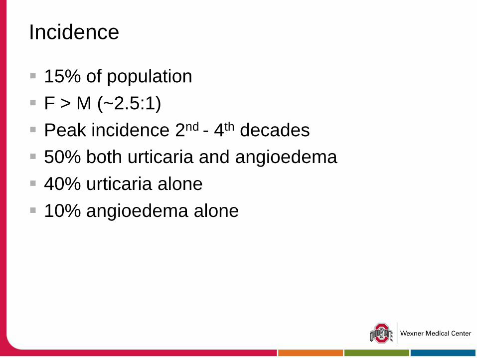
Incidence
15% of population
F > M (~2.5:1)
Peak incidence 2nd - 4th decades
50% both urticaria and angioedema
40% urticaria alone
10% angioedema alone
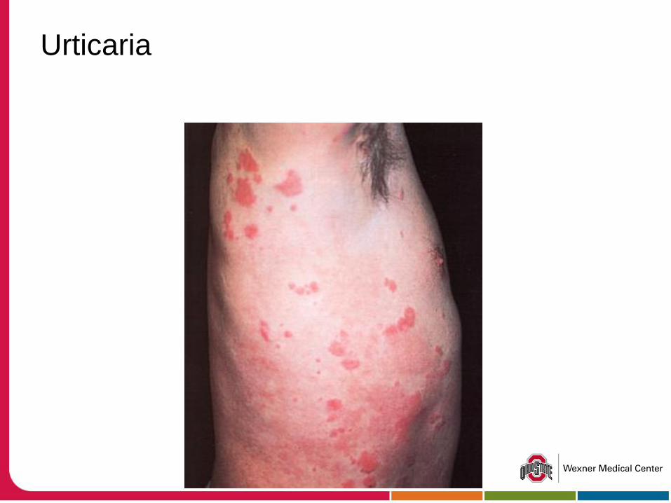
Urticaria/Angioedema
Urticaria
Pruritic, erythematous, cutaneous elevations that blanch with pressure, indicating the presence of dilated blood vessels and edema
Angioedema
Similar pathologic alterations in deep dermis and subcutaneous tissue; swelling is predominant manifestation, little or no pruritis; may be painful or burning
Angioedema
Unlike other forms of edema
Not characteristically in dependent areas
asymmetrically distributed
transient
Often seen with urticaria
Urticaria
Acute vs chronic
Urticaria that exceeds 6 weeks is arbitrarily designated chronic
Dermagraphism
Ability to write on skin: 2-5% of population
Only small fraction warrant chronic treatment with antihistamines
CASE 1: MJ
42 y/o w/m with CC: “whelps” x 2 months
Itching
1st episode: No lifestyle changes
Doctors didn’t help
Benadryl, Claritin, Tavist w/o relief
Lab work, x-rays normal
PE: 0.5-5 cm urticarial lesions
Urticaria
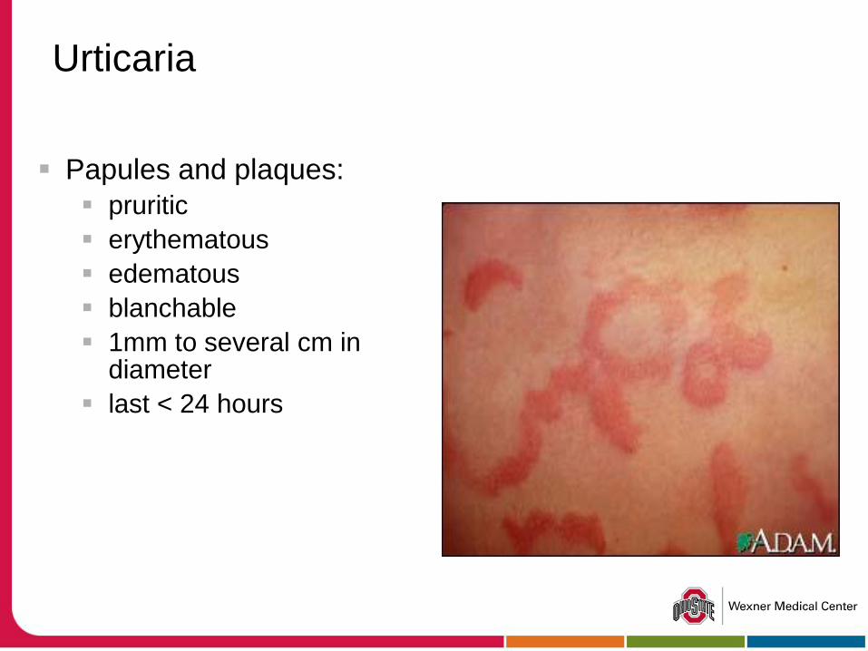
Urticaria
Papules and plaques:
pruritic
erythematous
edematous
blanchable
1mm to several cm in diameter
last < 24 hours
Urticaria Evaluation History
Duration - < or > 6 weeks
Triggers – identifiable cause more likely in acute but < 5% in chronic
ingestants, contactants, physical stimuli, infections
Lesional hx duration, purpura, pain
refer to Dermatology if suspected vasculitis for Bx
PMH/ROS suggestive of systemic disease
Urticaria Evaluation
Acute urticaria
systemic disease
acute infections
dermatographism
Chronic urticaria systemic disease
usually idiopathic/autoimmune
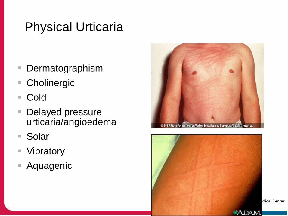
Physical Urticaria
Dermatographism
Cholinergic
Cold
Delayed pressure urticaria/angioedema
Solar
Vibratory
Aquagenic
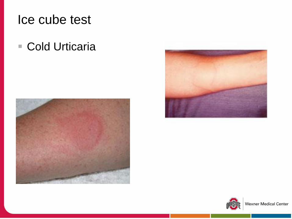
Ice cube test
Cold Urticaria
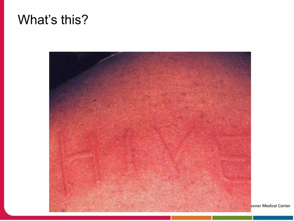
What’s this?
Causes of Urticaria
Infections
URI virus, HBV, EBV, b-Strep, Mycoplasma
Drugs (NSAIDS, Aspirin)
Foods or other ingestants (rare)
Contactants
soaps, perfumes, deodorants, insects
Systemic disease
thyroid, CTD, malignancy
Autoimmune
Idiopathic (>80% in chronic urticaria)
Urticaria Evaluation Labs
Skin tests
Seldom indicated
Of questionable value
can’t get the patient off antihistamines
many patients have dermatographism
Most urticaria is not triggered by food or aeroallergens
Labs as indicated by Hx/PE
TSH, CBC, LFT’s, ESR, ANA, C4
Skin Bx as indicated by Hx
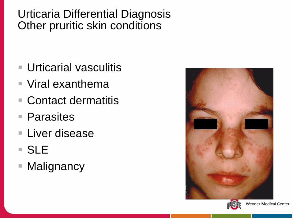
Urticaria Differential Diagnosis Other pruritic skin conditions
Urticarial vasculitis
Viral exanthema
Contact dermatitis
Parasites
Liver disease
SLE
Malignancy
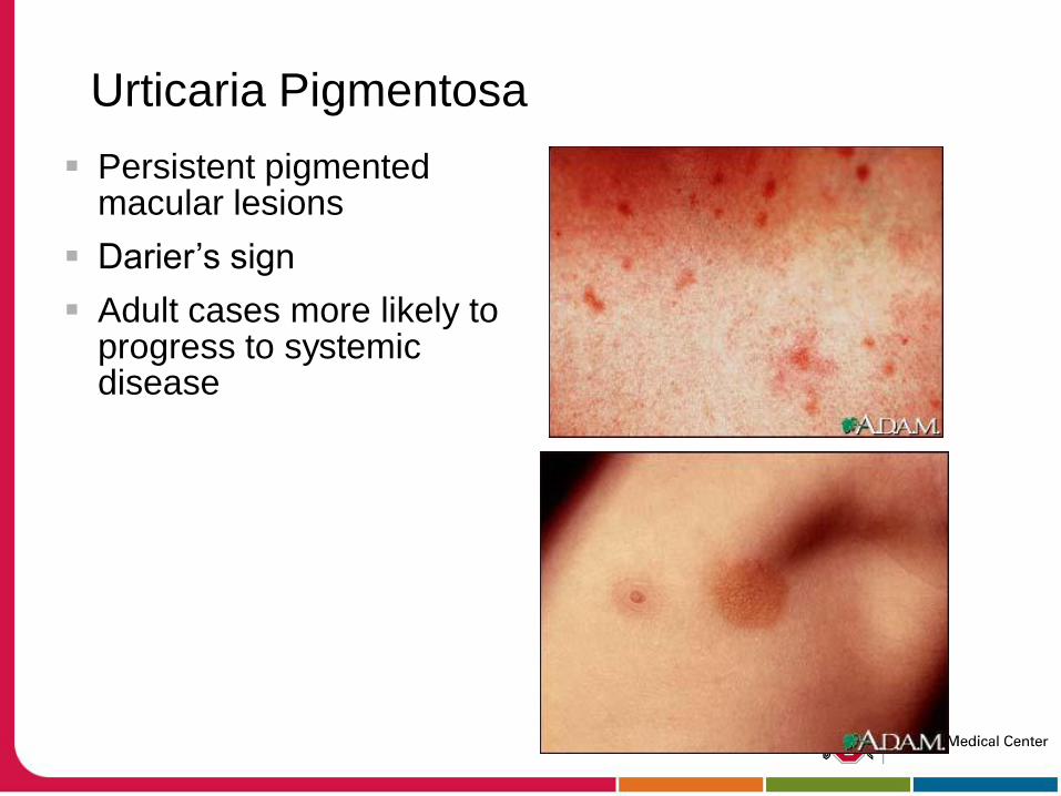
Urticaria Pigmentosa
Persistent pigmented macular lesions
Darier’s sign
Adult cases more likely to progress to systemic disease
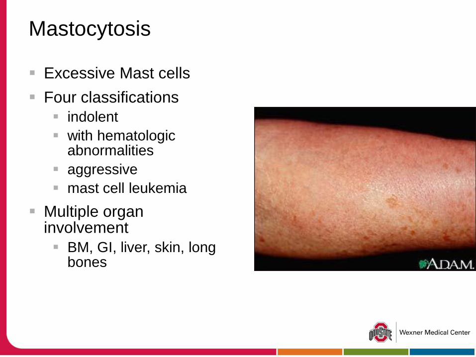
Mastocytosis
Excessive Mast cells
Four classifications
indolent
with hematologic abnormalities
aggressive
mast cell leukemia
Multiple organ involvement
BM, GI, liver, skin, long bones
Mastocytosis
Pruritis, flushing, urticaria, hypotension,
Idiopathic anaphylaxis
May progress to malignancy
Anemia is a poor prognosis
Treat with:
antihistamines, oral cromolyn, NSAID’s
possible use of LTRA
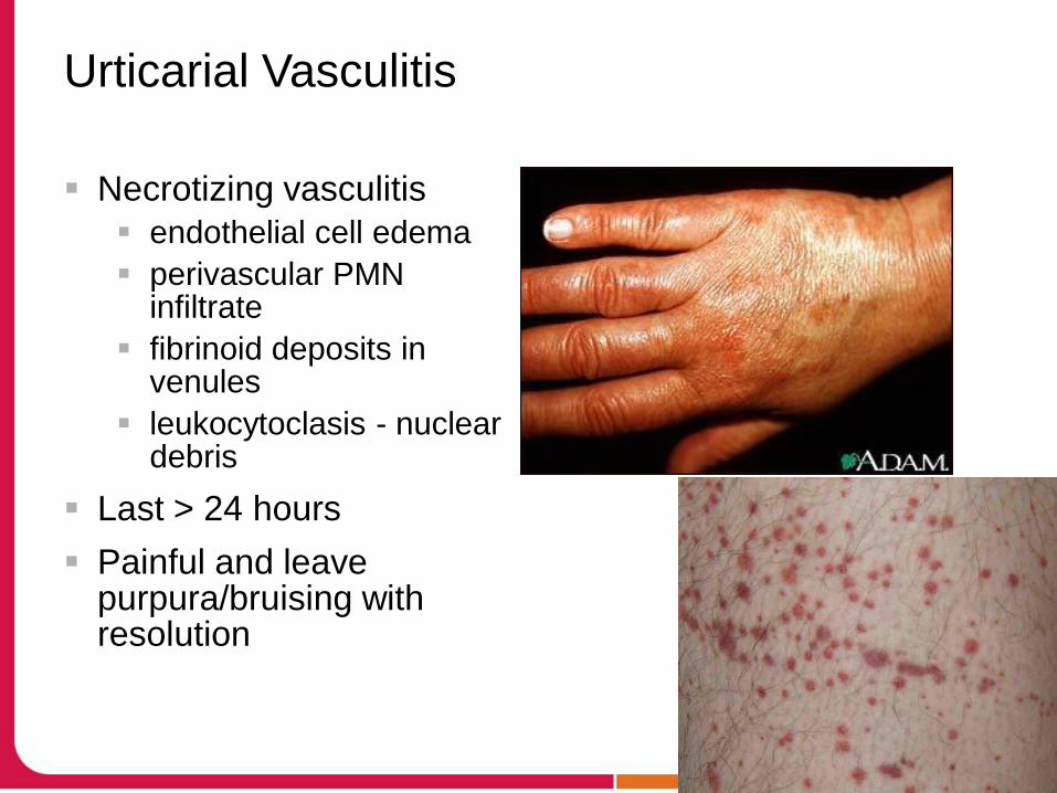
Urticarial Vasculitis
Necrotizing vasculitis
endothelial cell edema
perivascular PMN infiltrate
fibrinoid deposits in venules
leukocytoclasis - nuclear debris
Last > 24 hours
Painful and leave purpura/bruising with resolution
Urticaria: Treatment
Eliminate trigger factors
H-1 Antihistamines
combination H-1 & H-2 Antihistamines
Corticosteroids
Avoid ETOH, ASA, Tobacco
Avoid hot showers, hot tubs

Case 2: CW
37 y/o b/m with CC: “swelling, typically of 1 or both hands every 3-4 months x 18 yrs
Frightened by recent episode involving face/throat
Seen by a number of physicians, no definitive diagnosis, no treatment plan has helped. No current medications
PE: normal
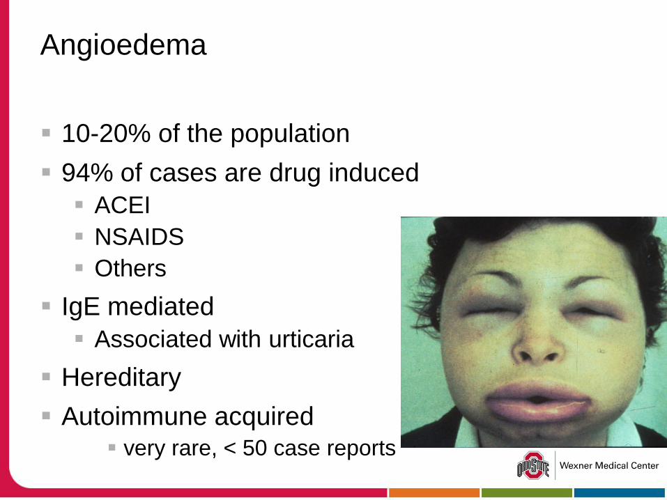
Angioedema
10-20% of the population
94% of cases are drug induced
ACEI
NSAIDS
Others
IgE mediated
Associated with urticaria
Hereditary
Autoimmune acquired very rare, < 50 case reports
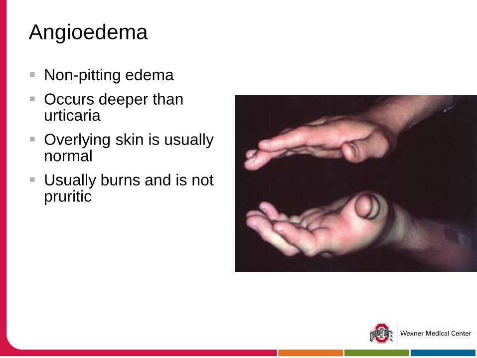
Angioedema
Non-pitting edema
Occurs deeper than urticaria
Overlying skin is usually normal
Usually burns and is not pruritic
ACEI Induced Angioedema
1-2 cases per 1000 persons
>70% symptomatic within first week of therapy
Likely precipitated by increased bradykinin
Angiotensin II inhibits bradykinin
ACEI blocks conversion of angiotensin I II
Vasodilatation, increased vascular permeability
Can lead to life-threatening upper airway obstruction
22% require intubation with11% mortality
Rare in Angiotensin II receptor blockers
Hereditary Angioedema
Rare (1/150,000)
Autosomal dominant
Onset in adolescence
Angioedema is
painless and non-pruritic
lasts 3-5 days
unrespsonsive to Epi, antihistamines, pred.
triggered by mild trauma
Hereditary Angioedema
C1 Inhibitor (C1-INH) deficiency
Type I (85%)
Quantitative deficiency (5-30% normal)
Type II (15%)
Qualitative deficiency
Quantity is normal or elevated
Functional activity is markedly reduced
Type III
Unknown cause
C1q, C1-INH, C4 normal with suggestive history
C4, C1-INH normal during attack
Hereditary Angioedema
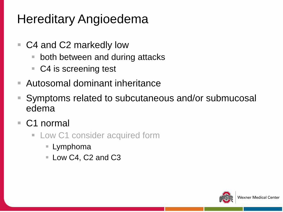
C4 and C2 markedly low
both between and during attacks
C4 is screening test
Autosomal dominant inheritance
Symptoms related to subcutaneous and/or submucosal edema
C1 normal
Low C1 consider acquired form
Lymphoma
Low C4, C2 and C3
Hereditary Angioedema
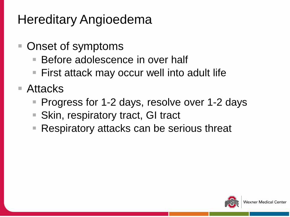
Onset of symptoms
Before adolescence in over half
First attack may occur well into adult life
Attacks
Progress for 1-2 days, resolve over 1-2 days
Skin, respiratory tract, GI tract
Respiratory attacks can be serious threat
Hereditary Angioedema: Treatments
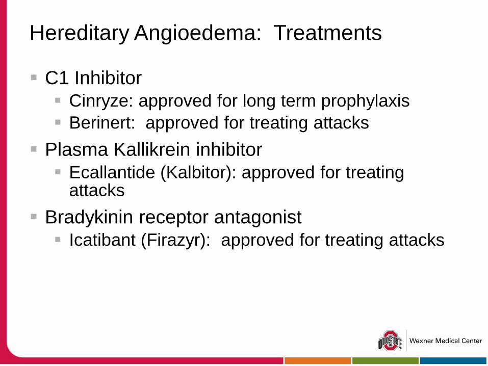
C1 Inhibitor
Cinryze: approved for long term prophylaxis
Berinert: approved for treating attacks
Plasma Kallikrein inhibitor
Ecallantide (Kalbitor): approved for treating attacks
Bradykinin receptor antagonist
Icatibant (Firazyr): approved for treating attacks
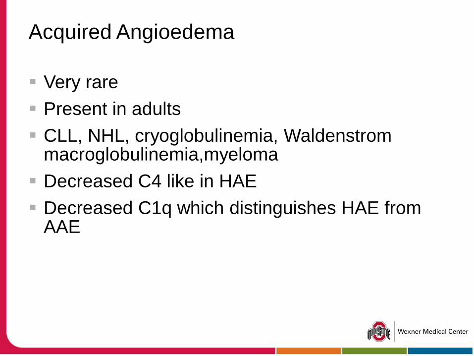
Acquired Angioedema
Very rare
Present in adults
CLL, NHL, cryoglobulinemia, Waldenstrom macroglobulinemia,myeloma
Decreased C4 like in HAE
Decreased C1q which distinguishes HAE from AAE
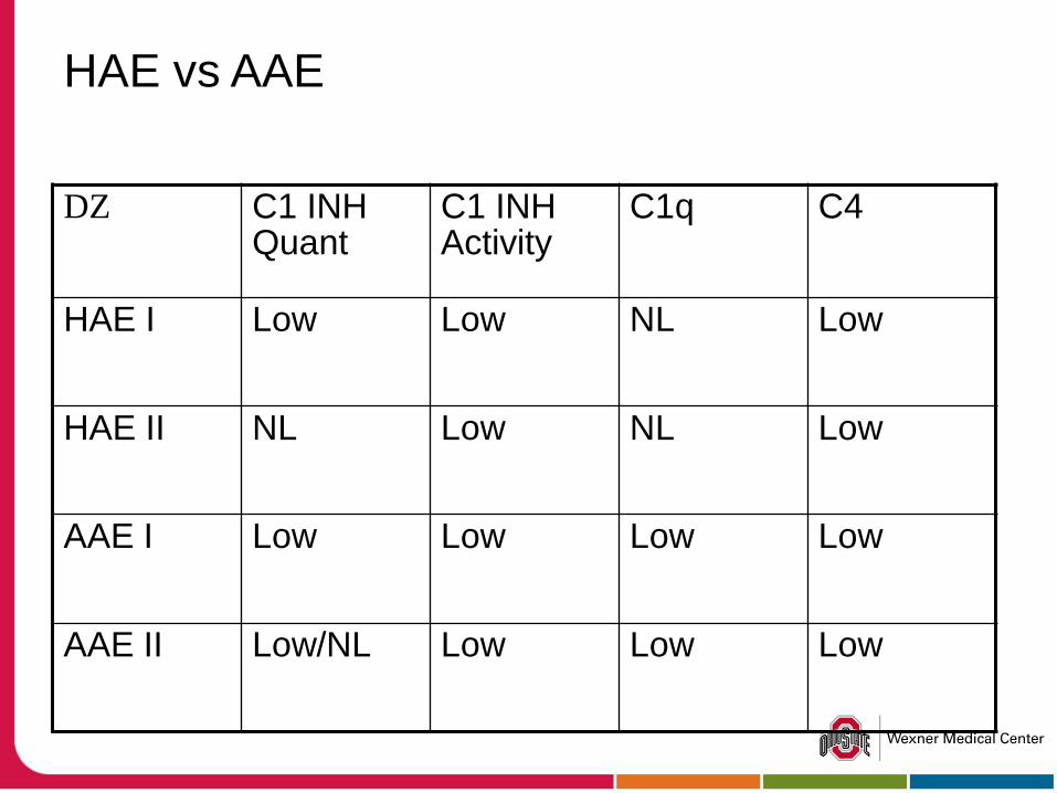
HAE vs AAE
DZ C1 INH Quant
C1 INH Activity
C1q C4
HAE I Low Low NL Low
HAE II NL Low NL Low
AAE I Low Low Low Low
AAE II Low/NL Low Low Low
Universal Precautions & Latex
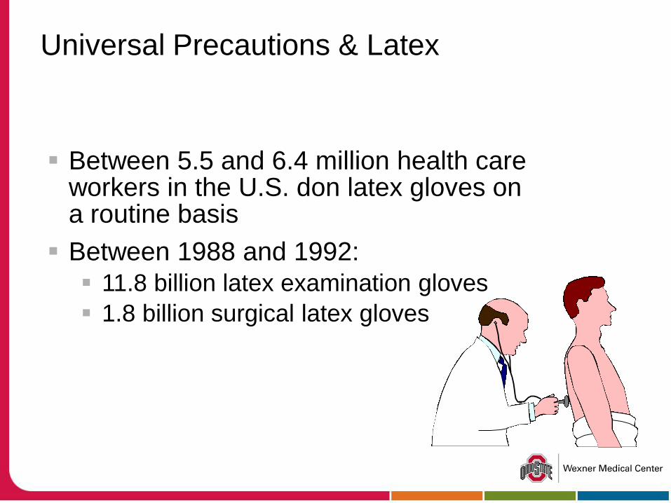
Between 5.5 and 6.4 million health care workers in the U.S. don latex gloves on a routine basis
Between 1988 and 1992:
11.8 billion latex examination gloves
1.8 billion surgical latex gloves
Latex Reactions
Irritant
Allergic CONTACT Dermatitis
Type I IgE mediated allergic reaction: systemic reaction
Latex Irritant Reaction
Non allergic cutaneous response
Erythema, chapping, cracking, dryness, rarely vesicles
Only latex exposed areas
Prolonged and repeated Latex exposure
Age: older skin is more easily irritated and heals more slowly
Allergic Contact Dermatitis
Type IV Gel & Coombs
Multiple exposures: weeks to months
Reaction to chemical additives in gloves
Reaction 12-24 hrs after exposure
may be 6-96 hours after exposure
Vesicular skin lesions
Hypoallergenic gloves appropriate
Patch test to ID culprit additive
Systemic Latex Reaction
Gel & Coombs type I
Anti-latex IgE
Occurs in minutes: rarely > 2 hours
Contact urticaria, rhinoconjunctivitis, anaphylaxis
Mucosal exposure > cutaneous exposure
Modest correlation between IgE concentration and severity
Reaction Severity
Future experience is NOT predicted by prior reactions
Severe anaphylactic reactions may occur following any type of exposure
In general, mucosal exposure results in more severe reactions than cutaneous exposure
Treatment: Latex Anaphylaxis
Avoid further exposure
Non Latex gloves
Hypoallergenic latex gloves are NOT APPROPRIATE
Medic Alert Bracelet
Carry epi and antihistamines
Pretreatment for necessary exposure
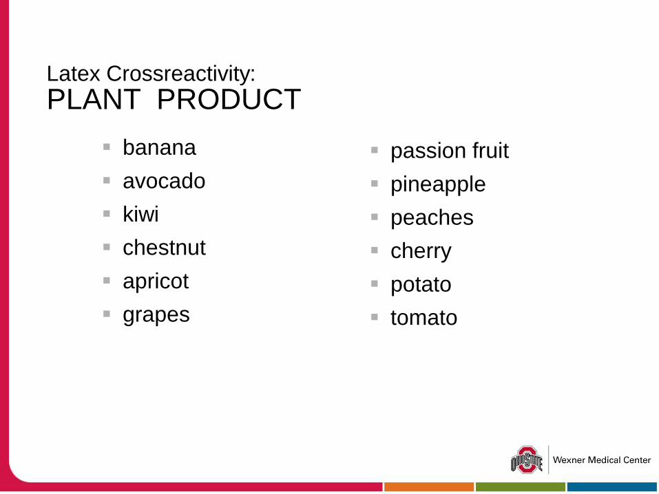
Latex Crossreactivity:
PLANT PRODUCT
banana
avocado
kiwi
chestnut
apricot
grapes
passion fruit
pineapple
peaches
cherry
potato
tomato

Most Frequent Signs and Symptoms of Anaphylaxis
MANIFESTATION PERCENT
Urticaria/Angioedema 88
Upper Airway Edema 56
Dyspnea/Wheeze 47
Flush 46
Hypotension 33
Gastrointestinal 30
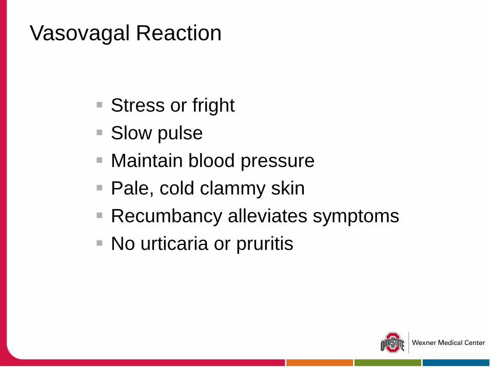
Vasovagal Reaction
Stress or fright
Slow pulse
Maintain blood pressure
Pale, cold clammy skin
Recumbancy alleviates symptoms
No urticaria or pruritis

DDX Anaphylaxis
System Anaphylaxis Vaso-Vagal Rxn
Cutaneous Urticaria, erythema Pale, clammy
Respiratory Globus, SOB wheezing, SPO2
Hyperventilation SPO2:
Cardiovascular Tachycardia, hypotension
Bradycardia, normotensive
G.I. N, V, D N, V, D
C.N.S. “Feeling of impending doom”
Light headed, confused

Anaphylaxis: Treatment
Stabilize airway
Epinephrine O2
Large gauge IV
Benadryl 50-100 mg IV or IM
Cimetidine 300 mg IV
Methyprednisolone 125mg IV
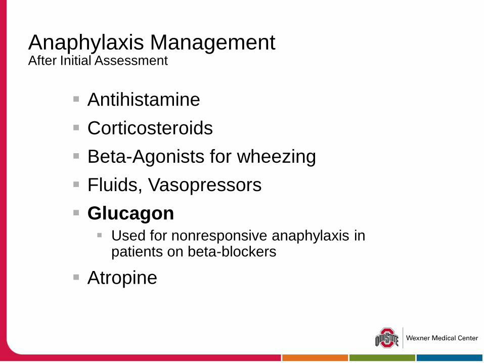
Anaphylaxis Management After Initial Assessment
Antihistamine
Corticosteroids
Beta-Agonists for wheezing
Fluids, Vasopressors
Glucagon Used for nonresponsive anaphylaxis in
patients on beta-blockers
Atropine
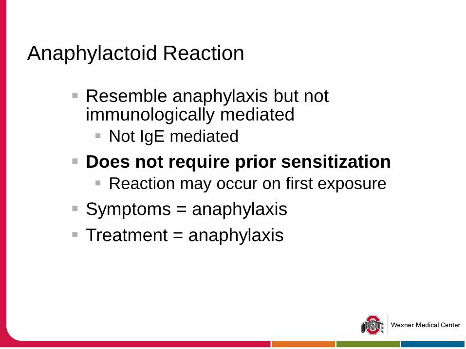
Anaphylactoid Reaction
Resemble anaphylaxis but not immunologically mediated
Not IgE mediated
Does not require prior sensitization
Reaction may occur on first exposure
Symptoms = anaphylaxis
Treatment = anaphylaxis
Anaphylactoid Reactions Non IgE mediated causes
Complement-mediated
Direct activation of mast cell-mediator release
Arachidonic acid metabolism
Unknown
Complement Mediated Anaphylactoid Reactions
Human plasma and blood products
Dialysis membranes
Direct activation of Mast Cell mediator release
Opiates
Vancomycin
Muscle-depolarizing drugs
Aminoglycosides
Radiocontrast media
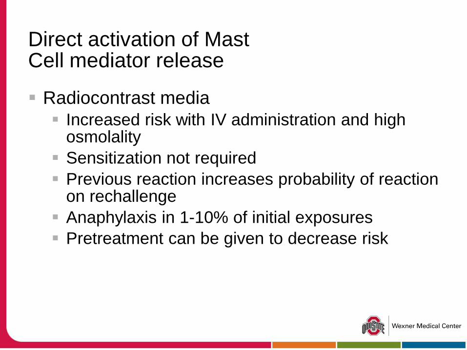
Direct activation of Mast Cell mediator release
Radiocontrast media
Increased risk with IV administration and high osmolality
Sensitization not required
Previous reaction increases probability of reaction on rechallenge
Anaphylaxis in 1-10% of initial exposures
Pretreatment can be given to decrease risk
Modulators of Arachidonic Acid Metabolism
Aspirin and Nonsteroidal drugs
Generally progresses more slowly
Less often hypotension
Bronchoconstriction, wheezing often begin within 30 minutes and progress for several hours
Anaphylaxis While Receiving Beta-blocker Therapy
Unusual severity
Bradycardia during profound hypotension
Severe sustained bronchospasm
Total body angioedema
Refractory to usual treatment
Glucagon is used for refractory cases
Treatment of Anaphylaxis: in presence of Beta-blockade
Aggressive and prompt support
Epinephrine
Large volume IV
Glucagon
Atropine
Increased dopamine or beta-agonist
Antishock trousers
Prevention of Anaphylaxis Radiocontrast Media (RCM)
Use non-ionic media
History
Premedicate before RCM Prednisone 50 mg PO 13, 7 and 1 hour before
procedure
Benadryl 1mg/kg 1 hour before procedure
Anaphylaxis: Differential Diagnosis
Vasodepressor Reaction
Flush syndrome
Restaurant Syndrome
Other forms of shock
Endogenous overproduction of histamine
Red-man syndrome
Pseudoanaphylaxis

Questions?