university of kentucky, lexington, ky, usa...
TRANSCRIPT
Assessment and Treatment of Scapular Dysfunction Using a Kinetic Chain Approach Tim L. Uhl PhD ATC PT, Division of Athletic Training, Department of Rehabilitation Sciences, University of Kentucky, Lexington, KY, USA ([email protected])
Description: The body functions as an integrated system, in sport and in work, we are task oriented. Understanding how the entire system works together as a functional unit within its environment is indispensable for appropriate evaluation and intervention to restore patients’ to their full functional level. An individual patient develops movement patterns and resting postures dependent on their physical characteristics (strength, flexibility, endurance) and their psychological state to meet the demands of the task (throwing, lifting packages into a truck, inserting rivets) and the environment the task is performed. In the common microtraumatic presentation of an injury our job is to determine which component(s) are creating the pathology, impairments and functional limitations. The focus of this presentation will address the biomechanical, anatomical, and the physiological considerations needed to create interventions to ultimately resolve the functional limitations to allow the patient to return to full function. Objectives:
1. Identify factors leading to upper extremity dysfunction (scapular dyskinesis) 2. Review normal biomechanical function and motor control shoulder and scapular dynamic
motion 3. Describe an evaluation procedure for upper extremity that incorporates the entire system
(kinetic chain) and incorporates scapular assessment 4. Identify indicators of impairments and/or functional limitations that may be addressed in
the intervention Incidence of Problem • Scapular dyskinesis Intimately involved with glenohumeral derangement
• 64% Instability
• 100% Impingement72
• Dyskinesis - Impairment of the power of voluntary movement resulting in fragmentary movements.16
Role of the Scapula29
• Scapular motion is critical for normal motion and function of upper extremity
• Critical link between trunk and upper extremity – Site of multiple muscular attachments
• Provide mobile base for the humerus to maintain glenohumeral stability
• Transmission of forces through the kinetic chain
• Scapular motion is critical for normal motion and function of the upper extremity24 Potential Scapular Dysfunction Factors • Glenohumeral pathology17, 44, 46 • Neuropathy64, 73 • Muscle weakness9 • Muscle tightness5 • Muscle fatigue 52 • Pain6

• Loss of neuromuscular control 65, 70 Role of Scapular Control in Injury • Upward rotation, external rotation, and posterior tilt necessary for typical arm elevation &
function • Scapular Kinematics altered in patients with RC tendonopathy
– Demonstrate diminished posterior tilting (~10) – Excessive superior translation of scapula significantly higher (~2cm)46
• Protracted posture decreases sub-acromial space67 • Altered muscular activations
– Elevated Upper Trapezius activity44 Neurological Deficits64 • Long thoracic / Spinal accesory nerve palsies
• Reduces mechanical stability of the shoulder • Highlights the role of neuromotor control of scapula and its effect on the entire upper
extremity • Pain inhibits motor activation6 Chronic Shoulder Pain Neuromuscular Adaptations • Swimmer’s with painful shoulder • Serratus Anterior inhibition • Substitution of upper trapezius and rhomboids65 • Delayed activation of Serratus anterior by 80 ms (* p< 0.05) indicating poor muscular
control70 Shoulder Instability in Pitching Affects Neuromuscular Control17
• Decreased activation of Serratus Anterior
• Increased activation of supraspinatus and biceps to stabilize humeral head
• Decreased activation of accelerators (internal rotators) – latissimus dorsi, subscapularis, & pectoralis major
Role of Scapular Control in Glenohumeral Instability • Abnormal motion can
• Overload joint capsule
• Impinge underlying rotator cuff & labral tissues • Hyperangulation of humerus
• Excessive horizontal abduction
• Macrotraumatically cause dislocation
• Microtraumatically leads to postererior impingement & subluxation7, 60 Poor Proximal Control • Without proximal control distal movement are compensating for a poor foundation • Single leg squat
• Women tend to go into more valgus75 • Single leg stance is not just hip abductor strength
• Hip abductor strength poorly correlated with 2D Hip Adduction12 Appreciate Normal Motor Control and Kinematics
• The ability to lift arm is dependent on proximal stability: glenohumeral joint, scapulothoracic, & spine
• Dynamic stability of proximal segments arises due to anticipatory postural adjustments10, 74
• The kinetic chain model is linked segments commonly used in biomechanics61
• The human body can be characterized as a kinetic chain
• Kinetic chain theory supports that distal force production is due to summation of forces in the proximal segments throughout the entire kinetic chain (hitting or kicking a ball)
• Breakdown along the chain Increase demand on segments such as shoulder
• Typical motor control pattern activates in a proximal – distal manner in Shoulder Elevation 10,
22
• Transverse abdominal and multifidus musculature precedes distal arm motion to stabilize trunk and prevent postural perturbation
Scapula part of the CHAIN • Critical links between trunk & upper extremity • Periscapular musculature act to transfer energy through the kinetic chain • Missing segment in link models • Throwers develop adaptations:54
• Increased upward rotation
• Increased internal rotation
• Increased retraction during scaption than non throwers 2-D Biomechanics of Scapula
• Scapulohumeral rhythm of 2:1 following a setting phase24
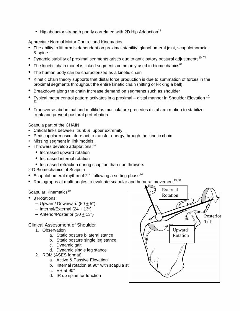
• Radiographs at multi-angles to evaluate scapular and humeral movement15, 59 Scapular Kinematics50
• 3 Rotations – Upward/ Downward (50 + 5) – Internal/External (24 + 13) – Anterior/Posterior (30 + 13)
Clinical Assessment of Shoulder
1. Observation a. Static posture bilateral stance b. Static posture single leg stance c. Dynamic gait d. Dynamic single leg stance
2. ROM (ASES format) a. Active & Passive Elevation b. Internal rotation at 90 with scapula stabilized
Upward Rotation
Posterior Tilt
External Rotation
c. ER at 90 d. IR up spine for function

e. GIRD = difference in degrees between dominant and non-dominant IR measures 3. Strength
a. Rotator cuff b. Scapular
4. Special test a. Scapular Assistance b. Scapular Retraction c. Kinematic analysis
i. Inclinometer ii. 3-D kinematics
Postural Assessment
-Cited as a potential cause of shoulder and neck pain 26 Slouched thoracic posture reduces humeral and scapular motion along with reducing strength27 -Recent report on short pectoralis minor has demonstrated reduced scapular motion 5
Slouched Thoracic Posture27 • Shoulder abduction ROM
• Erect: 157.5° (+ 10.8)
• Slouched: 133.9° (+ 13.7) • Abduction strength @ 90°
• Erect: 10.4kg (+ 4.5)
• Slouched: 8.7kg (+ 3.5) • Scapular Kinematics • Upward rotation:
• Erect: 43.1° (+7.5)
• Slouched: 37.9° (+6.5) • Posterior tilt
• Erect: 44.7° (+6.8)
• Slouched: 40.6° (+6.9)
Rounded Shoulder Posture5
• Report on short pectoralis minor has demonstrated reduced scapular motion
• Short pectoralis minor group at 90o elevation – 7o less External Rotation – 6o less Upward Rotation – 7o less Posterior Tilting
Clinical Measures3 • Scapular Index
• A) Distance between sternal notch and coracoid (cm) • r = .48 to pect minor shortness
• B) Distance between T-spine and lateral acromion • Scapular index (A/B) X 100 • r = .37 to pect minor index (tightness)

New Clinical Measure of Posture4
• Measure pectoralis minor length from 4th rib to coracoid
• High reliability ICC=.82
• Based on the sample of 26 subjects typical lengths were 16 + .3 cm Quantitative Assessment: Lateral Scapular Slide57 • Measure the distance (cm) from spinous process to inferior angle • Bilateral comparison in three position • Does not discriminant between injured and non-injured Scapular Assessment
• Observational static descriptions with arms at side 63
• Downward Rotation
• Depression
• Abduction
• Winging and tilting syndrome Scapular classification by observational analysis during AROM assessment 34 • Normal • Superior border pattern • Medial border pattern • Inferior angle pattern Results
• Moderate reliability • Intertester of therapists: k = .42 • Intratester of therapist: k = .49 • New observational system based on 3-D motion • 2-dimensional Video tape analysis • Further evidence to support observational system assessment
Follow up study of observational assessment method is better with 2 category system (sensitivity 75%)
– Presence of scapular dyskinesis – Absence of scapular dyskinesis
The dysfunction appears to be a loss of consistency (Abstract – ASES 2004) – Internal/external rotation – Upward rotation
Single leg stance series:
1 leg to evaluate balance and hip stability 1 legged squat for lower strength Without proximal control distal movement are compensating for a poor foundation
Assessment of Scapular Muscle Function 53
Lower Trapezius-Prone arm is abducted to 135o shoulder is flexed and scapula retracted, apply pressure in line with fibers of lower trapezius and anteriorly to move scapula anteriorly

Middle Trapezius and Rhomboids - Prone humerus is extended and scapula retracted (medial border near spine), apply pressure
Serratus Anterior - Supine arm is flexed to 90o with arm protracting so that scapular moves laterally along thoracic wall, apply pressure to resist protraction
Upper Trapezius - cervical spine side bent and rotated away with scapula shrugged, apply pressure to resist elevation and posterior occiput
Scapular Assessment Special Tests
Goal- Repositioning the scapula to open subacromial space Many impingement tests drive the humerus into the scapula where these tests consider
moving the scapula out of the way of the humerus Scapular Assistance Test31, 62 • Active elevation of the arm without scapula stabilized, patient reports pain • Stabilize the scapula by applying anterior and upward force on the inferior angle
as the arm is elevated • (+) Pain diminished • Indicates improving scapular motion may diminish symptoms Scapular Retraction Test • Perform provocative resistance test
• Empty can to assess strength of deltoid and rotator cuff • Have patient retract scapula actively and manually provide scapular stability then retest
strength • Improved symptoms (strength) indicates scapular muscular control is compromised
Ongoing study 20 pathological subjects improved strength 25%
The first step in rehabilitation is a complete and thorough assessment.30 A complete assessment of a patient with an upper extremity injury should include assessment and consideration of scapular dysfunction as part of a thorough evaluation.

Evaluation Format: Considerations Findings Observation Static posture bilateral stance Lateral view
Lower Extremity Hips/pelvis Thoracic Kyphosis – double square Shoulder/Scapula – tape measure Head
Anterior/Posterior view Feet Knee Hips Scapular position (Lateral Scapular Slide) Scolosis Atrophy Dynamic gait
AROM (standing) Add load for strong patients
5-10 forward flexion 5-10 scaption Trunk motion (4-6 directions)
Dysfunction Yes No Yes No
Core Stability (standing)
Single leg balance Hip adduction Ankle control Single leg squat Hip/pelvis rotation Knee valgus
Special Tests (standing)
Neer Hawkins-Kennedy Active compression Mayo shear Painful arc
Rotator Cuff/ Scapular Strength (standing)
Arm elevation thumb-up (Deltoid & Supraspinatus) External rotation at 45 (Flip sign) Lift-off / Belly press Arm elevation >120 (Serratus Anterior)
Scapular Tests (standing)
Scapular Assistance Test (+) reduction of pain with scapular support Scapular Retraction Test (+) reduction of pain and/or increase strength
Scapular Strength (lying)
Prone flexion at 135 abduction (Lower Trapezius) Medial border retraction (Rhomboids)
PROM/ Glenohumeral Instability (lying)
Glenohumeral internal rotation with scapula stabilized External rotation Elevation Apprehension Relocation (instability / labral) Crank Biceps Load

Clinical Tests – Evidence from the literature on Shoulder Clinical Tests (+) LR > 5 moderate shift toward presence of pathology. (-) LR <.02 moderate shift toward absence of pathology
Test Specificity (+) Likelihood Ratio Sensitivity
(-) Likelihood Ratio Reference
Anterior Glenohumeral instability tests
Anterior Release Test 89% 8.40 92% 0.09
Gross ML & Distefanco MC Clin Orth Rel Res 199718
Apprehension Test (apprehension) 96% 20.2 72% 0.29 Farber JBJS 2006 14 Relocation Test (relief of apprehension) 92% 10.4 81% 0.2 Farber JBJS 2006 14 Labral Tests Active Compression (O'Brien's Test) 98% 21.00 100% 0.01
O'Brien Am J Sports Med 199856
Crank Test 93% 13.00 91% 0.10 Liu SH, et al. AJSM 199643
Biceps Load Tests II 96% 26.00 90% 0.10 Kim Arthorscopy 200135
Biceps Load Tests I 98% 29.00 83% 0.09 Kim AM J Sports Med 199936
Resisted Sup_ER Test 82% 4.55 83% 0.21 Myers TH, et al., Am J Sports Med, 200555
Anterior Slide Test 92% 8.30 78% 0.20 Kibler WB, Arthroscopy, 199528
Active Compression Test 11% 0.88 78% 2.00 Myers TH, et al., Am J Sports Med, 200555
Active Compression Test 73% 2.33 63% 0.51 Guanche & Jones, Arthroscopy 200319
Posterior Glenohumeral instability tests
Jerk Test 98% 36.50 73% 0.28
Kim, SH, Am J Sports Med 2005 33(8)37
Impingement Tests
Hawkins 44% 1.64 92% 0.18
MacDonald PB et al JSES 9:299-301 200047
Hawkins 25% 1.23 92% 0.32 Calis M, et al., Ann Rheum Dis, 20008
Painful arc 81% 3.89 74% 0.32 Park HB et al., JBJS, 200558
3/7 positive Hawkins, Neer, Drop Arm, Horiz. Add., Speed, Yeargason, painful arc 44% 1.50 84% 0.36
Calis M, et al., Ann Rheum Dis, 20008
Neer 31% 1.28 89% 0.37 Calis M, et al., Ann Rheum Dis, 20008
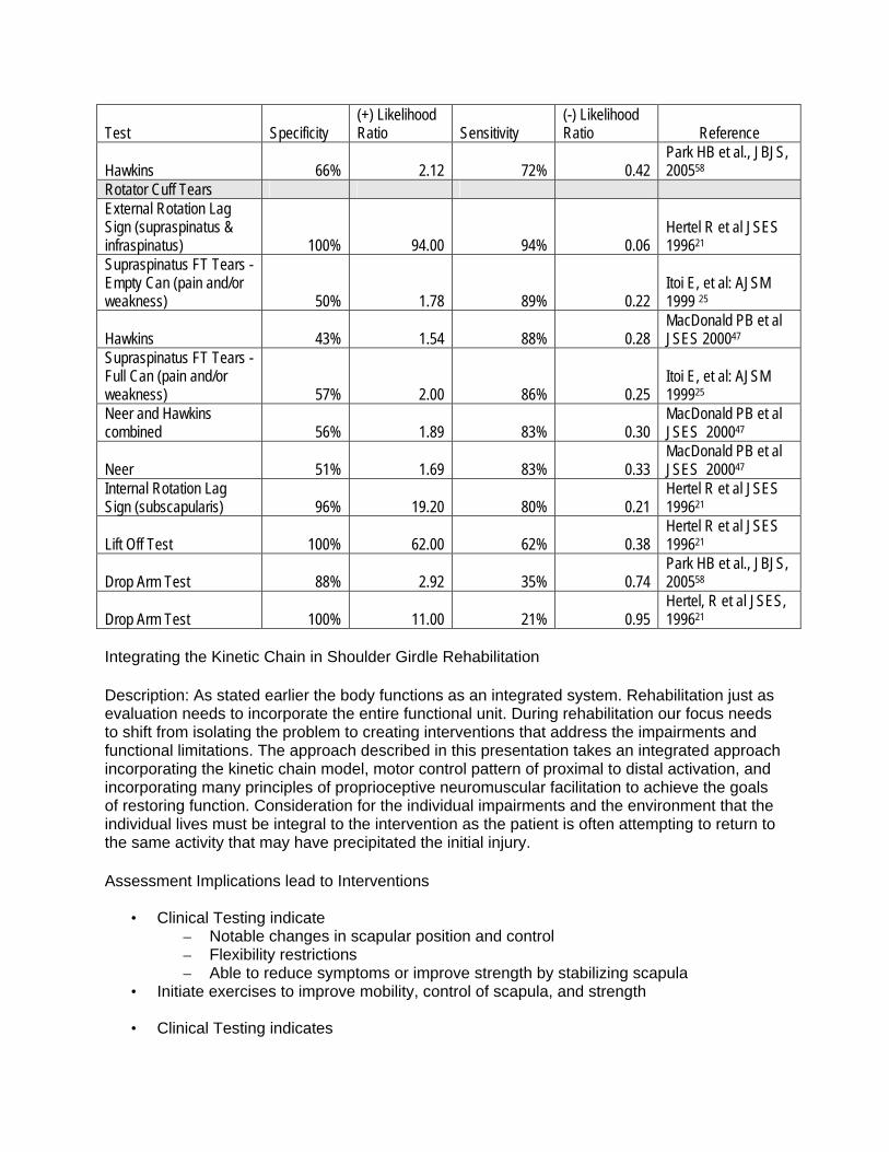
Test Specificity (+) Likelihood Ratio Sensitivity
(-) Likelihood Ratio Reference
Hawkins 66% 2.12 72% 0.42 Park HB et al., JBJS, 200558
Rotator Cuff Tears External Rotation Lag Sign (supraspinatus & infraspinatus) 100% 94.00 94% 0.06
Hertel R et al JSES 199621
Supraspinatus FT Tears - Empty Can (pain and/or weakness) 50% 1.78 89% 0.22
Itoi E, et al: AJSM 1999 25
Hawkins 43% 1.54 88% 0.28 MacDonald PB et al JSES 200047
Supraspinatus FT Tears - Full Can (pain and/or weakness) 57% 2.00 86% 0.25
Itoi E, et al: AJSM 199925
Neer and Hawkins combined 56% 1.89 83% 0.30
MacDonald PB et al JSES 200047
Neer 51% 1.69 83% 0.33 MacDonald PB et al JSES 200047
Internal Rotation Lag Sign (subscapularis) 96% 19.20 80% 0.21
Hertel R et al JSES 199621
Lift Off Test 100% 62.00 62% 0.38 Hertel R et al JSES 199621
Drop Arm Test 88% 2.92 35% 0.74 Park HB et al., JBJS, 200558
Drop Arm Test 100% 11.00 21% 0.95 Hertel, R et al JSES, 199621
Integrating the Kinetic Chain in Shoulder Girdle Rehabilitation Description: As stated earlier the body functions as an integrated system. Rehabilitation just as evaluation needs to incorporate the entire functional unit. During rehabilitation our focus needs to shift from isolating the problem to creating interventions that address the impairments and functional limitations. The approach described in this presentation takes an integrated approach incorporating the kinetic chain model, motor control pattern of proximal to distal activation, and incorporating many principles of proprioceptive neuromuscular facilitation to achieve the goals of restoring function. Consideration for the individual impairments and the environment that the individual lives must be integral to the intervention as the patient is often attempting to return to the same activity that may have precipitated the initial injury. Assessment Implications lead to Interventions
• Clinical Testing indicate – Notable changes in scapular position and control – Flexibility restrictions – Able to reduce symptoms or improve strength by stabilizing scapula
• Initiate exercises to improve mobility, control of scapula, and strength • Clinical Testing indicates

Positive signs for tissue dysfunction Minimal issues with motor control and mobility of shoulder girdle is adequate Unable to reduce symptoms by moving or stabilizing scapula MD consult for further diagnostic testing indicated
The Basis for using the Trunk in Upper Extremity Rehabilitation51 1.Kinetic Chain
• A model of linked segments commonly used in biomechanics
• Sport and work activities attempting to strike or throw at high velocities use the kinetic chain to impart these forces
• Acceleration of distal segment comes from the “controlled deceleration” of proximal
segments Breakdown anywhere along the kinetic chain can lead to
• Increase demand on shoulder musculature
• Change the biomechanical stresses
• Diminish performance61 2. Typical motor control pattern activates in a proximal – distal manner in Shoulder Elevation 10,
22
• Shoulder function depends on proximal stability
• Rehabilitation needs to establish proximal stability before distal mobility • Transverse abdominal and multifidus musculature precedes distal arm motion41, 74 • This activation provides trunk stabilization and prevents postural perturbation10, 22
• Trunk serves to generate, absorb, and distribute forces during all activities, particularly important in sports
• Inner Core musculature stabilize pelvis and lumbar spine and is activated prior to distal segment motion
• Outer Core musculature provides some global stability but is integral in generating and transmitting functional mobility
• Serape function of trunk musculature • Serape effect is described as the function of trunk muscles generating forces and
transferring it to the extremities40 • Combining all three planes of motion is functional and is a fundamental principle for
many power motions68 3. Applying PNF Principles to Kinetic Chain Rehabilitation39, 69
• Motor behavior is a sequence including head, trunk, and extremities
• Goal directed movements are dependent on synergies
• Normal motor development occurs in a proximal to distal manner
• Stronger components of a movement pattern facilitate weaker (irradiation)
• Clinician must help the patient relearn the movement pattern

– Selecting resistance or assistance
– Verbal cueing
– Manual contact
– Visual, auditory, and tactile feedback Isolating vs. Integrating Exercises • Prone Extension
– Posterior cuff (teres minor) and deltoid >60% MVIC2 • Standing shoulder extension with forward step
– Trunk / Scapular/ Cuff musculature • Two different approaches neither is wrong Integrating Entire Body with Elevation Exercise with good Core
• Step forward punch
• Activate legs trunk scapula & rotator cuff muscle in normal pattern – Proximal to distal emphasized
• Anterior / Acceleration Serape Management Strategies • Base intervention on level of tissue irritability
– More inflamed or reactive – less direct activity to tissue • Proximal control and alignment of spine a scapula are necessary prior to applying distal
stressors – motor control • Find appropriate intensity level for patient that does not create compensations
– Facilitate scapular motion • Thoracic extension or rotation • Scapular retraction & depression (external rotation & posterior tilt) • Support the weight of the arm to reduce compensations
• Rehabilitation environment must match the level of function – must constantly be challenged to improve function
• Continuum of functional rehabilitation is driven by patient’s response to exercise Rehabilitation Components
• Address – Spine posture – Proximal stability & balance – Position, motion, stability, & strength of all joints of the shoulder
• Postural correction
• Proximal stability
• Rehabilitation exercises should simulate neuromotor patterns
• Flexibility and mobility
• Pain management
• Exercise progressively increase demand Proximal Stability
• High tissue irritability

– Acute
• Addressing proximal control or posture issues is appropriate
• Patient is observed to have poor core control – Remedial core program
• Adequate control – Integrate into complex exercises
Kinetic Chain Exercises in Sling • EMG activity for subscapularis was moderate to high (30 – 70% MVC) • All other cuff and deltoid musculature low <20% MVC • Addition of step with exercise slightly increased EMG activity <10% • Lawnmower exercise keeping elbow low activated Serratus Anterior to very high levels >
80% while other muscles remain <20% • Lawnmower exercises are safe in early phases of shoulder rehab except for subscapularis
repairs66 Increasing Spine & Shoulder Mobility
• Shoulder elevation can be increased by increased thoracic and cervical extension
• Joint mobilization techniques – Decrease muscle guarding – Increase mobility
Posture can be Positively Effected 71 • 6 weeks program (3 x wk) • Stretching
– Pectoralis group – corner stretch • Hold 10 seconds – 10 repetitions
• Strengthening (Theraband) – Scapular elevation (shrug) – Horizontal Shoulder ADD (retraction) – ER @ 0o – Shoulder abduction
Results • Produced more erect thoracic spine • Increased shoulder strength • Improved scapular stability Forward Posture can be Reduced 38 • Competitive swimmers on 6 weeks program (3 x wk) • Stretching
– Pectoralis minor – partner stretching – Pectoralis major – ADB/ER
• Strengthening (Theraband) – ER @ 90o – Shoulder flexion – Shoulder Horizontal ADD (retraction)
• Reduced forward shoulder posture by 9mm
Taping to Improve Posture & Scapular Position42

• 60 patients with Impingement Syndrome and 60 without • Underwent taping and placebo taping correction of posture and scapular retraction • Kyphosis decreased by 6o
• Diminished lateral scapular displacement by 1.5cm • Increased shoulder elevation by 15o in flexion and scaption Limitations to Taping
• Taping requires the assistance of a knowledgeable aid and frequent re-application
• Skin breakdown can occur with repetitive taping
• Taping can loosen with time and become wet & loose with sweating Passive Techniques to Correct Posture • Devices for postural (passive positioning)
– Figure 8 clavicle straps – McConnell taping – Spine and Scapular Stabilizing Brace S3 (Alignmed, Santa Ana, CA)
• Research question: To determine if the Spine and Scapular Stabilizing Brace (S3)* has an effect on scapular kinematics at rest and during arm elevation – 15 healthy – 18 injured with scapular dyskinesis
Upward Rotation and Posterior Tilt 3 Relief of Painful Myofascial Spasms23
• Several combinations are effective at decreasing pain & sensitivity of trigger point – 1.Moist Hot pack > AROM > Ischemic Compression > TENS – 2. Moist Hot pack > AROM > Spray and Stretch > TENS – 3. Moist Hot pack > AROM > IFC > Myofascial release
• Ischemic compression @ 90 seconds provide relief of trigger point sensitivity Address Muscular Imbalances • Stretching for upper trapezius • Stretching for levator scapulae • Stretch for biceps and pect minor Pathomechanics of Tight Posterior Structures • Scapular motion particularly at end ranges influenced by capsular tension50 • Humeral head is pushed superiorly into acromion
– Anteriorsuperior20 – Posteriorsuperior7
Stretching the Posterior Tissues • Adaptive changes can occur shortening posterior cuff/capsule32 • Sleeper Stretch • Cross body adduction with scapula stable and humerus internally rotating • Cross body adduction increases IR motion by 20o .48 • Hold Stretch 30 seconds (No Pain)1

Rehabilitation Exercise
• Scapular substitution patterns present due to poor muscular performance and pain
• Rehabilitation exercise should progressively increase the stress on muscles
• Integrating – Posture and position of spine and scapula – Rehabilitation exercises should simulate normal neuromotor patterns – Exercise progression from low to high demand
Finding Appropriate Intensity Level of Exercise
• Standing - attempt to gain scapular control with sagittal plane elevation – Functional
• Standing - work on transverse plane
• Static arm – exercise trunk and possibly scapula
• Unload weight of arm or shorten lever arm
Serratus Exercise Progression
611
17 22 23
49
7280 82
91
0102030405060708090
100
Towel s
lide
Side-
lyin
g Ele
vatio
n
Supin P
ress
-up
Ball R
olls
Activ
e Ele
vatio
n
Forwar
d punch
Knee p
ush u
p plu
s (t
op to p
lus)
Push u
p plu
s
Mili
tary
pre
ss
Scaptio
n
EM
G (
%M
VIC
)
Lower Trapezius Progression
3 818
27
4550
61
79
97
0
20
40
60
80
100
120
Towel slid
e
Side-ly
ing E
levation
Wall s
lide
ER sidelyin
g
Unilate
ral r
ow
Scaption <
80°
Scaption >
120°
90° ABD E
xt. R
ot
EM
G (
% M
VIC
)
Regaining Scapular Control
• Often regaining scapular retraction control first step
• Static – Low Row
• Rotational plane –
• Sagittal plane - Lawnmower Applying PNF Principles & Kinetic Chain to Shoulder Rehabilitation • Start motion with trunk and with scapular muscles on stretch
– Extend and rotate spine – Scapula retracts – Shoulder externally rotates51
• Training Tip: • “Tuck Elbow in back pocket” • “Pull scapula back” • Exaggerating transverse plane Facilitate Scapular Muscle Activation of Retractors
• Stronger components of a movement pattern facilitate weaker (irradiation)

• Irradiation of loading proximal musculature to activate scapular musculature Kinetic Chain Scapular emphasis “Down and Back” • Active trunk extension (sternal lift) with scapular retraction • Facilitate proper posture with re-education techniques • Use trunk to activate scapular musculature and lengthening tight anterior musculature5 Identifying the Intensity • Low Row • Inferior Glide • Lawnmower • Robbery • These exercises are low to intermediate phase scapular strengthening33 Regaining Motor Control
• Patient awareness: Facilitate or Inhibit
– Tactile feedback – PNF – Rhythmic Initiation /Rhythmic stabilization
– EMG feedback
– Visual feedback • Mirror • Video
• Muscle re-education – Neuromuscular control
• Utilization of video to provide feedback
• Do not overly verbal correct
• Activation of Serratus facilitates scapular posterior tilt and external rotation Regain Motor Control: Rhythmic Initiation39 • Tactile feedback – PNF – Rhythmic Initiation /Rhythmic stabilization • A technique that assists in transition from PROM to AAROM to AROM exercises • Directing patients motion to facilitate voluntary motor pattern • Arm can be supported • Advantage to translate into manual resistive exercise without changing positions
– Intensity (painless ROM) – Time (3-4 mins / 3 x 10)
Scapulohumeral Relationship during Manual Stability Ex’s • Rhythmic stabilization - Isometric muscle contraction of antagnostic muscles to facilitate
dynamic joint stability39 • Musculature are activated due to a response to load • Emphasis is on holding a position • Watch level of effort might injure healing tissue • Constantly inspect patient position Combining Video and EMG Feedback with Exercise
• Supervise for proper form is sometimes not enough
• Incorporate real time feedback with video and EMG to correctly activate SA

Elevation Progression • Serratus Anterior needs facilitation17 • Exercises that emphasize Serratus over Upper Trapezius45 • Protraction bias upper Serratus fibers & elevation above 120o biases lower fibers13 • Be ever vigilant on compensations
Elevation with Kinetic Chain
• Active elevation without facilitation
• If see substitution you – Increase trunk facilitation (step) – Decrease distal load by support of surface
Applying Kinetic Chain to Punch • Scapular protraction weak in patients with impingement 9 • Scapular protraction activates Serratus Anterior11, 13 • Functional rehabilitation replicate normal neuromotor pattern 10, 74
– Legs drive arm in diagonal • Shoulder strengthening 3 x10 daily improves strength and function in patients with
impingement49
Progress to Long Lever Arm Ex’s Last
• Exercise place high demand on scapular musculature – Torque = Force x Distance
• Elevation above 120o bias lower fiber of Serratus Anterior
• Prone elevation at 135o activates Low Trap13 Exercise Matches Demands
• Special consideration needs to be given to athletes with no to little base of support – Volleyball – Swimming
• Unstable base facilitates trunk stabilizers – (non-ground based sports: volleyball)
Summary
• Management of scapular dysfunction – Accurate assessment – Respect irritable tissue – Work on proximal control and mobility first – Apply appropriate exercise intensity that does not produce compensations – Prescribe exercises along a continuum that meet the demands of the activity

Exercise examples Scapula depressed and retracted
Down and Back!
Sagital plane utilizing hip extension
Emphasizing transverse plane
Unload weight of arm

Vary angle of unloading to meet functional demand and patients’ impairment
Sitting unstable surface (Non-weightbearing activities)
Static Arm emphasizing trunk and scapular co-activation
Think Functional, Think Total Body, Have Fun
References 1. Bandy WD, Irion JM, Briggler M. The effect of time and frequency of static stretching on
flexibility of the hamstring muscles. Phys.Ther. 1997;77(10):1090-1096. 2. Blackburn TA, McLeod WD, White B, Wofford L. EMG analysis of posterior rotator cuff
exercises. Athletic Training. Spring 1990;25(1):40;42-45.
3. Borstad JD. Resting position variables at the shoulder: evidence to support a posture-impairment association. Phys Ther. Apr 2006;86(4):549-557.
4. Borstad JD. Measurement of pectoralis minor muscle length: validation and clinical application. J Orthop Sports Phys Ther. Apr 2008;38(4):169-174.
5. Borstad JD, Ludewig PM. The effect of pectoralis minor length on scapular kinematics in subjects without shoulder pathology. Journal of Orthopaedic and Sports Physical Therapy. 2004;34(1):A16.
6. Brox JI, Røe C, Saugen E, Vøllestad NK. Isometric abduction muscle activation in patients with rotator tendinosis of the shoulder. Arch Phys Med Rehabil. 1997;78(11):1260-1267.
7. Burkhart SS, Morgan CD, Kibler WB. The disabled throwing shoulder: spectrum of pathology Part I: pathoanatomy and biomechanics. Arthroscopy. 2003;19(4):404-420.
8. Calis M, Akgun K, Birtane M, Karacan I, Calis H, Tuzun F. Diagnostic values of clinical diagnostic tests in subacromial impingement syndrome. Ann Rheum Dis. Jan 2000;59(1):44-47.
9. Cools AM, Witvrouw E, Declercq GA, Vanderstraeten GG, Cambier DC. Evaluation of isokinetic force production and associated muscle activity in the scapular rotators during a protraction-retraction movement in overhead athletes with impingement symptoms. British Journal of Sports Medicine. 2004;38:64-68.
10. Cordo PJ, Nashner LM. Properties of postural adjustments associated with rapid arm movements. Journal of Neurophysiology. 1982;47(2):287-308.
11. Decker MJ, Hintermeister RA, Faber KJ, Hawkins RJ. Serratus anterior muscle activity during selected rehabilitation exercises. American Journal of Sports Medicine. 1999;27(6):784-791.
12. DiMattia MA, Livengood AL, Uhl TL, et al. What Are the Validity of the Single-Leg-Squat Test and Its Relationship to Hip-Abduction Strength? Rehabilitation concerns of the middle age athlete. 2005.
13. Ekstrom RA, Donatelli RA, Soderberg GL. Surface electromyographic analysis of exercises for the trapezius and serratus anterior muscles. Journal of Orthopaedic and Sports Physical Therapy. 2003;33(5):247-258.
14. Farber AJ, Castillo R, Clough M, Bahk M, McFarland EG. Clinical assessment of three common tests for traumatic anterior shoulder instability. J Bone Joint Surg Am. Jul 2006;88(7):1467-1474.
15. Freeman L, Munro RR. Scapular and glenohumeral movements. Journal of Bone and Joint Surgery, American Volume. 1966;48A((8)):1503-1510.
16. Friel JP. Dorland's Illustrated Medical Dictionary. Philadelphia: WB Saunders; 1974. 17. Glousman R, Jobe FW, Tibone JE, Moynes D, Antonelli D, Perry J. Dynamic
electromyographic analysis of the throwing shoulder with glenohumeral instability. Journal of Bone and Joint Surgery, American Volume. 1988;70A((2)):220-226.
18. Gross ML, Distefano MC. Anterior release test. A new test for occult shoulder instability. Clin Orthop Relat Res. Jun 1997(339):105-108.
19. Guanche CA, Jones DC. Clinical testing for tears of the glenoid labrum. Arthroscopy. May-Jun 2003;19(5):517-523.
20. Harryman DT, Sidles JA, Clark JM, McQuade KJ, Gibb TD, Matsen FA. Translation of the humeral head on the glenoid with passive glenohumeral motion. Journal of Bone and Joint Surgery, American Volume. 1990;72-A(9):1334-1343.
21. Hertel R, Ballmer FT, Lombert SM, Gerber C. Lag signs in the diagnosis of rotator cuff rupture. J Shoulder Elbow Surg. Jul-Aug 1996;5(4):307-313.
22. Hodges PW, Richardson CA. Feedforward contraction of transversus abdominus is not influenced by the direction of arm movement. Experimental Brain Research. 1997;114:362-370.

23. Hou CR, Tsai L, Cheung KF, Chung KC, Hong HM. Immediate effects on various physical therapeutic modalities on cervical myofacial pain and trigger-point sensitivity. Archieves of Physical Medicine and Rehabilitation. 2002;83:1406-1414.
24. Inman VT, Saunders M, Abbot LC. Observations of the function of the shoulder joint. Journal of Bone and Joint Surgery, American Volume. 1944;26(1):1-30.
25. Itoi E, Kido T, Sano A, Urayama M, Sato K. Which is more useful, the "full can test" or the "empty can test," in detecting the torn supraspinatus tendon? Am J Sports Med. Jan-Feb 1999;27(1):65-68.
26. Kamkar A, Irrgang JJ, Whitney SL. Nonoperative management of secondary shoulder impingement syndrome. Journal of Orthopaedic and Sports Physical Therapy. 1993;17(5):212-224.
27. Kebaetse M, McClure P, Pratt N. Thoracic position effect on shoulder range of motion, strength, and three-dimensional scapular kinetics. Archives of Physical Medicine and Rehabilitation. 1999;80:945-950.
28. Kibler WB. Specificity and sensitivity of the anterior slide test in throwing athletes with superior glenoid labral tears. Arthroscopy. 1995;11(3):296-300.
29. Kibler WB. The role of the scapula in athletic shoulder function. American Journal of Sports Medicine. 1998;26((2)):325-337.
30. Kibler WB, Livingston B, Bruce R. Current concepts in shoulder rehabilitation. Advances in Operative Orthopaedics. 1995;3:249-299.
31. Kibler WB, McMullen J. Scapular dyskinesis and its relation to shoulder pain. Journal of American Academy of Orthopaedic Surgeons. 2003;11:142-151.
32. Kibler WB, McQueen C, Uhl T. Fitness evaluations and fitness findings in competitive junior tennis players. Clin.Sports.Med. 1988;7(2):403-416.
33. Kibler WB, Sciascia AD, Uhl TL, Tambay N, Cunningham T. Electromyographic analysis of specific exercises for scapular control in early phases of shoulder rehabilitation. Am J Sports Med. Sep 2008;36(9):1789-1798.
34. Kibler WB, Uhl TL, Maddux JQ, McMullen J, Brooks PV, Zeller B. Qualitative clinical evaluation of scapular dysfunction. A reliability study. Journal of Shoulder and Elbow Surgery. 2002;11(6):550-556.
35. Kim SH, Ha KI, Ahn JH, Choi HJ. Biceps load test II: A clinical test for SLAP lesions of the shoulder. Arthroscopy. Feb 2001;17(2):160-164.
36. Kim SH, Ha KI, Han KY. Biceps load test: a clinical test for superior labrum anterior and posterior lesions in shoulders with recurrent anterior dislocations. Am J Sports Med. May-Jun 1999;27(3):300-303.
37. Kim SH, Park JS, Jeong WK, Shin SK. The Kim test: a novel test for posteroinferior labral lesion of the shoulder--a comparison to the jerk test. Am J Sports Med. Aug 2005;33(8):1188-1192.
38. Kluemper M, Uhl TL, Hazelrigg H. Effect of stretching and strengthening shoulder muscles on forward shoulder posture in competitive swimmers. Journal of Sport Rehabilitation. 2006;15(1):58-70.
39. Knott M, Voss DE. Proprioceptive Neuromuscular Facilitation Patterns and Techniques. Philadelphia: Harper & Row; 1968.
40. Konin JG, Beil N, Werner G. Facilitating the serape effect to enhance extremity force production. Athletic Therapy Today. 2003;8(2):54-56.
41. Le Bozec S, Lesne J, Bouisset S. A sequence of postural muscle excitation precedes and accompanies isometric ramp efforts performed while sitting in human subjects. Neuroscience Letters. 2001;303:72-76.
42. Lewis JS, Wright C, Green A. Subacromial impingement syndrome: the effect of changing posture on shoulder range of movement. Journal of Orthopaedic and Sports Physical Therapy. 2005;35(2):72-87.

43. Liu SH, Henry MH, Nuccion SL. A prospective evaluation of a new physical examination in predicting glenoid labral tears. The American Journal of Sports Medicine. 1996;24(6):721-725.
44. Ludewig PM, Cook TM. Alterations in shoulder kinematics and associated muscle activity in people with symptoms of shoulder impingement. Phys.Ther. 2000;80(3):276-291.
45. Ludewig PM, Hoff MS, Osowski EE, Meschke SA, Rundquist PJ. Relative balance of serratus anterior and upper trapezius muscle activity during push-up exercises. American Journal of Sports Medicine. 2004;32(2):484-493.
46. Lukasiewicz AC, McClure P, Michener L, Pratt N, Sennett B. Comparison of 3-dimensional scapular position and orientation between subjects with and without shoulder impingement. Journal of Orthopaedic and Sports Physical Therapy. 1999;29(10):574-586.
47. MacDonald PB, Clark P, Sutherland K. An analysis of the diagnostic accuracy of the Hawkins and Neer subacromial impingement signs. J.Shoulder.Elbow.Surg. 2000;9(4):299-301.
48. McClure P, Balaicuis J, Heiland D, Broersma ME, Thorndike CK, Wood A. A randomized controlled comparison of stretching procedures for posterior shoulder tightness. J Orthop Sports Phys Ther. Mar 2007;37(3):108-114.
49. McClure PW, Michener LA, Karduna AR. Shoulder function and 3-dimensional scapular kinematics in people with and without shoulder impingement syndrome. Phys Ther. Aug 2006;86(8):1075-1090.
50. McClure PW, Michener LA, Sennett BJ, Karduna AR. Direct 3-dimensional measurement of scapular kinematics during dynamic movements in vivo. J.Shoulder.Elbow.Surg. 2001;10(3):269-277.
51. McMullen J, Uhl TL. A kinetic chain approach for shoulder rehabilitation. Journal of Athletic Training. 2000;35(3):329-337.
52. McQuade KJ, Dawson JD, Smidt GL. Scapulothoracic muscle fatique associated with alterations in scapulohumeral rhythm kinematics during maximum resistive shoulder elevation. Journal of Orthopaedic and Sports Physical Therapy. 1998;28(2):74-80.
53. Michener LA, Boardman ND, Pidcoe PE, Breath KJ. Scapula muscle tests in subjects with shoulder pain and functional loss: reliability and construct validity. Physical Therapy. 2005;85(11):1128-1138.
54. Myers JB, Laudner KG, Pasquale MR, Bradley JP, Lephart SM. Scapular position and orientation in throwing athletes. Am J Sports Med. Feb 2005;33(2):263-271.
55. Myers TH, Zemanovic JR, Andrews JR. The resisted supination external rotation test. The American Journal of Sports Medicine. 2005;33(9):1315-1320.
56. O'Brien SJ, Pagnani MJ, Fealy S, McGlynn SR, Wilson JB. The active compression test: a new land and effective test for diagnosing labral tears and acromioclavicular joint abnormality. American Journal of Sports Medicine. 1998;26(5):610-613.
57. Odom CJ, Taylor AB, Hurd CE, Denegar CR. Measurement of scapular asymmetry and assessment of shoulder dysfunction using the lateral scapular slide test: a reliability and validity study. Physical Therapy. 2001;81(2):799-809.
58. Park HB, Yokota A, Gill HS, El Rassi G, McFarland EG. Diagnostic accuracy of clinical tests for the different degrees of subacromial impingement syndrome. J Bone Joint Surg Am. Jul 2005;87(7):1446-1455.
59. Poppen NK, Walker PS. Normal and abnormal motion of the shoulder. Journal of Bone and Joint Surgery, American Volume. 1976;58A((2)):195-201.
60. Pradhan RL, Itoi E, Hatakeyama Y, Urayama M, Sato K. Superior labral strain during the throwing motion. A cadaveric study. Am J Sports Med. Jul-Aug 2001;29(4):488-492.
61. Putnam CA. Sequential motions of body segments in striking and throwing skills: Description and explanations. Journal of Biomechanics. 1993;26:125-135.
62. Rabin A, Irrgang JJ, Fitzgerald GK, Eubanks A. The intertester reliability of the Scapular Assistance Test. J Orthop Sports Phys Ther. Sep 2006;36(9):653-660.
63. Sahrmann SA. Diagnosis and Treatment of Movement Impairment Syndromes. St. Louis: Mosby; 2002.
64. Schultz JS, Leonard JA, Jr. Long thoracic neuropathy from athletic activity [see comments]. Archives of Physical Medicine and Rehabilitation. 1992;73(1):87-90.
65. Scovazzo ML, Browne A, Pink M, Jobe FW, Kerrigan J. The painful shoulder during freestyle swimming, an electromyographic cinematographic analysis of twelve muscles. American Journal of Sports Medicine. 1991;19((6)):577-582.
66. Smith J, Dahm DL, Kotajarvi BR, et al. Electromyographic activity in the immobilized shoulder girdle musculature during ipsilateral kinetic chain exercises. Arch Phys Med Rehabil. Nov 2007;88(11):1377-1383.
67. Solem Bertoft E, Thuomas KA, Westerberg CE. The influence of scapular retraction and protraction on the width of the subacromial space. An MRI study. Clin.Orthop. 1993(296):99-103.
68. Verstegen M, Williams P. Core Performance: the revolutionary workout program to transform your body and your life: Rodale; 2004.
69. Voss DE, Knott M, Kabat H. The application of neuromuscular facilitation in the treatment of shoulder disabilities. Physical Therapy Review. 1953;33(10):536-541.
70. Wadsworth DJ, Bullock-Saxton JE. Recruitment patterns of the scapular rotator muscles in freestyle swimmers with subacromial impingement. International Journal of Sports Medicine. 1997;18(8):618-624.
71. Wang CH, McClure P, Pratt N, Nobilini R. Stretching and strengthening exercises: their effect on three-dimensional scapular kinematics. Archives of Physical Medicine and Rehabilitation. 1999;80 923-929.
72. Warner JJP, Micheli LJ, Arslanian LE, Kennedy J, Kennedy R. Scapulothoracic motion in normal shoulders and shoulders with glenohumeral instability and impingement syndrome. Clinical Orthopaedics and Related Research. 1992;285(191):199.
73. Warner JJP, Navarro RA. Serratus anterior dysfunction. Clinical Orthopaedics and Related Research. 1998;349:139-148.
74. Zattara M, Bouisset S. Posturo-kinetic organisation during the early phase of voluntary upper limb movement. 1 Normal subjects. Journal of Neurology, Neurosurgery, and Psychiatry. 1988;51:956-965.
75. Zeller BL, McCrory JL, Kibler WB, Uhl TL. Differences in kinematics and electromyographic activity between men and women during the single-legged squat. American Journal of Sports Medicine. 2003;31(3):449-456.