9 : 394-404 ... · franz schelling fingstr. 32, 6974 gaissau, austria abstract chronic...
TRANSCRIPT

Journal Identification = STV Article Identification = 0727 Date: February 8, 2013 Time: 1:37 pm
doi:10.1684/stv.2012.0727
394 To cite this article: Schelling F. Chronic cerebrospinal venous insufficiency in multiple sclerosis: Weighing the findings. Sang Thrombose Vaisseaux 2012 ; 24 (9):394-404doi:10.1684/stv.2012.0727
Mini-revueSang Thrombose Vaisseaux 2012 ;24, no 9 : 394-404
Chronic cerebrospinal venous insufficiency in multiplesclerosis: Weighing the findingsFranz Schelling
Fingstr. 32, 6974 Gaissau, Austria<[email protected]>
Abstract Chronic cerebrospinal venous insufficiency (CCSVI) proposeshindrances to the venous drainage of the brain and spine. Clinicallydiagnosed or definite multiple sclerosis (CDMS) is diagnosed by a “dis-semination in space and time” of brain and spinal cord lesions in wantof a specific characterization. This definition of CDMS and its attribu-tion to auto-immune mediated demyelination (somehow also destructingaxons) has never been proven and complicates the assessment of its rolein CCSVI and other venous anomalies in the development of conditionscategorized as CDMS.In this review, post mortem and serial MRI observations made in his-torically specific instances of multiple sclerosis are focused on. Theydemonstrate the existence of venodynamic lesions that can only beexplained by the forceful impacts of fleeting venous flow inversions(FVFIs) which may be of very short duration. To understand the veno-dynamics of this typical, or “VDMS” it is necessary to determine thesource of the venous impacts and how they are directed and limited tothe lesion domains. Understanding these venous flow dynamics is criticalfor evaluating the significance of CCSVI in general; it further seems indis-pensable for ensuring the success of venoplasties; and it is crucial to thedevelopment of alternative rational treatment options in those instancesof CDMS in which interventions for CCSVI do not achieve the intendedresults.Key words: chronic cerebrospinal venous insufficiency, clinically definite multiple scle-rosis
RésuméIVCSC dans la SEP : évaluation des découvertesL’insuffisance veineuse chronique cérébrale et spinale (IVCCS) est liéeà la présence d’obstacles au drainage veineux du cerveau et de lamoelle épinière. Du point de vue clinique, la sclérose en plaques (SEP)est définie comme une « dissémination dans le temps et l’espace » delésions qui ne présentent aucun caractère distinctif spécifique, considé-rées comme secondaires à une inflammation démyélinisante. Cette déf-inition « simpliste » de la SEP rend délicate l’interprétation du rôlede l’IVCCS déjà soulevé il y a de nombreuses années, comme causeReprints :
F. Schelling

Journal Identification = STV Article Identification = 0727 Date: February 8, 2013 Time: 1:37 pm
STV, vol. 24, no 9, novembre 2012 395
potentielle de cette affection. Pourtant un certain nombre d’observationsanatomopathologiques post-mortem et d’imagerie par résonnance mag-nétique mettent en évidence des anomalies propres à la SEP qui nepourraient être expliquées que par l’impact de l’inversion brutale du fluxsanguin au sein de certaines structures du drainage veineux du cerveauet de la moelle. Une analyse fine de la pathologie, notamment par l’IRM,peut fournir des indications sur le ou les siège(s) de l’inversion du fluxveineux, préalable indispensable à toute intervention endovasculairesusceptible de rétablir une hémodynamique veineuse satisfaisante.
Mots clés : insuffisance veineuse cérébrospinale chronique, sclérose en plaques
CCSVI and CDMS: An uneasy relationship
CCSVI
Prototypical CCSVI (chronic cerebrospinal venous insuf-ficiency) is flow reversals in inner jugular, vertebral, orinner cerebral veins and signs of hindrances to normalflow in the inner jugular, vertebral and azygos veins invarying associations [1, 2]. Mainly due to the difficultiesin evaluating intracranial venous flow, the original dia-gnostic criteria for CCSVI have, as a rule, been substantiallytruncated.
CDMS
Clinically diagnosed or definite multiple sclerosis (CDMS),on the other hand, grounds in a clinical convention. In1965 a dozen of neurologists agreed to clinically rede-fine MS as a matter of days and months through whichunexplained neurological dysfunctions persist and pause.The reason for this step was to facilitate the selection ofcomparable populations for MS drug trials [3]. CDMS cri-teria went on to be continually modified in adding andchanging complementary laboratory and imaging criteria[4, 5] for diagnosing MS on the all-embracing principleof “lesion dissemination in time and space” [3-5]. Auto-cratic neurological authorities asserted that CDMS wasattributable to a process of inflammatory demyelination,further specified as immunological autoaggression. Thisclinical conception and its pathogenic interpretation denywell documented and commonly quoted post mortem obser-vations on multiple sclerosis. As there is no evidence fora distinct CDMS pathology or agent, CDMS is a dubi-ous basis for evaluating the pathogenic significance ofCCSVI.
Historic evidence on MS of the brain
Observations of a vein- (not venule !) dependent lesion-spread in exemplary MS specimens, are key-evidence forunderstanding the significance of CCSVI. A comprehensivereview of pertinent literature reveals this [6] (figure 1).
Pioneering post mortem observations
Figure 1(A) is the earliest illustration of the way in whichMS invades the cerebral hemispheres [7] [6: Plate IV,figure 1]. The lesion is seen to have spread out from the cen-ter into the periphery of the inner cerebral veins’ territory(Charcot 1866, 1867, 1884). First to notice this relation-ship was Alexander Bruce (Dawson 1916). Later accountsshow grotesque deformations and dilations of lesion veinsand their perivascular spaces (Dawson 1916, Putnam &Adler Alexandra 1937, Fog 1965, Allen Ingrid 1981). Attimes, blood or fibrin is seen leaking from veins (Adams1989) which elsewhere give rise to typically eccentric lesionexpansions bursting sometimes forth just to one side of thevein (Scheinker 1947, Fog 1965). The findings can only beexplained by crude physical impacts exerted from insidethese veins [6].
Cranial MRI evidence
From 1981 onwards, magnetic resonance imaging (MRI)confirmed and expanded on these post mortem findings:In the brain, the outer angle of the lateral cerebral ventri-cles is obviously the area being primarily affected. Thissite, the zone of convergence of all the tributary vesselsof the brain’s large inner collecting veins, is referred toas “Steiner’s Wetterwinkel” (“Wetterwinkel”, in German,is a corner particularly aimed at by tempests and deluges)

Journal Identification = STV Article Identification = 0727 Date: February 8, 2013 Time: 1:37 pm
396 STV, vol. 24, no 9, novembre 2012
B
C
A
Figure 1. Lesions peculiar to MS in cerebral hemisphere and spinal cord. Sketch by J.M. Charcot of 1866. A) Lateral cerebral ventricle;roof from below: Scarring about periventricular collecting veins. B) Upper cervical cord lesion, left side (above). C) Its cross-section (below)shows a scar connecting both sides and posterior midline [6: Plate IV, Figure 1].
[6-10]. Here lesions are often seen to start as tiny spikesand ripples. But there also arise more extensive lesionslikened to knuckles, fingers, bumps or waves – so-calledDawson’s finger formations. Such tend to persist whileseparate peripheral lesions (Steiner’s splashes) rather dis-appear, reappear, varying in form and enhancement pattern[8-10]. The only explanation possible for all these differentlesion eruptions is fleeting venous flow inversions (FVFIs).They burden their paths’ wide proximal parts first and thenspread peripherally simply depending on the momentarybalancing of intra- and perivenous pressures. In consider-ing the dynamics of FVFIs, three observations are of specialinterest:(1) Destruction of tissues tends to be of a higher degreenot in larger but smaller lesions [11]. This speaks for ahigher prevalence of small volume high speed FVFIs beingable to push farther back into the venous periphery. In fact,lesions deep in the substance of the brain were seen to belocalized to bends or ramifications of veins, as modeled anddescribed by Fog [6]. Such findings reflect a plain principleof physics: The higher the speed of a FVFI, the more itsthrust will become manifest at deviations, bifurcations andnarrowings of its course.
(2) Lesions do not usually turn up in showers, or rash like,many at a time [10]. Here the strict limitation to the volumeof any FVFI in the rigidly enclosed cranio-vertebral spaceagain comes into play. This volume can be used up in asingle large lesion expansion burdening some particularlyquickly and easily reached rootstock of veins in isolation.Slower, more voluminous, FVFIs appear apt to cause large,well circumscribed lesion expansions showing throughoutthe same, often remarkably slight tissue changes (so called“shadow plaques”). If they form just a peripheral halo offibrillary gliosis this is simply a reaction to less intensivemechanical strains [6].(3) Experimental evidence for the role of FVFIs in VDMSis this: While injecting cerebral veins at post mortemSchlesinger realized: Lesion patterns as found in MS can beproduced by retrograde overfilling of inner cerebral veins[6].Lesion veins first expand, and then they dwindle away. Astudy of the inner cerebral and peripheral lesion-penetratingveins on enhanced MR angiography in obvious instances ofVDMS revealed this. The internal draining system of thecerebral hemispheres is involved as a whole. In early andespecially in active stages, the inner cerebral veins and

Journal Identification = STV Article Identification = 0727 Date: February 8, 2013 Time: 1:37 pm
STV, vol. 24, no 9, novembre 2012 397
their main tributaries tend to slightly dilate; periventric-ular veins, especially those penetrating lesions, show upin larger numbers, dilate and elongate. With older, inac-tive lesions, all the veins, from largest to smallest, begin todwindle away, large veins become unsmooth and slightlydiscontinuous, small peripheral ones appear reduced innumber, thinned and shortened; and all this even if lesionscould be detected in the spinal cord only, forming lengthystripes involving its broadest part (details below) [12].
Spinal cord lesions due to ligaments
Medical history shows it was peculiar scars in the spinalcord discovered together with a scattering of roundishlesions over the pons that established MS as a distinct condi-tion. Discovered in post mortem specimens in the 1830s, thefindings became the subject of lithographs by Robert Car-swell and Jean Cruveilhier. While Lumsden subsequentlyrealized that pons and brainstem lesions are equally vein-related as those in the cerebral hemispheres, the specialnature of spinal VDMS was commonly overlooked.
Piecemeal lesion specification
Spinal VDMS was first recognized as something distinc-tive on account of its somewhat jagged out scars whichextend along and into the two sides of the spinal cord.Similar changes were found to occur along especially pos-terior but also anterior midline of the spinal cord [6, platesiii, iv, v]. Charcot twice depicted the same kind of dam-ages to the spinal cord. Each time, the bilateral spinal cordarea affected ended abruptly with the dentate ligament’suppermost insertion point [6, 7] (figure 1(B)).A tendency for right and left-sided spinal cord lesionsto interconnect or also join posterior lesions in crossinggrey matter was highlighted by Charcot (figure 1B) andFalkiewicz [13].David Oppenheimer finally realized, in 1978: Spinal MSrelates to the dentate ligament [14].
Analogous cord injuries
Lesion patterns as described above are known to result frombrusque shifting of the spinal cord in relation to its sheaths[15, 16], showing the same early outgrowth of astrogliafibrils from affected framework structures [17] which From-mann had found in spinal MS ([6]; plate ix]). This led to thecondition’s early interpretation as a primary scarring pro-cess, turning brain tissues into leather and the spinal cordinto a stick.
Cord injury by subarachnoid volume shifts
Intensified subarachnoid flow readily damages the surfaceof the medulla [6: review by Mayer]. Bulk flow rather strainsthe spinal cord along and between its stronger outer fixa-tions. Scars were accordingly seen to extend both acrossthe cord and occasionally all along the line of insertion ofthe dentate ligaments in severe instances of VDMS [7, 13].Also in the absence of crude outer impacts, the spinalcanal can become the site of intense volume shifts [18, 19].These will mainly be caused by abrupt compressions ofengorged prevertebral collecting veins (cava, azygos, renaland ascending lumbar veins).Quite remarkably, histologists focusing on the spinal cordspoke of MS as a (primary) scarring process. Those whostudied the brain spoke of (primary) demyelination [6]. Nowonder: In VDMS of the brain it is the vein walls whichbear the brunt of the physical impact from the FVFIs. InVDMS of the spinal cord it is the fibrous structures joiningor connecting strict fixation points which are strained first.
CDMS: Not the Gold Standard for ratingCCSVI
CCSVI in CDMS: Parallel neurological andvenous dysfunction?
Speculation rather than insight governs discussions of howCCSVI might further manifestations and progression ofCDMS. Nothing is said on the point of how CCSVI maycontribute to the so-called “CDMS typical” lesion devel-opments. The issue of how CCSVI relates to brain andspinal cord pathology must not be sidestepped. Dwellingon commonplace phenomena or aspiring simply to showtherapeutic success is a shortsighted approach, particularlygiven the declared lack of neurological professional inter-est in advancing the cause of CCSVI. No scheme used fordiagnosing CDMS can tell us anything about the nature ofCCSVI anyhow. And to be frank: Standing for ‘unexplainedneurological dysfunctions finding no better explanationthan MS’, a diagnosis of CDMS demonstrates only thatthe neurologist could reach no better verdict.
CCSVI a hoax?
What led to the discovery of CCSVI was a sincere attemptto trace the reasons for venous findings in CDMS. Usingthe ambiguous term of venous insufficiency in namingand interpreting the given observations as a state of sta-sis has resulted in confusion. Obstructions of main venouspathways of brain and spine by themselves cannot cause

Journal Identification = STV Article Identification = 0727 Date: February 8, 2013 Time: 1:37 pm
398 STV, vol. 24, no 9, novembre 2012
the lesions of VDMS. If a venous occlusion damagesthe brain it effects a correspondingly localised conges-tive encephalopathy, which may be attended by bleedingsor intracranial hypertension; in affecting the spinal cordit causes a congestive myelopathy attended rather by acompressive myelopathy due to complicating bleeding.Claiming CCSVI was a hoax was made all too easy: Neitherthe fluctuation in the symptoms nor the sporadic eruptionsof lesions on serial MRIs in CDMS match a static venousanomaly.
Zamboni’s liberating coup
There is a further paradox which seems, at first sight, towholly cut the ground from under the very founding premiseof CCSVI: It is the remarkably low trans-stenotic (resting!)pressures measured in affected jugular and azygos veins.This finding nonetheless offers the key to solving the riddle.For a narrowing of the transverse sinus, high up in thevenous drainage of the brain, to be (usually rightly) con-sidered to play a part in the emergence of intracranialhypertension, its trans-stenotic pressure gradient has toexceed 10 mm Hg and may even reach 50 mm Hg beforebeing deemed pathogenically relevant.In CCSVI, trans-stenotic (resting) pressure gradients of amere 1 or 2 mm Hg were found to attain significance, inmany patients with CDMS, as far down as the lower endof a small left internal jugular vein. That pressures havingsuch a limited peripheral effect and resulting in such a neg-ligible degree of stasis can constitute a threat on accountof intervening events and circumstances was shown onlyby drastic and lasting improvements in many a CDMS (orrather VDMS) patient’s neurological and mental state afterangioplasty for CCSVI [1, 2, 20].
From CCSVI to FVFI: Understanding VDMS
Meaningful functional findings
The first two of Zamboni’s five criteria for diagnosingCCSVI do not refer to signs of venous obstruction butto venous reflux in the internal jugular (and/or vertebral)vein and further, most importantly, to reflux in deep cere-bral veins [1, 2]. This reflux in intracerebral veins isscarcely studied in patients. Yet it is the very CCSVI findingwhich shows what damages the brain in VDMS (and mostinstances of CDMS).
Venous reflux or FVFI
Venous reflux is extremely changeable. It ranges from slightand slow regurgitation to hard back jet, can be an acci-
dent or recur as series of ‘battering ram’ impulses. At timesreflux is understood as normal venous return or some persis-tent venous flow reversal. To avoid such misunderstandingsa short pushing back of blood in any venous periph-ery is here defined as a “fleeting venous flow inversion”(FVFI).
CCSVI paradox
Understanding the interrelationship of CCSVI, FVFIs andVDMS depends on answering the question: How can triflingdegrees of overfilling and negligible increases in the out-flow resistance of internal jugular or azygos veins enhanceFVFIs to such an extent that they may injure the brainand/or spinal cord? The problem has not been researched.The circumstances in which the emergence, reach, intensityand eventual injuriousness of FVFIs depend shall now bereviewed.
FVFI basics
Compression of a vein that, as seen in CCSVI, cannot emptywith normal ease in the direction of the heart first drivesits blood into low pressure collateral vessels. Dependingon these vessels’ width and reserve capacity the shuntingof FVFI volumes varies widely. FVFIs having sufficientvolume and pressure reach out into the venous periphery.The peripheral spread and effects of FVFIs thus depend onsuprastenotic peak pressure and volume and their ultimatespeed. All of these are critically modified by the degree ofprefilling up to prestressing of the involved venous periph-ery. These principles apply:With a rising degree of suprastenotic engorgement FVFIsfirst attain larger volumes but then, with increasing pre-stressing of the venous periphery, instead develop higherspeeds. A blood column translating a sudden rise insuprastenotic pressure into the venous periphery may exertconsiderable thrust.Within the firmly enclosed craniovertebral space the expo-nentially diminishing compliance (potential to yield in formof a compensatory venting) of separate vascular compart-ments and an as yet unreached venous periphery eventuallyputs a stop to any FVFI’s advance – the larger the FVFI-burdened venous territory, the sooner this happens. Whatdefinitely limits the spread of any FVFI into the brain orspinal canal is therefore, if its volume or momentum isnot exhausted before, the balancing of its thrust by its owninstantaneous raising of perivascular pressure. Up to thismoment FVFIs first overload large proximal veins; then, thehigher their speed the more specifically, they affect bends,ramifications or narrowings of their path.
Journal Identification = STV Article Identification = 0727 Date: February 8, 2013 Time: 1:37 pm
STV, vol. 24, no 9, novembre 2012 399
CCSVI no must for VDMS, VDMS no mustin CCSVI!
Critical permanent co-factors
Underdevelopment or lack of ordinary channels of collat-eral venous drainage plays a decisive role in limiting thespread of FVFIs in a particular part of the brain. Weak orabsent venous interconnections at the so-called confluenceof sinuses are of particular interest: This not only because oflimitations to the cross-over flow from one to the other lat-eral sinus respectively internal jugular vein via confluenceof sinuses. What is an even greater risk are predisposi-tions to a preferential burdening of the small and ratherstrictly demarcated territory of the internal cerebral veins.This is not so much because its collecting veins lack inbroader interconnections and because FVFIs here hardlyencounter sharp angles or turns in their paths, so that theveins branching out around lateral and third ventricle can beeasily assailed. What makes matters far worse is the dispo-sition to a copious compensatory emptying of venous bloodfrom the wide meshwork of large cortical veins: The buildup of perivenous pressures capable to stop the advance ofan FVFI may become fatally delayed.Far off vein stenoses in venous drainages of the spine mayraise the risk in enlarging the blood volume which suchFVFIs can displace out of the craniovertebral space; as anyFVFI’s vein of origin is intermittently exposed to pressuresof up to hundreds of mm Hg, 1 or 2 mm Hg of increasedoutflow resistance from well filled, compensatorily empty-ing, spinal epidural veins in a stenosed azygos or left renalvein are no longer relevant.
Functional versus preformed stenoses
The preformed, mainly intramural vein stenoses of CCSVIcause permanent, usually slight, increases in suprastenoticvein volume and pressure. In restraining the speeding upof the normally directed flow during vein compressionsthe venous stenoses’ efficacy is multiplied. This shortterm effect attains special significance with ‘functional’stenoses: Dependent on movements and positions, all sortof organs and tissues, above all muscles, exert pressure onextra-cranial and prevertebral veins for varying periods oftime. Resultant functional stenoses can work as a factorby themselves or interact with CCSVI stenoses, variouslymodifying their efficacy. They may even contribute moreto VDMS than the preformed stenoses of CCSVI. Corre-sponding sporadic narrowings play a role mainly along theinternal jugular and deep cervical vein, and to some degreealong the prevertebral veins.
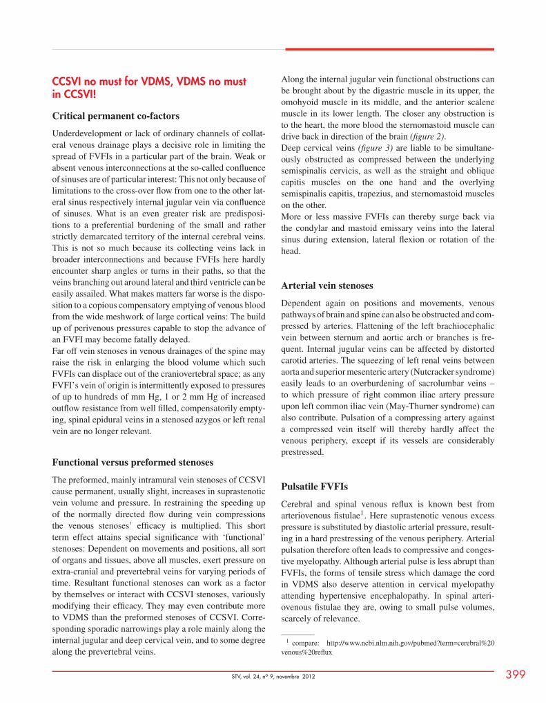
Along the internal jugular vein functional obstructions canbe brought about by the digastric muscle in its upper, theomohyoid muscle in its middle, and the anterior scalenemuscle in its lower length. The closer any obstruction isto the heart, the more blood the sternomastoid muscle candrive back in direction of the brain (figure 2).Deep cervical veins (figure 3) are liable to be simultane-ously obstructed as compressed between the underlyingsemispinalis cervicis, as well as the straight and obliquecapitis muscles on the one hand and the overlyingsemispinalis capitis, trapezius, and sternomastoid muscleson the other.More or less massive FVFIs can thereby surge back viathe condylar and mastoid emissary veins into the lateralsinus during extension, lateral flexion or rotation of thehead.
Arterial vein stenoses
Dependent again on positions and movements, venouspathways of brain and spine can also be obstructed and com-pressed by arteries. Flattening of the left brachiocephalicvein between sternum and aortic arch or branches is fre-quent. Internal jugular veins can be affected by distortedcarotid arteries. The squeezing of left renal veins betweenaorta and superior mesenteric artery (Nutcracker syndrome)easily leads to an overburdening of sacrolumbar veins –to which pressure of right common iliac artery pressureupon left common iliac vein (May-Thurner syndrome) canalso contribute. Pulsation of a compressing artery againsta compressed vein itself will thereby hardly affect thevenous periphery, except if its vessels are considerablyprestressed.
Pulsatile FVFIs
Cerebral and spinal venous reflux is known best fromarteriovenous fistulae1. Here suprastenotic venous excesspressure is substituted by diastolic arterial pressure, result-ing in a hard prestressing of the venous periphery. Arterialpulsation therefore often leads to compressive and conges-tive myelopathy. Although arterial pulse is less abrupt thanFVFIs, the forms of tensile stress which damage the cordin VDMS also deserve attention in cervical myelopathyattending hypertensive encephalopathy. In spinal arteri-ovenous fistulae they are, owing to small pulse volumes,scarcely of relevance.
1 compare: http://www.ncbi.nlm.nih.gov/pubmed?term=cerebral%20venous%20reflux

Journal Identification = STV Article Identification = 0727 Date: February 8, 2013 Time: 1:37 pm
400 STV, vol. 24, no 9, novembre 2012
a
d
cb
Figure 2. Zones of intermittent muscular obstruction of the internal jugular vein: Overcrossing by digastric (a) and omo-hyoid muscle (b);undercrossing by upper anterior scalene muscle (c) under overlying sternomastoid muscle (d).

Journal Identification = STV Article Identification = 0727 Date: February 8, 2013 Time: 1:37 pm
STV, vol. 24, no 9, novembre 2012 401
a
b
h g
fe
d
cab
c
d
J D
A
B
J S
C D
C S
M D
M S
Q (mm4) : 0 1 2 4 6.5 10 20 40 65 100 200 400 650
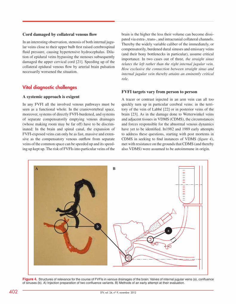
Figure 3. Connections of deep cervical vein (a) to lateral sinus (b). A) Condylar (c) and mastoid emissary veins (d), seen from behind. B)Conductivity of the osseous bottlenecks in the venous drainage of the posterior cranial fossa. Origin of right (JD) and left internal jugularvein in sigmoid sinus (JS) compared to right (CD) and left condylar emissary vein (CS) and right (MD) and left mastoid emissary vein (MS).
Journal Identification = STV Article Identification = 0727 Date: February 8, 2013 Time: 1:37 pm
402 STV, vol. 24, no 9, novembre 2012
Cord damaged by collateral venous flow
In an interesting observation, stenosis of both internal jugu-lar veins close to their upper bulb first raised cerebrospinalfluid pressure, causing hypertensive hydrocephalus. Dila-tion of epidural veins bypassing the stenoses subsequentlydamaged the upper cervical cord [21]. Speeding up of thecollateral epidural venous flow by arterial brain pulsationnecessarily worsened the situation.
Vital diagnostic challenges
A systemic approach is exigent
In any FVFI all the involved venous pathways must beseen as a functional whole. In the craniovertebral space,moreover, systems of directly FVFI-burdened, and systemsof separate compensatorily emptying venous drainages(whose making room may be far off) have to be discrim-inated: In the brain and spinal canal, the expansion ofFVFI-exposed veins can only be as fast, massive and exten-sive as the compensatory venous outflow from separateveins of the common space can be speeded up and its speed-ing up kept up. The risk of FVFIs into particular veins of the
brain is the higher the less their volume can become dissi-pated via extra-, trans-, and intracranial collateral channels.Thereby the widely variable caliber of the immediately, orcompensatorily, burdened dural sinuses and emissary veins(and their bony bottlenecks in particular), assume criticalimportance. In two cases out of three, the straight sinusrelates the left rather than the right internal jugular vein.How exclusive the connection between straight sinus andinternal jugular vein thereby attains an eminently criticalrole.
FVFI targets vary from person to person
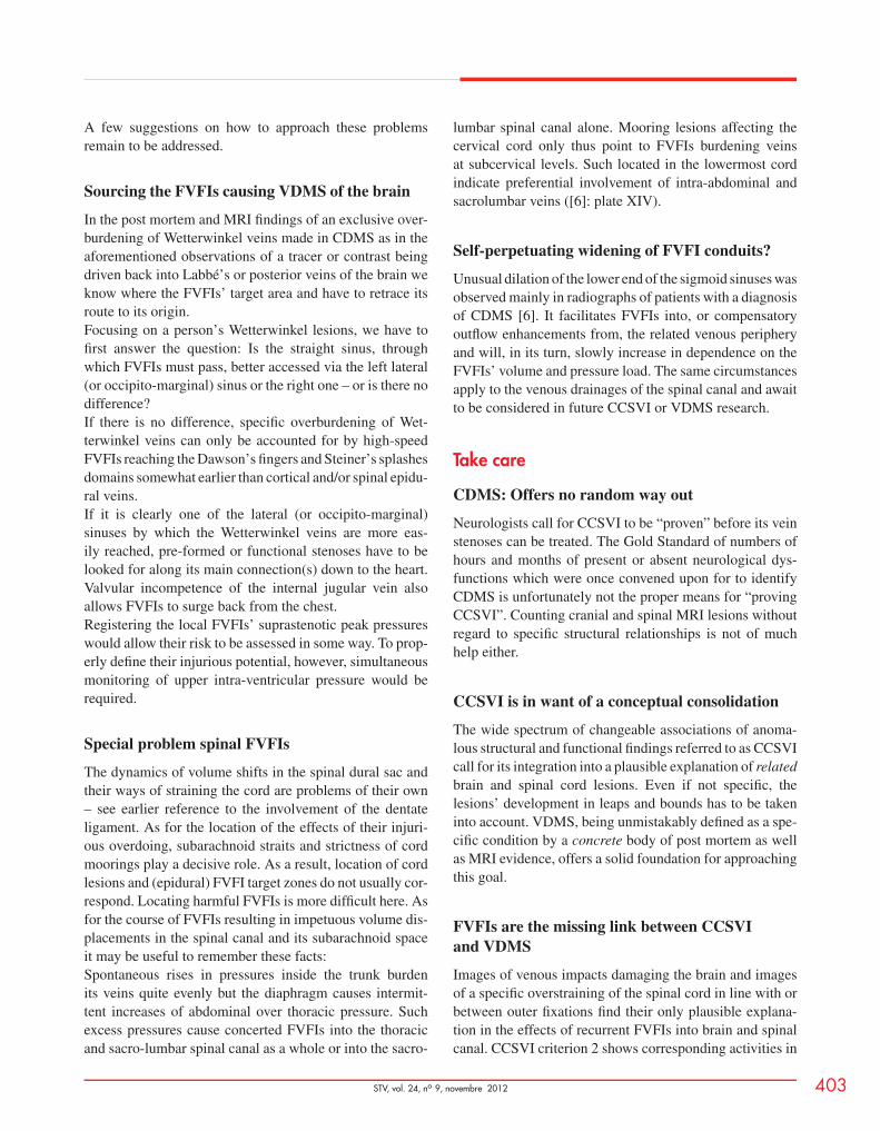
A tracer or contrast injected in an arm vein can all tooquickly turn up in particular cerebral veins: in the terri-tory of the vein of Labbé [22] or in posterior veins of thebrain [23]. As in the damage done to Wetterwinkel veinsand adjacent tissues in VDMS (CDMS), the circumstancesand forces responsible for the abnormal venous dynamicshave yet to be identified. In1982 and 1989 early attemptsto address these questions, starting with post mortems inCDMS in seeking to find instances of VDMS (figure 4),met with resistance on the grounds that CDMS (and therebyalso VDMS) were assumed to be autoimmune in origin.
ab
A B
Figure 4. Structures of relevance for the course of FVFIs in venous drainages of the brain: Valves of internal jugular veins (a), confluenceof sinuses (b). A) Injection preparation of two confluence variants. B) Methods of an early attempt at their evaluation.
Journal Identification = STV Article Identification = 0727 Date: February 8, 2013 Time: 1:37 pm
STV, vol. 24, no 9, novembre 2012 403
A few suggestions on how to approach these problemsremain to be addressed.
Sourcing the FVFIs causing VDMS of the brain
In the post mortem and MRI findings of an exclusive over-burdening of Wetterwinkel veins made in CDMS as in theaforementioned observations of a tracer or contrast beingdriven back into Labbé’s or posterior veins of the brain weknow where the FVFIs’ target area and have to retrace itsroute to its origin.Focusing on a person’s Wetterwinkel lesions, we have tofirst answer the question: Is the straight sinus, throughwhich FVFIs must pass, better accessed via the left lateral(or occipito-marginal) sinus or the right one – or is there nodifference?If there is no difference, specific overburdening of Wet-terwinkel veins can only be accounted for by high-speedFVFIs reaching the Dawson’s fingers and Steiner’s splashesdomains somewhat earlier than cortical and/or spinal epidu-ral veins.If it is clearly one of the lateral (or occipito-marginal)sinuses by which the Wetterwinkel veins are more eas-ily reached, pre-formed or functional stenoses have to belooked for along its main connection(s) down to the heart.Valvular incompetence of the internal jugular vein alsoallows FVFIs to surge back from the chest.Registering the local FVFIs’ suprastenotic peak pressureswould allow their risk to be assessed in some way. To prop-erly define their injurious potential, however, simultaneousmonitoring of upper intra-ventricular pressure would berequired.
Special problem spinal FVFIs
The dynamics of volume shifts in the spinal dural sac andtheir ways of straining the cord are problems of their own– see earlier reference to the involvement of the dentateligament. As for the location of the effects of their injuri-ous overdoing, subarachnoid straits and strictness of cordmoorings play a decisive role. As a result, location of cordlesions and (epidural) FVFI target zones do not usually cor-respond. Locating harmful FVFIs is more difficult here. Asfor the course of FVFIs resulting in impetuous volume dis-placements in the spinal canal and its subarachnoid spaceit may be useful to remember these facts:Spontaneous rises in pressures inside the trunk burdenits veins quite evenly but the diaphragm causes intermit-tent increases of abdominal over thoracic pressure. Suchexcess pressures cause concerted FVFIs into the thoracicand sacro-lumbar spinal canal as a whole or into the sacro-
lumbar spinal canal alone. Mooring lesions affecting thecervical cord only thus point to FVFIs burdening veinsat subcervical levels. Such located in the lowermost cordindicate preferential involvement of intra-abdominal andsacrolumbar veins ([6]: plate XIV).
Self-perpetuating widening of FVFI conduits?
Unusual dilation of the lower end of the sigmoid sinuses wasobserved mainly in radiographs of patients with a diagnosisof CDMS [6]. It facilitates FVFIs into, or compensatoryoutflow enhancements from, the related venous peripheryand will, in its turn, slowly increase in dependence on theFVFIs’ volume and pressure load. The same circumstancesapply to the venous drainages of the spinal canal and awaitto be considered in future CCSVI or VDMS research.
Take care
CDMS: Offers no random way out
Neurologists call for CCSVI to be “proven” before its veinstenoses can be treated. The Gold Standard of numbers ofhours and months of present or absent neurological dys-functions which were once convened upon for to identifyCDMS is unfortunately not the proper means for “provingCCSVI”. Counting cranial and spinal MRI lesions withoutregard to specific structural relationships is not of muchhelp either.
CCSVI is in want of a conceptual consolidation
The wide spectrum of changeable associations of anoma-lous structural and functional findings referred to as CCSVIcall for its integration into a plausible explanation of relatedbrain and spinal cord lesions. Even if not specific, thelesions’ development in leaps and bounds has to be takeninto account. VDMS, being unmistakably defined as a spe-cific condition by a concrete body of post mortem as wellas MRI evidence, offers a solid foundation for approachingthis goal.
FVFIs are the missing link between CCSVIand VDMS
Images of venous impacts damaging the brain and imagesof a specific overstraining of the spinal cord in line with orbetween outer fixations find their only plausible explana-tion in the effects of recurrent FVFIs into brain and spinalcanal. CCSVI criterion 2 shows corresponding activities in

Journal Identification = STV Article Identification = 0727 Date: February 8, 2013 Time: 1:37 pm
404 STV, vol. 24, no 9, novembre 2012
the brain, in which criterion 1(a) may play a role. CCSVIcriteria 3 to 5 are evidence of important but not always deci-sive preconditions to the emergence of FVFIs in large neckand prevertebral veins.
Clinical CCSVI trials: Statistical or rationalevaluation?
The peremptory demand that CCSVI be evaluated but onthe basis of its stenoses’ tentative dilations, reasoning exclu-sively in the domains of clinical trials and evidence-basedmedicine sidestep the issue that the nature of “(CD)MS”has not been properly defined. Interventions having no clearindication and sound rationale are an undue threat to bothpatients and interventional radiologists. Is a sink that inter-mittently overflows best dealt with by exploring means toachieve a statistically significant reduction in the frequencyof the unwanted incidents – or by competent plumbing?
Paying heed to the non-responders’ plight
Far too many patients with a diagnosis of CDMS, or evenone of CCSVI, are not really helped or are even furtherburdened by the present treatment schemes. Five decades ofclinical drug trials on CDMS have hardly improved on thesenon-responders dismal fate. Only using the best available,and in the future achievable, technology to identify andprevent the FVFIs of VDMS will ease the lot of patients.The first tentative steps in this direction have been made[1, 2, 20, 24].
Acknowledgements. It is thanks to Dr. Jean-Marc Pernèsthat this paper appears. Alison Fisher, Carol Schumacher,John Christensen, and especially also Michele Findlayworking with Kevin Campbell did their best to make itsunderstanding easier. Son Matthias and Bernhard gavetechnical support. Dr. Cathrin Korz of Elsevier Publish-ing Group is owed the copyright permission for adaptingand reproducing old friend and artist Franz Batke’s deepcervical vein illustration. !
Conflicts of interest : none
References1. Zamboni P, Menegatti E, Bartolomei I, et al. Intracranial venous haemo-dynamics in MS. Curr Neurovasc Res 2007 ; 4 : 252-8.
2. Zamboni P, Galeotti R, Menegatti E, et al. CCSVI in patients with MS.J Neurol Neurosurg Psychiatry 2009 ; 80 : 392-9.
3. Schumacher GA, Beebe G, Kibler RF, et al. Problems of experi-mental trials of therapy in MS: Report by the panel on the evaluationof experimental trials of therapy in MS. Ann New York Acad Sciences1965 ; 122 : 552-66.
4. Confavreux C, Vukusic S, Achiti J. [MS:] Diagnostic criteria of differ-ent clinical forms. Rev Neurol (Paris) 2001 ; 157 : 907-13.
5. Diagnostic criteria [of] MS [on] MRI. Literature Review. Pubmed,http://www.ncbi.nlm.nih.gov/pubmed?term=diagnostic%20criteria%20ms%20mri [Accessed June 11, 2012].
6. Schelling FA. Multiple sclerosis: The image and its message. E-book,online since 2002. http://www.ms-info.net/ms_040504.pdf.
7. Schelling F. CCSVI in MS: Why all the confusion? In: 2nd AnnualISNVD Scientific Meeting Orlando, Florida USA; Abstracts, posters,consensus notes. ISNVD 2012, 1-4.
8. Young IR, Hall AS, Pallis CA, Legg NJ, Bydder GM, Steiner RE. NMRIof the brain in multiple sclerosis. Lancet 1981 ; ii : 1063-6.
9. Johnson MA, Li DKB, Bryant DJ, Payne JA. MRI: Serial observationsin MS. Am J Neuroradiol 1984 ; 5 : 495-9.
10. Koopmans RA, Li DKB, Oger JJF, Mayo J, Paty DW. The lesionof MS: Imaging of acute and chronic stages. Neurology 1989 ; 39 :959-63.
11. Meier DS, Weiner HL, Guttmann CR. MRI intensity modeling ofdamage and repair in MS. Am J Neuroradiol 2007 ; 28 : 1956-63.
12. Zeng C, Chen X, Li Y, Ouyang Y, Lv F, Rumzan R, Wang Z. Cere-bral vein changes in RRMS demonstrated by three-dimensional enhancedT2*-weighted angiography at 3.0 T. Eur Radiol, 12 September 2012.http://www.springerlink.com/content/x74m208nl60j78p0/?MUD=MP.
13. Falkiewicz T. Zur Pathogenese der MS. Arb. a. d. neurol. Inst. a. d.Wien. Univ 1926 ; 28 : 172-96.
14. Oppenheimer DR. The cervical cord in MS. Neuropath Appl Neurobiol1978 ; 4 : 151-2.
15. Schmaus H. Beiträge zur pathologischen Anatomie der Rückenmark-serschütterung. Virchows Arch path Anat Physiol 1890 ; 122 : 326-56.
16. Kahn EA. The role of the dentate ligaments in spinal cord compressionand the syndrome of lateral sclerosis. J Neurosurg 1947 ; 4 : 191-9.
17. Mussen A. The finer histological changes in the traumatic degenera-tions of the spinal cord, following bullet wounds of the cord substance,or shock to the vertebral column. Rev Neurol Psychiat 1916; 14: 417-46;Plates 7, 8.
18. Reitan H. On movements of fluid inside the cerebro-spinal space. ActaRadiol 1941 ; 22 : 762-79.
19. Strenta-Filstroff C. Les mouvements du liquide céphalo-rachidien etdu sac dural à la lumière de la radiculographie. Marseille, Thèse de doctoraten médecine, 1970.
20. Hubbard D, Ponec D, Saxon R, Sander H, Haacke M. J Vasc InterventRad, 28.08.2012 (In press). Abstract at http://www.jvir.org/article/S1051-0443(12)00708-7/abstract.
21. Vardiman AB, Dickman CA, Spetzler RF, Heiserman JE,Thompson BG: Cervical myelopathy from dilated epidural veins:Case report of intracranial outflow obstruction treated with a sig-moid sinus-to-internal jugular vein bypass. http://www.thebarrow.org/education_and_resources/barrow_quarterly/204853.
22. Friedman BH, Lovegrove FTA, Wagner HN. An unusual variant incerebral circulation studies. J Nucl Med 1974 ; 15 : 363-5.
23. Chen JY, Mamourian AC, Messe SR, Wolf RL. Pseudopathologicbrain parenchyma enhancement due to venous reflux from left-sided injec-tion and brachiocephalic vein narrowing. AJNR 2010 ; 31 : 86-7.
24. Sclafani SJ. Intravascular ultrasound in the diagnosis and treatment ofCCSVI. Tech Vasc Interv Radiol 2012 ; 15 : 131-42.