chf6001, a novel highly potent and selective...
TRANSCRIPT
JPET#220541
1
CHF6001, a novel highly potent and selective phosphodiesterase 4 inhibitor with
robust anti-inflammatory activity and suitable for topical pulmonary
administration
Nadia Moretto, Paola Caruso, Raffaella Bosco, Gessica Marchini, Fiorella Pastore, Elisabetta
Armani, Gabriele Amari, Andrea Rizzi, Eleonora Ghidini, Renato De Fanti, Carmelida Capaldi,
Laura Carzaniga, Emilio Hirsch¶, Carola Buccellati§, Angelo Sala§, Chiara Carnini, Riccardo
Patacchini, Maurizio Delcanale, Maurizio Civelli, Gino Villetti, Fabrizio Facchinetti
Corporate Pre-Clinical R&D, Chiesi Farmaceutici S.p.A., Largo Belloli 11/A, 43122 Parma,
Italy (N.M., P.C., R.B., G.M., F.P., E.A., A.R., E.G., R.D.F., C.C., L.C., C.C., R.P. M.D., M.C.,
G.V., M.C.); Molecular Biotechnology Center, University of Turin, Turin, Italy (E.H.),
Dipartimento di Scienze Farmacologiche e Biomolecolari, Via Balzaretti 9, Milan, Italy (C.B.,
A.S.)
This article has not been copyedited and formatted. The final version may differ from this version.JPET Fast Forward. Published on January 9, 2015 as DOI: 10.1124/jpet.114.220541
at ASPE
T Journals on June 15, 2018
jpet.aspetjournals.orgD
ownloaded from

JPET#220541
2
Running Title: CHF6001: in vitro characterization
Corresponding Author:
Fabrizio Facchinetti, PhD
Corporate Pre-clinical R&D
Chiesi Farmaceutici S.p.A.
Largo Belloli 11/A, 43122,
Parma, Italy.
Phone: +39-0521-279215.
FAX +39-0521-279549.
E-mail: [email protected]
text pages: 27
number of tables: 3
number of figures: 7
number of references: 39
number of words in the Abstract: 216
number of words in the Introduction: 636
number of words in the Discussion: 1096
Non standard abbreviations: PDE, phosphodiesterase; CHF6001, (S)-3,5-dichloro-4-(2-(3-
(cyclopropylmethoxy)-4-(difluoromethoxy)phenyl)-2-(3-(cyclopropylmethoxy)-4-
(methylsulfonamido)benzoyloxy)ethyl)pyridine 1-oxide; high (HARBS) and low (LARBS)
affinity rolipram binding site; PBMCs, peripheral blood mononucleate cells; interferon-γ (IFN-γ)
This article has not been copyedited and formatted. The final version may differ from this version.JPET Fast Forward. Published on January 9, 2015 as DOI: 10.1124/jpet.114.220541
at ASPE
T Journals on June 15, 2018
jpet.aspetjournals.orgD
ownloaded from

JPET#220541
3
ABSTRACT
This study describes the pharmacological characterization of (S)-3,5-dichloro-4-(2-(3-
(cyclopropylmethoxy)-4-(difluoromethoxy)phenyl)-2-(3-(cyclopropylmethoxy)-4-
(methylsulfonamido)benzoyloxy)ethyl)pyridine 1-oxide (CHF6001), a novel PDE4 inhibitor,
designed for treating pulmonary inflammatory diseases via inhaled administration. CHF6001
was 7- and 923-fold more potent than roflumilast and cilomilast respectively, in inhibiting PDE4
enzymatic activity (IC50= 0.026±0.006 nM). CHF6001 inhibited PDE4 isoforms A-D with equal
potency, showed an elevated high (HARBS) versus low (LARBS) affinity rolipram binding site
ratio (>40) and displayed >20,000-fold selectivity versus PDE4 in comparison with a panel of
PDEs. CHF6001 effectively inhibited (sub-nanomolar IC50s values) the release of tumor necrosis
factor-α (TNF-α) from human peripheral blood mononucleate cells (PBMCs), THP-1 monocyte-
derived macrophages and rodent macrophages (RAW 264.7 and NR8383). Moreover, CHF6001
potently inhibited the activation of neutrophils and eosinophils (fMLP-evoked oxidative burst),
neutrophil chemotaxis and the release of interferon-γ (IFN-γ) from CD4+ T cells. In all these
functional assays, CHF6001, was more potent than previously described PDE4 inhibitors,
including roflumilast, UK-500,001 and cilomilast, and comparable to GSK256066. When
administered intratracheally to rats as a micronized dry powder, CHF6001 inhibited LPS-
induced pulmonary neutrophilia (ED50=0.205 μmoles/kg) and leukocytes infiltration
(ED50=0.188 μmoles/kg) with an efficacy comparable to a high dose of budesonide (1 μmole/kg,
i.t.). In sum, CHF6001 has the potential to be an effective topical treatment for conditions
associated with pulmonary inflammation, including asthma and chronic obstructive pulmonary
disease (COPD).
This article has not been copyedited and formatted. The final version may differ from this version.JPET Fast Forward. Published on January 9, 2015 as DOI: 10.1124/jpet.114.220541
at ASPE
T Journals on June 15, 2018
jpet.aspetjournals.orgD
ownloaded from

JPET#220541
4
INTRODUCTION
Phoshodiesterases (PDEs) form a superfamily of at least 11 intracellular iso-enzymes which are
involved in the modulation of signal transduction processes via the degradation of cyclic
nucleotides (cAMP and/or cGMP) (Bender and Beavo, 2006). Type 4 cyclic nucleotide
phosphodiesterases (PDE4) are a family of cAMP-specific PDEs encoded by four genes
(PDE4A, PDE4B, PDE4C, and PDE4D) sharing a highly conserved catalytic domain and
abundantly expressed in leukocytes (Srivani et al., 2008). By increasing intracellular cAMP
levels, PDE4 inhibitors show a broad spectrum of anti-inflammatory effects in almost all cells of
the immune system. In particular PDE4 is a major player in regulating pro-inflammatory cellular
functions, such as proliferation and cytokine secretion, chemotaxis, degranulation, antibody IgE
release and generation of lipid mediators (Spina, 2003). PDE4 regulates also the function of
several structural cells that control lung functions such as airway smooth muscle, airway
epithelium, vascular endothelium and airway sensory nerves (Fan Chung, 2006). This, together
with a large body of evidence supporting potent anti-inflammatory activity of PDE4 inhibitors in
various experimental models, including models of pulmonary inflammation (Hatzelmann et al.,
2010), has generated a considerable interest in targeting PDE4 in chronic inflammatory and
obstructive airway diseases (Spina, 2003). Clinical investigations of second-generation oral
PDE4 inhibitors, e.g., cilomilast, Ariflo (Barnette et al., 1998), and roflumilast, Daxas
(Hatzelmann et al., 2001), have demonstrated efficacy in chronic lung diseases such as chronic
obstructive pulmonary disease (COPD) and asthma (Press and Banner, 2009). However, the
development of these compounds has been hampered by dose limiting adverse events which are
mechanism-related, in particular gastrointestinal disturbances such as nausea, diarrhea,
abdominal pain, vomiting and dyspepsia (Calvelry et al., 2009). Roflumilast has recently been
This article has not been copyedited and formatted. The final version may differ from this version.JPET Fast Forward. Published on January 9, 2015 as DOI: 10.1124/jpet.114.220541
at ASPE
T Journals on June 15, 2018
jpet.aspetjournals.orgD
ownloaded from
JPET#220541
5
approved in EU and USA for once-daily treatment of severe COPD associated with chronic
bronchitis and frequent exacerbations as add-on to bronchodilator treatment. However, target-
related gastrointestinal side effects may limit roflumilast dosage and thus optimal clinical
efficacy (Giembycz, 2010; Page and Spina, 2012). In line with this consideration, the active
doses of roflumilast determined in experimental models (Martorana et al., 2005; Hatzelmann et
al., 2010) hardly support the daily dose of 0.5 mg approved in COPD patients.
In the respiratory field, a logical way to improve the therapeutic index is to deliver drugs directly
into the lung via the inhalation route allowing high topical efficacy coupled with low oral
availability and limited systemic exposure. The PDE4 inhibitor UK-500,001 developed for
inhaled administration was tested in a double-blind, placebo-controlled, 6-week trial in 209
patients with moderate-to-severe COPD but with disappointing results and the study was
stopped following a planned interim analysis for futility (Vestbo et al., 2009). Lack of efficacy in
phase II trials in asthma and COPD led to the discontinuation also of AWD 12-281 and
tofimilast (Pages et al., 2009). To date, the best in class among inhaled PDE4 inhibitors is
GSK256066, which appears to be more potent than all the other PDE4 inhibitors described so
far (Nials et al., 2011; Tralau-Stewart et al., 2011), is well tolerated in COPD patients (Watz et
al., 2013) and is effective in reducing allergen challenge responses in asthma patients (Singh et
al., 2010).
In the present study we report the pharmacological characterization of (S)-3,5-dichloro-4-(2-(3-
(cyclopropylmethoxy)-4-(difluoromethoxy)phenyl)-2-(3-(cyclopropylmethoxy)-4-
(methylsulfonamido)benzoyloxy)ethyl)pyridine 1-oxide (CHF6001), a novel highly potent and
selective inhaled PDE4 inhibitor which was previously selected and optimized for inhaled
delivery (Armani et al., 2013). In addition to defining its profile as PDE4 inhibitor, we
exensively studied anti-inflammatory activities of CHF6001 in several in vitro cellular models
This article has not been copyedited and formatted. The final version may differ from this version.JPET Fast Forward. Published on January 9, 2015 as DOI: 10.1124/jpet.114.220541
at ASPE
T Journals on June 15, 2018
jpet.aspetjournals.orgD
ownloaded from
JPET#220541
6
(neutrophils, lymphocytes, monocytes, macrophages) in direct comparison with two oral,
roflumilast and cilomilast, and two inhaled, GSK256066 and UK500,001, PDE4 inhibitors.
Moreover the anti-inflammatory potency and efficacy of CHF6001 was demonstrated in vivo in a
rat model of endotoxin-induced acute pulmonary inflammation. Some of these data were
presented in a poster form at the 2014 American Thoracic Society conference (Moretto et al.,
2014).
This article has not been copyedited and formatted. The final version may differ from this version.JPET Fast Forward. Published on January 9, 2015 as DOI: 10.1124/jpet.114.220541
at ASPE
T Journals on June 15, 2018
jpet.aspetjournals.orgD
ownloaded from
JPET#220541
7
Materials and Methods
Chemicals
(S)-3,5-dichloro-4-(2-(3-(cyclopropylmethoxy)-4-(difluoromethoxy)phenyl)-2-(3-
(cyclopropylmethoxy)-4-(methylsulfonamido)benzoyloxy)ethyl)pyridine 1-oxide, CHF6001
(Armani et al, 2014), 6-((3-(dimethylcarbamoyl)phenyl)sulfonyl)-4-((3-methoxyphenyl)amino)-
8-methylquinoline-3-carboxamide, GSK256066 (Tralau-Stewart et al., 2011), 3-
(cyclopropylmethoxy)-N-(3,5-dichloropyridin-4-yl)-4-(difluoromethoxy)benzamide, roflumilast
(Hatzelmann et al., 2001), (1s,4s)-4-cyano-4-(3-(cyclopentyloxy)-4-methoxyphenyl)cyclohexane-
1-carboxylic acid, cilomilast (Pagès et al., 2009), 6-((3-(dimethylcarbamoyl)phenyl)sulfonyl)-4-
((3-methoxyphenyl)amino)-8-methylquinoline-3-carboxamide, UK-500,001 (Pagès et al., 2009),
were synthesized at Chiesi Farmaceutici S.p.A., Parma, Italy. Unless otherwise stated, all other
chemical reagents used in this study were purchased from Sigma-Aldrich (St. Louis, MO).
PDE4 enzymatic assays from U937 extracts. PDE4 activity was determined in U937 human
monocytic supernatants cells lysate (ATCC, Manassas, USA). Cells were cultured, harvested
and supernatant fraction prepared essentially as previously described (Barnette et al. 1996). All
the materials used for cell culture were from Gibco (Monza, Italy). U937 cells were grown at
37°C, 5% CO2 in RPMI-1640 with GlutaMAX™-I medium supplemented with 10% fetal
bovine serum and 100 μg/mL Pen-strep. Cells were harvested and washed twice by
centrifugation (150 x g, 8 min) in cold PBS. Washed cells were resuspended in cold Krebs-
Ringer-Henseleit buffer at a final concentration 20 x 106 cells /mL and sonicated. After
centrifugation at 15,000 x g for 20 min, the supernatants were pooled, divided in aliquots and
stored at -80°C. PDE4 activity was determined in cells supernatants by assaying cAMP
This article has not been copyedited and formatted. The final version may differ from this version.JPET Fast Forward. Published on January 9, 2015 as DOI: 10.1124/jpet.114.220541
at ASPE
T Journals on June 15, 2018
jpet.aspetjournals.orgD
ownloaded from
JPET#220541
8
disappearance from the incubation mixtures. The concentration of the test compounds ranged
between 10-12 M and 10-6 M. Reactions were stopped by enzyme heat inactivation (2.5 minutes
at 100°C) and residual cAMP content was determined using the ‘LANCE ™cAMP Detection
kit’ from PerkinElmer (Milan, Italy) following the provider instructions. IC50 values were
determined from concentration-response curves by nonlinear regression analysis.
Rolipram Binding Assay. The affinity against HARBS has been evaluated in a radioligand
binding assay performed in rat brain membranes, using [3H]-rolipram as radioligand. Fresh rat
brains were homogenized in 20 volumes of ice-cold 50 mM Tris-HCl (pH 8.0) buffer containing
1.2 mM MgCl2 in a polytron PT-10 homogenizer (Brinkman Instruments). The resulting
homogenate was centrifuged at 30,000g for 20 min at 4 °C. The pellet was washed by
resuspension in 20 volumes of fresh buffer and recovered by centrifugation as before. The final
pellet was suspended in Tris buffer (0.5 mg of protein/mL) for binding experiments. Incubation
mixtures in duplicates consisted of 0.1 mL of (+) [3H]rolipram (2 nM final), 0.02 mL of
inhibitor, and 0.9 mL of membrane preparation (added last). Rolipram (10 μM) was used for
nonspecific binding. After 60 min incubation at 4 °C the contents of the incubation tubes were
filtered through a Whatman GF/C glass filter. The membranes were washed three times with 3
mL of ice-cold buffer, and radioactivity on the separated filter disks was determined in a liquid
scintillation counter. IC50 values were determined from semilog graphs of percent inhibition
versus concentration.
PDE4 Enzyme Assays. PDE4 assays were performed using recombinant PDE enzymes
expressed in a baculoviral system. The radiometric assay method is a modification of the two-
step method of Thompson and Appleman (1971) as described by Mackenzie et al., 2010. All the
This article has not been copyedited and formatted. The final version may differ from this version.JPET Fast Forward. Published on January 9, 2015 as DOI: 10.1124/jpet.114.220541
at ASPE
T Journals on June 15, 2018
jpet.aspetjournals.orgD
ownloaded from

JPET#220541
9
assays use a substrate concentration below the Km determined for each enzyme so that ki=IC50.
The PDE4 inhibitors were prepared as stocks at a concentration of 40 mM in 100% DMSO and
were tested at 11 concentrations (0.5% final DMSO concentration) in duplicate with a starting
concentration of 1μM and a 1:10 serial dilution against human PDE4A4, PDE4B2, PDE4C2,
PDE4D3. Concentration up to 5% DMSO are tolerated in this assay as reported previously
(MacKenzie et al., 2010), a notion confirmed also by a preliminary study aimed at evaluating
tolerance to DMSO (estimated up to 5%).
PDEs Enzyme Assays. PDE1 was prepared from bovine brain (Gietzen, 1982). PDE2, PDE3
and PDE5 were purified from human platelets (Schudt, 1991). PDE6 was purified from bovine
retina (Baehr W, 1979). Recombinant human PDE7A, PDE8A1, PDE9A2, PDE10A2 and
PDE11A4 were expressed in a baculovirus/ insect cell system. The radiometric assay method is
a modification of the two-step method of Thompson and Appleman (1971). Briefly, the assay
mixture contained 50 mM Tris (pH 7.4), 5 mM MgCl2, 0.5 mM cAMP or cGMP, and [3H]cAMP
or [3H]cGMP (1 μM/mL), the indicated concentration of the inhibitor and an aliquot of the
enzyme solution. CHF6001 was prepared as stock at a concentration of 0.3 mM in 100% DMSO
and tested at 5 concentrations (1% final DMSO concentration) in duplicate starting at 30 μM
and a 1:10 serial dilution against human PDEs. PDE1 isoenzyme was assayed in the presence of
Ca2+ (2 mM) and calmodulin (100 U/ml) using cAMP as substrate. PDE2, PDE3, PDE7A,
PDE8A1, PDE10A2 and PDE11A4 were assayed in the presence of cAMP as substrate. PDE5,
PDE6 and PDE9A2 were assayed using cGMP as substrate.
PBMCs. Cells were purchased from Lonza (Basel, CH), washed, resuspended in RPMI 1640
medium (w/o Phenol Red) supplemented with 10% FBS, 2 mM glutamine, 100 U penicillin and
This article has not been copyedited and formatted. The final version may differ from this version.JPET Fast Forward. Published on January 9, 2015 as DOI: 10.1124/jpet.114.220541
at ASPE
T Journals on June 15, 2018
jpet.aspetjournals.orgD
ownloaded from

JPET#220541
10
100 µg/mL streptomycin, and plated in 96-well tissue culture plates at the density of 105
cells/well, in an atmosphere of 95% air and 5% CO2 at 37°C. Cells were treated with different
concentrations of PDE4 inhibitors (10-15 M-10-8 M, 0.2% final DMSO concentration) 1h before
stimulation with lipolysaccharide (LPS) from Escherichia coli (3 ng/mL for 18h). Increasing
concentrations of DMSO were tested and no significant effects of DMSO up to 0.4% on cell
viability and TNFα release were noticed. Human TNFα in the supernatant was assayed using a
paired antibody quantitative ELISA kit (Bender Medsystem, Austria).
Human myeloid leukemia THP-1 cell line. THP-1 cell line was obtained from Sigma (St.
Louis, MO) and cultured in RPMI 1640 medium supplemented with 10% FBS, 2 mM
glutamine, 100 U penicillin and 100 µg/ml streptomycin, in an atmosphere of 95% air and 5%
CO2 at 37°C. To induce differentiation into adherent macrophages, cells were plated in 48-well
plates (2.5x10⁵ cells/well) and incubated for 4 days (THP-1) with 50 nM of phorbol 12-myristate
13-acetate, as previously described (Daigneault et al., 2010). Subsequently, cells were treated
with different concentration of PDE4 inhibitors (10-12M-10-6M, 0.2% final DMSO
concentration), stimulated with LPS (100 ng/ml final concentration) and incubated for 18 hours
in RPMI (w/o Phenol Red) supplemented with 10% FBS. Human TNFα in the supernatant
determined by quantitative ELISA (Bender Medsystem, Vienna, Austria).
Murine (RAW264.7) and rat (NR8383) macrophagic cell lines. RAW264.7 and NR8383
cells were purchased from ATCC and cultured in RPMI 1640 medium (w/o Phenol Red)
supplemented with 10% FBS, 2 mM glutamine, 100 U penicillin and 100 µg/ml streptomycin, in
an atmosphere of 95% air and 5% CO2 at 37°C. RAW264.7 and NR8383 cells were seeded in
RPMI (w/o Phenol Red) containing 10% FBS in 48-well tissue culture plates at the density of
This article has not been copyedited and formatted. The final version may differ from this version.JPET Fast Forward. Published on January 9, 2015 as DOI: 10.1124/jpet.114.220541
at ASPE
T Journals on June 15, 2018
jpet.aspetjournals.orgD
ownloaded from

JPET#220541
11
7.5 x 10⁴ cells/well and grown for 24 hours at 37°C with 5% CO2. Subsequently, cells were
treated with different concentration of PDE4 inhibitors (10-12M-10-6 M, 0.2% final DMSO
concentration), stimulated with LPS (10 ng/ml and 100 ng/ml final concentration for RAW264.7
and NR8383 cells, respectively) and incubated for 18 hours in RPMI (w/o Phenol Red)
supplemented with 10% FBS. Murine and rat TNFα in the supernatant were determined by
quantitative ELISA kit (Bender Medsystem, Vienna, Austria). As an indirect index of nitric
oxide (NO), accumulation of nitrite in the medium production was measured by a colorimetric
assay method based on the Griess reaction as previously described (Facchinetti et al., 2004).
CD4+ T Lymphocytes. CD4+ T lymphocytes were purchased from Lonza (Base, CH). CD4+ T
lymphocytes were cultured in Lymphocyte Growth Medium supplemented with 10% FBS,
albumin, insulin, transferrin and gentamicin. Cells were stimulated via the T-cell receptor CD3
and CD28 co-receptor by using selective mAbs (Hatzelmann and Schudt, 2001). For this
purpose, 96-well microtiter plates were prepared on the day before cell plating: each well was
incubated with 50 μl of PBS containing 6 μg/ml of anti-CD3 mAb (Orthoclone OKT-3; Janssen-
Cilag, Neuss, Germany) for about 2.5 h at 37°C; plates were then stored overnight at 4°C and
washed three times with PBS (200 μl) before use. CD4+ T lymphocytes were plated at the
density of 105 cells/well in 96-well tissue culture plates, pre-coated with the anti-CD3 mAb, and
treated with different concentrations of PDE4 inhibitors (10-13 M-10-8 M, 0.2% final DMSO
concentration). Subsequently, anti-CD28 (clone CD28.2, Coulter-Immunotech Diagnostics,
Hamburg, Germany) was added to the final concentration of 3 μg/ml and the plates were further
incubated at 37°C and 5% CO2 for 72 h. Human IFNγ in the supernatant was assayed using a
paired antibody quantitative ELISA kit (Life Technologies, Grand Island, NY).
This article has not been copyedited and formatted. The final version may differ from this version.JPET Fast Forward. Published on January 9, 2015 as DOI: 10.1124/jpet.114.220541
at ASPE
T Journals on June 15, 2018
jpet.aspetjournals.orgD
ownloaded from
JPET#220541
12
Human primary peripheral eosinophils. Human primary peripheral eosinophils were
purchased from 3H Biomedical (Uppsala, Sweden). Human primary eosinophils were washed,
resuspended in RPMI 1640 medium (w/o Phenol Red) supplemented with 10% FBS, 2 mM
glutamine, 100 U penicillin and 100 µg/ml streptomycin and plated in 96-well white OptiPlate
at the density of 105 cells/well. Cells were pretreated with different concentration of PDE4
inhibitors (10-13 M-10-8 M, 0.2% final DMSO concentration) for 30 min, primed with
cytochalasin B (CB, 5 μM) for 15 minutes and stimulated with fMLP (1 μM) in RPMI (w/o
Phenol Red) supplemented with 10% FBS. Following the addition of fMLP, cells were
incubated with the chemiluminescence probe L-012 (500 nM) (Wako Chemicals GmbH, Neuss,
Germany) and ROS generation was recorded repeatedly over 1 hour (one reading every minute)
with a Luminescence reader (Centro LB 960, Berthold Technologies). During luminescence
measurement, the plate was rotatively agitated at 37°C. ROS generation was determined by
calculating the area under the curve.
Neutrophils oxidative burst. Human neutrophils (PMNL) were purified from diluted “buffy
coat” (Blood Bank, University of Milan) centrifuged on a discontinuous Percoll gradient (∂
1,077-1,098, 4-10°C), followed by red cells lysis (1 volume 0,2% NaCl, balanced with 1 volume
1,6% NaCl and 0.2% saccarose, 4-10°C). Handling in sterile buffer prevented spontaneous
activation of PMNL. Purified neutrophils (1x 10⁶/ml) were suspended in D-PBS (Sigma-D1408)
supplemented with CaCl2, MgCl2, glucose (0.9, 0.5 e 7.5 mM respectively) and Cytochrome C
(50 μM, Sigma-C2506). The cell suspension was dispensed in microplate (200μl /well), gently
shaken at 37°C and absorbance read at λ550-490 by a spectrophotometer (Spectramax190
microplate reader). Neutrophils suspensions (1x 10⁶/ml) were then incubated with the direct
inhibitor of neutrophil NAD(P)H-oxidase diphenyleneiodonium (DPI), the PDE4 inhibitors
This article has not been copyedited and formatted. The final version may differ from this version.JPET Fast Forward. Published on January 9, 2015 as DOI: 10.1124/jpet.114.220541
at ASPE
T Journals on June 15, 2018
jpet.aspetjournals.orgD
ownloaded from
JPET#220541
13
compounds or its vehicle, DMSO, for 10 min at 37°C before challenge with fMLP (1 μM), and
ROS production monitored by measuring the variation in absorbance of the reduced cytochrome
c: duplicate samples containing SOD (180U/ml) were used as blanks. As PMNL suspension
obtained from different donors, significantly differs in response to challenge, results were
expressed as percentage of inhibition of ROS release. Each treatment had 4 replicates with cells
from the same donor and concentration-response curves were originated using the average
values from at least 3 different donors.
Neutrophil chemotaxis. Male C57BL/6J mice were bred in the animal house of the Center for
Molecular Biotechnology (University of Turin, Italy) and maintained on a standard diet with tap
water ad libitum and a 12:00 h light /dark cycle. Mice were sacrified by cervical dislocation. The
femur and the tibia bones from both hind legs were removed and freed of soft tissue attachments,
and the extreme distal tip of each extremity was cut off. Saline with 0.1% Fetal Bovine Serum
was forced through the bone with a syringe. After dispersing cell clumps, the cell suspension
was centrifuged (260 g, 12 min at 4°C) and resuspended in 2 ml of saline-0.1%FBS. Cells were
then treated on a three-layer Percoll gradient (72%-64%-52%). Neutrophils were collected from
the ring between 72%-64%, counted and concentrated at 2x10⁶ cells/ml in RPMI medium. Cells
were incubated with the compound at different concentration for 1h at 37°C and then let migrate
against C5a 50nM for 2h in the 48 multiwell chamber. The inhibition of the compound was
evaluated on the number of the cells still able to migrate towards C5a.
LPS-induced neutrophilia in rat. Non-fasted Wistar rats (150-250g) were weighed,
individually identified on the tail with a permanent marker and intra-tracheally dosed with
vehicle (lactose), CHF6001 (CHF, 0.01, 0.1, 0.3, 1 μmoles/kg), GSK256066 (1 μmol/kg) or
This article has not been copyedited and formatted. The final version may differ from this version.JPET Fast Forward. Published on January 9, 2015 as DOI: 10.1124/jpet.114.220541
at ASPE
T Journals on June 15, 2018
jpet.aspetjournals.orgD
ownloaded from
JPET#220541
14
Budesonide (1 μmol/kg), 1 hour prior and 6 hours after LPS exposure. Dry powder formulations
were prepared by blending of mainly coarse respiratory grade lactose and micronized test
compound. For the challenge, rats were placed into a perspex chamber and exposed to LPS
(salmonella enterica serotype) or 0.9% saline. The LPS was prepared in a solution of 0.1 mg/mL
and aerosolised using a De Vibliss ultrasonic nebuliser 2000, so that 7 mL of the solution was
aerosolised during a 30 min exposure period. Compressed air at 6 L min-1 was passed through
the nebulizer and the output of the nebulizer was passed into the box containing the rats. 24
hours after LPS challenge the animals were culled by an intraperitoneal injection of 1.0 mL of
pentobarbitone sodium (lethobarb). The trachea was then isolated by a midline incision in the
neck and separation of the muscle layers. A small incision was made into the trachea and a
plastic cannula was inserted and secured in place with a suture. The airway was then lavaged
using 2.5 mL of sterile PBS at room temperature. The PBS was left in the airway for 10 seconds
before being removed. From this resulting lavage an aliquot (1.5 mL) was centrifuged (1200 rpm
for 2 min) and 2 samples (300 μL) of the resulting supernatant were aliquoted and stored at -
80oC for cytokine analysis. Total cell counts of the BALF samples were measured using a
Neubaur haemocytometer. Results were expressed as cells/mL. Cytospin slides were also
prepared by adding a 100 μl aliquot of BALF into cytospin funnels in a Shandon Cytospin4
operated at 1,200 rpm for 2 min at room temperature and stained using a DiffQuik stain system.
100 cells were counted on each slide under a microscope. Cells were classified as neutrophils,
eosinophils or mononuclear cells (monocytes, macrophages and lymphocytes) based on
morphological criteria. Erythrocytes and epithelial cells were ignored. The number of each cell
type were quantified by expressing the cell number as a percentage of the total count.
This article has not been copyedited and formatted. The final version may differ from this version.JPET Fast Forward. Published on January 9, 2015 as DOI: 10.1124/jpet.114.220541
at ASPE
T Journals on June 15, 2018
jpet.aspetjournals.orgD
ownloaded from
JPET#220541
15
Statistical analysis. All values are expressed as means ± SEM of the given number (n) of
independent experiments. IC50 values were calculated by the analysis of the sigmoidal dose-
response curve (variable slope) elaborated by Graph Pad PRISM4 program. Statistical analysis
was performed using one-way analysis of variance (ANOVA) followed by Dunnett´s post-hoc
test for multigroup comparisons.
This article has not been copyedited and formatted. The final version may differ from this version.JPET Fast Forward. Published on January 9, 2015 as DOI: 10.1124/jpet.114.220541
at ASPE
T Journals on June 15, 2018
jpet.aspetjournals.orgD
ownloaded from
JPET#220541
16
RESULTS
CHF6001 selection
The structure of CHF6001 is shown in figure 1. It was identified as a potent inhibitor of PDE4
activity (compound 32a) from a series of novel ester derivatives of 1-(S)-(3-
(cyclopropylmethoxy)-4-(difluoromethoxy)phenyl)-2-(3,5-dichloropyridin-4-yl) ethanol (Armani
et al., 2014). In particular, esters of variously substituted benzoic acids were explored and
structural modifications of the benzoic moiety were performed in order to maximize the
inhibitory potency. Besides CHF6001, several other novel PDE4 inhibitors with potent anti-
inflammatory activity in vitro were obtained (Armani et al., 2014).
Activity of CHF6001 and reference compounds in PDE enzyme assays
PDE4 isoenzymes exist in both LARBS and HARBS conformations. CHF6001 enzymatic
activity was tested both in a LARBS conformation by utilizing an assay with PDE4 extracts from
U937 cells and in HARBS conformation by utilizing rolipram binding assay (see Materials and
Methods) in comparison with reference compounds GSK256066, roflumilast, UK 500,001 and
cilomilast (Table 1).
The IC50 value for the inhibition of enzymatic activity of PDE4 by CHF6001 in the LARBS
assay is 0.026+0.006 nM. The most potent comparator, as expected, resulted GSK256066 (IC50
=0.025+0.01 nM), whereas the other PDE4 inhibitors tested showed IC50 values ranging
between 0.17 and 24 nM (Table 1).
CHF6001 inhibited [3H] rolipram binding in rat brain cytosol (HARBS) with a IC50 of 1.05 nM,
giving a HARBS ratio (HARBS IC50/ LARBS IC50) of about 40, a value higher than GSK256066
This article has not been copyedited and formatted. The final version may differ from this version.JPET Fast Forward. Published on January 9, 2015 as DOI: 10.1124/jpet.114.220541
at ASPE
T Journals on June 15, 2018
jpet.aspetjournals.orgD
ownloaded from
JPET#220541
17
ratio (11), whereas roflumilast, UK 500,001 and cilomilast showed ratios comprised between 2
and 4 (Table 1).
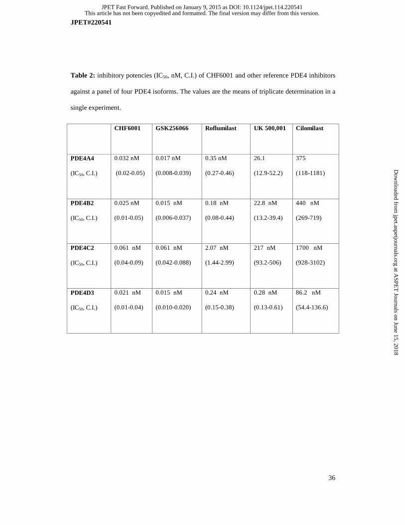
CHF6001 inhibited most of PDE4 splicing variants at sub-nanomolar concentrations (Table 2)
and, similarly to GSK256066 and roflumilast, CHF6001 did not show PDE4 subtype selectivity
against different PDE isoenzymes. Roflumilast is ten-fold less potent than CHF6001 with
respect to PDE4 isoenzymes inhibition, apart from PDE4C, which is inhibited with a slightly
lower potency. In contrast, UK 500,001 and cilomilast showed some subtype selectivity for
PDE4D (Table 2).
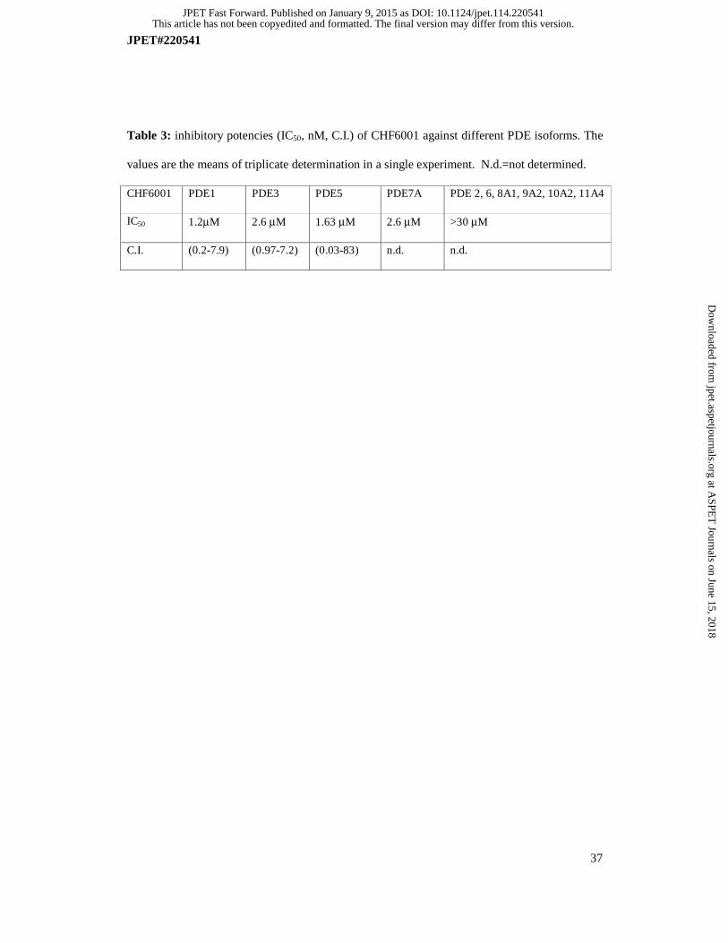
CHF6001 is at least 25,000 fold less potent against the following panel of PDE isoenzymes:
PDE1, PDE2, PDE3, PDE5, PDE6, PDE7A, PDE8A1, PDE9A2, PDE10A2 and PDE11A4,
therefore showing a marked selectivity for PDE4 (Table 3).
Activity of CHF6001 and reference compounds in human PBMCs, THP-1 monocyte-
derived macrophages and CD4+ T lymphocytes
We selected two in vitro models of LPS-stimulated TNF-α to evaluate the anti-inflammatory
potency of CHF6001 in comparison with other known PDE4 inhibitors, including GSK256066,
which is the most potent PDE4 inhibitor so far described in the literature, UK500,001,
roflumilast (the only clinically approved PDE4 inhibitor) and cilomilast. All the compounds
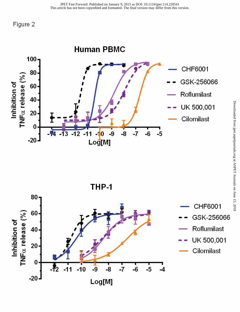
tested inhibited TNF-α release up to a maximum of about 90% in PBMC, an outcome in line
with the notion that PDE4 is the predominant isoform expressed in monocytes (Figure 2, upper
panel). In PBMCs, basal release of TNF-α (mean±s.d.) was of 98.6±6.1 pg/ml, while upon LPS
stimulation became 40146±2227 pg/ml. The rank order of potency of the compounds
(confidence interval values given in parenthesis) is the following: GSK256066, IC50=3 pM (2-
4), CHF6001, IC50=28 pM (21-40), roflumilast IC50=1.77 nM (0.45-6.83), UK500,001,
This article has not been copyedited and formatted. The final version may differ from this version.JPET Fast Forward. Published on January 9, 2015 as DOI: 10.1124/jpet.114.220541
at ASPE
T Journals on June 15, 2018
jpet.aspetjournals.orgD
ownloaded from
JPET#220541
18
IC50=12.1 nM (2.9-50.1) and cilomilast IC50=165.3 nM (61.8-442.6). These results were well in
agreement with the previously reported potencies of GSK256066, roflumilast and cilomilast
against TNF-α release in PBMCs (Hatzelmann and Schudt, 2001; Tralau-Stewart et al., 2011).
In human THP-1 monocytic-derived macrophages, basal release of TNF-α (mean±s.d.) was of
10.3±5.2 pg/ml, while upon LPS stimulation became 18945±838 pg/ml. All the compounds
tested inhibited TNF-α release up to a maximum of 65-70% (Figure 2, lower panel) with the
following potencies: GSK256066, IC50=16 pM (8-35), CHF6001, IC50=43 pM (15-122), UK
500,001, IC50=3.60 nM (1.22-10.45); roflumilast, IC50=4.41 nM (1.0-19.3), cilomilast,
IC50=264.0 nM (121.2-575.0). The observed potency ranking in THP-1 monocyte-derived
macrophages is similar to that observed in PBMCs with both CHF6001 and GSK256066
displaying a potency in the low picomolar range.
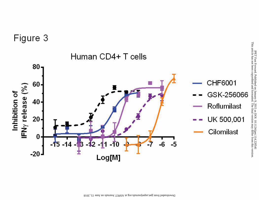
PDE4s are critical regulators in TCR signaling and their inhibition blunts T cell cytokine
production (Abrahamsen et al., 2004). To mimic the physiological conditions of T cell
activation, we stimulated the CD4+ T lymphocytes via the TCR (by plate-bound anti-CD3
antibody) and the CD28 co-receptor (by soluble anti-CD28 antibody). Basal release of IFN-γ
(mean±s.d.) was of 54±8.5 pg/ml, while upon LPS stimulation became 3036±489 pg/ml. All the
PDE4 inhibitors tested, including CHF6001, inhibited IFN-γ release up to a maximum of 50-
60% (Figure 3) with the following potencies: GSK256066, IC50=3 pM (1-12); CHF6001,
IC50=62 pM (13-289); roflumilast, IC50=0.42 nM (0.09-1.87); UK 500,001, IC50=9.4 nM (73-
120); cilomilast, IC50=386.7 nM (149.8-998.5). The observed potency ranking in activated
CD4+ T cells is similar to that observed in PBMCs and THP-1 with both CHF6001 and
GSK256066 being active in the low picomolar range.
Activity of CHF6001 and reference compounds in rodent macrophagic cell lines
This article has not been copyedited and formatted. The final version may differ from this version.JPET Fast Forward. Published on January 9, 2015 as DOI: 10.1124/jpet.114.220541
at ASPE
T Journals on June 15, 2018
jpet.aspetjournals.orgD
ownloaded from

JPET#220541
19
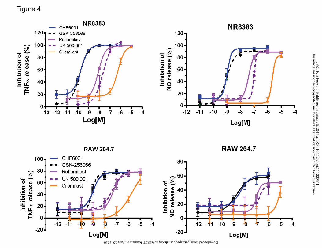
In two different rodent macrophagic cell lines, namely RAW 264.7 (mouse) and NR8383 (rat),
all the PDE4 inhibitors tested inhibited the release of TNF-α and NO evoked by LPS. In RAW
264.7, basal release of TNF-α (mean±s.d.) was of 10.3±5.2 pg/ml, while upon LPS stimulation
became 18945±838 pg/ml. The following inhibitory potencies (confidence interval values given
in parenthesis) against TNF-α (Figure 4A) were observed: GSK256066, IC50=0.82 nM (0.3-2.1),
CHF6001, IC50=1 nM (0.7-1.5), roflumilast, IC50=54.8 nM (23-130), UK 500,001, IC50=36,60
nM (11.3-181); cilomilast, IC50=830 nM (340-2000). Similar results were obtained in NR8383
cells where the inhibition potencies against TNF-α release (Figure 4C) were the following:
GSK256066, IC50=0.08 nM (0.06-0.1), CHF6001, IC50=0.19 nM (0.18-0.2), roflumilast,
IC50=8.3 nM (5.5-12.4), UK 500,001, IC50=19.8 nM (3.2-123); cilomilast, IC50=548 nM (353-
852). In NR8383, basal release of TNF-α (mean±s.d.) was of 1.2±0.5 pg/ml, while upon LPS
stimulation became 1184±111 pg/ml.
In both NR8383 and RAW 264.7 the potencies of all the tested inhibitors showed slightly higher
IC50 values against NO release (Figure 4B and D) but the ranking order observed in the previous
assays was maintained.
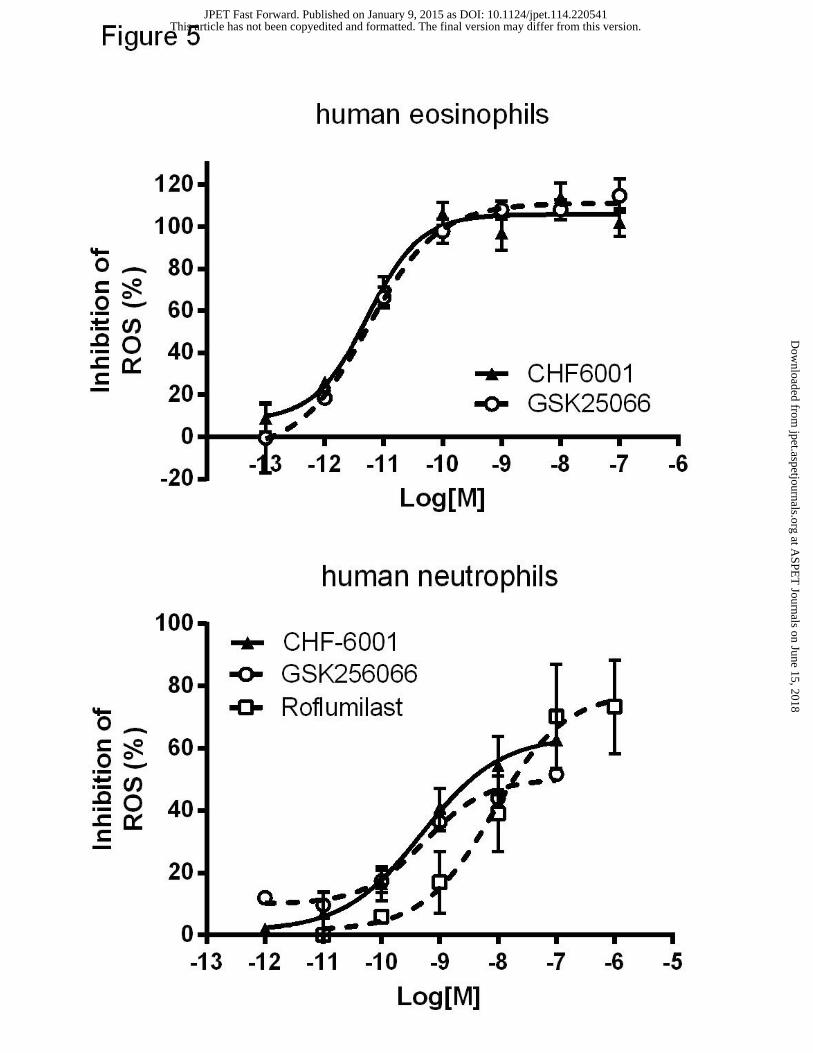
Activity of CHF6001 and reference compounds in human eosinophils and neutrophils
Since PDE4 inhibitors have been shown to negatively modulate fMLP-stimulated release of
reactive oxygen species (ROS) from human eosinophils and neutrophils (Hatzelmann and Shudt,
2001; Press and Banner, 2009), we evaluated the ability of CHF6001 in inhibiting fMLP-
induced ROS production. CHF6001 and GSK256066 fully inhibited fMLP-induced ROS
generation in human eosinophils, at low picomolar concentrations (Figure 5, upper panel), with
IC50s of 5 pM (0.002-0.013) and 5 pM (0.002-0.015) respectively. Similarly, in human
neutrophils, CHF6001 and GSK256066 inhibited fMLP-induced ROS generation (Figure 5,
This article has not been copyedited and formatted. The final version may differ from this version.JPET Fast Forward. Published on January 9, 2015 as DOI: 10.1124/jpet.114.220541
at ASPE
T Journals on June 15, 2018
jpet.aspetjournals.orgD
ownloaded from
JPET#220541
20
lower panel) with IC50s of 0.55 nM (0.07-4.20) and 0.49 nM (0.17-1.43) respectively.
Roflumilast, with an IC50=8.51 nM (1.51-47.88), resulted again less potent than CHF6001 and
GSK256066.
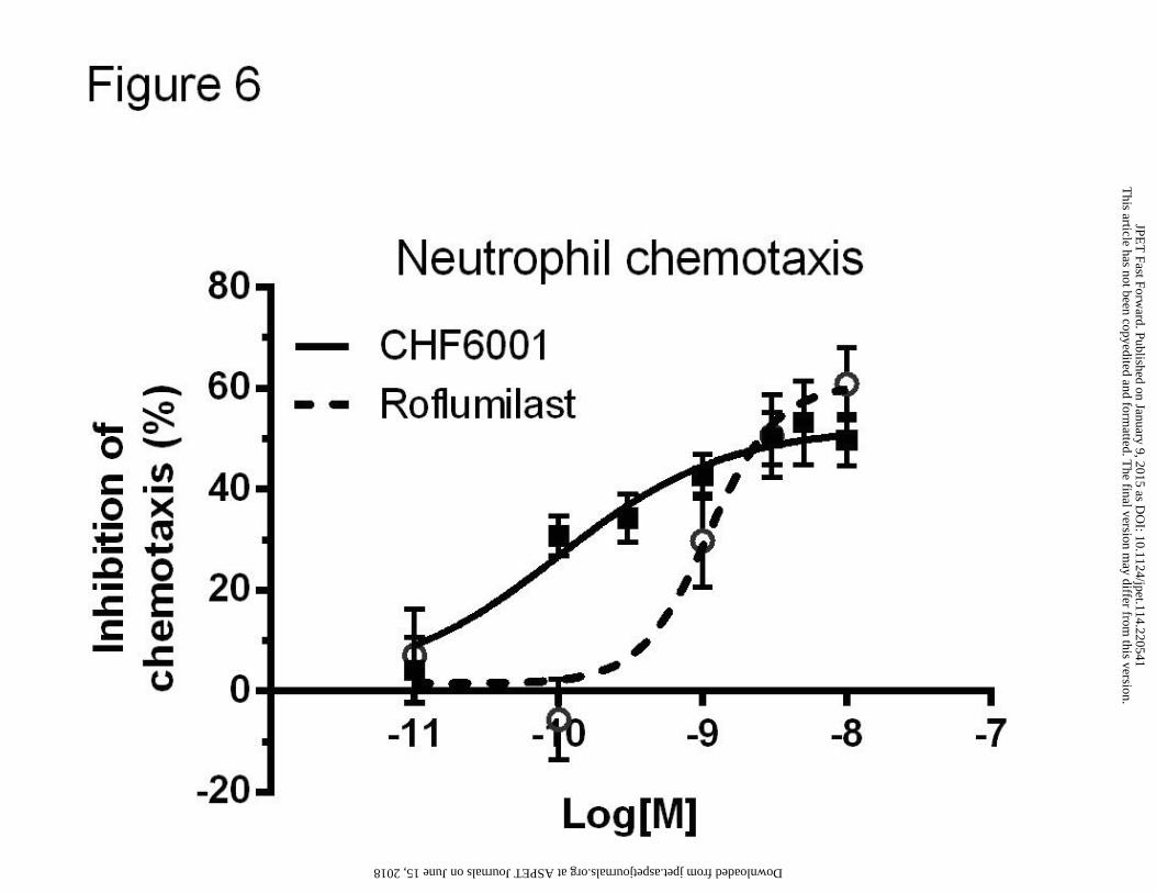
Activity of CHF6001 and roflumilast on neutrophilic chemotaxis
Neutrophil-dominant pulmonary inflammation is an important feature of COPD. Since PDE4
inhibitors have a functional role in mediating chemotaxis (Ariga et al., 2004), the ability of
CHF6001 in inhibiting neutrophilic chemotaxis was tested in comparison with roflumilast in
vitro in a Boyden two-chamber chemotactic assay. CHF6001 inhibited of about 60%
neutrophilic migration with a subnanomolar potency, IC50=0.093 nM (0.036-0.241).
Roflumilast, by showing an IC50=0.937 nM (0.678-1.296), resulted to be less potent than
CHF6001 (Figure 6).
CHF6001 inhibits LPS-induced pulmonary leukocytes infiltration in rat
CHF6001 (CHF, 0.01, 0.1, 0.3, 1 μmoles/kg), GSK256066 (GSK, 1 μmoles/kg) or Budesonide
(Bude, 1 μmoles/kg) were administered intra-tracheally as micronized dry powder 1 hour before
and 6 hours after LPS challenge. Broncho-alveolar lavage fluid (BALF) was collected 24 hours
after challenge and total and differential cell counts performed.
When administered 1 hour before and 6 hours after the inflammatory stimulus, CHF6001 elicited
a dose-dependent inhibition of pulmonary neutrophilia which reached a statistical significance at
0.1 μmoles/kg (p<0.05) and was maximal at 1 μmoles/kg (77%) similarly to budesonide at 1
μmoles/kg (77%). CHF6001 elicited also a dose-dependent inhibition of pulmonary white blood
cell counts in BALF which reached significance at 0.3 μmoles/kg (p<0.01) and was maximal at
1 μmoles/kg (61%), similarly to budesonide at 1 μmoles/kg (62%). Calculated ED50 values for
This article has not been copyedited and formatted. The final version may differ from this version.JPET Fast Forward. Published on January 9, 2015 as DOI: 10.1124/jpet.114.220541
at ASPE
T Journals on June 15, 2018
jpet.aspetjournals.orgD
ownloaded from

JPET#220541
21
CHF6001 were 0.188 μmoles/kg against total white blood cells and 0.205 μmoles/kg against
neutrophilia. Similarly to CHF6001, GSK256066, when tested at 1 μmoles/kg, showed an
inhibitory effect on LPS-evoked pulmonary neutrophilia (70% inhibition) and white blood cells
infiltration (60% inhibition) (Figure 7).
This article has not been copyedited and formatted. The final version may differ from this version.JPET Fast Forward. Published on January 9, 2015 as DOI: 10.1124/jpet.114.220541
at ASPE
T Journals on June 15, 2018
jpet.aspetjournals.orgD
ownloaded from

JPET#220541
22
DISCUSSION
The recent approval of roflumilast as an oral drug for COPD treatment defines the standing of
PDE4 as a therapeutic target in respiratory diseases. Nevertheless, this second-generation PDE4
inhibitor is still not without side effects which may compromise optimal dosing and affect
patient compliance. Several strategies have been proposed to minimize PDE4 inhibition-related
side effects, including designing inhibitors suitable for inhaled delivery that would limit
systemic exposure, while increasing the pulmonary anti-inflammatory activity achievable. In the
present study we report the pharmacological characterization of a novel highly potent and
selective PDE4 inhibitor (CHF6001) optimized for inhaled delivery through a rationale drug-
design and screening program (Armani et al., 2014). CHF6001 inhibitory potency was evaluated
in enzymatic cell free assays and subsequently in several cell-based assays taking advantage of
different immuno-competent cells known to play key roles in asthma and COPD, including
monocytes, macrophages, CD4+ T cells, neutrophils and eosinophils. Finally, for testing how in
vitro anti-inflammatory potency and efficacy translates in vivo, CHF6001 was delivered as
micronized dry powder in a rat model of endotoxin-induced acute neutrophilia. A further
preclinical in vivo safety and efficacy profile is presented in a companion paper.
CHF6001 was compared with two oral (roflumilast and cilomilast) and two inhaled
(GSK256066 and UK500,001) PDE4 inhibitors already described in detail in the scientific
literature. Roflumilast was chosen because is the only PDE4 inhibitor approved in EU and USA
for once-daily treatment of severe COPD, cilomilast being the first oral PDE4 that reached
preregistration and the inhaled PDE4 inhibitors UK 500,001 and GSK256066 because were
both tested in controlled clinical trials of phase II in COPD and asthma respectively. Head to
head comparison with the four reference compounds show that CHF6001 is highly potent in
This article has not been copyedited and formatted. The final version may differ from this version.JPET Fast Forward. Published on January 9, 2015 as DOI: 10.1124/jpet.114.220541
at ASPE
T Journals on June 15, 2018
jpet.aspetjournals.orgD
ownloaded from
JPET#220541
23
inhibiting all the four PDE4 (A-D) isoforms tested with a potency comparable to that of
GSK256066, which is the most potent inhibitor described in the literature so far, and 10 to 100-
fold more potent than roflumilast. Indeed, the following ranking could be determined for
inhibition of PDE4 isoforms: GSK256066≥ CHF6001> roflumilast> UK500,001> cilomilast.
Such ranking was substantially maintained when CHF6001 inhibitory PDE4 activity was
determined in U937 human monocytic cells lysate.
Two distinct PDE4 conformers can be distinguished based on their affinity to bind the
prototypic inhibitor rolipram: the LARBS conformer, encompassing the catalytic domain of
PDE4 and the HARBS conformer, which requires both amino terminal and the catalytic domain
and present in the brain but not in peripheral tissues (Barnette et al., 1996; Rocque et al., 1997).
Targeting the conformer of PDE4 associated with the LARBS over the HARBS may retain anti-
inflammatory activity and have a reduced capacity to cause nausea and emesis (Souness and
Rao, 1997). Interestingly CHF6001 results in a HARBS/LARBS ratio of 40, more than ten-fold
higher than roflumilast, UK 500,001 and cilomilast, a characteristic suggestive of a potentially
wider therapeutic window.
CHF6001 was tested in several functional cellular assays designed to evaluate its anti-
inflammatory activities. CHF6001 shows sub-nanomolar potency in inhibiting the release of the
clinically relevant pro-inflammatory cytokine TNF-α from PBMCs stimulated with LPS.
Macrophage numbers are markedly increased in the lung and in alveolar space of patients with
COPD and are preferentially localized to sites of alveolar destruction. Therefore, alveolar
macrophages are critical players in the pathophysiology of COPD and a major target for future
anti-inflammatory therapy. Indeed, CHF6001 was also tested in THP-1 monocytic-derived
macrophages and two macrophagic cell lines, namely RAW 264.7 which is derived from
peritoneal mouse macrophages and NR8383 which is derived from rat alveolar macrophages.
This article has not been copyedited and formatted. The final version may differ from this version.JPET Fast Forward. Published on January 9, 2015 as DOI: 10.1124/jpet.114.220541
at ASPE
T Journals on June 15, 2018
jpet.aspetjournals.orgD
ownloaded from

JPET#220541
24
Again, the anti-inflammatory effects of CHF6001 were evident in all macrophagic cell lines as
TNF-α and NO release were inhibited by CHF6001 with potencies slightly inferior (2 to 10 fold
potency difference) to those observed in PBMCs. This probably reflects different PDE4 levels
of expression between macrophages and monocytes (Gantner et al., 1997).
CHF6001 was >6000-fold more potent than cilomilast in inhibiting release of IFN-γ in CD4+ T
cells which are known to play critical in the initiation and propagation of immune response in
asthma (Kumar et al., 2006). CHF6001 was extremely potent (IC50 in the low picomolar range)
in inhibiting fMLP-induced eosinophil activation, a finding consistent with the notion that
PDE4 is prominently expressed in eosinophils.
Neutrophil-dominant pulmonary inflammation is an important feature of COPD (Watt et al.,
2005; Quint and Wedzicha , 2007) and we found that CHF6001 was highly potent in inhibiting
fMLP-evoked ROS production from human neutrophils (IC50=0.005 nM) and C5a-induced
chemotaxis in mouse neutrophils (IC50=0.093 nM). Overall, these findings suggest that
CHF6001 targets neutrophils both through direct inhibition of oxidative burst and chemotaxis,
two biological responses known to be scarcely sensitive to glucocorticoids (Kubo et al., 2012).
This underlines a therapeutic potential for CHF6001, as well as PDE4 inhibitors, in treating
COPD which is often associated with lung neutrophilia.
The high potency of CHF6001 in all the in vitro assays utilized in this study translates well in
anti-inflammatory efficacy in vivo when administered intratracheally as a micronized dry
powder. By using an endotoxin rat model of pulmonary neutrophilia, we compared CHF6001
head-to-head with the corticosteroid budesonide and GSK256066 (same formulation and same
delivery system). GSK256066 was chosen since has been previously reported to be the most
potent PDE4 inhibitor so far described in the literature and it is effective in reducing allergen
challenge responses in mild asthmatics (Singh et al., 2010). CHF6001 was highly potent and
This article has not been copyedited and formatted. The final version may differ from this version.JPET Fast Forward. Published on January 9, 2015 as DOI: 10.1124/jpet.114.220541
at ASPE
T Journals on June 15, 2018
jpet.aspetjournals.orgD
ownloaded from

JPET#220541
25
efficacious in inhibiting LPS-induce neutrophilia (full inhibitory effect equal to a maximal dose
of budesonide or GSK256066). Potency is an important feature for an inhaled compound and
UK500,001 failure in phase II clinical development may be, at least in part, a consequence of
insufficient potency as suggested by the head-to-head comparisons made in this study. Low
systemic exposure is important in determining the therapeutic index of a PDE4 inhibitor being
emesis and gastrointestinal disturbances directly associated with the mechanism of action. In a
recent safety and tolerability study in healthy volunteers, CHF6001, administered as a inhaled
dry powder formulation for 7-day, proved to be well tolerated up to 1.6 mg (Esposito et al.,
2013). A complete preclinical in vivo safety and activity profiling of CHF6001 is presented in
the companion article by Villetti (2014). Given its excellent pharmacological potency and
efficacy as topical anti-inflammatory agent, CHF6001 holds promises as a novel inhaled PDE4
inhibitor for treating lung inflammatory diseases such as asthma and COPD. The testing of
CHF6001 in clinical trials is currently ongoing (clinicaltrials.gov).
Authorship Contributions
Partecipated in the research design: Armani, Rizzi, Amari, Patacchini, Delcanale, Carnini,
Hirsch, Civelli, Villetti, Facchinetti
Conducted the experiments: Moretto, Caruso, Bosco, Marchini, Pastore, Buccellati
Performed data analysis: Moretto, Bosco, Marchini, Facchinetti, Pastore
Contributed with reagents and analytic tools: Amari, Armani, Ghidini, De Fanti, Capaldi,
Carzaniga, Rizzi
Wrote or contributed to the writing of the manuscript: Moretto, Sala, Carnini, Civelli, Villetti,
Facchinetti
This article has not been copyedited and formatted. The final version may differ from this version.JPET Fast Forward. Published on January 9, 2015 as DOI: 10.1124/jpet.114.220541
at ASPE
T Journals on June 15, 2018
jpet.aspetjournals.orgD
ownloaded from

JPET#220541
26
References
Abrahamsen H, Baillie G, Ngai J, Vang T, Nika K, Ruppelt A, Mustelin T, Zaccolo M, Houslay
M and Tasken K (2004) TCR- and CD28-mediated recruitment of phosphodiesterase 4 to
lipid rafts potentiates TCR signaling. J Immunol 173:4847-4858.
Ariga M, Neitzert B, Nakae S, Mottin G, Bertrand C, Pruniaux MP, Jin SL and Conti M (2004)
Nonredundant function of phosphodiesterases 4D and 4B in neutrophil recruitment to the
site of inflammation. J Immunol 173:7531-7538.
Armani E, Amari G, Rizzi A, De Fanti R, Ghidini E, Capaldi C, Carzaniga L, Caruso P, Guala M,
Peretto I, La Porta E, Bolzoni PT, Facchinetti F, Carnini C, Moretto N, Patacchini R,
Bassani F, Cenacchi V, Volta R, Amadei F, Capacchi S, Delcanale M, Puccini P,
Catinella S, Civelli M, Villetti G. (2014) Novel Class of Benzoic Acid Ester Derivatives
as Potent PDE4 Inhibitors for Inhaled Administration in the Treatment of Respiratory
Diseases. J Med Chem. 57: 793-816
Baehr W, Devlin MJ and Applebury ML (1979) Isolation and characterization of cGMP
phosphodiesterase from bovine rod outer segments. J Biol Chem 254:11669-11677.
Barnette MS, Bartus JO, Burman M, Christensen SB, Cieslinski LB, Esser KM, Prabhakar US,
Rush JA and Torphy TJ (1996) Association of the anti-inflammatory activity of
phosphodiesterase 4 (PDE4) inhibitors with either inhibition of PDE4 catalytic activity or
competition for [3H]rolipram binding. Biochem Pharmacol 51:949-956.
Barnette MS, Christensen SB, Essayan DM, Grous M, Prabhakar U, Rush JA, Kagey-Sobotka A
and Torphy TJ (1998) SB 207499 (Ariflo), a potent and selective second-generation
phosphodiesterase 4 inhibitor: in vitro anti-inflammatory actions. J Pharmacol Exp Ther
284:420-426.
This article has not been copyedited and formatted. The final version may differ from this version.JPET Fast Forward. Published on January 9, 2015 as DOI: 10.1124/jpet.114.220541
at ASPE
T Journals on June 15, 2018
jpet.aspetjournals.orgD
ownloaded from

JPET#220541
27
Bender AT and Beavo JA (2006) Cyclic nucleotide phosphodiesterases: molecular regulation to
clinical use. Pharmacol Rev 58:488-520.
Boswell-Smith V and Page CP (2006) Roflumilast: a phosphodiesterase-4 inhibitor for the
treatment of respiratory disease. Expert Opin Investig Drugs 15:1105-1113.
Calverley PM, Rabe KF, Goehring UM, Kristiansen S, Fabbri LM and Martinez FJ (2009)
Roflumilast in symptomatic chronic obstructive pulmonary disease: two randomised
clinical trials. Lancet 374:685-694.
Daigneault M, Preston JA, Marriott HM, Whyte MK and Dockrell DH (2010) The identification
of markers of macrophage differentiation in PMA-stimulated THP-1 cells and monocyte-
derived macrophages. PLoS One 5:e8668.
Esposito O, Mariotti F, Acerbi D, Compagnoni A and Nandeuil MA. (2013) Safety, tolerability
and pharmacokinetics of a new inhaled once-daily phosphodiesterase (PDE) 4 inhibitor in
healthy volunteers. ERJ 42: Suppl 57, P736
Facchinetti F, Del Giudice E, Furegato S, Passarotto M, Arcidiacono D and Leon A. (2004)
Dopamine inhibits responses of astroglia-enriched cultures to ipopolysaccharide via a
beta-adrenoreceptor-mediated mechanism. J Neuroimmunol 150: 29-36.
Fan Chung K (2006) Phosphodiesterase inhibitors in airways disease. Eur J Pharmacol 533:110-
117.
Gantner F, Kupferschmidt R, Schudt C, Wendel A and Hatzelmann A (1997) In vitro
differentiation of human monocytes to macrophages: change of PDE profile and its
relationship to suppression of tumour necrosis factor-alpha release by PDE inhibitors. Br
J Pharmacol 121:221-231.
Giembycz MA and Field SK (2010) Roflumilast: first phosphodiesterase 4 inhibitor approved for
treatment of COPD. Drug Des Devel Ther 4:147-158.
This article has not been copyedited and formatted. The final version may differ from this version.JPET Fast Forward. Published on January 9, 2015 as DOI: 10.1124/jpet.114.220541
at ASPE
T Journals on June 15, 2018
jpet.aspetjournals.orgD
ownloaded from
JPET#220541
28
Gietzen K, Sadorf I and Bader H (1982) A model for the regulation of the calmodulin-dependent
enzymes erythrocyte Ca2+-transport ATPase and brain phosphodiesterase by activators
and inhibitors. Biochem J 207:541-548.
Hatzelmann A, Morcillo EJ, Lungarella G, Adnot S, Sanjar S, Beume R, Schudt C and Tenor H
(2010) The preclinical pharmacology of roflumilast--a selective, oral phosphodiesterase 4
inhibitor in development for chronic obstructive pulmonary disease. Pulm Pharmacol
Ther 23:235-256.
Hatzelmann A and Schudt C (2001) Anti-inflammatory and immunomodulatory potential of the
novel PDE4 inhibitor roflumilast in vitro. J Pharmacol Exp Ther 297:267-279.
Kubo S, Kobayashi M, Iwata M, Miyata K, Takahashi K and Shimizu Y (2012) Anti-neutrophilic
inflammatory activity of ASP3258, a novel phosphodiesterase type 4 inhibitor. Int
Immunopharmacol 12:59-63.
Kumar RK, Webb DC, Herbert C and Foster PS (2006) Interferon-gamma as a possible target in
chronic asthma. Inflamm Allergy Drug Targets 5:253-256.
Pagès L, Gavaldà A, Lehner MD (2009) PDE4 inhibitors: a review of current developments
(2005 - 2009). Expert Opin. Ther. Patents 19:1501-1519.
MacKenzie SJ, Hastings SF, and Well C. (2010) Cyclic Nucleotide Phosphodiesterase Assay
Technology. Curr. Prot. Pharmacol. Suppl. 49:12.1-26.
Martorana PA, Beume R, Lucattelli M, Wollin L and Lungarella G (2005) Roflumilast fully
prevents emphysema in mice chronically exposed to cigarette smoke. Am J Respir Crit
Care Med 172:848-853.
Moretto N, Caruso P, Marchini G, R. Bosco R, Carnini C, Civelli M, G. Villetti G, Facchinetti F.
CHF6001 is a novel highly potent and selective phosphodiesterase 4 (pde4) inhibitor with
a robust in vitro anti-inflammatory profile. Am J Respir Crit Care Med 189:2014:A6725
This article has not been copyedited and formatted. The final version may differ from this version.JPET Fast Forward. Published on January 9, 2015 as DOI: 10.1124/jpet.114.220541
at ASPE
T Journals on June 15, 2018
jpet.aspetjournals.orgD
ownloaded from
JPET#220541
29
Nials AT, Tralau-Stewart CJ, Gascoigne MH, Ball DI, Ranshaw LE and Knowles RG (2011) In
vivo characterization of GSK256066, a high-affinity inhaled phosphodiesterase 4
inhibitor. J Pharmacol Exp Ther 337:137-144.
Page CP and Spina D (2011) Phosphodiesterase inhibitors in the treatment of inflammatory
diseases. Handb Exp Pharmacol 204:391-414.
Pages L, Gavalda A and Lehner MD (2009) PDE4 inhibitors: a review of current developments
(2005 - 2009). Expert Opin Ther Pat 19:1501-1519.
Press NJ and Banner KH (2009) PDE4 inhibitors - a review of the current field. Prog Med Chem
47:37-74.
Quint JK and Wedzicha JA (2007) The neutrophil in chronic obstructive pulmonary disease. J
Allergy Clin Immunol 119:1065-1071.
Rocque WJ, Tian G, Wiseman JS, Holmes WD, Zajac-Thompson I, Willard DH, Patel IR, Wisely
GB, Clay WC, Kadwell SH, Hoffman CR and Luther MA (1997) Human recombinant
phosphodiesterase 4B2B binds (R)-rolipram at a single site with two affinities.
Biochemistry 36:14250-14261.
Schudt C, Winder S, Forderkunz S, Hatzelmann A and Ullrich V (1991) Influence of selective
phosphodiesterase inhibitors on human neutrophil functions and levels of cAMP and Cai.
Naunyn Schmiedebergs Arch Pharmacol 344:682-690.
Singh D, Petavy F, Macdonald AJ, Lazaar AL and O'Connor BJ (2010) The inhaled
phosphodiesterase 4 inhibitor GSK256066 reduces allergen challenge responses in
asthma. Respir Res 1:11-26.
Souness JE, Houghton C, Sardar N and Withnall MT (1997) Evidence that cyclic AMP
phosphodiesterase inhibitors suppress interleukin-2 release from murine splenocytes by
This article has not been copyedited and formatted. The final version may differ from this version.JPET Fast Forward. Published on January 9, 2015 as DOI: 10.1124/jpet.114.220541
at ASPE
T Journals on June 15, 2018
jpet.aspetjournals.orgD
ownloaded from

JPET#220541
30
interacting with a 'low-affinity' phosphodiesterase 4 conformer. Br J Pharmacol 121:743-
750.
Souness JE and Rao S (1997) Proposal for pharmacologically distinct conformers of PDE4 cyclic
AMP phosphodiesterases. Cell Signal 9:227-236.
Spina D (2003) Phosphodiesterase-4 inhibitors in the treatment of inflammatory lung disease.
Drugs 63:2575-2594.
Srivani P, Usharani D, Jemmis ED and Sastry GN (2008) Subtype selectivity in
phosphodiesterase 4 (PDE4): a bottleneck in rational drug design. Curr Pharm Des
14:3854-3872.
Thompson WJ and Appleman MM (1971) Multiple cyclic nucleotide phosphodiesterase activities
from rat brain. Biochemistry 10:311-316.
Tralau-Stewart CJ, Williamson RA, Nials AT, Gascoigne M, Dawson J, Hart GJ, Angell AD,
Solanke YE, Lucas FS, Wiseman J, Ward P, Ranshaw LE and Knowles RG (2011)
GSK256066, an exceptionally high-affinity and selective inhibitor of phosphodiesterase 4
suitable for administration by inhalation: in vitro, kinetic, and in vivo characterization. J
Pharmacol Exp Ther 337:145-154.
Vestbo J, Tan L, Atkinson G and Ward J (2009) A controlled trial of 6-weeks' treatment with a
novel inhaled phosphodiesterase type-4 inhibitor in COPD. Eur Respir J 33:1039-1044.
Watt AP, Schock BC and Ennis M (2005) Neutrophils and eosinophils: clinical implications of
their appearance, presence and disappearance in asthma and COPD. Curr Drug Targets
Inflamm Allergy 4:415-423.
Watz H, Mistry SJ and Lazaar AL (2013) Safety and tolerability of the inhaled phosphodiesterase
4 inhibitor GSK256066 in moderate COPD. Pulm Pharmacol Ther 26:588-595.
This article has not been copyedited and formatted. The final version may differ from this version.JPET Fast Forward. Published on January 9, 2015 as DOI: 10.1124/jpet.114.220541
at ASPE
T Journals on June 15, 2018
jpet.aspetjournals.orgD
ownloaded from

JPET#220541
31
This work was supported by institutional funds of Chiesi Farmaceutici S.p.A. and by Drug
Innovation and Discovery –DruIDi network, Regione Piemonte (D.D. 164, July 10th, 2008).
This article has not been copyedited and formatted. The final version may differ from this version.JPET Fast Forward. Published on January 9, 2015 as DOI: 10.1124/jpet.114.220541
at ASPE
T Journals on June 15, 2018
jpet.aspetjournals.orgD
ownloaded from

JPET#220541
32
Aknowledgments
We thank Ornella Azzolino and Serena Bertolini for skilful technical assistance.
This article has not been copyedited and formatted. The final version may differ from this version.JPET Fast Forward. Published on January 9, 2015 as DOI: 10.1124/jpet.114.220541
at ASPE
T Journals on June 15, 2018
jpet.aspetjournals.orgD
ownloaded from
JPET#220541
33
Figure legends
Figure 1. Chemical structure of CHF6001.
Figure 2. Inhibitory activity of CHF6001, GSK256066, roflumilast, UK 500,001 and
cilomilast on LPS-evoked TNF-α release in human blood mononucleate cells. Concentration-
response curve for the inhibition of LPS-induced TNFα in PBMCs (A) and in THP-1 monocyte-
derived macrophages cells (B). Each point represents the mean ± SEM of n=3 independent
experiments performed in quadruplicate.
Figure 3. Inhibitory activity of CHF6001, GSK256066, roflumilast, UK 500,001 and
cilomilast on IFN-γ release in human CD4+ T cells. Concentration-response curve for the
inhibition of IFN-γ release stimulated by anti CD3/CD28. Each point represents the mean ±
SEM of n=3 independent experiments performed in quadruplicate.
Figure 4. Inhibitory activity of CHF6001, GSK256066, roflumilast, UK 500,001 and
cilomilast on TNF-α and NO release in mouse (RAW 264.7) and rat (NR8383) macrophagic
cell lines. Concentration-response curves for the inhibition of LPS-induced TNFα (A) and NO
(B) release in mouse RAW264.7 and TNFα (C) and NO (D) release in rat NR8383 macrophagic
cell lines. Each data point represents the mean ± SEM of n=3 independent experiments
performed in quadruplicate.
This article has not been copyedited and formatted. The final version may differ from this version.JPET Fast Forward. Published on January 9, 2015 as DOI: 10.1124/jpet.114.220541
at ASPE
T Journals on June 15, 2018
jpet.aspetjournals.orgD
ownloaded from
JPET#220541
34
Figure 5. Inhibitory activity of CHF6001 and GSK256066 on ROS production in human
eosinophils and neutrophils. Concentration-response curves for CHF6001 and GSK256066
against the inhibition of CB/fMLP-induced ROS in human eosinophils (above) and neutrophils
(below, roflumilast is also included). AUCs for chemiluminescence were recorded for 5 min
after stimulation with CB 5 μM and fMLP 1 μM. Each point represents the mean ± SEM of n=3
independent experiments performed in quadruplicate.
Figure 6. Effects of CHF6001 and roflumilast on mouse neutrophilic chemotaxis.
Concentration-response inhibitory curves of CHF6001 and roflumilast versus C5a-induced
chemotaxis of mouse neutrophils. Each point represents the mean ± SEM of n=3 independent
experiments performed in quadruplicate.
Figure 7. Inhibition of neutrophils and total leukocytes cell count in bronchoalveolar lavage
fluid in endotoxin (LPS) exposed rats treated with CHF6001. CHF6001 (CHF, 0.01, 0.1, 0.3,
1 μmoles/kg), GSK256066 (GSK, 1 μmoles/kg) or Budesonide (Bude, 1 μmoles/kg) were
administered intra-tracheally as micronized dry powder (vehicled in lactose) 1 hour before and 6
hours after LPS challenge. BALF was collected 24 hours after challenge and total and
differential cell counts performed. Bars are the counts of neutrophils (left) and total white blood
cell (right) in BALF. Treatment groups were compared with LPS/vehicle treated group by using
ANOVA followed by Dunnett´s t test. Significance *P<0.05 and **P<0.01 for treatment groups
compared to vehicle + LPS control group. Values are expressed as the mean ± SEM values of
each treatment group (n=8).
This article has not been copyedited and formatted. The final version may differ from this version.JPET Fast Forward. Published on January 9, 2015 as DOI: 10.1124/jpet.114.220541
at ASPE
T Journals on June 15, 2018
jpet.aspetjournals.orgD
ownloaded from

JPET#220541
35
TABLES
Table 1: inhibitory potencies (IC50, nM ± S.E.M.) of CHF6001 and other reference PDE4
inhibitors against the enzymatic activity of HARBS and LARBS conformers.
(nM) CHF-6001 GSK256066 Roflumilast UK 500,001 Cilomilast
LARBS 0.026±0.006
(n=6)
0.025±0.010
(n=3)
0.176±0.095
(n=5)
1.28±0.168
(n=2)
24.17 ± 7.83
(n=2)
HARBS 1.05±0.195
(n=6)
0.274±0.115
(n=2)
0.784±0.191
(n=3)
2.84±0.413
(n=2)
50.9 ± 10
(n=2)
Ratios 40.6 11.0 4.4 2.2 2.1
This article has not been copyedited and formatted. The final version may differ from this version.JPET Fast Forward. Published on January 9, 2015 as DOI: 10.1124/jpet.114.220541
at ASPE
T Journals on June 15, 2018
jpet.aspetjournals.orgD
ownloaded from
JPET#220541
36
Table 2: inhibitory potencies (IC50, nM, C.I.) of CHF6001 and other reference PDE4 inhibitors
against a panel of four PDE4 isoforms. The values are the means of triplicate determination in a
single experiment.
CHF6001
GSK256066 Roflumilast UK 500,001 Cilomilast
PDE4A4
(IC50, C.I.)
0.032 nM
(0.02-0.05)
0.017 nM
(0.008-0.039)
0.35 nM
(0.27-0.46)
26.1
(12.9-52.2)
375
(118-1181)
PDE4B2
(IC50, C.I.)
0.025 nM
(0.01-0.05)
0.015 nM
(0.006-0.037)
0.18 nM
(0.08-0.44)
22.8 nM
(13.2-39.4)
440 nM
(269-719)
PDE4C2
(IC50, C.I.)
0.061 nM
(0.04-0.09)
0.061 nM
(0.042-0.088)
2.07 nM
(1.44-2.99)
217 nM
(93.2-506)
1700 nM
(928-3102)
PDE4D3
(IC50, C.I.)
0.021 nM
(0.01-0.04)
0.015 nM
(0.010-0.020)
0.24 nM
(0.15-0.38)
0.28 nM
(0.13-0.61)
86.2 nM
(54.4-136.6)
This article has not been copyedited and formatted. The final version may differ from this version.JPET Fast Forward. Published on January 9, 2015 as DOI: 10.1124/jpet.114.220541
at ASPE
T Journals on June 15, 2018
jpet.aspetjournals.orgD
ownloaded from
JPET#220541
37
Table 3: inhibitory potencies (IC50, nM, C.I.) of CHF6001 against different PDE isoforms. The
values are the means of triplicate determination in a single experiment. N.d.=not determined.
CHF6001 PDE1 PDE3 PDE5 PDE7A PDE 2, 6, 8A1, 9A2, 10A2, 11A4
IC50 1.2μM 2.6 μM 1.63 μM 2.6 μM >30 μM
C.I. (0.2-7.9) (0.97-7.2) (0.03-83) n.d. n.d.
This article has not been copyedited and formatted. The final version may differ from this version.JPET Fast Forward. Published on January 9, 2015 as DOI: 10.1124/jpet.114.220541
at ASPE
T Journals on June 15, 2018
jpet.aspetjournals.orgD
ownloaded from

This article has not been copyedited and form
atted. The final version m
ay differ from this version.
JPET
Fast Forward. Published on January 9, 2015 as D
OI: 10.1124/jpet.114.220541
at ASPET Journals on June 15, 2018 jpet.aspetjournals.org Downloaded from
This article has not been copyedited and formatted. The final version may differ from this version.JPET Fast Forward. Published on January 9, 2015 as DOI: 10.1124/jpet.114.220541
at ASPE
T Journals on June 15, 2018
jpet.aspetjournals.orgD
ownloaded from
This article has not been copyedited and form
atted. The final version m
ay differ from this version.
JPET
Fast Forward. Published on January 9, 2015 as D
OI: 10.1124/jpet.114.220541
at ASPET Journals on June 15, 2018 jpet.aspetjournals.org Downloaded from
This article has not been copyedited and form
atted. The final version m
ay differ from this version.
JPET
Fast Forward. Published on January 9, 2015 as D
OI: 10.1124/jpet.114.220541
at ASPET Journals on June 15, 2018 jpet.aspetjournals.org Downloaded from
This article has not been copyedited and formatted. The final version may differ from this version.JPET Fast Forward. Published on January 9, 2015 as DOI: 10.1124/jpet.114.220541
at ASPE
T Journals on June 15, 2018
jpet.aspetjournals.orgD
ownloaded from
This article has not been copyedited and form
atted. The final version m
ay differ from this version.
JPET
Fast Forward. Published on January 9, 2015 as D
OI: 10.1124/jpet.114.220541
at ASPET Journals on June 15, 2018 jpet.aspetjournals.org Downloaded from

This article has not been copyedited and form
atted. The final version m
ay differ from this version.
JPET
Fast Forward. Published on January 9, 2015 as D
OI: 10.1124/jpet.114.220541
at ASPET Journals on June 15, 2018 jpet.aspetjournals.org Downloaded from