dorothee heemskerk maxine caws jeremy farrar tuberculosis in adults and children · 2018-07-27 ·...
TRANSCRIPT

123
S P R I N G E R B R I E F S I N P U B L I C H E A LT H
Dorothee HeemskerkMaxine CawsBen MaraisJeremy Farrar
Tuberculosis in Adults and Children

SpringerBriefs in Public Health

SpringerBriefs in Public Health present concise summaries of cutting-edge researchand practical applications from across the entire field of public health, withcontributions from medicine, bioethics, health economics, public policy, biostatis-tics, and sociology.
The focus of the series is to highlight current topics in public health of interest to aglobal audience, including health care policy; social determinants of health; healthissues in developing countries; new research methods; chronic and infectiousdisease epidemics; and innovative health interventions.
Featuring compact volumes of 50 to 125 pages, the series covers a range of contentfrom professional to academic. Possible volumes in the series may consist of timelyreports of state-of-the art analytical techniques, reports from the field, snapshots ofhot and/or emerging topics, elaborated theses, literature reviews, and in-depth casestudies. Both solicited and unsolicited manuscripts are considered for publication inthis series.
Briefs are published as part of Springer’s eBook collection, with millions of usersworldwide. In addition, Briefs are available for individual print and electronicpurchase.
Briefs are characterized by fast, global electronic dissemination, standard publish-ing contracts, easy-to-use manuscript preparation and formatting guidelines, andexpedited production schedules. We aim for publication 8–12 weeks afteracceptance.
More information about this series at http://www.springer.com/series/10138

Dorothee Heemskerk • Maxine CawsBen Marais • Jeremy Farrar
Tuberculosis in Adultsand Children
Dorothee HeemskerkTuberculosis (TB) GroupOxford University Clinical Research UnitHo Chi Minh CityVietnam
Maxine CawsSchool of Clinical SciencesLiverpool School of Tropical MedicineLiverpoolUK
Ben MaraisPaediatrics and Child HealthThe Children’s Hospital at WestmeadSydneyAustralia
Jeremy FarrarGibbs BuildingWellcomeTrustLondonUK
ISSN 2192-3698 ISSN 2192-3701 (electronic)SpringerBriefs in Public HealthISBN 978-3-319-19131-7 ISBN 978-3-319-19132-4 (eBook)DOI 10.1007/978-3-319-19132-4
Library of Congress Control Number: 2015943842
Springer Cham Heidelberg New York Dordrecht London© The Author(s) 2015. The book is published with open access at SpringerLink.com.Open Access This book is distributed under the terms of the Creative Commons Attribution Noncom-mercial License which permits any noncommercial use, distribution, and reproduction in any medium,provided the original author(s) and source are credited.All commercial rights are reserved by the Publisher, whether the whole or part of the material isconcerned, specifically the rights of translation, reprinting, reuse of illustrations, recitation, broadcasting,reproduction on microfilms or in any other physical way, and transmission or information storage andretrieval, electronic adaptation, computer software, or by similar or dissimilar methodology now knownor hereafter developed.The use of general descriptive names, registered names, trademarks, service marks, etc. in this publi-cation does not imply, even in the absence of a specific statement, that such names are exempt from therelevant protective laws and regulations and therefore free for general use.The publisher, the authors and the editors are safe to assume that the advice and information in thisbook are believed to be true and accurate at the date of publication. Neither the publisher nor theauthors or the editors give a warranty, express or implied, with respect to the material contained herein orfor any errors or omissions that may have been made.
Printed on acid-free paper
Springer International Publishing AG Switzerland is part of Springer Science+Business Media(www.springer.com)

Preface
This monograph is written for healthcare workers in any setting who are faced withthe complex care for patients with tuberculosis. Prevention, diagnosis and treatmentof tuberculosis are fraught with challenges that are often reflective of problems insociety as a whole. Significant progress has been made since the millennium;Global TB incidence has been reduced, access to rapid molecular diagnosis for bothTB and drug resistance has been scaled up, and two new TB drugs have beenapproved in Europe and the USA. However, major political and socio-economicobstacles remain in the translation of these and other advances into equitable TBhealthcare access for all. Access to information on developments in TB care is onesuch barrier, and by summarizing the most recent advances in disease epidemiol-ogy, scientific achievements in treatment and diagnosis and current recommenda-tions for all forms of tuberculosis, we hope to improve the dissemination of accessto the latest evidence base for the care of individuals with tuberculosis.
v

Contents
1 Epidemiology . . . . . . . . . . . . . . . . . . . . . . . . . . . . . . . . . . . . . . . . 11.1 Tuberculosis in History . . . . . . . . . . . . . . . . . . . . . . . . . . . . . . 11.2 Pathogen . . . . . . . . . . . . . . . . . . . . . . . . . . . . . . . . . . . . . . . . 21.3 Epidemiology. . . . . . . . . . . . . . . . . . . . . . . . . . . . . . . . . . . . . 41.4 Prognosis . . . . . . . . . . . . . . . . . . . . . . . . . . . . . . . . . . . . . . . 7
2 Pathogenesis. . . . . . . . . . . . . . . . . . . . . . . . . . . . . . . . . . . . . . . . . 92.1 Transmission . . . . . . . . . . . . . . . . . . . . . . . . . . . . . . . . . . . . . 92.2 The Innate Immune Response . . . . . . . . . . . . . . . . . . . . . . . . . 102.3 The Adaptive Immune Reponse . . . . . . . . . . . . . . . . . . . . . . . . 122.4 The Complex Role of TNF and Its Genetic Control . . . . . . . . . . 122.5 The Tuberculoma . . . . . . . . . . . . . . . . . . . . . . . . . . . . . . . . . . 132.6 Vitamin D and the Immune Response . . . . . . . . . . . . . . . . . . . . 14
2.6.1 Vitamin D Metabolism . . . . . . . . . . . . . . . . . . . . . . . . . 142.6.2 Antimicrobial Effects of Vitamin D . . . . . . . . . . . . . . . . 152.6.3 Vitamin D Deficiency and Susceptibility
to Tuberculosis . . . . . . . . . . . . . . . . . . . . . . . . . . . . . . 15
3 Clinical Manifestations . . . . . . . . . . . . . . . . . . . . . . . . . . . . . . . . . 173.1 Primary Tuberculosis . . . . . . . . . . . . . . . . . . . . . . . . . . . . . . . 173.2 Pulmonary Tuberculosis . . . . . . . . . . . . . . . . . . . . . . . . . . . . . 18
3.2.1 Parenchymal Disease . . . . . . . . . . . . . . . . . . . . . . . . . . 183.2.2 Endobronchial Tuberculosis . . . . . . . . . . . . . . . . . . . . . . 193.2.3 Intra-Thoracic Lymphnode Disease . . . . . . . . . . . . . . . . . 20
3.3 Extra-Pulmonary Tuberculosis . . . . . . . . . . . . . . . . . . . . . . . . . 203.3.1 Pleural Tuberculosis . . . . . . . . . . . . . . . . . . . . . . . . . . . 213.3.2 Miliary Tuberculosis . . . . . . . . . . . . . . . . . . . . . . . . . . . 213.3.3 Extra-Thoracic Lymphnode Disease . . . . . . . . . . . . . . . . 223.3.4 Central Nervous System Tuberculosis . . . . . . . . . . . . . . . 23
vii

3.3.5 Tuberculous Pericarditis . . . . . . . . . . . . . . . . . . . . . . . . 253.3.6 Spinal Tuberculosis . . . . . . . . . . . . . . . . . . . . . . . . . . . 253.3.7 Other Forms of Extra-Pulmonary Tuberculosis . . . . . . . . . 26
4 Diagnosis . . . . . . . . . . . . . . . . . . . . . . . . . . . . . . . . . . . . . . . . . . . 274.1 Smear Microscopy . . . . . . . . . . . . . . . . . . . . . . . . . . . . . . . . . 274.2 Mycobacterial Culture . . . . . . . . . . . . . . . . . . . . . . . . . . . . . . . 314.3 Nucleic Acid Amplification Tests . . . . . . . . . . . . . . . . . . . . . . . 324.4 Diagnosing Drug-Resistant Tuberculosis . . . . . . . . . . . . . . . . . . 344.5 Other Diagnostic Methods . . . . . . . . . . . . . . . . . . . . . . . . . . . . 354.6 Diagnosing Latent Tuberculosis Infection . . . . . . . . . . . . . . . . . 36
5 Treatment . . . . . . . . . . . . . . . . . . . . . . . . . . . . . . . . . . . . . . . . . . 395.1 First-Line Antituberculous Treatment . . . . . . . . . . . . . . . . . . . . 395.2 HIV Associated Tuberculosis . . . . . . . . . . . . . . . . . . . . . . . . . . 435.3 Treatment of Drug-Resistant Tuberculosis . . . . . . . . . . . . . . . . . 445.4 The Role of Fluoroquinolones . . . . . . . . . . . . . . . . . . . . . . . . . 475.5 Bedaquiline . . . . . . . . . . . . . . . . . . . . . . . . . . . . . . . . . . . . . . 485.6 Delamanid . . . . . . . . . . . . . . . . . . . . . . . . . . . . . . . . . . . . . . . 49
6 Prevention . . . . . . . . . . . . . . . . . . . . . . . . . . . . . . . . . . . . . . . . . . 516.1 Prophylactic Treatment . . . . . . . . . . . . . . . . . . . . . . . . . . . . . . 516.2 Prophylactic Treatment in Multi-drug Resistant Tuberculosis . . . . 526.3 Vaccines . . . . . . . . . . . . . . . . . . . . . . . . . . . . . . . . . . . . . . . . 536.4 Concluding Remarks. . . . . . . . . . . . . . . . . . . . . . . . . . . . . . . . 54
References. . . . . . . . . . . . . . . . . . . . . . . . . . . . . . . . . . . . . . . . . . . . . 57
Index . . . . . . . . . . . . . . . . . . . . . . . . . . . . . . . . . . . . . . . . . . . . . . . . 65
viii Contents

Chapter 1Epidemiology
Abstract This chapter will describe the pathogen which causes tuberculosis:Mycobacterium tuberculosis. It will give an overview of the historical context, themolecular and clinical epidemiology of tuberculosis in adults and children globallyand describes how other epidemics, such as HIV and diabetes, influence diseasecontrol. It also summarizes the current efforts of the WHO to curtail the pandemic.
Keywords Tuberculosis � Mycobacterium tuberculosis � Lineage � Virulence �Drug-resistance � Epidemiology � HIV � Prognosis
1.1 Tuberculosis in History
Tuberculosis (TB) has caused more deaths through the last 200 years than any otherinfectious disease, and has been with us since ancient times (Paulson 2013).Evidence of tuberculosis has been found in 9,000 year old mummies. There areconflicting theories of the timing of the emergence of Mycobacterium tuberculosis(M.tuberculosis) as a human pathogen with two recent theories proposing70,000 years ago (Comas et al. 2013) or 6,000 years ago. The later study proposedthat seals first transmitted the disease to humans (Bos et al. 2014).
Tuberculosis (TB) is a chronic granulomatous disease caused by the bacteriumM. tuberculosis, and more rarely, other species of the Mycobacterium tuberculosiscomplex including Mycobacterium bovis and Mycobacterium africanum. The term“tubercle” in the context of consumptive (“wasting”) disease was first coined byFransiscus de la Boë (also known as Sylvius of Leyden), a Dutch anatomist in the17th century. He found tubercles (from: tuberculum, “small lump” in Latin) in thelungs of most consumptives. Before the discovery of the pathogen in 1882 byRobert Koch, the spectrum of diseases caused by the mycobacteria were known bymany names including: consumption, phtisis (from Greek “phtinein” to wasteaway), scrofula (swelling of lymphnodes, especially in the neck), Pott’s disease(tuberculous spondylitis, named after a British orthopedic surgeon Percivall Pott, in
© The Author(s) 2015D. Heemskerk et al., Tuberculosis in Adults and Children,SpringerBriefs in Public Health, DOI 10.1007/978-3-319-19132-4_1
1

the 18th century, but found in Egyptian mummies and art as early as 1000 BC),yaksma (from Sanskrit: gradual destruction) and shaky oncay (Incan), balasa(Hindu: swelling). The European epidemic in the 17th century was known as “thewhite plague” (Fig. 1.1).
1.2 Pathogen
TB is caused by bacteria of the Mycobacterium tuberculosis complex, mostlyM.tuberculosis, but rarely also M.canetti, M.microti, M.africanum, and M.bovis(de Jong et al. 2010). Mycobacteria are non-motile, non spore-forming, aerobic,rod-shaped bacteria of 2–4 µm in length and possess a unique lipid-rich cell wallwhich gives the ‘acid-fast’ property by which they are known (acid-fast bacilli, or
THE CAPTAIN OF ALL THESE MEN OF DEATH: Deaths from Infectious Diseases in last 200 years
Fig. 1.1 The burden of tuberculosis. From Paulson T. Nature, 2013. Reprinted with permission
2 1 Epidemiology

AFBs) and renders them resistant to many disinfectants and antibiotics. They can bedivided into slow growing or rapid growing species (Image 1.1).
M. tuberculosis is slow-growing, non-pigmented and appears as cream coloured‘breadcrumbs’ on culture, often also described as ‘rough, tough and buff’ (Collins1997) (Image 1.2). Other mycobacteria are variously described by the synonymousterms non-tuberculous mycobacteria (NTM), mycobacteria other than tuberculosis(MOTT) and atypical mycobacteria. NTM management is complex and poorlystandardized due to differences in disease presentation and available treatmentoptions. This book will focus on TB; for guidance on NTM management refer to theAmerican Thoracic Society (ATS) guidelines: http://www.thoracic.org/statements/resources/mtpi/nontuberculous-mycobacterial-diseases.pdf. The only other majorhuman pathogen of the mycobacteria genus is M. leprae, which causes leprosy and isnot discussed further (White and Franco-Paredes 2015).
The whole genome ofM. tuberculosis (laboratory strain H37Rv) was sequenced in1998 (Cole et al. 1998). Subsequent sequencing of clinical strains from around theworld has illuminated pathogen diversity, evolution and spread (Comas et al. 2013).Six major geographic lineages of M. tuberculosis have been identified: the
Image 1.1 Transmission electron microscope image of Mycobacterium tuberculosis. The Blackarrow indicates the thick myolic acid layer. The n. indicates the nucleide (from Srinivasan et al.,Arch Microbiol, 2014, reprinted with permission)
Image 1.2 Mycobacterium tuberculosis colonies on solid Lowenstein Jensen medium (courtesyof Dr. Dang Thi Minh Ha)
1.2 Pathogen 3

Euro-American, Indo-Oceanic, East-Asian (including Beijing strains), West-African1 and 2, and East-African-Indian. Many studies have attempted to identifylineage-specific differences in clinical virulence and/or transmissibility, but resultshave been conflicting. These different findings may be the result of differences in theparticular strains used for comparison, variation in host genetics, environmentalinfluences or different study methodologies.
Some strains (e.g. Beijing and Haarlem strains) have been associated withincreased drug resistance. This may result from intrinsic factors such as increasedgenetic mutation rates, intrinsic drug tolerance, lower fitness cost associated withresistance-conferring mutations (Ford et al. 2013), or from environmental factorsthat facilitated its emergence and spread. Current typing methods such as spoli-gotyping, IS6110 restriction fragment length polymorphism (RFLP) and variablenumber tandem repeat (VNTR) have value for outbreak investigations and studiesof population transmission, but do not offer any information to guide treatment.Advances in the speed and cost of whole genome sequencing will soon supersedeother typing techniques and would be far more informative, facilitating detailedtransmission mapping and proving information on likely drug-resistance to guideclinical management (Anderson et al. 2014; Comas et al. 2013; Barry et al. 2012;Borrell and Gagneux 2009; Borrell et al. 2013; Cohen et al. 2011; Coll et al. 2013;Steiner et al. 2014).
1.3 Epidemiology
Although TB is often thought of as a historical disease in the developed world, this isnot the case. Globally in 2012 there were an estimated 8.6 million new cases ofactive TB and 1.3 million deaths; therefore there is one new TB case every 4 s andmore than two TB deaths every minute. Twenty-two high-burden countries accountfor 80 % of all TB cases, with India and China alone contributing almost 40 % (26and 12 % respectively). The TB incidence per 100,000 population varies dramati-cally, from less than 10 per 100,000 in developed countries such as Japan, the UnitedStates, Western Europe and Australia, to rates exceeding 1000 per 100,000 in SouthAfrica and Swaziland (WHO 2014). Overall, it is estimated that just 64 % of incidentTB cases were notified to National TB Programmes in 2013 (WHO 2014).
In high burden settings, TB has its peak incidence in early adulthood, affectingthe most economically productive age-groups. Whilst in low burden countries, TBis more common in the elderly; also in immigrant populations and the sociallydestitute. In the United States 63 % of the 9945 TB cases (a rate of 3.2 cases per100,000 persons) reported in 2012 were among immigrants; with case rates 11times higher than among US-born citizens (http://www.cdc.gov/tb/statistics/reports/2012/default.htm). In a Dutch study on long-term travellers to TB endemic coun-tries the overall TB was estimated to be 3.5 per 1000 person-months of travel(Cobelens et al. 2000). TB notifications are usually higher among men than womenin a ratio of approximately 2:1. Despite this, TB is a leading non-obstetric cause of
4 1 Epidemiology

death in women from TB endemic areas (WHO 2002). Various theories have beenproposed to account for this difference including differences in smoking rates,occupational lung damage, social networking patterns and immune function. It islikely that the causes are multifactorial and include potential detection bias insettings where women have greater difficulty in accessing health care.
Infection with human-immunodeficiency virus (HIV) greatly increases thechances of an individual developing active TB following exposure, or of havingreactivation of latent disease, with the probability increasing as immunosuppressionadvances (Lin and Flynn 2010). For an HIV uninfected individual with latent TBthere is a 10% lifetime risk of developing active TB disease, while for those with HIVthere is a 10 % annual risk (WHO 2008). 1.1 million (13 %) of the incident TB casesin 2012 were in people living with HIV/AIDS and 75 % of these were in sub-SaharanAfrica. TB is the leading cause of death among HIV-infected patients, with an esti-mated one in four HIV-related deaths attributed to TB (WHO 2008) (Fig. 1.2).
Young children with TB are generally less infectious and due to the difficulty ofconfirming a TB diagnosis in this age group, data has not been systematicallycollected on the TB disease burden suffered by children and many are treated withoutnotification. However, since 2010 countries have been encouraged to report agedisaggregated data to WHO for children less than 5 years and 5–14 years of age.Despite being limited by poor case ascertainment and incomplete reporting, WHOestimates that 530,000 children developed TB during 2012; resulting in 74,000deaths among HIV-uninfected (and many more among HIV-infected) children(WHO 2013). The contribution of TB to child mortality is undetermined, particularly
Fig. 1.2 Estimated TB incidence rates. Source WHO, reprinted with permission
1.3 Epidemiology 5

in TB endemic areas. More recent estimates are that*1 million incident cases occuramong children every year (Jenkins et al. 2014), while the contribution of TB tounder-5 mortality is likely to be underestimated in TB endemic areas, especiallyamong children dying from pneumonia, malnutrition and meningitis (Graham et al.2014). Pooled analysis of autopsy studies identified TB in *10 % of 811 children(both HIV-infected and -uninfected) who died from respiratory disease in fiveAfrican countries (Marais et al. 2014). Of the estimated 1.3 million deaths in childrenattributed to pneumonia in 2011, most occurred among young children living in TBendemic areas (Zar et al. 2013). Apart from its contribution to “pneumonia deaths”,TB may also be the underlying cause in a substantial number of children dying frommeningitis, presumed sepsis, HIV/AIDS or severe malnutrition.
Smoking, diabetes and other co-morbidities increase susceptibility to active TB.The increasing prevalence of diabetes, particularly in developing Asian countriessuch as India and China has focused attention on the link between diabetes and TBsusceptibility and in 2011 WHO issued guidelines for the integrated management ofTB among diabetes patients (WHO 2011). It has been predicted that global diabetesprevalence will increase by 69 % by 2030, with 80 % of prevalent cases in thedeveloping world (Shaw et al. 2009). Individuals living with diabetes have a 2–3times higher risk of developing active TB; around 10 % of TB cases globally are nowlinked to diabetes (WHO 2011). The Stop TB Strategy was launched in 2006 and nowaims to eliminate TB (defined as <1 case/million population) by 2050 (http://www.who.int/tb/features_archive/global_plan_to_stop_tb/en/). Efforts towards elimina-tion are challenged by the HIV pandemic and the increasing prevalence of drugresistant strains of M. tuberculosis (Fig. 1.3).
Fig. 1.3 Percentage of new TB cases with MDR-TB. Source WHO, reprinted with permission
6 1 Epidemiology

1.4 Prognosis
TB is a curable disease. The fact that it remains the most pressing public healthproblem for a significant proportion of the world, despite the availability of a cureand knowledge on prevention of transmission shows how medicine can fail withoutcommitment at all levels of the community. The distribution of the TB pandemicpainfully demonstrates the inequalities in health care delivery globally. Over 95 %of cases and deaths are in low and middle income countries. In general, prognosisof outcome is dependent on a multitude of factors: host factors (genetic variance,co-morbidities, HIV-coinfection, treatment adherence, access to healthcare) andpathogen factors (pathogen virulence, drug-resistance) and the site of the infection(pulmonary or extrapulmonary). The principle factors in a favourable outcome areearly recognition, drug susceptibility and appropriate treatment. Without treatment,the case fatality for sputum culture positive (HIV negative) patients is estimated tobe 70 %, in contrast with sputum culture negative patients for whom it is estimatedto be 20 % (Tiemersma et al. 2011). The treatment success rate (either cured orfinished a full course of treatment) for newly diagnosed sputum positive TB patientsreported for the US in 2011 (according to WHO) was 78 %. For new smearnegative and extrapulmonary TB, treatment success rate is 85 % (http://www.who.int/gho/tb/epidemic/treatment/en/).
TB is the most common cause of death among HIV patients, estimated to cause26 % of AIDS related deaths. The treatment success rate globally for all new TBpatients without HIV was 87 %, in contrast with a 73 % success rate for new TBpatients with HIV (Getahun et al. 2010). The most lethal form of TB is TB men-ingitis, which, when treated, has a mortality of approximately 25 % in HIV negativepatients and can exceed 60 % in HIV positive patients. Half of TB meningitissurvivors will suffer neurological sequelae (Thwaites et al. 2004; Torok et al. 2011).
Drug resistant TB carries a higher mortality than drug susceptible TB. Of the34,000 MDR patients enrolled on treatment in 2010, only 48 % successfullycompleted treatment and 15 % died. Among 795 XDR cases, mortality wasapproximately 50 %.
The key to maintaining the momentum towards achieving the STOPTB target ofglobal TB eradication by 2050 will be sustained commitment from donors, gov-ernments, national TB programmes, researchers and other stakeholders at all levelsof society.
Open Access This chapter is distributed under the terms of the Creative Commons AttributionNoncommercial License, which permits any noncommercial use, distribution, and reproduction inany medium, provided the original author(s) and source are credited.
1.4 Prognosis 7

Chapter 2Pathogenesis
Abstract In this section the different phases of infection with Mycobacteriumtuberculosis will be reviewed. Starting from transmission by inhalation, to theinnate and adaptive immune response and the dual role of tuberculoma formation inwalling off infection, but also providing an advantageous environment for bacilli tosurvive and multiply. Recent data has shown the role of Tumour Necrosis Factoralpha (TNF-α) in tuberculoma maintenance and its genetic control is more complexthan previously thought. The role of vitamin D in susceptibility to tuberculosis alsoan area which has seen a resurgence of interest and new evidence emerging thattargeted vitamin D therapy may have a role in improving TB outcomes.
Keywords Transmission � Innate immune response � Aadaptive immuneresponse � Tumor necrosis factor (TNF) � Tuberculoma � Granuloma � Vitamin D �Susceptibility � Interferron gamma (IFN)
2.1 Transmission
Transmission of TB is by inhalation of infectious droplet nuclei containing viablebacilli (aerosol spread). Mycobacteria-laden droplet nuclei are formed when apatient with active pulmonary TB coughs and can remain suspended in the air forseveral hours. Sneezing or singing may also expel bacilli. Factors influencing thechance of transmission include the bacillary load of the source case (sputumsmear-positive or lung cavities on chest radiograph), as well as the proximity andduration of exposure (Escombe et al. 2008). Transmission is dramatically andrapidly reduced with effective treatment (Dharmadhikari et al. 2014). In general, therisk of infection among household contacts of TB patients is *30 % (Singh et al.2005) (Fig. 2.1).
For reasons not clearly understood, the majority of individuals infected withM. tuberculosis (*90 %) do not develop disease. Following inhalation ofM. tuberculosis an individual may have one of the following outcomes: (1) fail to
© The Author(s) 2015D. Heemskerk et al., Tuberculosis in Adults and Children,SpringerBriefs in Public Health, DOI 10.1007/978-3-319-19132-4_2
9

register an infection, (2) become infected but then clear the infection, (3) success-fully contain the infection but continue to harbour bacilli in the absence ofsymptomatic disease (latent TB infection), or (4) develop progressive TB disease(Saenz et al. 2013). It has been estimated that one-third of the world populationhave latent TB infection and may be at risk to develop TB disease as they age, orbecome immunocompromised in the future. The factors resulting in reactivation oflatent TB infection in the absence of overt immune suppression are not wellunderstood, but the huge reservoir of individuals with latent TB infection representsa major barrier to TB elimination (Dye and Williams 2010).
Susceptibility to TB is influenced by environmental, host and pathogen factors.Innate immune responses play a crucial role in host defense against mycobacteria(Fig. 2.2). Although numerous gene polymorphisms have been identified whichinfluence host susceptibility to TB, it is apparent that in the vast majority of casessusceptibility is polygenetic (Fitness et al. 2004). The complex interplay of multiplegenetic variants has yet to be fully elucidated. On-going genome wide associationstudies (GWAS) studies should better illuminate genetic determinants of TB sus-ceptibility and disease severity (O’Garra et al. 2013). In children immune maturationis a major determinant of risk with infants (<2 years of age) being at highest risk ofdisease development and potential dissemination (Perez-Velez and Marais 2012).
2.2 The Innate Immune Response
The key players in the innate defence against M. tuberculosis are the alveolarmacrophages and dendritic cells. Macrophages, dendritic cells and other immunecells recognize mycobacterial structures, pathogen associated molecular patterns(PAMPs) with membrane associated pattern recognition receptors (PRRs), of whichthe most studied are the Toll-like receptors (TLR2, TLR4, TLR9). PAMPs such aslipoarabinomannan, phosphatidylinositol and heat shock proteins (Hsp65 andHsp70), and mycobacterial nucleic acids, such as the CpG motif, are recognized byTLRs. On interaction with the TLRs, signalling pathways are activated which leadto the production of predominantly proinflammatory cytokines, such as TNF,IL-1B, IL-12 and nitric oxide (Kleinnijenhuis et al. 2009; van Crevel et al. 2002).
Fig. 2.1 Transmission of TBbacilli. Source CDC
10 2 Pathogenesis

PRR-mediated phagocytosis of the pathogen by macrophages is an essentialfeature of the innate immune response. Ingested bacteria are then destroyed throughphagosome-lysosome fusion and acidification (by H2O2 and other reactive oxygenintermediates) however M. tuberculosis may subvert this process and evade
Fig. 2.2 Host genotype influences response to treatment with adjunctive steroids in Vietnamesepatients with TB meningitis. A Humans may have polymorphisms in the LTA4H gene locus,which influence the severity of the inflammatory response. A process which is thought to beanalogous to the susceptibility of zebrafish to Mycobacterium marinum infection. B Patients withthe TT (high inflammatory) genotype, respond well to adjunctive treatment with dexamethasone.From Tobin et al., Cell, 2012, reprinted with permission
2.2 The Innate Immune Response 11

destruction (Sullivan et al. 2012). Essentially the innate immune response mediatedthrough macrophages can have three major results; (1) cell necrosis, (2) apoptosis(3) survival of the infected macrophages. If the cell undergoes necrosis, myco-bacteria are released and may infect new macrophages or disseminate whereas anapoptotic cell membrane is not compromised and the bacteria are destroyed with themacrophage. Survival of infected macrophages enables the mycobacteria to persistand even proliferate before the adaptive immune response is activated by specificT-cells that have been selected in the regional lymph nodes; generally 2–3 weeksafter primary infection (Saenz et al. 2013).
2.3 The Adaptive Immune Reponse
Dendritic cells are an important mediator between the innate and adaptive immuneresponse which in addition to phagocytosis, present live mycobacteria to naïveT cells after migrating to regional lymph nodes. After antigen presentation in lymphnodes, CD4+ T cells are activated, and migrate to the lungs to impede mycobac-terial progressive growth. The crucial role of T-cells in immunity to mycobacteria isevidenced by the dramatically increased susceptibility of individuals with HIVinfection. Susceptibility to TB increases as the CD4 cell count decreases. IFN-γ,produced by activated T-cells, has a crucial role in protection against TB.IFN-knock-out mice, and humans with impaired IFN-γ genes are highly susceptibleto severe TB disease (van Crevel et al. 2002). IFN-γ is essential in macrophageactivation and intracellular mycobacterial killing (Flynn et al. 1993). TNF-α isanother key cytokine produced by macrophages, dendritic cells and T cells andplays a central role in granuloma formation, macrophage induction and hasimmunoregulatory properties. Patients using TNF suppressing agent are atincreased risk of infection and reactivation. A Cochrane review of TNF-α inhibitorsgiven for any indication found a summary risk estimate odds ratio [OR] of 4.68,[95 %CI: 1.18–18.60] for reactivation of TB compared to control groups (Singhet al. 2011). However, TNF may also contribute to deleterious inflammatoryresponses in patients with progressive disease.
2.4 The Complex Role of TNF and Its Genetic Control
Although it is observed that TNF suppression can cause more rapid progression toTB disease, many aspects of the diverse functions of this proinflammatory factorhave yet to be elucidated (Souto et al. 2014; Murdaca et al. 2015). Currently it isproposed that the effect of TNF on containment of mycobacterial infection isachieved by mediating the maintenance of granuloma integrity by regulatingcell-adhesion proteins, chemokine attraction, and preventing T-cell dependentgranuloma disintegration and inflammatory destruction by regulating IFN
12 2 Pathogenesis

producing CD4+ and CD8+ T cells. A second mechanism is by promoting apop-tosis of mycobacteria containing macrophages, rather than non-apoptotic death,thus preventing intercellular spread of bacteria (Miller and Ernst 2008).
It has been shown that in a Vietnamese population with TB meningitis that apolymorphism in the LTA4H gene which leads to either excessive or deficientTNF-α production can determine the response to adjunctive dexamethasone ther-apy. This polymorphism was initially identified in a zebrafish model of myco-bacterial infection (Cronan and Tobin 2014). TB meningitis patients with anexcessive TNF-α genotype appeared to benefit from adjunctive corticosteroids, withdecreased mortality. While for those with a low TNF-genotype, steroids mayactually be harmful, with increased mortality observed in this group when receivingsteroids. It is possible that similar naturally occurring variants in the LTA4Hgenotype in all individuals exposed to TB may influence susceptibility and diseaseprogression. It is now becoming apparent that rather than a simplistic model of highpro-inflammatory response being protective, the most protective response is bal-anced between pro-and anti-inflammatory mediators, or ‘just right’, which has ledto the term ‘Goldilocks’ gene (Tobin et al. 2013).
2.5 The Tuberculoma
The hallmark of mycobacterial infection is the tuberculoma or granuloma. Ourcurrent knowledge on granuloma development in the human in the different stagesof disease stems from meticulous post-mortem studies performed more than acentury ago. Granulomas are described by gross pathological appearance: solid ornon-necrotic, caseous or necrotic, or end-stage cavitary. Depending on the degree ofliquefaction, the caseum (from Latin, cheese-like), can be referred to as liquid/softor solid/hard. It is thought that in solid necrosis, the mycobacteria are more effi-ciently contained, and generally less viable mycobacterium are found in hard ca-seum. If sufficiently large, the granulomas may drain their (liquid) content into thebronchial tree, releasing viable bacilli into the airways, to be aspirated into otherparts of the lung or coughed up and transmitted. If associated with parenchymaldestruction it heralds the onset of lung cavities, where extra-cellular bacilli multiplyexponentially. It has long been assumed that the granuloma formation serves thehost in containing the bacilli and preventing bacterial spread but it may also beexploited by the bacilli to proliferate (Ramakrishnan 2012). Indeed many peoplehave evidence of healed granulomas, without having experienced active tubercu-lous disease. However, it is evident that control of infection within granulomas arenot necessarily homogeneous within the same individual and ineffective in a sub-stantial proportion of the global population.
On microscopic level the tuberculous granuloma is an organized aggregation ofimmune cells and debris. It contains macrophages that have undergone morpho-logical change into epithelioid cells which form into zipper-like arrays around thenecrotic centre. They retain the ability to phagocytise mycobacteria. Macrophages
2.4 The Complex Role of TNF and Its Genetic Control 13

can also fuse to form multinucleated giant cells and foam cells, which have highlipid contents, but only few bacteria and their protective role is uncertain. Other celltypes surrounding the granuloma are dendritic cells, neutrophils, B cells, T cells,natural killer (NK) cells, fibroblasts. Epithelial cells often are found in the outerlayer of the granuloma. Mycobacteria are concentrated in the periphery of thecentral necrotic area.
2.6 Vitamin D and the Immune Response
In the pre-antibiotic era TB patients were often treated with cod-liver oil andsunshine, both sources of 25-hydroxyvitamin-D, which has immunomodulatoryproperties. Currently the interest in the role of vitamin D-status in susceptibility toTB and the use of vitamin D adjunctive to antimycobacterial treatment has beenre-ignited (Nunn et al. 2013). Particularly in the context of multi drug resistance,adjunctive treatment with vitamin D may be of importance in TB patients as,second-line treatment regimens are less bactericidal and should be paired with anoptimal immune response in order to effectively eliminate infection.
2.6.1 Vitamin D Metabolism
Vitamin D is historically associated with bone disease for its role in maintenance ofcalcium homeostasis by promoting calcium absorption in the intestines and boneresorption, processes which are regulated by parathyroid hormone. However, theanti-inflammatory properties of vitamin D are increasingly being investigated byresearchers globally in the context of other conditions, such as diabetes, infectiousand autoimmune diseases and cardiovascular disease (Theodoratou et al. 2014).
Dietary sources of vitamin D are limited, however fish liver oils and fatty fishnaturally contain vitamin D. It is difficult however to get the acquired intake ofvitamin D solely from natural dietary sources. Sunlight is a another source ofVitamin D, as after exposure to ultraviolet B, 7-dehydrocholesterol in the plasmamembrane of human keratinocytes is converted to previtamin D3, from whichvitamin D3 (cholecalciferol) is formed. Vitamin D is fat soluble and is carried in thecirculation by hepatically produced vitamin D-binding protein. In the liver, vitaminD is hydroxylated to form 25-hydroxyvitamin (25(OH)D) (also known as calcidiol,the serum measure of vitamin D), which is converted to the steroid hormone1,25-dihydroxyvitamin D (calcitriol, the biologically active metabolite) in thekidneys. The actions of the hormone are mediated either through ligation with anuclear vitamin D-receptor (VDR) to regulate gene transcription, resulting ingenomic responses, or via membrane rapid-response receptors (Ralph et al. 2013;Coussens et al. 2014).
14 2 Pathogenesis

2.6.2 Antimicrobial Effects of Vitamin D
Several mechanisms are proposed by which vitamin D may exert antimycobacterialproperties and enhances the immune response. In particular the transcription ofcathelecidin is completely dependant on sufficient levels of 1,25-hydroxyvitamin D(Aranow 2011). Cathelecidin destroys microbial membranes in the phagolysosomein macrophages.
2.6.3 Vitamin D Deficiency and Susceptibilityto Tuberculosis
Vitamin D deficiency has been implicated to play a role in increased susceptibilityto active TB disease in numerous studies.
25-hydroxyvitamin-D receptors are present on all immune cells, includingmacrophages, and are upregulated after stimulation of Toll-like receptors, whichplay a central role in mycobacterial recognition. Polymorphisms in the VDRreceptor potentially modulate the activity of the receptor and thus the action ofVitamin D. A meta-analysis of the most widely studied polymorphisms in the VDR(FokI, TaqI, ApaIand BsmI) and susceptibility to TB, showed that among Asians,the FokIff genotype had a positive association (OR 2.0, 95 %CI 1.3–3.2) with TB,whereas, a significant inverse association was observed for the BsmI bb genotype(OR 0.5, 95 %CI 0.4–0.8). Marginal significant associations were found for TaqIand ApaI polymorphisms (Gao et al. 2010).
A meta analysis of 7 studies on vitamin D status and susceptibility to TB,including 531 individuals found that patients with tuberculosis have lower averagepre-treatment serum levels of vitamin D than healthy controls matched on sex, age,ethnicity, diet and geographical location. The pooled effect size in random effectsmeta-analysis was 0.68 (95 %CI 0.43–0.93) (Nnoaham and Clarke 2008).
A systematic Cochrane review in 2011 found no consistent evidence of bene-ficial impact on TB treatment outcomes for micronutrient supplementation,including vitamin D (Sinclair et al. 2011). Supplementation of patients on treatmentfor pulmonary TB has been associated with accelerated resolution of inflammatoryresponses (Coussens et al. 2012). Supplementation led to accelerated clinical andradiological recovery as well as an enhanced immune response in those withdeficient serum vitamin D at diagnosis in a recent trial (Salahuddin et al. 2013)(Fig. 2.3).
2.6 Vitamin D and the Immune Response 15

Open Access This chapter is distributed under the terms of the Creative Commons AttributionNoncommercial License, which permits any noncommercial use, distribution, and reproduction inany medium, provided the original author(s) and source are credited.
Fig. 2.3 Schematic representation of basic immunological antimycobacterial mechanisms in thelung and lymphnode. Macrophages and dendritic cells initially encounter Mycobacteriumtuberculosis (M.TB) in the lung. A After ingestion, macrophages can undergo apoptosis ornecrosis. After necrosis, bacterial spread may ensue. Surviving macrophages assist in earlygranuloma formation, either leading to elimination or clinical latency. B The mycobacteria canevade the immune response by inhibiting phagolysosome formation and apoptosis, as well asblocking the response of macrophages to IFNγ. C Resident dendritic cells of the lung can travel toregional lymphnodes, presenting live mycobacteria and mycobacterial antigen, activating naïveT-cells, B-cells and regulatory T-cells. D In the lung, activated T-cells and B-cells (attracted to thelung by chemokines) control bacterial growth by production of cytokines and antibodies.Regulatory T-cells control the inflammation through the production of IL-10 and TGF-β. Adaptedfrom Saenz et al. Tuberculosis, 2013, adapted and reprinted with permission
16 2 Pathogenesis

Chapter 3Clinical Manifestations
Abstract In this chapter we will review the clinical manifestations of tuberculosisdisease.
Keywords Symptoms � Primary tuberculosis � Pulmonary tuberculosis � ChestX-ray � Endobronchial tuberculosis � Lymphnode tuberculosis � Extra-pulmonarytuberculosis � Pleural tuberculosis � Miliary tuberculosis � Central nervous systemtuberculosis � Tuberculous meningitis � Spinal tuberculosis
3.1 Primary Tuberculosis
Primary (initial) infection is usually indicated by tuberculin skin test (TST) orinterferon-gamma release assay (IGRA) conversion, which reflects a delayed typehypersensitivity reaction to protein products of M. tuberculosis. TST conversionusually occurs 3–6 weeks after exposure/infection; guidelines for its correct inter-pretation can be found at: http://www.cdc.gov/tb/publications/factsheets/testing/skintesting.htm. Primary infection remains undiagnosed in the majority of cases, assymptoms are mild, non-specific and usually self-resolving. A primary (Ghon)complex is formed, consisting of a granuloma, typically in the middle or lowerzones of the lung (primary or Ghon focus) in combination with transient hilarand/or paratracheal lymphadenopathy and some overlying pleural reaction. Theprimary complex usually resolves within weeks or months, leaving signs of fibrosisand calcification detectable on chest X-ray. In general the risk of disease progres-sion following primary infection is low, but young children and immunocompro-mised patients are at increased risk.
The natural history of a re-infection event is not well described, since we have nogood measure of its occurrence. We know it is likely to be common in TB endemicareas, since molecular epidemiological evidence suggests that many disease epi-sodes (the vast majority in some settings) result from currently circulating strains,representing recent infection/re-infection. A re-infection event probably triggers
© The Author(s) 2015D. Heemskerk et al., Tuberculosis in Adults and Children,SpringerBriefs in Public Health, DOI 10.1007/978-3-319-19132-4_3
17

very similar responses to those observed with primary (first-time) infection and therisk of subsequent disease progression seems to be substantially reduced. However,re-infection is likely to occur multiple times during the lifetime of an individualliving in a TB endemic area, which explains its large contribution to the diseaseburden observed.
Reactivation disease or post-primary TB are often used interchangeably for TBoccurrence after a period of clinical latency. However, since reactivation disease isclinically indistinguishable from progressive primary disease or re-infection disease(DNA fingerprinting is required to distinguish reactivation from re-infection) theterminology is not descriptive or clinically useful. True reactivation disease is oftenpreceded by an immunological impetus. Patients with immunocompromise due tosevere malnutrition, HIV-infection, chronic hemodialysis, immunosuppressivetherapy, diabetes or silicosis etc. are at increased risk.
3.2 Pulmonary Tuberculosis
TB symptoms are usually gradual in onset and duration varying from weeks tomonths, although more acute onset can occur in young children or immunocom-promised individuals. The typical triad of fever, nightsweats and weightloss arepresent in roughly 75, 45 and 55 % of patients respectively, while a persistentnon-remitting cough is the most frequently reported symptom (95 %) (Davies et al.2014). Approximately 20 % of active TB cases in the US are exclusively extra-pulmonary (EPTB), with an additional 7 % of cases having concurrent pulmonaryand EPTB (Peto et al. 2009).
3.2.1 Parenchymal Disease
Patients with cavitary lung disease typically present with (chronic) cough, mostlyaccompanied by fever and/or nightsweats and weightloss. Cough may benon-productive or the patient may have sputum, that can be mucoid, mucopurulent,blood-stained or have massive haemoptysis. Other symptoms may be chest pain, inpatients with subpleural involvement, or dyspnoea, however rare. Upon ausculta-tion, the findings in the chest may be disproportionally normal to the findings onchest X-ray. The results of the chest X-ray may be critical for treatment initiationfor those patients who are sputum smear negative. In particular in low resourcecountries, chest X-ray interpretation is often done by non-expert medical staff, andmissed diagnosis is common. Typical findings include normal chest X-ray, focalupper lobe opacities, diffuse opacities, consolidation, reticulonodular opacities,cavities (Fig. 3.1a), nodules, miliary pattern (Fig. 3.1b), intrathoracic lymphade-nopathy, pleural effusion. In HIV-infected patients, smear yield is lower andradiological abnormalities may be less typical, frustrating diagnosis. Severely
18 3 Clinical Manifestations

immune-suppressed patients and young children are less likely to present withcavitation on chest X-ray, and more frequently have miliary (disseminated) disease.
3.2.2 Endobronchial Tuberculosis
Endobronchial TB is a specific form of pulmonary TB affecting the trachea and majorbronchi. It is often misdiagnosed as bronchial asthma or bronchial malignancy. Ifunrecognized, the endobronchial lesions progress and cause stenosis. Symptoms areas those of pulmonary TB, however examination may include wheezing and dysp-noea may be more prominent. There may be a female predominance, with a male:
(a) (b)
(d)(c)
Fig. 3.1 a Chest X-ray showing cavitary lung lesions (white arrow) and upper lobe opacities(smaller red arrows) in 46 year old male. b Chest X-ray with the classic ‘scattered millet seed’appearance of milliaryTB 49 year old female. c Magnetic resonance image (MRI) of a 35 year oldfemale with spinal TB, showing destruction of thoracic vertebral bodies (T8 and T9) andcompression of the spinal cord. d MRI scan showing tuberculoma (large white arrow), basalmeningeal enhancement (small red arrows) and hydrocephalus (blue arrow) in a 2 year old childwith tuberculous meningitis
3.2 Pulmonary Tuberculosis 19

female ratio of 1:2 (Qingliang and Jianxin 2010; Xue et al. 2011). Bronchoscopy andbiopsy is the most useful diagnostic tool and to establish a prognosis depending onwhich histological subtype is found. Sputum smear and culture should be performed,but varying test sensitivities are reported. Early therapy is needed in order to preventstrictures, treatment with standard first-line short-course regimen (see Treatmentsection), but treatment prolongation may be considered on a case by case basis, forthose patients with intractable disease (Xue et al. 2011).
3.2.3 Intra-Thoracic Lymphnode Disease
Following first-time infection the regional lymph nodes form part of the primary(Ghon) complex. Progressive disease may occur within these affected regionallymph nodes and is typically seen in young children. Symptoms are similar to thosedescribed for other forms of pulmonary TB, although the cough is rarely productiveor the sputum blood-stained. Young children are unable to expectorate and theorganism load is greatly reduced compared to adults with lung cavities, whichcomplicates diagnosis (Perez-Velez and Marais 2012). Enlarged peri-hilar and/orparatracheal lymph nodes may obstruct large airways with resultant collapse orhyperinflation of distal lung segments, form cold abscesses with persistent highfever, or erode into surrounding anatomical structures such as the pericardiumleading to TB pericarditis. Peri-hilar and/or paratracheal lymph node enlargementwith/without airway compression is the cardinal sign of intra-thoracic lymph nodedisease. Lymph nodes may also erupt into the airways with aspiration of infectiouscaseum leading to lobar consolidation and an expansile caseating pneumonia if theairway is completely obstructed.
3.3 Extra-Pulmonary Tuberculosis
20 3 Clinical Manifestations

3.3.1 Pleural Tuberculosis
Between 3 and 25 % of TB patients will have tuberculouspleuritis or pleural TB. Aswith all forms of extrapulmonary TB, incidence is higher in HIV-infected patients.In some high burden countries, TB is the leading cause of pleural effusions. Typicalpresentation is acute with fever, cough and localized pleuritic chest pain. It mayfollow recent primary infection or result from reactivation. If part of primaryinfection, the effusion may be self-limiting. However, if it occurs in pregnancy itsignals a potential risk to foetus, since recent primary infection is frequentlyassociated with the occult dissemination. TB pleural effusions are usually unilateraland of variable size. Approximately 20 % of patients have concurrent parenchymalinvolvement on chest X-ray, however CT-scans have higher sensitivity and maydetect parenchymal lesions in up to 80 % of patients (Light 2010). HIV infectedpatients may present with atypical symptoms, often with less pain and longerduration of illness and more generalized signs.
Pleural fluid is mostly lymphocytic with high protein content. Bacillary load isgenerally low and smear is typically negative, although this may be higher in HIVpositive patients, in whom diagnostic yield from smear may be as high as 50 %.Elevated levels of adenosine deaminase (ADA) may be indicative; sensitivity andspecificity estimates from a meta-analysis of published studies were 92 and 90 %respectively, with a cut-off value of 40U/l (Liang et al. 2008). However ADA levelscan be increased in other diseases, such as empyema, lymphomas, brucellosis, andQ fever, and the test cannot differentiate between these diseases. A negative resultsuggests that TB is unlikely, but should always be interpreted in the clinical context.Pleural biopsy may show granuloma in the parietal pleura and are highly suggestiveof TB, even in the absence of caseation or AFB. Stain and culture of the pleuralbiopsy is reported to have a higher yield than pleural fluid (positive results inapproximately 25 and 56 % of biopsies respectively) (Light 2010).
3.3.2 Miliary Tuberculosis
Miliary TB can occur during primary infection and in post-primary disease. Itindicates dissemination of disease and arises from the haematogenous spread ofbacilli, which may occur shortly after primary infection or from any active diseasesite. Miliary granulomas are 1–3 mm in diameter (the size of a millet seed (Latin:milia)), are widespread and may be found in any visceral organ (Davies et al. 2014).In immunocompetent patients, miliary TB accounts for approximately 3 % of TBcases and is more commonly found in immunocompromised patients (>10 % ofHIV-infected patients) and in young children (Sharma et al. 2005).
Clinical symptoms are mostly constitutional, including malaise, fever, weight-loss, sweats, anorexia. Pulmonary signs may be similar but often less pronouncedthan in uncomplicated pulmonary TB. If the brain is involved, neurological
3.3 Extra-Pulmonary Tuberculosis 21

symptoms may include headache, reduced consciousness and cranial nerve palsies.Involvement of other organs usually does not elicit localized symptoms. Inimmunocompromised patients physical signs may be less apparent and includedyspnoea, wasting, lymphnode enlargement, hepatosplenomegaly, cutaneouslesions. These patients are more at risk of meningeal involvement. Cutaneousinvolvement is rare (tuberculosis miliaria cutis), but if present may provide avaluable clue to the diagnosis. Rare complications including adult respiratory dis-tress syndrome (ARDS), pneumothorax, cardiac and multi-organ dysfunction havebeen described. Due to the non-specific symptomatology, miliary TB is often onlybe discovered at post-mortem. A chest radiograph is pivotal in diagnosis, but isnotoriously treacherous (Fig. 3.1b). A high index of suspicion is needed to be ableto perceive the fine nodular lesions in more obscure cases. In uncertain cases, (highresolution) CT-scan is more sensitive in detecting the miliary lung nodularity(Sharma et al. 2005). Miliary TB may be accompanied by consolidation (30 %),parenchymal lung cavities (3–12 %), or mediastinal and/or hilar lymphadenopathy(15 %) on chest X-ray (Sharma et al. 2005). A missed diagnosis is grave, asuntreated miliary TB often leads to TB meningitis and can be rapidly fatal.
Rapid diagnostic confirmation is not easily achieved, since cough is oftennon-productive, the sensitivity of conventional sputum smear is low. Smear may beperformed on other bodily fluids such as gastric fluid, urine, cerebrospinal fluid,bronchial lavage and pleural fluid. Sputum culture may be positive in 30–60 % ofpatients. Tissue biopsy or fine needle aspiration may be indicated and should besent for smear and biopsies examined for granulomatous disease. In tissue biopsies(liver, bonemarrow, transbronchial, pleura or lymphnode) confirmation rate is highand a diagnosis may be found in up to 83 % of cases.
3.3.3 Extra-Thoracic Lymphnode Disease
Cervical lymphadenitis (scrofula) is the most common form of extra-pulmonaryTB. In the middle-ages it was known as ‘the King’s evil’ because it was believedthe touch of royalty could cure the disease. Before the pasteurization of milk, themore likely causative agent wasMycobacterium bovis, which is non-distinguishablefrom M. tuberculosis on ZN stain. Some non-tuberculous mycobacteria (NTM) areknown to cause lymphadenitis: Mycobacterium scrofulaceum, Mycobacteriumavium-intracellular complex, Mycobacterium malmoense, Mycobacterium fortui-tum, Mycobacterium chelonei and Mycobacterium kansassi, of whichMycobacterium avium-intracellular complex is the most common causative agent(Handa et al. 2012). The route of entry is thought to be through ingestion, via theoropharyngeal mucosa or tonsils, or through skin abrasions.
In the US, lymphadenitis accounts for 40 % of extra-pulmonary TB cases. Themost common site is the cervical region, followed by mediastinal, axillary, mes-enteric, hepatic portal, peripancreatic, and inguinal lymphnodes (Rieder et al.1990). Lymph node involvement may follow first-time infection as part of the
22 3 Clinical Manifestations

primary (Ghon) focus, with subsequent haematogenous or lymphatic spread, withreactivation of a dormant focus or with direct extension of a contiguous focus.
The patient usually presents with a palpable (lymph node) mass greater than2 × 2 cm and mostly in the cervical area (60 %), either in the jugular, posteriortriangle or supraclavicular region, with or without fistula or sinus formation (Handaet al. 2012). Other complications are overlying violaceous skin inflammation andcold abscess formation. Tenderness or pain is not typically described, unless there issecondary bacterial infection. Generalised constitutional symptoms and pulmonarysymptoms or signs may be absent, but are more often reported in HIV-infectedpatients. The differential diagnosis includes bacterial adenitis, fungal infection, viralinfection, toxoplasmosis, cat-scratch disease, neoplasms (lymphoma, metastaticcarcinoma, Hodgkin’s disease, sarcoma), sarcoidosis, drug reactions andnon-specific hyperplasia.
The history is important and chest X-ray should be obtained but may be normalin the majority. TST may be helpful in non-endemic countries, reported positive inover 85 % of patients, however it may be negative in patients with HIV infectionand non-tuberculous lymphadenitis (Razack et al. 2014).
Diagnosis is classically confirmed by excisional biopsy and histological andmicrobiological examination. Incisional biopsy has been associated with increasedrisk of sinus tract formation and is not recommended. Caseating granulomatousinflammation with Langhans and giant cells is highly suggestive of TB. Positiveculture from biopsies are reported in between 60 and 80 %, with even higher ratesreported fine needle aspiration biopsy (FNAB), which has replaced more invasivebiopsies as the diagnostic procedure of choice (Handa et al. 2012). The diagnosis oflymph node TB can be achieved with a combination of FNAB cytology (detectionof epithelioid cells), AFB smear, PCR and culture in over 80 % of cases (Razacket al. 2014).
3.3.4 Central Nervous System Tuberculosis
The most common clinical manifestation of central nervous system (CNS) TB istuberculous meningitis (TBM). Other entities are CNS tuberculoma, which may bepresent without symptoms or rarely with seizures, tuberculous encephalopathy(rare, only described in children) and tuberculous radiculomyelitis. Pathogenesis isthought to be through a two-step process, in which heamotogenous spread leads to atuberculous focus (Rich focus) in the brain, which then invades and release bacilliin the subarachnoid space (Donald et al. 2005). In HIV-infected patients and youngchildren it is more often associated with miliary disease, which may indicate moredirect haematogenous spread in these patients. TBM is the most lethal form of TB.Almost a third of HIV uninfected patients, and more than half of patients that areco-infected with HIV die from TBM, despite treatment. Half of the survivors sufferfrom permanent neurological impairment (Thwaites et al. 2004).
3.3 Extra-Pulmonary Tuberculosis 23

Early recognition and appropriate treatment are key to improved outcome. Earlysymptoms are non-specific, including the suggestive triad of fever, nightsweats andweightloss and headache of increasing intensity. A duration of symptoms (headacheand fever) of more than 5 days should prompt clinicians to include TBM in thedifferential diagnosis. In the more advanced stages patients become more confused,present with reduced consciousness, hemiplegia, paraplegia and urinary retention(seen with spinal involvement) and cerebral nerve palsies, most frequently involvedis nerve VI (up to 40 % of cases), but also III and VII. Seizures are not frequently apresenting symptom in adults (seen in less than 5 % of cases), however oftenreported in children (50 % of TBM cases). Movement disorders may be seen andare associated with typical basal ganglia involvement. Upon examination, nuchalrigidity is typically less pronounced than in acute bacterial meningitis. Sixth nervepalsy is pathognomonic (Thwaites and Tran 2005).
Diagnosis is often based on a clinical algorithm rather than mycobacterial iso-lation. Typical features on cerebral imaging on presentation are basal meningealenhancement, hydrocephalus, and tuberculoma solitary or multiple (MRI shown inFig. 3.1d). Cerebral infarction may occur during treatment, mostly in the basalganglia, or paradoxical tuberculoma may form. The cerebrospinal fluid (CSF) ispaucibacillary, thus diagnosis confirmed by AFB smear of the CSF is relatively rare(less than 20 % in most laboratories). CSF cellularity is typically lymphocytic(although neutrophils may predominate in the early stages), has raised proteincontent and moderately raised lactate (typically between 3 and 8 mmol/l), in con-trast with bacterial meningitis in which lactate is generally higher. Raised ADAmay aid diagnosis, however is not specific, particularly for the differentiation ofbacterial meningitis (Tuon et al. 2010). If TBM is suspected, large volumes (>6 ml)of CSF should be drawn and concentrated by centrifugation in order to facilitatemicrobiological confirmation. Meticulous examination of the smear for up to30 min can significantly increase detection to over 60 % of those clinically diag-nosed (Thwaites et al. 2004).
In contrast to pulmonary TB where sputum smear is less often positive inHIV-infected individuals, CSF is more often positive in HIV-infected individualswith TBM. Liquid culture still provides the ‘gold standard’ (positive cultures foundin approximately 65 % of clinical TBM cases), however results take 2–4 weeks andshould not be awaited for treatment initiation. Xpert MTB/RIF is more sensitive thanconventional smear and WHO currently recommends this PCR based test for thediagnosis of TBM (Nhu et al. 2013; World Health Organization 2013). The currenttreatment guidelines are extrapolated from pulmonary regimens, with durationsvarying from 9 to 12 months of at least 4 first-line agents and including adjunctivecorticosteroids (Prasad and Singh 2008; Chiang et al. 2014). However a recent studysuggests that the addition of fluoroquinolones and higher doses of rifampicin mayimprove treatment outcome, since CSF penetration of most of the first-line TB drugs(particularly rifampicin, streptomycin and ethambutol) is poor (Ruslami et al. 2012).Surgical intervention may be indicated in cases with severe non-communicatinghydrocephalus and large tuberculousabcesses.
24 3 Clinical Manifestations
3.3.5 Tuberculous Pericarditis
Cardiac TB most frequently involves the pericardium. TB endocarditis orinvolvement of the myocardium is extremely rare. Clinical progression is charac-terized by insidious onset, classically with a presentation with fever of unknownorigin. Upon examination a pericardial friction rub may be auscultated. ECGchanges consist of diffuse ST elevations, without reciprocal changes, T waveinversion, PR segment deviations. Typical changes as found in acute pericarditis(The PR-segment deviation and ST-segment elevation) are only found in roughly10 % of cases (Mayosi et al. 2005). Usually the rise in erythrocyte sedimentationrate (ESR) and C-reactive protein (CRP) are less marked compared to the sameparameters measured in viral or bacterial pericarditis. A chest X-ray may reveal leftpleural effusion, however this is a non-specific finding. Echocardiogram is centralin diagnosis, revealing effusion and if present, tamponade. Confirmation of diag-nosis is by demonstration of AFB in the pericardial aspirate by smear. In pericardialTB the sensitivity of smear is 15–20 % and of mycobacterial culture 30–75 % (Gooiand Smith 1978). The presence of cardiac tamponade is the most predictive sign oflater development of constrictive pericarditis.
The optimal treatment duration remains uncertain, but suggested treatmentregimens range from 6 to 12 months. The addition of corticosteroids as an adjuvantto prevent further accumulation of fluid and the development of constrictive peri-carditis is recommended (Fowler 1991; Mayosi et al. 2005). Open surgical drainagemay be indicated to prevent tamponade, however little data exists on the benefit ofclosed percutaneous drainage (Reuter et al. 2007).
3.3.6 Spinal Tuberculosis
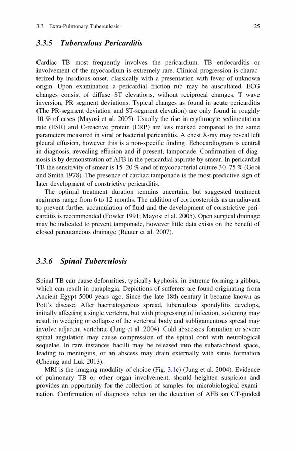
Spinal TB can cause deformities, typically kyphosis, in extreme forming a gibbus,which can result in paraplegia. Depictions of sufferers are found originating fromAncient Egypt 5000 years ago. Since the late 18th century it became known asPott’s disease. After haematogenous spread, tuberculous spondylitis develops,initially affecting a single vertebra, but with progressing of infection, softening mayresult in wedging or collapse of the vertebral body and subligamentous spread mayinvolve adjacent vertebrae (Jung et al. 2004). Cold abscesses formation or severespinal angulation may cause compression of the spinal cord with neurologicalsequelae. In rare instances bacilli may be released into the subarachnoid space,leading to meningitis, or an abscess may drain externally with sinus formation(Cheung and Luk 2013).
MRI is the imaging modality of choice (Fig. 3.1c) (Jung et al. 2004). Evidenceof pulmonary TB or other organ involvement, should heighten suspicion andprovides an opportunity for the collection of samples for microbiological exami-nation. Confirmation of diagnosis relies on the detection of AFB on CT-guided
3.3 Extra-Pulmonary Tuberculosis 25

tissue biopsies or abscess aspirates. Treatment regimens are as for pulmonary TB,however some advocate longer duration of treatment. Based on the results of aseries of randomized clinical trials conducted by the MRC Working party on TB ofthe spine, spanning a period of 15 year follow up, it is currently accepted that earlyand mild disease, without significant neurological deficits, may be treated conser-vatively with anti-tuberculous chemotherapy without operative intervention.Patients treated with debridement alone or combined with spinal fixation (withanterior strut graft) had the tendency to earlier resolution of abscesses, earlier bonyfusion and less kyphotic deformity (Mak and Cheung 2013). It is important toidentify the poor prognostic factors that are associated with severe kyphosisdevelopment, such as the degree of vertebral body loss before treatment, and theseparation of facet joints, to identify patients that would benefit for operativeintervention by reducing kyphotic deformity.
3.3.7 Other Forms of Extra-Pulmonary Tuberculosis
Tuberculous arthritis, almost always affects only a single joint, usually the hip andknee. It can be diagnosed by examination of synovial fluid or synovial tissuebiopsies. Gastrointestinal TB may mimic Crohn’s disease, both clinically andradiographically. Preferred sites are the ileocecum, ileum and jejunum and isusually associated with peritonitis. Barium contrast studies can reveal ulceration,strictures, bowel wall thickening, skip lesions and fistulae. In endemic countries,diagnosis is usually made on clinical suspicion. Biopsies may be useful in estab-lishing the diagnosis (Nagi et al. 2002, 2003).
Urogenital TB is notoriously asymptomatic. TB of the urinary tract, occasionallycauses flank pain or present with a renal or pelvic mass. Persistent “sterile” pyuriaon urine analysis, especially early morning samples, require further investigationwith urine AFB smear, PCR and culture. Further investigations include intravenousurography (Merchant et al. 2013a, b).
Laryngeal TB is one of the most infectious forms of TB. Sputum smear is reportedpositive in up to 70 % of cases. It can result from primary infection with infecteddroplet nuclei or secondary to pulmonary disease. Hoarseness and dysphagia can beamong the presenting signs. Laryngeal TB can be primary, when bacilli directlyinvade the larynx or secondary from bronchial spread of advanced pulmonary TB(Benwill and Sarria 2014). It presents with hoarseness and dysphagia, or chroniccough if associated with pulmonary TB (Michael and Michael 2011). It should bedifferentiated from laryngeal malignancy. TB can potentially affect any organ in thehuman body, further discussion of all rare forms fall beyond the scope of this chapter.
Open Access This chapter is distributed under the terms of the Creative Commons AttributionNoncommercial License, which permits any noncommercial use, distribution, and reproduction inany medium, provided the original author(s) and source are credited.
26 3 Clinical Manifestations

Chapter 4Diagnosis
Abstract At the turn of the century, it was widely recognized that an accuratepoint-of care test for TB was required to make significant reductions in the pan-demic. At this time, many novel tests had been developed by research groups orsmall biotech companies, but had never been standardized or evaluated for scale-upand application in low-resource, high-burden settings where the need is greatest.This motivated a major drive to systematically evaluate existing tests such ascommercial liquid culture and nucleic acid amplification tests (NAAT), and todevelop new approaches, principally led by the Foundation for Innovative NewDiagnostics (FIND www.finddiagnostics.org) in collaboration with industry, gov-ernment and clinical partners. The evidence generated by this renewed focus onnovel TB diagnostic tests, processes and algorithms has led to a substantial numberof policy revisions and new WHO recommendations (Table 4.1, see also www.tbevidence.org).
Keywords Smear microscopy � Ziehl neelsen stain � Mycobacterial culture �Nucleic acid amplification tests (NAAT) � Xpert MTB/RIF � GeneXpert � Lineprobe assay � Drug-resistant tuberculosis � Interferon gamma release assays (IGRA)
4.1 Smear Microscopy
The confirmation of TB disease still rests upon identification or isolation ofM. tuberculosis bacilli from a clinical sample. This can be achieved by smearmicroscopy for acid-fast bacilli (AFB), mycobacterial culture or nucleic acidamplification (NAAT) tests. The appropriate sample will depend upon the sus-pected site of disease. The quality of the sample may greatly affect the chances of apositive result therefore care should be taken to instruct the patient in producing asputum sample. Children are often unable to produce sputum and in young childrengastric aspirate is usually necessary.
© The Author(s) 2015D. Heemskerk et al., Tuberculosis in Adults and Children,SpringerBriefs in Public Health, DOI 10.1007/978-3-319-19132-4_4
27

Tab
le4.1
Laboratorytestsfordiagno
sisof
activ
etuberculosisanddrug
resistance
Diagn
ostic
testsforactiveTB
Testtype
Principalcommercial
tests
WHO
policy
recommendatio
nAdv
antages
Lim
itatio
ns
Smearmicroscop
yNon
-com
mercial
Recom
mended
Inexpensive,
simple,
rapid,
specific
Canno
tdifferentiate
NTM
a
andM.tuberculosis
LED
microscop
yRecom
mended
Inexpensive,
simple,
rapid
Canno
tdifferentiate
NTM
a
andM.tuberculosis
Autom
ated
real-tim
enu
cleicacid
amplification
GeneX
pertMTB/RIF
Recom
mended
Rapid
(2hto
result).Detects
smear-negativ
eTB.Alsodetects
RIF
resistance
Highercostthan
smear
Loo
p-mediated
isotherm
alam
plificatio
ntestkitforTB
LAMPassay
Not
recommended.
Und
erfurther
developm
ent
Rapid,simple
Subjectiv
einterpretatio
nand
poor
specificity
Rap
idspeciation
strip
techno
logy
Recom
mended
Forrapiddifferentiatio
nof
NTM
a
andM.tuberculosis
Exp
ensive
Serodiagno
stic
tests
Over20
commercial
variants
Not
recommended
Poor
sensitivity
andspecificity
Interferon
-Gam
ma
releaseassays
QuantiFERON-TB
GoldIn-Tub
etest,
T-Spo
ttest
Not
recommendedfordiagno
sisof
activ
eTB
Com
plex
toperform
and
indeterm
inateresults
relativ
ely
common
Drugsusceptib
ility
tests
Testtype
Principal
Com
mercial
tests
WHO
policy
recommendatio
nDrugs
tested
Adv
antages
Lim
itatio
ns
Pheno
typicDST
onsolid
orliq
uid
media
Non
-com
mercial
Recom
mendedfor
USE
Alldrug
sbGold-standard
Extremelylong
timeto
result
(6–12
weeks)
Com
mercial
liquid
culturean
dDST
system
s
BactecMGIT
Recom
mendedfor
USE
STR,IN
H,RIF,
EMB,PZ
AFaster
than
solid
cultu
remedia.Ten
days
ifdirect
testing
Exp
ensive
(con
tinued)
28 4 Diagnosis

Tab
le4.1
(con
tinued)
Drugsusceptib
ility
tests
Testtype
Principal
Com
mercial
tests
WHO
policy
recommendatio
nDrugs
tested
Adv
antages
Lim
itatio
ns
Lineprob
eassay
first-lin
eMTBDR-Plus;
INNO
LiPA-RIF
TB
Recom
mendedfor
USE
onsm
ear-po
sitive
samples
RIF,IN
HResultin
2days
Exp
ensive
Lineprob
eassay
second
-line
MTBDRsl
Not
yet
recommendeddu
eto
insufficient
evidence
Fluo
roqu
inolon
es,
aminog
lycosides
andEMB
Resultin
2days
Low
sensitivity
forethambu
tol
Autom
ated
real-tim
enu
cleic
acid
amplification
GeneX
pert
MTBRIF
Recom
mendedfor
USE
RIF
Resultin
2h
Cartridge
priceredu
ctions
only
availablein
low
middleincome
coun
tries
Microscop
icob
servationdrug
susceptibility
(MODS)
Non
-com
mercial
Recom
mendedfor
USE
RIF,IN
HLow
-tech.
10–14
days
forresult
Subjectiv
einterpretatio
n.Laborious
manualplate
readingc
Colom
etricredo
xindicator(CRI)
Non
-com
mercial
Not
yet
recommendeddu
eto
insufficient
evidence
RIF,IN
HLow
-tech.
10–14
days
forresult
Subjectiv
einterpretatio
n
Nitrateredu
ctase
assays
(NRA)
Non
-com
mercial
Not
yet
recommendeddu
eto
insufficient
evidence
RIF,IN
HLow
-tech10–14
days
forresult
Subjectiv
einterpretatio
n
(con
tinued)
4.1 Smear Microscopy 29

Tab
le4.1
(con
tinued)
Drugsusceptib
ility
tests
Testtype
Principal
Com
mercial
tests
WHO
policy
recommendatio
nDrugs
tested
Adv
antages
Lim
itatio
ns
Phageassays
FAST
plaque,
lucerferase
repo
rter
phage
assay
Not
recommended
RIF,IN
HN/a
Poor
specificity
Sequ
encing
Non
-com
mercial
Nopo
licy
Depends
ongene
region
ssequ
enced
Can
prov
ide
inform
ationon
multip
ledrug
ssimultaneou
sly
Requiresspecialist
interpretatio
n.Not
generally
availableou
tsideresearch
centres
Detailsof
policygu
idance
at:http://www.who
.int/tb/labo
ratory/en/
a NTM:no
n-tuberculou
smycob
acteria
b Reliableforfirst-lin
edrug
s(exceptpy
razinamide),flu
oroq
uino
loes
andam
inog
lycosides.
Second
-lineDST
shou
ldbe
interpretedin
contextof
treatm
ent
historyandlocalprevalence
ofresistance
(ifkn
own)
c Ind
icator
wellmustbe
incorporated
todifferentiate
NTM
from
M.tuberculosis
30 4 Diagnosis

Diagnosis for the majority of patients worldwide suspected of TB is still madeby sputum smear microscopy for acid-fast bacilli. The test, which was developed100 years ago by Franz Ziehl and Frederick Neelsen, is inexpensive, simple, rapidand specific but is only positive in around half of patients with active TB. TheZiehl-Neelsen smear exploits the acid-fast property of mycobacteria by stainingbacilli with carbol-fuschin, using gentle heat to facilitate penetration of the dye, andthen using a decolorising acid solution, which fails to penetrate the mycobacteria,leaving them stained red while other bacilli are decolorised. The slide is usuallythen counterstained with methylene blue to improve visualization of the myco-bacteria (World Health Organisation 1998). The Kinyoun stain is an alternativecold-stain method. The sensitivity of the test is substantially lower in children andpatients with HIV. In addition the test is not specific for M. tuberculosis, but detectsall acid-fast bacilli including NTMs. Sensitivity may be increased by concentrationof samples prior to microscopy, usually by centrifugation or filtration (Van Deunet al. 2000) but direct (unconcentrated) ZN stain is the most widely appliedmethodology due to resource limitations.
Traditional TB control focused on the identification and treatment of sputumsmear-positive TB patients, considered to be most infectious cases, in the mistakenbelief that systematic identification and treatment of smear-positive cases would besufficient to reach eventual TB elimination. Recent efforts to improve the sensitivityof basic smear microscopy have developed improved fluorescent microscopes todecrease the reading time and increase the sensitivity of smear microscopy withoutsignificantly affecting specificity if training and quality control are maintained.WHO now recommends the replacement of conventional microscopy with fluo-rescent microscopy wherever possible, using rugged and energy efficient LEDfluorescent microscopes that can be battery operated. In a further policy change,WHO recommended in 2010 that two sputum samples are sufficient, rather than thestandard three samples (spot-morning-spot) which had been recommended forseveral decades (World Health Organisation 2010). This is due to the low diag-nostic yield of a third sputum sample and the resource limitations of TB pro-grammes. If clinical suspicion is high repeated testing may still be warranted.A single positive smear is now also considered sufficient for a TB diagnosis(Bonnet et al. 2007; Mase et al. 2007).
4.2 Mycobacterial Culture
Culture of M. tuberculosis is a more sensitive technique for diagnosis but due to theslow growth of the organism (replication time of 24–30 h) sputum cultures take4–6 weeks to become positive on solid media and 10–21 days in liquid media. Solidculture is usually performed on Lowenstein Jensen (LJ), Ogawa or Middlebrook7H10/11 agar media. Liquid culture of M. tuberculosis is more sensitive and rapidthan solid culture but can be prone to contamination in some laboratories. Earlycommercial automated liquid culture systems for mycobacteria used radiometric
4.1 Smear Microscopy 31

assay but have now been replaced with fluorescence based quenching systemswhich has improved safety. The most widely used system is the BactecMycobacterial Growth Indicator Tube (MGIT) (Becton Dickinson, Sparks,Massachusetts) system which can also be used for susceptibility testing to first linedrugs using a commercially available kit. A culture is necessary to confirm drugsusceptibility, particularly for second-line drugs in cases of multi-drug resistance(MDR TB). M. tuberculosis culture and phenotypic DST requires significanttraining, infrastructure, strict infection control and on-going quality assurance,which is only available in regional reference laboratories in most countries.
4.3 Nucleic Acid Amplification Tests
Various commercial and in-house nucleic acid amplification tests (NAAT) havebeen available since the 1990s. Detection of M. tuberculosis in clinical samples isgenerally less sensitive than NAAT for other pathogens due to the relatively lownumbers of bacilli present and the difficulty of efficiently extracting DNA from thetough mycobacteria. The development of Line Probe assays (LPA) allowed thesimultaneous detection of M. tuberculosis and determination of resistance to rif-ampicin and later isoniazid. However these tests are only endorsed for use on smearpositive sputum and therefore do not aid greatly in the diagnosis of TB itself. TheMTBDR-Plus assay (HainLifesciences, Nehren, Germany) has recently beenadapted to enhance detection for use on smear negative sputum samples butlarge-scale evaluation data is not yet available. The use of LPA for detection of drugresistance is discussed in more detail in the section on diagnosis of drug resistancebelow.
The most significant advance in the diagnosis of TB in the last decade has beenthe advent of the GeneXpert MTB/RIF test (Cepheid, California, USA). This testsystem was originally developed for testing for the presence of anthrax spores in theUnited States bioterrorism-scares. A specific cartridge was later developed to detectM. tuberculosis and simultaneously determine resistance to rifampicin. In 2010results of a multi-country demonstration study sponsored by FIND demonstratedthat the Xpert MTB/RIF test detected TB and rifampicin resistance with highsensitivity and specificity compared to liquid culture; confirmed by a Cochranereview in 2013. The test was officially endorsed by WHO, followed by anunprecedented rapid scale up of the new technology [http://who.int/tb/laboratory/mtbrifrollout/en/]. A key factor in wide-scale implementation was a negotiated pricereduction facilitated by a large guaranteed buy-down from UNITAID, USAID,PEPFAR and the Bill and Melinda Gates Foundation which reduced the price percartridge from more than 40 USD to less than 10 USD for public health facilities in141 low and middle income countries. A major advantage of the Xpert MTB/RIFtest is the ability to detect smear negative TB in HIV-infected individuals (WorldHealth Organization 2013).
32 4 Diagnosis

In 2013 WHO issued updated policy guidance on the use of Xpert MTB/RIFadditionally endorsing its application for extrapulmonary and pediatric samples.This policy update expanded the recommended application of Xpert MTB/RIF toinclude pediatric and extrapulmonary samples, including gastric aspirate, lymphaspirate, pleural fluid and cerebrospinal fluid. There was insufficient evidence toestimate sensitivity with urine, pericardial fluid and ascitic fluid, although speci-ficity is generally high with these sample types. Optimal sample processing forblood and stool samples has not been determined and therefore the Xpert MTB/RIFtest is not recommended pending further research. Full recommendations can befound at http://tbevidence.org/wp-content/uploads/2013/11/WHOstat.pdf.
By facilitating early detection of TB, prior to smear positivity, the application ofXpert MTB/RIF should have a significant impact on transmission chains and pushback the epidemic. However, many of the patients diagnosed by Xpert MTB/RIFwould have been initiated on treatment due to chest X-ray findings or clinicalfindings consistent with TB and the extent to which the use of Xpert MTB/RIF willincrease case finding is not yet clear. Theoretical modelling studies suggest that theapplication of the test will improve targeting of treatment, with less patients who donot have TB incorrectly started on treatment and a greater number of smear negative‘true TB’ cases detected. South Africa has implemented the Xpert MTB/RIF testnationwide and data on the cost-effectiveness and impact on the epidemic are
Fig. 4.1 Current development in TB diagnostics. Only the Xpert® MTB/RIF has received WHOendorsement. From UNITAID, Diagnostic technology and market landscape, 3rd edition, 2014.Reprinted with permission
4.3 Nucleic Acid Amplification Tests 33

eagerly awaited. Although relatively simple to perform and rapid, the XpertMTB/RIF is not a true point-of-care test and many challenges have been encoun-tered during scale-up. The need for a reliable electricity supply is a major barrier insome settings, problems with module calibration and maintenance, the need for thebulky cartridges to be stored below 30 °C, determining optimal testing algorithmsand logistics of kit supply have been some of the challenges encountered(Abdurrahman et al. 2014) (Fig. 4.1).
4.4 Diagnosing Drug-Resistant Tuberculosis
A major impact of the scale-up of Xpert MTB/RIF is increased detection of RIFresistance, which is a surrogate marker for MDR TB. Classical diagnosis of drugresistance in M. tuberculosis involves culture of the bacilli on solid or liquid mediaand comparison of growth between drug-free and drug containing media. Even withthe advent of direct liquid culture methodology, detection of drug resistance takesover two weeks, and with indirect methods, two months or more. Standardisation ofdrug susceptibility testing for the antituberculous drugs is difficult and should beperformed in a biosafety level 3 laboratory by trained personnel participating in anexternal quality assurance scheme. Isoniazid, rifampicin and streptomycin are themost reliable drug susceptibility tests.
Molecular detection of drug resistance mutations provides a rapid alternative, butthe accuracy of these tests varies according to the drug. Rifampicin resistancedetection is the most accurate, as 95 % of phenotypically rifampicin resistant strainscarry a mutation in the 81 base pair rifampicin—resistance-determining-region(RRDR) of the rpoB gene. For isoniazid, molecular methods can detect approxi-mately 75 % of phenotypically resistant strains by detecting mutations in the katGgene or InhA promoter region. Development of commercial NAAT for the otherantituberculous drugs has been hampered by incomplete understanding of themolecular mechanisms of resistance. The principal commercial NAAT for drugresistance are the Xpert MTB/RIF test and the line probe assays. The most recentpooled estimates for M. tuberculosis detection by Xpert MTB/RIF were sensitivityof 88 % (95 %CI; 83–92%) and specificity of 98 % (95 %CI; 97–99 %); forrifampicin resistance sensitivity 94 % (95 %CI; 87–97 %) and specificity 98 %(95 %CI; 97–99 %) (Steingart et al. 2014). Line Probe assays detect both rifampicinand isoniazid resistance simultaneously and the MTBDR-sl assay detects resistanceto fluoroquinolones, ethambutol and aminoglycosides. The MTBDR-sl test has highsensitivity for fluoroquinolones, but low sensitivity for aminoglycoside and eth-ambutol resistance (Feng et al. 2013). However, specificity is high for all drugs andtherefore the test can be used to detect resistance but should not be used to rule-outresistance. Unfortunately, the need remains to confirm susceptibility by laboriousphenotypic DST. Rapid sequencing techniques provide more comprehensive drugsusceptibility data but are not yet widely available beyond research settings.
34 4 Diagnosis

Several non-commercial phenotypic DST approaches have been developedincluding microscopic observation drug susceptibility testing (MODS), nitratereductase assay (NRA) and colorimetric redox indicator (CRI) tests. A MODS testkit is now available to improve standardization (Hardy diagnostics). In 2010 WHOissued a recommendation that MODS could be used as an ‘interim’ approach forincreased DST in high-burden countries but concluded that there was insufficientdata to recommend NRA or CRI. Reservations particularly around biosafety andquality control have limited scale-up of the techniques (Image 4.1).
4.5 Other Diagnostic Methods
In 2011 WHO issued an unprecedented negative advisory on the use of serodiag-nostic tests for TB (Steingart et al. 2011). These tests are appealing because of theyare simple, rapid, inexpensive and non-invasive and are marketed with claims ofhigh sensitivity and specificity. However, systematic evaluation of 19 commerciallyavailable tests using a well-characterised serum bank, and systematic review of allpublished studies concluded that none was accurate for use in clinical practice(Steingart et al. 2011). The search for accurate biomarkers for use in serodiagnostictests continue, but has so far yielded little promise. Tests under evaluation by the
Image 4.1 Mycobacterial cording in MODS. Image courtesy of Dr. Dang Thi Minh Ha
4.4 Diagnosing Drug-Resistant Tuberculosis 35
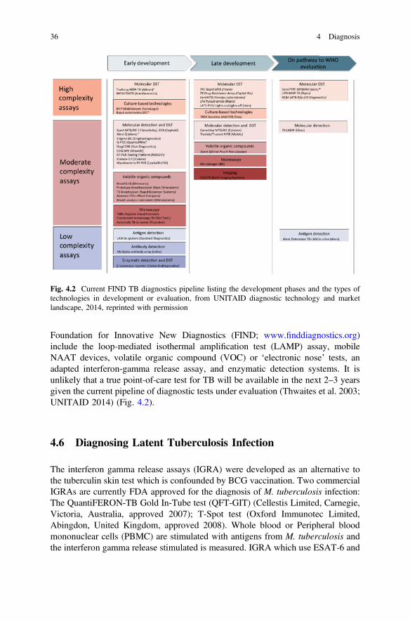
Foundation for Innovative New Diagnostics (FIND; www.finddiagnostics.org)include the loop-mediated isothermal amplification test (LAMP) assay, mobileNAAT devices, volatile organic compound (VOC) or ‘electronic nose’ tests, anadapted interferon-gamma release assay, and enzymatic detection systems. It isunlikely that a true point-of-care test for TB will be available in the next 2–3 yearsgiven the current pipeline of diagnostic tests under evaluation (Thwaites et al. 2003;UNITAID 2014) (Fig. 4.2).
4.6 Diagnosing Latent Tuberculosis Infection
The interferon gamma release assays (IGRA) were developed as an alternative tothe tuberculin skin test which is confounded by BCG vaccination. Two commercialIGRAs are currently FDA approved for the diagnosis of M. tuberculosis infection:The QuantiFERON-TB Gold In-Tube test (QFT-GIT) (Cellestis Limited, Carnegie,Victoria, Australia, approved 2007); T-Spot test (Oxford Immunotec Limited,Abingdon, United Kingdom, approved 2008). Whole blood or Peripheral bloodmononuclear cells (PBMC) are stimulated with antigens from M. tuberculosis andthe interferon gamma release stimulated is measured. IGRA which use ESAT-6 and
Fig. 4.2 Current FIND TB diagnostics pipeline listing the development phases and the types oftechnologies in development or evaluation, from UNITAID diagnostic technology and marketlandscape, 2014, reprinted with permission
36 4 Diagnosis

CFP-10 antigens for stimulation are not confounded by prior BCG vaccinationbecause these antigens are found in a region (RD1) of the M. tuberculosis genomewhich is deleted from BCG and thought to be partially responsible for the loss ofvirulence.
In the United States, IGRA are widely used for the diagnosis of latent TBinfection and are recommended by CDC guidelines (available at: http://www.cdc.gov/mmwr/pdf/rr/rr5905.pdf). WHO do not recommend the use of IGRA inendemic settings or for the diagnosis of active TB. It should be noted that 95 % ofIGRA positive individuals do not go on to develop active TB and therefore thepredictive value of a positive IGRA is extremely low; no statistically significantdifference in the incidence of active TB between IGRA positive and IGRA negativeindividuals has been demonstrated in the small number of studies which haveaddressed this question.
Open Access This chapter is distributed under the terms of the Creative Commons AttributionNoncommercial License, which permits any noncommercial use, distribution, and reproduction inany medium, provided the original author(s) and source are credited.
4.6 Diagnosing Latent Tuberculosis Infection 37

Chapter 5Treatment
Abstract In this chapter the treatment of drug sensitive and drug resistant TB andtiming of antiretroviral treatment for HIV infected patients will be reviewed.Emphasis is placed on results of recent trials of fluoroquinolones for treatmentshortening of drug sensitive TB. The use of two relatively novel agents in MDR-TBtreatment, bedaquiline and delamanid, will be discussed.
Keywords First-line antituberculous treatment � Rifampicin � Isoniazid �Pyrazinamide � Streptomycin � Ethambutol � HIV associated tuberculosis �Antiretroviral therapy (ARV, ART) � Treatment of drug-resistant tuberculosis �Fluoroquinolones � Bedaquiline � Delamanid
5.1 First-Line Antituberculous Treatment
The introduction of rifampicin to the first-line combination regimen in the late1970s allowed the shortening of treatment for TB from 18–24 months to 6 months.This “short-course” regimen, consists of an ‘intensive phase’ of 4 first line drugs;ethambutol (EMB) [or streptomycin (SM)], isoniazid (INH), pyrazinamide (PZA)and rifampicin (RIF) for 2 months followed by a ‘continuation phase’ of 2 monthsof RIF and INH (Table 5.1). The continuation phase may be extended in morecomplex cases and some countries still use an 8-month standard regimen which isno longer recommended by WHO. Ethambutol should be used in place of theinjectable streptomycin where possible for HIV-infected individuals. Since thedevelopment of the short-course regimen, standard TB-treatment has remainedlargely unchanged for the past 40 years. Daily, directly observed therapy (DOT) ispreferable to intermittent regimens and fixed dose combination (FDC) drugs may beused to ensure multi-drug therapy (Nunn et al. 2014). Pyridoxine should beadministered with isoniazid to prevent peripheral neuropathy. The historic eventsthat have led to the current treatment schedule are comprehensively reviewed byDiacon and colleagues (Diacon et al. 2012).
© The Author(s) 2015D. Heemskerk et al., Tuberculosis in Adults and Children,SpringerBriefs in Public Health, DOI 10.1007/978-3-319-19132-4_5
39

Table 5.1 Drugs used in the treatment of tuberculosis
Drug Adult dosemg/kg (range);[maximum dose]
Pediatric dosemg/kg (range);[maximumdose]
Common sideeffectsa
Group 1: first-line oral drugs
Isoniazidb 5 (4–6) [300 mg] 10 (7–15)[300 mg]
Hepatitis; peripheralneuropathy
Rifampicin(rifapentine/rifabutinalternative rifamycins)
10 (8–12)[600 mg]
15 (10–20)[600 mg]
Hepatitis; orangediscoloration ofsecretions;drug-druginteractions
Pyrazinamide 25 (20–30) 35 (30–40)[2000 mg]
Hepatitis; arthralgia
Ethambutol 15(15–20 mg/kg)
20 (15–25)[1200 mg]
Visual disturbance(acuity, colourvision)
Second-line drugs (group 2, injectables)
Streptomycin (S) 15 (12–18)[1000 mg]
Notrecommendedas first-line.15 (12–18)[1000 mg]
Auditory nervedamage
Kanamycin (Km) 15–20 mg/kg[1000 mg]
15–30 [1000] Renal failure(usually reversible)
Amikacin (Am) 15–20 mg/kg[1000 mg]
15–22.5[1000]
Proteinuria, serumelectrolytedisturbancesincludinghypokalaemia andhypomagnesaemia
Capreomycin (Cm) 15–20 mg/kg[1000 mg]
15–30 [1000] Nephrotoxicity(20–25 %), tubulardysfunction,azotaemia,proteinuria, urticariaor maculopapularrash
Group 3: fluoroquinolones
Levofloxacin (Lfx) 750 mg[1000 mg]
7.5–10 Generally welltolerated
Moxifloxacin (Mfx) 400 mg dailydose
7.5–10
Ofloxacin (Ofx) 800 mg[1000 mg]
15–20 [800]
(continued)
40 5 Treatment

Table 5.1 (continued)
Drug Adult dosemg/kg (range);[maximum dose]
Pediatric dosemg/kg (range);[maximumdose]
Common sideeffectsa
Group 4: oral bacteriostatic second-line
Ethionamide (Eto) 15–20 mg/kg[1000 mg]
15–20 mg/kg[1000 mg]
Severegastrointestinalintolerance (nausea,vomiting, diarrhoea,abdominal pain,excessive salivation,metallic taste,stomatitis, anorexiaand weight loss.Unable to tolerate 1g as a single dose
Prothionamide (Pto) 15–20 mg/kg[1000 mg]
15–20 mg/kg[1000 mg]
Terizidone (Trd) 15–20 mg/kg[900 mg]
10–20 [1000] Neurological andpsychiatricdisturbances,including suicidaland psychoticepisodes
Cycloserine (Cs) 15–20 mg/kg[1000 mg]
10–20 [1000] Neurological andpsychiatricdisturbances,includingheadaches,irritability, sleepdisturbances,aggression, andtremors, guminflammation, paleskin, depression,confusion,dizziness,restlessness,anxiety, nightmares,severe headache,drowsiness
Para-aminosalicylic acid(PAS)
150 mg/kg [8 g] 150 mg/kg[8 g]
Gastrointestinalintolerance(anorexia anddiarrhoea);hypo-thyroidism(increased risk withconcomitant use ofethionamide)
(continued)
5.1 First-Line Antituberculous Treatment 41

WHO treatment guidelines can be found at http://www.who.int/tb/publications/tb_treatmentguidelines/en/index.html. Treatment recommendations for pediatric TBwere revised in 2010, with an increased dose of all first-line drugs (http://whqlibdoc.who.int/publications/2010/9789241500449_eng.pdf) and a request notto use streptomycin as a first-line drug in children (World Health Organisation
Table 5.1 (continued)
Drug Adult dosemg/kg (range);[maximum dose]
Pediatric dosemg/kg (range);[maximumdose]
Common sideeffectsa
Group 5: Agents with unclear role in treatment of drug resistant-TBc
Clofazimine (Cfz) 100 mg daily Ichthyosis, and dryskin; pink tobrownish-blackdiscoloration ofskin, cornea, retinaand urine; anorexiaand abdominal pain
linezolid (Lzd) 600 mg daily Gastrointestinaldisturbance, visiondisturbances,anaemia
Amoxicillin/clavulanate(Amx/Clv)
875 twice daily Gastrointestinaldisturbance,psychiatricdisturbance, sleepdisturbance
Thioacetazone (Thz)d 2.5 mg/kg [150] Gastrointestinaldisturbance,arthralgia, seizures,hepatitis
High-dose isoniazid(high-dose H)
16–20 mg/kg/day Hepatitis; peripheralneuropathy
Clarithromycin (Clr) 500 mg daily Gastrointestinaldisturbance
NovelAgents
Bedaquiline 400 mg oncedaily
Nausea, arthralgia,headache, vomiting,gastrointestinaldisturbance, QTprolongation
Delaminid 200 mg twicedaily
QT prolongation
aHypersensitivity reactions and drug rashes may occur with any anti-tuberculous drugbPyridoxine should be given with isoniazid to prevent peripheral neuropathy. Guidelines variouslyrecommend 10 or 25 mg/kg dailycOptimal dose and long-term safety not well established for group 5 drugsdDo not use thioazetone for HIV-infected individuals (significant risk of Stevens-Johnsonsyndrome)
42 5 Treatment

2014). This followed systematic review of evidence showing that children achieveinadequate serum exposure when receiving the same dose/kg as adults and therewas no evidence of adverse toxicity from the higher doses. Pharmacokinetic datafrom South Africa showed substantially improved serum levels with the novelregimen but it remains to be demonstrated if these doses are sufficient to achieveimproved outcomes (Thee et al. 2011).
5.2 HIV Associated Tuberculosis
The advent of HIV has severely impacted the burden of TB. All HIV patientsshould be screened for TB and all TB patients should be offered HIV testing. Dualtreatment of HIV and TB is complex, as patients are faced with a higher pill-burden,increased risk of toxicity, drug-interactions and IRIS (Lawn et al. 2013; Lai et al.2013). Routine administration of co-trimoxazole (960 mg/day) is recommended inall patients with HIV-associated TB, since it has been shown to substantially reducemortality in patients in Sub Saharan Africa. Rifampicin is an inducer of the cyto-chrome P450 2B6 enzyme, which is the main pathway for the non-nucleasidereverse transcriptase inhibitors (NNRTIs) and protease inhibitors (PIs) either ofwhich are the basis of ARV treatment (Bonnet et al. 2013). The most commonlyused NNRTI is efavirenz. Although the US FDA recommends that the dose ofefavirenz should be increased when co-administered with rifampicin, this is notsubstantiated in the most recent clinical trials, which show excellent virologicalresponse in those patients receiving standard dose (600 mg) of efavirenz (Kwaraet al. 2010). Nevarapine is an alternative NNRTI used for those patients who cannottolerate efavirenz, but plasma levels are very low in patients receiving rifampicinduring the lead-in phase of nevarapine. This may predispose to resistance formationand virological failure. Dose increase is not recommended because concerns oftoxicity, however it is plausible to omit the 14 day lead-in phase of nevirapinedosing after the CARENIMO trial showed that nevirapine was well tolerated at fulldose when introduced in patients with CD4 cell counts <250/mm3 while receivingrifampicin (Bonnet et al. 2013). The co-administration of second-line ARV regi-mens containing PIs remains a challenge and there is an urgent need for clinicaltrials evaluating safety and efficacy. ‘Superboosting’ of ritonavir or doubling doseof lopinavir/ritanovir combination formulation are suggested, as is substituting ri-fabutin for rifampicin (Lawn et al. 2013).
Timing of initiation of ARV treatment in ARV-naïve TB/HIV patients has beenevaluated in a recent series of clinical trials published in 2011 (SAPIT[NCT00398996], CAMELIA[NCT01300481] and the AIDS clinical trial groupstudy A5221). Results showed that there is a reduction in mortality in patients withCD4 cell counts lower that 200/mm3 (CAMELIA trial) or 50/mm3 (other two trials)when ARV treatment was initiated within 2 weeks of TB treatment (GRADE A3),(Abdool Karim et al. 2010, 2013; Blanc et al. 2011). However this carries anincreased risk of IRIS and treatment toxicity. The management in patients with
5.1 First-Line Antituberculous Treatment 43

higher CD4 cell counts is less clear and it may be acceptable to defer ARV treat-ment until the continuation phase of TB treatment. The majority of patients in thesetrials had pulmonary TB. For patients with TB meningitis, a recent largeVietnamese randomised controlled trial showed no benefit of early initiation ofARVs and deferred treatment at 2 months was associated with less toxicity and lessoccurrence of IRIS (Torok et al. 2011). Patients in this cohort had extremely lowmedian CD4 count and the results may not be generalizable to other populations.
5.3 Treatment of Drug-Resistant Tuberculosis
The use of multi-drug therapy for TB treatment from the early days of discovery ofantituberculous drugs has actually preserved the efficacy of the five first line agents forTB remarkably well for over 50 years. However, drug resistance inM. tuberculosis isincreasing and has now reached alarming levels, particularly in the former Sovietstates, India, South Africa and part of Asia. Approximately 8 % of M. tuberculosiscases globally are now resistant to isoniazid. Multi-drug resistant TB (MDR TB) isdefined as TB resistant to at least isoniazid and rifampicin, the two key first line drugsin the treatment regimen. MDR TB is much harder to treat, requiring a minimum of18 months treatment with expensive, toxic and weak second line drugs. Worldwide,3.6 % of newly diagnosed and 20 % of patients previously treated for TB haveMDR TB but there are dramatic regional variations with the highest proportion inEastern Europe and Central Asia (WHO 2014).
In 2012 there were an estimated 450,000 new cases of MDR TB worldwide and170,000 deaths but only 17 % were diagnosed and enrolled into high qualitytreatment programmes. This figure does however represent a 42 % increase on2011, reflecting scale-up efforts for MDR detection and treatment. In 2006 WHOdefined a new category of drug resistant TB as extensively drug resistant TB (XDRTB). XDR TB is MDR TB additionally resistant to a fluoroquinolone and a secondline injectable drug (amikacin, capreomycin or kanmycin). This followed a docu-mented outbreak in Kwa-Zulu Natal province of South Africa in which 53 cases ofXDR TB were identified among HIV patients, all but one of whom died with amedian survival time of 16 days from diagnosis. Following acceptance of thedefinition of XDR TB, 92 countries have reported cases by 2013 (Fig. 5.1). It isestimated 10 % of MDR cases are in fact XDR and it is now clear that XDR TB ispresent in almost every country although the extent of transmission within com-munities is not established due to the limited availability of second-line drug sus-ceptibility testing for M. tuberculosis. The term totally drug resistant TB has beenused in the literature to describe M. tuberculosis strains resistant to all first andsecond line drugs but the use of this term is not recognised by WHO due to thedifficulty of standardising drug susceptibility testing for second line drugs and theintroduction of treatment options (Cegielski et al. 2012).
Drug resistant TB should be treated with at least four drugs to which theorganism is susceptible, prioritising any first-line agents and then including a
44 5 Treatment

fluoroquinolone and an injectable agent. In order to protect against drug resistanceamplification, a single drug should never be added to a failing regimen (Daley andCaminero 2013). Key principles of MDR treatment are summarised in Table 5.2(Kaufmann et al. 2014; Ulrich et al. 2006).
Comprehensive CDC guidelines on both susceptible and drug resistant TB canbe found on http://www.cdc.gov/mmwr/preview/mmwrhtml/rr5211a1.htm.
The painstakingly sluggish decline in incidence of TB and the rise of MDR- andXDR-TB underlines the inadequacies of current treatment and prevention measures.Priority questions for research, in addition to the search for novel agents includeshortened regimens, optimal regimens for all forms of drug resistant TB, optimaldose of drugs (especially rifampicin), and treatment of HIV-associated TB.
Several existing and newly developed compounds are under evaluation for drug-sensitive and drug-resistant TB (Fig. 5.2). In light of the growing epidemic ofMDR-TB and lack of new drug development there has been renewed interest inobsolete compounds for TB (Chang et al. 2013). In 2010, the Damien Foundationpublished the results of an observational study, comparing six regimens forMDR-TB, derived by sequential modification. The addition of gatifloxacin and useof clofozimine throughout the regimen apparently allowed a reduced duration oftreatment, to between 9–12 months, depending on time to smear conversion. All427 patients received clofazimine in the intensive phase and 206 patients received ashortened regimen of 9–12 months, using clofazimine throughout treatment [4(+)KCGEHZP, 5 GCEZ]. Treatment success-rate was highest in the short regimen
Fig. 5.1 Percentage of previously treated TB cases with multidrug-resistant TB. Source WHO,reprinted with permission
5.3 Treatment of Drug-Resistant Tuberculosis 45

treatment group with a relapse-free cure of 87.9 % (95 %CI 82.7–91.6), withrelatively good tolerability (Van Deun et al. 2010). This shortened ‘Bangladesh’regimen is now being subjected to a large multicentre (including sites in Ethiopia,India, South Africa, Vietnam) randomised controlled trial, known as the STREAMtrial (ISRCTN78372190). The STREAM trial is being conducted by the BritishMedical Research Council and will compare the standard MDR regimen (accordingto local National TB program guidelines, duration ranging from 18–24 months)with a modified form of the Bangladesh regimen. Moxifloxacin, clofazimine, eth-ambutol and pyrazinamide will be given for nine months, supplemented by kana-mycin, isoniazid and prothionamide in the four months of the intensive phase. Alldrugs are given daily except for kanamycin which is administered thrice weeklyafter 12 weeks. The target is to include at least 400 participants. The results of thismajor trial are expected in October 2016.
Table 5.2 Guidelines for themanagement of tuberculosiscaused by drug-resistantorganisms
Do not add a single drug to a failing regimen
When starting or modifying therapy, add three previouslyunused drugs (one an injectable) to which there is likely to besusceptibility
Use any of the first-line oral agents (Group 1) that are likely tobe effective
In multidrug-resistant tuberculosis (MDR TB), when there isresistance to additional first-line drugs, treat with 4–7 drugs(dependent on degree of confidence in susceptibility andstrength of derived regimen)
Patients with MDR TB have the highest priority for directlyobserved therapy because treatment failure may be associatedwith extensively drug-resistant TB
Intermittent therapy should not be used (except for injectablesafter 2–3 months)
Do not use drugs to which the M. tuberculosis isolate isresistant. Low-level resistance to isoniazid may be an exception
There is cross-resistance among rifamycins but not betweenstreptomycin and other aminoglycosides. Amikacin orkanamycin are preferred aminoglycosides for MDR as mostMDR strains are resistant to streptomycin
Drug susceptibility testing for pyrazinamide is complextechnically and not performed in most laboratories.Monoresistance to pyrazinamide suggests M. bovis
Drug susceptibility results for second line agents (exceptfluoroquinolones and aminoglycosides) may be unreliable andshould be interpreted within context of treatment history
Levofloxacin, or moxifloxacin are preferred flouroquinolones.Ciprofloxacin should not be used to treat TB
46 5 Treatment

5.4 The Role of Fluoroquinolones
The fluoroquinolones levofloxacin, gatifloxacin and moxifloxacin have strong an-timycobarterial activity with an early bactericidal activity (EBA) similar to that ofisoniazid and are the strongest of the second-line agents. Fluoroquinolones are thekeystone of MDR treatment regimens and should also be considered in cases ofnon-MDR drug resistant TB requiring additional agents or if a first-line agent in thestandard regimen is not tolerated, particularly in case of hepatotoxicity.Fluoroquinolones are excreted via the kidneys and thus have a lower potential todisturb liver function. They inhibit bacterial replication via the DNA gyrase, amechanism distinct from those of the first-line agents. They have broad-spectrumantibacterial activity, particularly against gram-negative bacteria, favourable phar-macokinetic profile, are relatively safe, have good tissue penetration and highin vitro activity against M. tuberculosis. Fluoroquinolones may therefore worksynergistically to the other TB agents and penetrate well in the tuberculoma.A recent Cochrane review, evaluating RCTs on ofloxacin, levofloxacin,moxifloxacin and gatifloxacin, reported that there is insufficient evidence to be clearwhether fluoroquinolones, either added to the first-line regimen or as a substitutionfor ethambutol or isoniazid, may prevent relapse or death, or increase sputumculture conversion at 8 weeks (Ziganshina et al. 2013). Three large multicountrytrials have been established to determine if the use of fluoroquinolones can shortentreatment regimens to four months rather than six: OFLOTUB (NCT00216385),REMOX TB (NCT00864383) and RIFAQUIN (ISRCTN44153044) studies (Merleet al. 2014; Gillespie et al. 2014; Jindani et al. 2013). The RIFAQUIN trial reportedin April 2013 and showed inferiority of the four month regimen; Two months ofdaily ethambutol, moxifloxacin, rifampicin and pyrazinamide followed by twomonths of twice weekly moxifloxacin (500 mg) and rifapentine (900 mg) comparedto the standard 6-month WHO regimen (2HRZE/4HR).
The OFLOTUB trial which tested a 4 month regimen of 2 months gatifloxacin(400 mg/day), isoniazid, rifampicin and pyrazinamide followed by two monthsgatifloxacin, isoniazid and rifampicin reported in September 2013 and(2GHRZ/2GHR) failed to show non-inferiority of the Gatifloxacin regimen at thepre-specified 6 % margin (Merle et al. 2014). Although Gatifloxacin has beenbanned by the FDA due to toxicity concerns, there was no increased risk of dis-gylcemia or QTc prolongation with the gatifloxacin regimen. The REMOX trialwhich tested two moxifloxacin containing regimens lasting 17 weeks, reported in2014. The moxifloxacin-containing regimens produced a more rapid initial declinein bacterial load, as compared with the control group. However, noninferiority forthese regimens was not shown, which indicates that shortening treatment to4 months was not effective in this setting (Gillespie et al. 2014). Several existingand newly developed compounds are under evaluation for drug- sensitive anddrug-resistant TB (Fig. 5.2) (Ma et al. 2010; Chang et al. 2013; Gopal et al. 2013).
5.4 The Role of Fluoroquinolones 47

5.5 Bedaquiline
Bedaquiline (trade name Sirtuto) is a diarylquinoline drug, formerly known underits code name TMC207, which was discovered in 2005 by Koen Andries andcolleagues in Belgium. It has a novel mode of action, acting by inhibiting ATPsynthase, and is active against replicating and non-replicating bacilli. The first phaseII trial on 47 patients was published in 2009 in the New England Journal ofMedicine and showed that addition of TMC207 to a standard second line regimenled to a higher proportion of patients with sputum conversion at 8 weeks oftreatment (48 % vs. 9 %), (Diacon et al. 2012). The long-term 2 year follow upresults for this pilot trial were published in 2012 and showed a significant reductionin time to sputum conversion in the TMC207 patients compared to placebo and lessacquisition of resistance to companion drugs in the TMC207 group, however thisdid not reach statistical significance. With the exception of nausea, which was morefrequently reported in the TMC207 patients, the occurrence of adverse events wasnot different between the two groups (Diacon et al. 2014). These results combinedwith two other phase II trials have led to accelerated approval of the drug by theFDA in 2012. This was the first new TB drug added to the arsenal of agents indecades. FDA approval was conditional upon prioritisation of phase three trials, butthese have not yet started. The WHO and CDC have published provisionalguidelines on the use of bedaquiline: http://www.who.int/tb/challenges/mdr/bedaquiline/en/index.html and http://www.cdc.gov/mmwr/preview/mmwrhtml/rr6209a1.htm?s_cid=rr6209a1_e (Provisional CDC guidelines for the use andsafety monitoring of bedaquiline fumarate (Sirturo) for the treatment ofmultidrug-resistant tuberculosis 2013).
Fig. 5.2 TB drug discovery pipeline
48 5 Treatment

5.6 Delamanid
Delamanid, (OPC 67683) is a nitro-dihydro-imidazooxazole derivative which actsby blocking the synthesis of myolic acids, thus interfering with cell-wall integrity ofthe mycobacteria. It was first proposed to be a potential new candidate for treatmentof TB in 2006 by Japanese scientists working for Otsuka Pharmaceutical.Subsequently it has shown promising results in phase IIa and b trials. In a seminalRCT including 481 patients recruited in 17 centers in 9 countries, the proportion ofsputum positive MDR-TB patients that converted to sputum negative after 2 monthsof treatment with delamanid (100 mg BID) added to an optimised backgroundregimen (n = 161) was 45.4 % opposed 29.6 % in those patients receiving abackground regimen and placebo (n = 160) (p = 0.008). The patients receivingdelamanid had significantly shorter time to sputum negativity and, additionally, amortality benefit was observed for those in the active arms of this trial (Gler et al.2012). Phase III trials are underway.
The European Medicines Agency (EMA) approved the drug for use in MDR-TBin November 2013, making Delamanid the second drug to be approved for use in(MDR-)TB in 50 years. It will be manufactured under the name of Deltyba.
Open Access This chapter is distributed under the terms of the Creative Commons AttributionNoncommercial License, which permits any noncommercial use, distribution, and reproduction inany medium, provided the original author(s) and source are credited.
5.6 Delamanid 49

Chapter 6Prevention
Abstract Prevention is the key to stop transmission of TB. It consists of earlydiagnosis and treatment of active TB to stop infectiousness, the prevention of activedisease in exposed or known latently infected individuals and vaccination.Vaccination with the Bacillus Calmette-Guerin (BCG) vaccine is unfortunatelylargely ineffective in interrupting transmission. However a more powerful vaccinewill have the potential to cause a major shift in the management of TB. In thischapter prophylactic treatment in latently infected and HIV infected patients isreviewed. Additionally the prevention of active disease in MDR-exposed personsand vaccine development will be discussed.
Keywords Prophylactic treatment � Prophylactic treatment in multi-drug resistanttuberculosis � Vaccine � Latent tuberculosis � Prevention
6.1 Prophylactic Treatment
TB transmission to susceptible contacts primarily occurs in enclosed, poorly ven-tilated locations. High risk locations for transmission are high density congregateliving environments such as hospitals, care homes, prisons or student/migrantworker hostels. Transmission within institutional environments such as hospitalscan be reduced by implementing effective infection control policies coveringadministrative, environmental and respiratory protection (refer to CDC guidelinesfor detailed recommendations: http://www.cdc.gov/tb/publications/guidelines/infectioncontrol.htm).
The standard regimen for treatment of latent TB infection is nine months iso-niazid, also known as isoniazid prophylaxis therapy (IPT). Pyrodoxine should begiven with isoniazid (Udani et al. 1971). In 2011 a trial of a novel 12 dose regimenof isoniazid and rifapentine showed higher completion rates and no loss of efficacy
© The Author(s) 2015D. Heemskerk et al., Tuberculosis in Adults and Children,SpringerBriefs in Public Health, DOI 10.1007/978-3-319-19132-4_6
51

compared to the standard 9-month isoniazid treatment (Sterling et al. 2011). The12-dose regimen has since been incorporated into US recommendations but is notrecommended for children younger than 2 years of age, people with HIV/AIDSwho are taking antiretroviral therapy (ART), people presumed to be infected withINH or rifampin-resistant M. tuberculosis or pregnant women, or women expectingto become pregnant while taking this regimen: http://www.cdc.gov/tb/publications/ltbi/treatment.htm.
A recent mass isoniazid prevention trial in South African mine workers showedno reduction in TB incidence and prevalence beyond the 9 months of IPT, sug-gesting that in high TB burden areas, transmission is not interrupted and theunchanged incidence is due to reinfection (Churchyard et al. 2014).
HIV is the strongest risk factor for developing TB in those with latent or recentM. tuberculosis infection, between 20 and 37 times the risk for those without HIV.To avoid treating an active TB case with monotherapy, HIV-infected individualsshould be screened for active TB before the administration of IPT (Rangaka et al.2014).
There is considerable debate around the duration of IPT in HIV-infected indi-viduals since the trial data from South African miners who are heavily exposedshowed that the protective effect of IPT quickly wanes, with the protective effectdisappearing after one year. This is likely to be due to IPT clearing any latent infection,and therefore preventing reactivation disease, but not preventing re-infection aftercompletion of therapy. WHO recommends extended IPT (36 months) for individualsliving in areas with a high background prevalence of HIV and TB. There is concernthat the widescale application of long-term IPT will lead to increases in isoniazidresistance and decrease in efficacy over time, but there is no evidence that this isoccurring. Trials of long-term IPT are now underway in South Africa.
The reservoir of latently infected individuals is clearly a major barrier to eventualTB elimination, and shorter, less toxic regimens for latent TB will be a major boostto elimination efforts.
For more information on TB prevention: http://www.tbfacts.org/tb-prevention.html.
6.2 Prophylactic Treatment in Multi-drug ResistantTuberculosis
Evidence to guide the choice of regimen for prophylatic treatment of the contacts ofMDR and XDR TB patients is extremely limited. Derivation and testing ofstandardised regimens is complicated by the diverse spectrum of possible suscep-tibility patterns of an index case.
52 6 Prevention

The American Thoracic Society (ATS) and the US Centers for Disease Controland Prevention (CDC) recommend prophylactic therapy for MDR TB contacts withthe regimen to be used determined by the drug susceptibility profile of the potentialsource case. WHO and European guidelines [The International Standards for TBCare (ISTC) and European Union Standards for TB Care (ESTC)] favour carefulclinical follow-up for a period of at least two years.
The European Centre for Disease prevention and control issued guidance onmanagement of contacts of MDR and XDR patients in 2012 based upon systematicreview of the available evidence: http://www.ecdc.europa.eu/en/publications/publications/201203-guidance-mdr-tb-contacts.pdf. The guidance recommendsthat each case must be considered individually with a comprehensive assessment ofthe likely risks and benefits of preventative therapy of unknown efficacy versusintensive clinical monitoring for signs of disease. The risk assessment shouldinclude consideration of any known individual risk factors for progression to activedisease, the drug susceptibility pattern of the index case and any known risk factorsfor adverse events with the prophylactic regimen. A recent prospective observa-tional study on an island in Micronesia, followed MDR-TB contacts who wereoffered 12-month preventive treatment with a fluoroquinolone. Among the 119infected contacts, 15 refused, while 104 began treatment for MDR latent TBinfection. None of the 104 contacts who undertook treatment with fluoroquinolonesdeveloped MDR-TB disease; however, 3 of 15 contacts who refused and 15unidentified contacts developed MDR-TB disease (Bamrah et al. 2014). These dataare compelling and warrant randomized studies, as the ability to prevent MDR-TBtransmission would be the paradigm shift in MDR-TB management.
6.3 Vaccines
The only vaccine currently available for TB is the bacillus Calmette-Guerin(BCG) vaccine developed by serial passage ofMycobacterium bovis and introducedin 1921. BCG is the most widely used vaccine in the world but measures ofeffectiveness have varied widely, between 0 and 80 %. Studies have however,consistently shown a protective effect against the most severe forms of childhoodTB, including TB meningitis. Meta-analysis of all published studies produced anestimate of 50 % for overall efficacy and 80 % efficacy in preventing TB meningitis(Colditz et al. 1994). BCG should not be administered to HIV-infected individuals(Nuttall and Eley 2011). Several theories have been proposed for the differences inobserved effectiveness, including the use of different strains of BCG, variation inearly exposure to non-tuberculous mycobacteria prior to vaccination, host genetic
6.2 Prophylactic Treatment in Multi-drug Resistant Tuberculosis 53

variation and pathogen genetic variation but none of these theories has strongsupporting evidence to date.
Clearly, given the huge burden of global TB and the major barrier to eliminationthat the ‘silent’ reservoir of latently infected individuals represents, an effective TBvaccine would be a major advance in the battle to eradicate TB. However, thecorrelates of protective immunity in TB are not understood which represents amajor hurdle to developing an effective vaccine. The most advanced vaccine can-didate for a primary TB vaccine, MVA85A, failed to show any efficacy in a phaseIIb trial in HIV-uninfected infants published in 2013 (Tameris et al. 2013). This trialshould however provide valuable data to guide further development of novelvaccine candidates.
The current pipeline includes three distinct approaches to the development of aTB vaccine, known as Prime, boost or immunotherapy. An alternative strategy alsobeing explored is post-infection vaccination. Vaccine types currentlyunder-development include (1) modification of the BCG vaccine (recombinantBCG), (2) Boosting of BCG with adjuvants, (3) incorporation of TB antigenexpression into vaccine vectors or (4) killed whole cell or extracts (Kaufmann et al.2014).
6.4 Concluding Remarks
TB is a curable disease. The fact that it remains the most pressing public healthproblem for a significant proportion of the world, despite the availability of a cureand knowledge on prevention of transmission shows how medicine can fail withoutcommitment at all levels of the community. The distribution of the TB pandemicpainfully demonstrates the inequalities in health care delivery globally. Over 95 %of cases and deaths are in low and middle income countries. In general, prognosisof outcome is dependent on a multitude of factors: host factors (genetic variance,co-morbidities, HIV-coinfection, treatment adherence, access to healthcare) andpathogen factors (pathogen virulence, drug-resistance) and the site of the infection(pulmonary or extrapulmonary). The principle factor in a favourable outcome for allforms is early recognition and appropriate treatment. TB is the most common causeof death among HIV patients, estimated to cause a quarter of AIDS related deaths.
Drug resistant TB is a growing problem and threatens to reverse the recent gainsin global TB control. In regions of the former soviet states MDR TB is found in overhalf of all new TB cases and threatens a return to untreatable strains of TB dis-seminating globally without immediate and sustained action. Of the 34,000 MDRpatients enrolled on treatment in 2010, only 48 % successfully completed treatmentand 15 % died. Among 795 XDR cases, mortality was approximately 50 %.
The key to maintaining the momentum towards achieving the STOPTB target ofglobal TB eradication by 2050 will be sustained commitment from donors, gov-ernments, national TB programmes, researchers and other stakeholders at all levelsof society.
54 6 Prevention

Table of useful online TB resources
Topic Source Website
Non-tuberculousmycobacterial disease
American ThoracicSociety (ATS)
http://www.thoracic.org/statements/resources/mtpi/nontuberculous-mycobacterial-diseases.pdf
United States TB statistics Center for DiseaseControl (CDC)
http://www.cdc.gov/tb/statistics/reports/2012/default.htm
Tuberculin skin test(TST) interpretation
CDC http://www.cdc.gov/tb/publications/factsheets/testing/skintesting.htm
Novel tuberculosisdiagnostics
Foundation forInnovative NewDiagnostics (FIND)
http://www.finddiagnostics.org
Evidence-based TBdiagnosis
McGill University http://www.tbevidence.org
TB diagnostics;XpertMTB/RIF Roll-out
World HealthOrganisation (WHO)
http://who.int/tb/laboratory/mtbrifrollout/en/
Xpert MTB/RIF fordiagnosis of pulmonaryand extrapulmonary TB inadults and children PolicyUpdate
WHO http://tbevidence.org/wp-content/uploads/2013/11/WHOstat.pdf
IGRA for detectinginfection with Mtb
CDC http://www.cdc.gov/mmwr/pdf/rr/rr5905.pdf
TB diagnostics andlaboratory strengthening
WHO http://www.who.int/tb/laboratory/en/
TB treatment guidelines WHO http://www.who.int/tb/publications/tb_treatmentguidelines/en/index.html
TB treatment in children WHO http://whqlibdoc.who.int/publications/2010/9789241500449_eng.pdf
Comprehensive treatmentsusceptible and drugresistant TB
CDC http://www.cdc.gov/mmwr/preview/mmwrhtml/rr5211a1.htm
Use of bedaquiline inMDR TB
WHO http://www.who.int/tb/challenges/mdr/bedaquiline/en/index.html
Use of bedaquiline inMDR TB
CDC http://www.cdc.gov/mmwr/preview/mmwrhtml/rr6209a1.htm?s_cid=rr6209a1_e
TB infection control andprevention guidelines
CDC http://www.cdc.gov/tb/publications/guidelines/infectioncontrol.htm
Management of contactsof MDR TB and XDR TBpatients
European Centre forDisease prevention andControl (ECDC)
http://www.ecdc.europa.eu/en/publications/publications/201203-guidance-mdr-tb-contacts.pdf
TB vaccine resources StopTB partnership http://www.stoptb.org/wg/new_vaccines/documents.asp
Global Plan to stop TB2006–2015
WHO http://www.who.int/tb/features_archive/global_plan_to_stop_tb/en/
Tuberculosis treatmentsuccess rate
WHO http://www.who.int/gho/tb/epidemic/treatment/en/
6.4 Concluding Remarks 55

Open Access This chapter is distributed under the terms of the Creative Commons AttributionNoncommercial License, which permits any noncommercial use, distribution, and reproduction inany medium, provided the original author(s) and source are credited.
56 6 Prevention

References
Abdool Karim, S. S., Naidoo, K., Grobler, A., Padayatchi, N., Baxter, C., Gray, A., et al. (2010).Timing of initiation of antiretroviral drugs during tuberculosis therapy. New England Journalof Medicine, 362(8), 697–706.
Abdool Karim, S. S., Naidoo, K., Grobler, A., Padayatchi, N., Baxter, C., Gray, A. L., et al.(2013). Integration of antiretroviral therapy with tuberculosis treatment. New England Journalof Medicine, 365(16), 1492–1501.
Abdurrahman, S. T., Emenyonu, N., Obasanya, O. J., Lawson, L., Dacombe, R., Muhammad, M.,et al. (2014). The hidden costs of installing xpert machines in a tuberculosis high-burdencountry: Experiences from Nigeria. The Pan African Medical Journal, 18, 277.
Anderson, L. F., Tamne, S., Brown, T., Watson, J. P., Mullarkey, C., Zenner, D., & Abubakar, I.(2014). Transmission of multidrug-resistant tuberculosis in the UK: A cross-sectionalmolecular and epidemiological study of clustering and contact tracing. The Lancet InfectiousDiseases, 14(5), 406–415.
Aranow, C. (2011). Vitamin D and the immune system. Journal of Investigative Medicine, 59(6),881–886.
Bamrah, S., Brostrom, R., Dorina, F., Setik, L., Song, R., Kawamura, L. M., et al. (2014).Treatment for LTBI in contacts of MDR-TB patients, Federated States of Micronesia,2009-2012. The International Journal of Tuberculosis and Lung Disease, 18(8), 912–918.
Barry, P. M., Gardner, T. J., Funk, E., Oren, E., Field, K., Shaw, T., & Langer, A. J. (2012).Multistate outbreak of MDR TB identified by genotype cluster investigation. EmergingInfectious Diseases, 18(1), 113–116.
Benwill, J. L., & Sarria, J. C. (2014). Laryngeal tuberculosis in the United States of America: Aforgotten disease. Scandinavian Journal of Infectious Diseases, 46(4), 241–249.
Blanc, F. X., Sok, T., Laureillard, D., Borand, L., Rekacewicz, C., Nerrienet, E., et al. (2011).Earlier versus later start of antiretroviral therapy in HIV-infected adults with tuberculosis. NewEngland Journal of Medicine, 365(16), 1471–1481.
Bonnet, M., Bhatt, N., Baudin, E., Silva, C., Michon, C., Taburet, A. M., et al. (2013). Nevirapineversus efavirenz for patients co-infected with HIV and tuberculosis: A randomisednon-inferiority trial. The Lancet Infectious Diseases, 13(4), 303–312.
Bonnet, M., Ramsay, A., Gagnidze, L., Githui, W., Guerin, P. J., & Varaine, F. (2007). Reducingthe number of sputum samples examined and thresholds for positivity: An opportunity tooptimise smear microscopy. The International Journal of Tuberculosis and Lung Disease,11(9), 953–958.
Borrell, S., & Gagneux, S. (2009). Infectiousness, reproductive fitness and evolution ofdrug-resistant Mycobacterium tuberculosis. The International Journal of Tuberculosis andLung Disease, 13(12), 1456–1466.
Borrell, S., Teo, Y., Giardina, F., Streicher, E. M., Klopper, M., Feldmann, J., et al. (2013).Epistasis between antibiotic resistance mutations drives the evolution of extensivelydrug-resistant tuberculosis. Evolution, Medicine, and Public Health, 2013(1), 65–74.
© The Author(s) 2015D. Heemskerk et al., Tuberculosis in Adults and Children,SpringerBriefs in Public Health, DOI 10.1007/978-3-319-19132-4
57

Bos, K.I., Harkins, K.M., Herbig, A., Coscolla, M., Weber, N., Comas, I., et al. (2014).Pre-Columbian mycobacterial genomes reveal seals as a source of New World humantuberculosis. Nature.
Cecchini, D., Ambrosioni, J., Brezzo, C., Corti, M., Rybko, A., Perez, M., et al. (2007).Tuberculous meningitis in HIV-infected patients: Drug susceptibility and clinical outcome.AIDS, 21(3), 373–374.
Cegielski, P., Nunn, P., Kurbatova, E. V., Weyer, K., Dalton, T. L., Wares, D. F., et al. (2012).Challenges and controversies in defining totally drug-resistant tuberculosis. EmergingInfectious Diseases, 18(11), e2.
Chang, K. C., Yew, W. W., Tam, C. M., & Leung, C. C. (2013). WHO group 5 drugs and difficultmultidrug-resistant tuberculosis: A systematic review with cohort analysis and meta-analysis.Antimicrobial Agents and Chemotherapy, 57(9), 4097–4104.
Cheung, W. Y., & Luk, K. D. (2013). Clinical and radiological outcomes after conservativetreatment of TB spondylitis: iIs the 15 years’ follow-up in the MRC study long enough?European Spine Journal, 22(Suppl 4), 594–602.
Chiang, S. S., Khan, F. A., Milstein, M. B., Tolman, A. W., Benedetti, A., Starke, J. R., & Becerra,M. C. (2014). Treatment outcomes of childhood tuberculous meningitis: A systematic reviewand meta-analysis. The Lancet Infectious Diseases, 14(10), 947–957.
Churchyard, G. J., Fielding, K. L., Lewis, J. J., Coetzee, L., Corbett, E. L., Godfrey-Faussett, P.,et al. (2014). A trial of mass isoniazid preventive therapy for tuberculosis control. NewEngland Journal of Medicine, 370(4), 301–310.
Ciardo, D. E., Burger, S., Payer, M., Lee, C., & McCallum, N. (2010). GeneXpert capturesunstable methicillin-resistant Staphylococcus aureus prone to rapidly losing the mecA gene.Journal of Clinical Microbiology, 48(8), 3030–3032.
Cobelens, F. G., van Deutekom, H., Draayer-Jansen, I. W., Schepp-Beelen, A. C., van Gerven, P. J.,van Kessel, R. P., & Mensen, M. E. (2000). Risk of infection with Mycobacterium tuberculosisin travellers to areas of high tuberculosis endemicity. The Lancet, 356(9228), 461–465.
Cohen, T., Murray, M., Abubakar, I., Zhang, Z., Sloutsky, A., Arteaga, F., et al. (2011). Multipleintroductions of MDR TB into households, Lima, Peru. New England Journal of Medicine,17(6), 969–975.
Colditz, G. A., Brewer, T. F., Berkey, C. S., Wilson, M. E., Burdick, E., Fineberg, H. V., &Mosteller, F. (1994). Efficacy of BCG vaccine in the prevention of tuberculosis. Meta-analysisof the published literature. JAMA, 271(9), 698–702.
Cole, S. T., Brosch, R., Parkhill, J., Garnier, T., Churcher, C., Harris, D., et al. (1998). Decipheringthe biology of Mycobacterium tuberculosis from the complete genome sequence. Nature,393(6685), 537–544.
Coll, F., Preston, M., Guerra-Assuncao, J. A., Hill-Cawthorn, G., Harris, D., Perdigao, J., et al.(2013). PolyTB: A genomic variation map for Mycobacterium tuberculosis. Tuberculosis(Edinb), 94(3), 346–354.
Collins, C. H., Yates, M. D., & Grange, J. M. (1997). Tuberculosis bacteriology: Organisationand practice. UK: CRC Press.
Comas, I., Coscolla, M., Luo, T., Borrell, S., Holt, K. E., Kato-Maeda, M., et al. (2013).Out-of-Africa migration and Neolithic coexpansion of Mycobacterium tuberculosis withmodern humans. Nature Genetics, 45(10), 1176–1182.
Coussens, A. K., Martineau, A. R., & Wilkinson, R. J. (2014). Anti-inflammatory andantimicrobial actions of vitamin D in combating TB/HIV. Scientifica (Cairo), 2014, 903680.
Coussens, A. K., Wilkinson, R. J., Hanifa, Y., Nikolayevskyy, V., Elkington, P. T., Islam, K.,et al. (2012). Vitamin D accelerates resolution of inflammatory responses during tuberculosistreatment. Proceedings of the National Academy of Sciences, 109(38), 15449–15454.
Cronan, M. R., & Tobin, D. M. (2014). Fit for consumption: Zebrafish as a model for tuberculosis.Disease models & mechanisms, 7(7), 777–784.
Daley, C. L., & Caminero, J. A. (2013). Management of multidrug resistant tuberculosis. Seminarsin Respiratory and Critical Care Medicine, 34(1), 44–59.
58 References

Davies, P. D. O., Gordon, S. B., & Davies, G. R. (2014). Clinical tuberculosis (5th ed.). BocaRaton: CRC Press.
de Jong, B. C., Antonio, M., & Gagneux, S. (2010). Mycobacterium africanum-review of animportant cause of human tuberculosis in West Africa. PLoS Neglect Tropical Diseases, 4(9),e744.
Dharmadhikari, A. S., Mphahlele, M., Venter, K., Stoltz, A., Mathebula, R., Masotla, T., et al.(2014). Rapid impact of effective treatment on transmission of multidrug-resistant tuberculosis.The International Journal of Tuberculosis and Lung Disease, 18(9), 1019–1025.
Diacon, A. H., Donald, P. R., Pym, A., Grobusch, M., Patientia, R. F., Mahanyele, R., et al.(2012). Randomized pilot trial of eight weeks of bedaquiline (TMC207) treatment formultidrug-resistant tuberculosis: Long-term outcome, tolerability, and effect on emergence ofdrug resistance. Antimicrobial Agents and Chemotherapy, 56(6), 3271–3276.
Diacon, A.H., Pym, A., Grobusch, M.P., de los Rios, J.M., Gotuzzo, E., Vasilyeva, I., et al. (2014).Multidrug-resistant tuberculosis and culture conversion with bedaquiline. New EnglandJournal of Medicine 371(8), 723–732.
Diacon, A. H., von Groote-Bidlingmaier, F., & Donald, P. R. (2012). From magic mountain totable mountain. Swiss Medical Weekly, 142, w13665.
Donald, P. R., Schaaf, H. S., & Schoeman, J. F. (2005). Tuberculous meningitis and miliarytuberculosis: The rich focus revisited. Journal of Infection, 50(3), 193–195.
Dye, C., & Williams, B. G. (2010). The population dynamics and control of tuberculosis. Science,328(5980), 856–861.
Escombe, A. R., Moore, D. A., Gilman, R. H., Pan, W., Navincopa, M., Ticona, E., et al. (2008).The infectiousness of tuberculosis patients coinfected with HIV. PLoS Medical, 5(9), e188.
Feng, Y., Liu, S., Wang, Q., Wang, L., Tang, S., Wang, J., & Lu, W. (2013). Rapid diagnosis ofdrug resistance to fluoroquinolones, amikacin, capreomycin, kanamycin and ethambutol usinggenotype MTBDRsl assay: A meta-analysis. PLoS ONE, 8(2), e55292.
Fitness, J., Floyd, S., Warndorff, D. K., Sichali, L., Malema, S., Crampin, A. C., et al. (2004).Large-scale candidate gene study of tuberculosis susceptibility in the Karonga district ofNorthern Malawi. American Journal of Tropical Medicine and Hygiene, 71(3), 341–349.
Flynn, J. L., Chan, J., Triebold, K. J., Dalton, D. K., Stewart, T. A., & Bloom, B. R. (1993). Anessential role for interferon gamma in resistance to Mycobacterium tuberculosis infection.Journal of Experimental Medicine, 178(6), 2249–2254.
Ford, C. B., Shah, R. R., Maeda, M. K., Gagneux, S., Murray, M. B., Cohen, T., et al. (2013).Mycobacterium tuberculosis mutation rate estimates from different lineages predict substantialdifferences in the emergence of drug-resistant tuberculosis. Nature Genetics, 45(7), 784–790.
Fowler, N. O. (1991). Tuberculous pericarditis. JAMA, 266(1), 99–103.Gao, L., Tao, Y., Zhang, L., & Jin, Q. (2010). Vitamin D receptor genetic polymorphisms and
tuberculosis: Updated systematic review and meta-analysis. The International Journal ofTuberculosis and Lung Disease, 14(1), 15–23.
Getahun, H., Gunneberg, C., Granich, R., & Nunn, P. (2010). HIV infection-associatedtuberculosis: The epidemiology and the response. Clinical Infectious Diseases, 50(Suppl 3),S201–S207.
Gillespie, S. H., Crook, A. M., McHugh, T. D., Mendel, C. M., Meredith, S. K., Murray, S. R.,et al. (2014). Four-month moxifloxacin-based regimens for drug-sensitive tuberculosis. NewEngland Journal of Medicine, 371, 1577–1587.
Gler, M. T., Skripconoka, V., Sanchez-Garavito, E., Xiao, H., Cabrera-Rivero, J. L.,Vargas-Vasquez, D. E., et al. (2012). Delamanid for multidrug-resistant pulmonary tubercu-losis. New England Journal of Medicine, 366(23), 2151–2160.
Gooi, H. C., & Smith, J. M. (1978). Tuberculous pericarditis in Birmingham. Thorax, 33(1), 94–96.Gopal, M., Padayatchi, N., Metcalfe, J. Z., & O’Donnell, M. R. (2013). Systematic review of
clofazimine for the treatment of drug-resistant tuberculosis. The International Journal ofTuberculosis and Lung Disease, 17(8), 1001–1007.
References 59

Graham, S. M., Sismanidis, C., Menzies, H. J., Marais, B. J., Detjen, A. K., & Black, R. E. (2014).Importance of tuberculosis control to address child survival. Lancet, 383(9928), 1605–1607.
Handa, U., Mundi, I., & Mohan, S. (2012). Nodal tuberculosis revisited: A review. The Journal ofInfection in Developing Countries, 6(1), 6–12.
Jenkins, H. E., Tolman, A. W., Yuen, C. M., Parr, J. B., Keshavjee, S., Perez-Velez, C. M., et al.(2014). Incidence of multidrug-resistant tuberculosis disease in children: Systematic reviewand global estimates. Lancet, 383(9928), 1572–1579.
Jindani, A., et al. (2013). A multicentre randomized clinical trial to evaluate high-dose rifapentinewith a quinolone for treatment of pulmonary TB: The RIFAQUIN trial. In: Program andAbstracts of the Twentieth Conference on Retroviruses and Opportunistic Infections, Atlanta,USA.
Jung, N. Y., Jee, W. H., Ha, K. Y., Park, C. K., & Byun, J. Y. (2004). Discrimination oftuberculous spondylitis from pyogenic spondylitis on MRI. AJR. American Journal ofRoentgenology, 182(6), 1405–1410.
Kaufmann, S. H., Lange, C., Rao, M., Balaji, K. N., Lotze, M., Schito, M., et al. (2014). Progressin tuberculosis vaccine development and host-directed therapies-a state of the art review.Lancet Respiratory Medicine, 2(4), 301–320.
Kleinnijenhuis, J., Joosten, L. A., van de Veerdonk, F. L., Savage, N., van Crevel, R., Kullberg, B.J., et al. (2009). Transcriptional and inflammasome-mediated pathways for the induction ofIL-1beta production by Mycobacterium tuberculosis. European Journal of Immunology, 39(7),1914–1922.
Kwara, A., Ramachandran, G., & Swaminathan, S. (2010). Dose adjustment of the non-nucleosidereverse transcriptase inhibitors during concurrent rifampicin-containing tuberculosis therapy:One size does not fit all. Expert Opinion on Drug Metabolic Toxicology, 6(1), 55–68.
Lai, R. P., Nakiwala, J. K., Meintjes, G., & Wilkinson, R. J. (2013). The immunopathogenesis ofthe HIV tuberculosis immune reconstitution inflammatory syndrome. European Journal ofImmunology, 43(8), 1995–2002.
Lawn, S. D., Meintjes, G., McIlleron, H., Harries, A. D., & Wood, R. (2013). Management ofHIV-associated tuberculosis in resource-limited settings: A state-of-the-art review. BMCMedical, 11, 253.
Liang, Q. L., Shi, H. Z., Wang, K., Qin, S. M., & Qin, X. J. (2008). Diagnostic accuracy ofadenosine deaminase in tuberculous pleurisy: A meta-analysis. Respiratory Medicine, 102(5),744–754.
Light, R. W. (2010). Update on tuberculous pleural effusion. Respirology, 15(3), 451–458.Lin, P. L., & Flynn, J. L. (2010). Understanding latent tuberculosis: A moving target. Journal of
Immunology, 185(1), 15–22.Ma, Z., Lienhardt, C., McIlleron, H., Nunn, A. J., & Wang, X. (2010). Global tuberculosis drug
development pipeline: The need and the reality. Lancet, 375(9731), 2100–2109.Mak, K. C., & Cheung, K. M. (2013). Surgical treatment of acute TB spondylitis: Indications and
outcomes. European Spine Journal, 22(4), 603–611.Marais, B. J., Schaaf, H. S., & Graham, S. M. (2014). Child health and tuberculosis. Lancet
Respiratory Medicine, 2(4), 254–256.Mase, S. R., Ramsay, A., Ng, V., Henry,M., Hopewell, P. C., Cunningham, J., et al. (2007). Yield of
serial sputum specimen examinations in the diagnosis of pulmonary tuberculosis: A systematicreview. The International Journal of Tuberculosis and Lung Disease, 11(5), 485–495.
Mase, S., et al. (2013). Provisional CDC guidelines for the use and safety monitoring ofbedaquiline fumarate (Sirturo) for the treatment of multidrug-resistant tuberculosis. MMWRRecomm Rep, 62(09), 1–12.
Mayosi, B. M., Burgess, L. J., & Doubell, A. F. (2005). Tuberculous pericarditis. Circulation,112(23), 3608–3616.
Merchant, S., Bharati, A., & Merchant, N. (2013). Tuberculosis of the genitourinarysystem-urinary tract tuberculosis: Renal tuberculosis-part I. Indian Journal of RadiologicalImaging, 23(1), 46–63.
60 References

Merchant, S., Bharati, A., & Merchant, N. (2013). Tuberculosis of the genitourinarysystem-Urinary tract tuberculosis: Renal tuberculosis-Part II. Indian Journal of RadiologicalImaging, 23(1), 64–77.
Merle, C. S., Fielding, K., Sow, O. B., Gninafon, M., Lo, M. B., Mthiyane, T., et al. (2014).A four-month gatifloxacin-containing regimen for treating tuberculosis. New England Journalof Medicine, 371(17), 1588–1598.
Michael, R. C., & Michael, J. S. (2011). Tuberculosis in otorhinolaryngology: Clinicalpresentation and diagnostic challenges. International Journal of Otolaryngology, 2011,686894.
Miller, E. A., & Ernst, J. D. (2008). Illuminating the black box of TNF action in tuberculousgranulomas. Immunity, 29(2), 175–177.
Murdaca, G., Spano, F., Contatore, M., Guastalla, A., Penza, E., Magnani, O., & Puppo, F. (2015).Infection risk associated with anti-TNF-alpha agents: A review. Expert Opinion on DrugSafety, 14, 1–12.
Nagi, B., Kochhar, R., Bhasin, D. K., & Singh, K. (2003). Colorectal tuberculosis. EuropeanRadiology, 13(8), 1907–1912.
Nagi, B., Lal, A., Kochhar, R., Bhasin, D. K., Thapa, B. R., & Singh, K. (2002). Perforations andfistulae in gastrointestinal tuberculosis. Acta Radiology, 43(5), 501–506.
Nhu, N. T., Heemskerk, D., Thudo, D. A., Chau, T. T., Mai, N. T., Nghia, H. D., et al. (2013).Evaluation of GeneXpert MTB/RIF for diagnosis of tuberculous meningitis. Journal ofClinical Microbiology, 52(1), 226–233.
Nnoaham, K. E., & Clarke, A. (2008). Low serum vitamin D levels and tuberculosis: A systematicreview and meta-analysis. International Journal of Epidemiology, 37(1), 113–119.
Nunn, A. J., Cook, S. V., Burgos, M., Rigouts, L., Yorke-Edwards, V., Anyo, G., et al. (2014).Results at 30 months of a randomised trial of FDCs and separate drugs for the treatment oftuberculosis. The International Journal of Tuberculosis and Lung Disease, 18(10), 1252–1254.
Nunn, A., Phillips, P. P., & Abubakar, I. (2013). Treatment of pulmonary tuberculosis. CurrentOpinion in Pulmonary Medicine, 19(3), 273–279.
Nuttall, J. J., & Eley, B. S. (2011). BCG vaccination in HIV-infected children. TuberculosisResearch and Treatment, 2011, 712736.
O’Garra, A., Redford, P. S., McNab, F. W., Bloom, C. I., Wilkinson, R. J., & Berry, M. P. (2013).The immune response in tuberculosis. Annual Reviews in Immunology, 31, 475–527.
Paulson, T. (2013). Epidemiology: A mortal foe. Nature, 502(7470), S2–S3.Perez-Velez, C. M., & Marais, B. J. (2012). Tuberculosis in children. New England Journal of
Medicine, 367(4), 348–361.Peto, H. M., Pratt, R. H., Harrington, T. A., LoBue, P. A., & Armstrong, L. R. (2009).
Epidemiology of extrapulmonary tuberculosis in the United States, 1993–2006. ClinicalInfectious Diseases, 49(9), 1350–1357.
Prasad, K., & Singh, M. B. (2008). Corticosteroids for managing tuberculous meningitis.Cochrane Database System Review, 1(1), CD002244.
Qingliang, X., & Jianxin, W. (2010). Investigation of endobronchial tuberculosis diagnoses in 22cases. European Journal of Medicinal Research, 15, 309–313.
Ralph, A. P., Lucas, R. M., & Norval, M. (2013). Vitamin D and solar ultraviolet radiation in therisk and treatment of tuberculosis. Lancet Infectious Disease, 13(1), 77–88.
Ramakrishnan, L. (2012). Revisiting the role of the granuloma in tuberculosis. Nature ReviewsImmunology, 12(5), 352–366.
Rangaka, M. X., Wilkinson, R. J., Boulle, A., Glynn, J. R., Fielding, K., van Cutsem, G., et al.(2014). Isoniazid plus antiretroviral therapy to prevent tuberculosis: A randomiseddouble-blind, placebo-controlled trial. Lancet, 384(9944), 682–690.
Razack, R., Louw, M., & Wright, C. A. (2014). Diagnostic yield of fine needle aspiration biopsy inHIV-infected adults with suspected mycobacterial lymphadenitis. South African MedicalJournal, 104(1), 27–28.
References 61

Reuter, H., Burgess, L. J., Louw, V. J., & Doubell, A. F. (2007). The management of tuberculouspericardial effusion: Experience in 233 consecutive patients. Cardiovascular Journal of SouthAfrica, 18(1), 20–25.
Rieder, H. L., Snider, D. E, Jr, & Cauthen, G. M. (1990). Extrapulmonary tuberculosis in theUnited States. The American Review of Respiratory Disease, 141(2), 347–351.
Ruslami, R., Ganiem, A. R., Dian, S., Apriani, L., Achmad, T. H., van der Ven, A. J., et al. (2012).Intensified regimen containing rifampicin and moxifloxacin for tuberculous meningitis: Anopen-label, randomised controlled phase 2 trial. Lancet Infectious Diseases, 13(1), 27–35.
Saenz, B., Hernandez-Pando, R., Fragoso, G., Bottasso, O., & Cardenas, G. (2013). The dual faceof central nervous system tuberculosis: A new Janus Bifrons? Tuberculosis (Edinb), 93(2),130–135.
Salahuddin, N., Ali, F., Hasan, Z., Rao, N., Aqeel, M., & Mahmood, F. (2013). Vitamin Daccelerates clinical recovery from tuberculosis: Results of the SUCCINCT study[Supplementary Cholecalciferol in recovery from tuberculosis]. A randomized,placebo-controlled, clinical trial of vitamin D supplementation in patients with pulmonarytuberculosis. BMC Infectious Diseases, 13, 22.
Sharma, S. K., Mohan, A., Sharma, A., & Mitra, D. K. (2005). Miliary tuberculosis: New insightsinto an old disease. Lancet Infectious Diseases, 5(7), 415–430.
Shaw, J. E., Sicree, R. A., & Zimmet, P. Z. (2009). Global estimates of the prevalence of diabetesfor 2010 and 2030. Diabetes Research and Clinical Practice, 87(1), 4–14.
Sinclair, D., Abba, K., Grobler, L., & Sudarsanam, T.D. (2011). Nutritional supplements forpeople being treated for active tuberculosis. Cochrane Database System Review (11),CD006086.
Singh, J.A., Wells, G.A., Christensen, R., Tanjong Ghogomu, E., Maxwell, L., Macdonald, J.K.,et al. (2011). Adverse effects of biologics: A network meta-analysis and Cochrane overview.Cochrane Database System Review (2), CD008794.
Singh, M., Mynak, M. L., Kumar, L., Mathew, J. L., & Jindal, S. K. (2005). Prevalence and riskfactors for transmission of infection among children in household contact with adults havingpulmonary tuberculosis. Archives of Disease in Childhood, 90(6), 624–628.
Souto, A., Maneiro, J. R., Salgado, E., Carmona, L., & Gomez-Reino, J. J. (2014). Risk oftuberculosis in patients with chronic immune-mediated inflammatory diseases treated withbiologics and tofacitinib: A systematic review and meta-analysis of randomized controlledtrials and long-term extension studies. Rheumatology (Oxford), 53(10), 1872–1885.
Steiner, A., Stucki, D., Coscolla, M., Borrell, S., & Gagneux, S. (2014). KvarQ: Targeted anddirect variant calling from fastq reads of bacterial genomes. BMC Genomics, 15, 881.
Steingart, K. R., Flores, L. L., Dendukuri, N., Schiller, I., Laal, S., Ramsay, A., et al. (2011).Commercial serological tests for the diagnosis of active pulmonary and extrapulmonarytuberculosis: An updated systematic review and meta-analysis. PLoS Medicine, 8(8),e1001062.
Steingart, K.R., Schiller, I., Horne D.J., Pai, M., Boehme, C.C., & Dendukuri, N. (2014). Xpert(R)MTB/RIF assay for pulmonary tuberculosis and rifampicin resistance in adults. CochraneDatabase System Review 1, CD009593.
Sterling, T. R., Villarino, M. E., Borisov, A. S., Shang, N., Gordin, F., Bliven-Sizemore, E., et al.(2011). Three months of rifapentine and isoniazid for latent tuberculosis infection. NewEngland Journal of Medicine, 365(23), 2155–2166.
Sullivan, J. T., Young, E. F., McCann, J. R., & Braunstein, M. (2012). The Mycobacteriumtuberculosis SecA2 system subverts phagosome maturation to promote growth in macro-phages. Infection and Immunity, 80(3), 996–1006.
Tameris, M. D., Hatherill, M., Landry, B. S., Scriba, T. J., Snowden, M. A., Lockhart, S., et al.(2013). Safety and efficacy of MVA85A, a new tuberculosis vaccine, in infants previouslyvaccinated with BCG: A randomised, placebo-controlled phase 2b trial. Lancet, 381(9871),1021–1028.
62 References

Thee, S., Seddon, J. A., Donald, P. R., Seifart, H. I., Werely, C. J., Hesseling, A. C., et al. (2011).Pharmacokinetics of isoniazid, rifampin, and pyrazinamide in children younger than two yearsof age with tuberculosis: Evidence for implementation of revised World Health Organizationrecommendations. Antimicrobial Agents Chemotherapy, 55(12), 5560–5567.
Theodoratou, E., Tzoulaki, I., Zgaga, L., & Ioannidis, J. P. (2014). Vitamin D and multiple healthoutcomes: Umbrella review of systematic reviews and meta-analyses of observational studiesand randomised trials. BMJ, 348, g2035.
Thwaites, G. E., Chau, T. T., & Farrar, J. J. (2004). Improving the bacteriological diagnosis oftuberculous meningitis. Journal of Clinical Microbiology, 42(1), 378–379.
Thwaites, G. E., Nguyen, D. B., Nguyen, H. D., Hoang, T. Q., Do, T. T., Nguyen, T. C., et al.(2004). Dexamethasone for the treatment of tuberculous meningitis in adolescents and adults.New England Journal of Medicine, 351(17), 1741–1751.
Thwaites, G.E., Simmons, C.P., Than Ha Quyen, N., Thi Hong Chau, T., Phuong Mai, P., ThiDung, N., et al. (2003). Pathophysiology and prognosis in vietnamese adults with tuberculousmeningitis. Journal of Infectious Diseases 188(8), 1105–1115.
Thwaites, G. E., & Tran, T. H. (2005). Tuberculous meningitis: Many questions, too few answers.Lancet Neurology, 4(3), 160–170.
Tiemersma, E. W., van der Werf, M. J., Borgdorff, M. W., Williams, B. G., & Nagelkerke, N.J. (2011). Natural history of tuberculosis: Duration and fatality of untreated pulmonarytuberculosis in HIV negative patients: A systematic review. PLoS ONE, 6(4), e17601.
Tobin, D. M., Roca, F. J., Oh, S. F., McFarland, R., Vickery, T. W., Ray, J. P., et al. (2013). Hostgenotype-specific therapies can optimize the inflammatory response to mycobacterialinfections. Cell, 148(3), 434–446.
Torok, M.E., Yen, N.T., Chau, T.T., Mai, N.T., Phu, N.H., Mai, P.P., et al. (2011). Timing ofinitiation of antiretroviral therapy in human immunodeficiency virus (HIV)-associatedtuberculous meningitis. Clinical Infectious Diseases 52(11), 1374–1383.
Tuon, F. F., Higashino, H. R., Lopes, M. I., Litvoc, M. N., Atomiya, A. N., Antonangelo, L., &Leite, O. M. (2010). Adenosine deaminase and tuberculous meningitis-a systematic reviewwith meta-analysis. Scandinavian Journal of Infectious Diseases, 42(3), 198–207.
Udani, P. M., Parekh, U. C., & Dastur, D. K. (1971). Neurological and related syndromes in CNStuberculosis. Clinical features and pathogenesis. Journal of Neurological Sciences, 14(3), 341–357.
Ulrich, M. P., Christensen, D. R., Coyne, S. R., Craw, P. D., Henchal, E. A., Sakai, S. H., et al.(2006). Evaluation of the Cepheid GeneXpert system for detecting Bacillus anthracis. Journalof Applied Microbiology, 100(5), 1011–1016.
UNITAID. (2014). Tuberculosis. Diagnostics technology and market landscape (3rd edn), Geneva(reprinted with permission).
van Crevel, R., Ottenhoff, T. H., & van der Meer, J. W. (2002). Innate immunity toMycobacterium tuberculosis. Clinical Microbiology Reviews, 15(2), 294–309.
Van Deun, A., Maug, A. K., Cooreman, E., Hossain, M. A., Chambuganj, N., Rema, V., et al.(2000). Bleach sedimentation method for increased sensitivity of sputum smear microscopy:Does it work? The International Journal of Tuberculosis and Lung Disease, 4(4), 371–376.
Van Deun, A., Maug, A. K., Salim, M. A., Das, P. K., Sarker, M. R., Daru, P., & Rieder, H. L.(2010). Short, highly effective, and inexpensive standardized treatment of multidrug-resistanttuberculosis. American Journal of Respiratory and Critical Care Medicine, 182(5), 684–692.
White, C., & Franco-Paredes, C. (2015). Leprosy in the 21st century. Clinical MicrobiologyReviews, 28(1), 80–94.
WHO. (2002). Tuberculosis and gender. Factsheet.WHO. (2008). Implementing the WHO stop TB strategy: A handbook for national tuberculosis
control programmes, 67.WHO. (2011). Tuberculosis and diabetes. Collaborative framework for care and control of
tuberculosis and diabetes.WHO. (2013). Combating tuberculosis in children. World Health Organisation Factsheet.
References 63

WHO. (2014). World Health Organisation. Global Tuberculosis Report 2014.World Health Organisation. (1998). Laboratory service in tuberculosis control: Part II, micros-
copy. WHO/TB/98.258. Available at http://www.who.int/tb/publications/1998/en/index2.html.Accessed March 3, 2010.
World Health Organisation. (2010). Same-day-diagnosis of tuberculosis by microscopy: Policystatement. Available at http://www.tbevidence.org/documents/policies/whopolicy_same-day-diagnosis_bymicroscopy_july10.pdf. Accessed March 28, 2011.
World Health Organisation. (2014). Guidance for national tuberculosis programmes on themanagement of tuberculosis in children (2nd ed.). Geneva, Switzerland. http://apps.who.int/iris/bitstream/10665/112360/1/9789241548748_eng.pdf. Accessed September 8, 2014.
World Health Organization. (2013). Automated real-time nucleic acid amplification technology forrapid and simultaneous detection of tuberculosis and rifampicin resistance: Xpert MTB/RIFassay for the diagnosis of pulmonary and extrapulmonary TB in adults and children: Policyupdate. Geneva, Switzerland: World Health Organization.
Xue, Q., Wang, N., Xue, X., & Wang, J. (2011). Endobronchial tuberculosis: An overview.European Journal of Clinical Microbiology and Infectious Diseases, 30(9), 1039–1044.
Zar, H. J., Madhi, S. A., Aston, S. J., & Gordon, S. B. (2013). Pneumonia in low and middleincome countries: Progress and challenges. Thorax, 68(11), 1052–1056.
Ziganshina, L.E., Titarenko, A.F., & Davies, G.R. (2013). Fluoroquinolones for treatingtuberculosis (presumed drug-sensitive). Cochrane Database System Review 6, CD004795.
64 References

Index
AAadaptive immune response, 12Antiretroviral therapy (ARV, ART), 44
BBedaquiline, 42, 48
CCentral nervous system tuberculosis, 23Chest X-ray, 17–19, 21, 23, 25
DDelamanid, 49Drug-resistance, 4, 7Drug-resistant tuberculosis, 34
EEndobronchial tuberculosis, 19Epidemiology, 4Ethambutol, 39, 40, 46, 47Extra-pulmonary tuberculosis, 20, 26
FFirst-line antituberculous treatment, 39Fluoroquinolones, 46, 47
GGranuloma, 12–14, 16
HHIV, 5–7HIV associated tuberculosis, 43
IInnate immune response, 10, 11Interferron gamma (IFN), 12, 16Interferon gamma release assays (IGRA), 36Isoniazid, 39, 42, 44, 46, 47
LLatent tuberculosis, 51Lineage, 3, 4Line probe assay, 32, 34Lymphnode tuberculosis, 22
MMiliary tuberculosis, 21Mycobacterial culture, 27, 31Mycobacterium tuberculosis, 1, 3
NNucleic acid amplification tests (NAAT), 32,
34
PPleural tuberculosis, 21Prevention, 52–54Primary tuberculosis, 17Prognosis, 7Prophylactic treatment, 51, 52Prophylactic treatment in multi-drug resistant
tuberculosis, 52Pulmonary tuberculosis, 18Pyrazinamide, 39, 40, 46, 47
RRifampicin, 43–45, 47
SSmear microscopy, 27, 28, 31Spinal tuberculosis, 25Streptomycin, 39, 40, 46Susceptibility, 10, 12–15Symptoms, 17–20, 23, 24
TTransmission, 9, 10Treatment of drug-resistant tuberculosis, 44, 52
© The Author(s) 2015D. Heemskerk et al., Tuberculosis in Adults and Children,SpringerBriefs in Public Health, DOI 10.1007/978-3-319-19132-4
65

Tuberculoma, 13Tuberculosis, 1, 2, 4, 5, 7Tuberculous meningitis, 19, 23Tumor necrosis factor (TNF), 10, 12
VVaccine, 53, 54Virulence, 4, 7
Vitamin D, 14, 15
XXpert MTB/RIF, GeneXpert, 28, 32
ZZiehl Neelsen stain, 31
66 Index