final supporting information 20150514 - the royal … figure s5. triplicate western blot analyses of...
TRANSCRIPT

S1
Supporting Information
Qian Zhao,a Yu Ding,b Zhangshuang Deng,c On-Yi Lee,a Peng Gao,d Pin Chen,a Rebecca J. Rose,e Hong
Zhao,f Zhehao Zhang,a Xin-Pei Tao,a Albert J.R. Heck,e Richard Kaod and Dan Yang*a
a Morningside Laboratory for Chemical Biology, Department of Chemistry, The University of Hong Kong, Hong
Kong, China.
b School of Life Sciences, Fudan University, Shanghai, China.
c Hubei Key Laboratory of Natural Product Research and Development, College of Biological and Pharmaceutical
Sciences, China Three Gorges University, Yichang, P. R. China
d Department of Microbiology, The University of Hong Kong, Hong Kong, China.
e Bijvoet Center for Biomolecular Research and Utrecht Institute for Pharmaceutical Sciences, Utrecht University;
Netherlands Proteomics Centre, the Netherlands.
f Department of Chemistry, The Chinese University of Hong Kong, Hong Kong, China.
*Corresponding author E-mail: [email protected]
Table of Contents
Table of Contents............................................................................................................................................. 1
Chemical Synthesis and Characterization of Triptolide Probes .................................................................... 3
General Methods and Materials .................................................................................................................... 17
Supplementary Data ...................................................................................................................................... 22
Figure S1. Silver stained gels of pull-down products of Biotin-TL using HeLa, HCT116 and HEK293T
cells. .......................................................................................................................................................... 22
Figure S2. A silver stained gel of pull-down products of Biotin-TL in the absence or presence of TL. 23
Figure S3. MALDI-TOF/MS analysis confirms the identity of 23-kDa protein as Prx I. ...................... 24
Figure S4. SDS-PAGE analysis of recombinant thioredoxin incubated with Cy3-TL. .......................... 25
Electronic Supplementary Material (ESI) for Chemical Science.This journal is © The Royal Society of Chemistry 2015

S2
Figure S5. Triplicate western blot analyses of recombinant hPrx I and mutants after incubation with
Biotin-TL. ................................................................................................................................................. 26
Figure S6. Kinetics curves show enzymatic activity of recombinant Prx I, in the presence or absence of
TL or adenanthin. ..................................................................................................................................... 27
Figure S7. Native mass spectrometry analysis on intact wildtype Prx I or mutants in absence or
presence of Cel and WA ........................................................................................................................... 28
Figure S8. LC-MS/MS analysis of tryptic peptide 169HGEVCWAPAGWKPGSDTIKPDVQK190 from
WA-treated Prx I. ..................................................................................................................................... 29
Figure S9. Aggregation of MDH in absence of Prx I and in presence of TL, Cel and WA as monitored
by light scattering. .................................................................................................................................... 30
Figure S10. Monitoring aggregation of Prx I in absence of MDH and in presence of TL, Cel and WA
by light scattering. .................................................................................................................................... 31
Table S1. Observed species in native mass spectrometry analysis of intact Prx I monomer and dimer in
absence or presence of TL. ....................................................................................................................... 31
Table S2. HCD MS/MS analysis: a summary of the assigned fragment ion peaks in Figure 2a of a
quaternary charged tryptic peptide, 169HGEVCTLPAGWKPGSDTIKPDVQK190, from the TL-treated
Prx I. ......................................................................................................................................................... 32
Table S3. HCD MS/MS analysis: summary of the assigned fragment ion peaks in Figure 4d of a tryptic
peptides, 79DSHFCWAHLAWVNTPK92, from the WA-treated Prx I. ..................................................... 33
References ...................................................................................................................................................... 33

S3
Chemical Synthesis and Characterization of Triptolide Probes
O
H
O
OMe
a
O
H
O
OMe OH
21
b
O
H
O
OMe O
3
c
O
H
O
OH O
4
d
O
H
O
OH O
5
e f g
I
O
H
O
OH O
6 7
THPO
O
H
O
OH O
THPO
O
H
O
OH O
HO
8
h
O
H
O
OH OH
HO
9
i
O
H
O
O
HO
10
O
j
O
H
O
O
HO
11
O
O +
O
H
O
O
HO
12
O
OO
k
O
H
O
O
O
O
OOO
OO2N
14
HNNH
S
O
HH H
N
O
OO
NH2
l
m
O
H
O
O
O
O
OONH
O
OO
HN
O
S
NHHN
O
HH
O
H
O
OH
O
O
OONH
O
OO
HN
O
S
NHHN
O
HH
15
biotin amine
Scheme S1. Chemical synthesis of biotin-triptolide (Biotin-TL)
Reagents and conditions: a) cerium(VI) ammonium nitrate, MeCN, 0 oC, 99 %; b) IBX, acetone, reflux, 98
%; c) AlCl3, MeCN, reflux, 100 %; d) Sc(OTf)3, NIS, AcOH, rt, 82 %; e) THPOCH2C≡CH, PdCl2(PPh3)2,
CuI, Et3N, THF, 35 oC, 97 %; f) H2, Pd/C, EtOH, 40-50 oC, 100 %; g) TsOH·H2O, MeOH, rt, 95 %; h)
NaBH4, EtOH, CH2Cl2, 0 oC, 99 %; i) NaIO4, MeOH, H2O, rt, 80 %; j) CF3COCH3, OXONE, NaHCO3,

S4
CH3CN, Na2(EDTA), 0 oC; k) p-nitrophenylchloroformate, pyridine, CH2Cl2, rt, the yield of 14 was 13%
from 10; l) DMAP, Et3N, CH2Cl2/MeOH, rt, 24 %; m) K-Selectride, THF, 40 oC, 39 %.
Biotin-amine used in step l was prepared according to procedure described in C. Gnaccarini et al. Bioorg. Med.
Chem. 2009, 17, 6354-6359.
Preparation of Compound 2
Compound 1 was synthesized in accordance with or by slight modification of the method disclosed in Gao,
Q.; Xue, J.; Zheng, B.; Liu, R. (Shanghai Haoyuan Chemexpress Co., Ltd., P.R. China) Patent application:
CN101638426 (2010) p. 13.
To a suspension of 1 (10.0 g, 35.2 mmol) in CH3CN (250 mL) was added ceric ammonium nitrate (CAN)
solution (40.5 g in 25 mL water) dropwise at 0 oC over a period of 30 min. The resulting mixture turned
transparent red, and was stirred for 30 min and monitored by TLC (EtOAc: Hexane = 3:7). When the
starting material was consumed and the mixture turned yellow, Na2SO3 solution (102 g Na2SO3 in 300 mL
H2O) was added. The mixture was stirred vigorously for 10 min and filtered through sand funnel. The
filtrate was extracted with CH2Cl2 (300 mL × 3), and the solid was washed with CH2Cl2 (200 mL × 3). The
combined organic layer was dried over anhydrous Na2SO4 and filtered via a short pad of silica gel. After
the solvent was removed by evaporation, compound 2 was obtained as a yellow powder (10.5 g, 99 %),
which was used directly in the next step without further purification.
Analytical TLC (silica gel 60), 40 % EtOAc in n-hexane, Rf = 0.16; 1H NMR (300 MHz) 7.30 (t, J = 8.0
Hz, 1H), 7.04 (d, J = 8.2 Hz, 1H), 6.83 (d, J = 8.1 Hz, 1H), 5.13 (t, J = 2.3 Hz, 1H), 4.80 (AB system, 2H),
3.88 (s, 3H), 3.173.12 (m, 1H), 2.542.40 (m, 3H), 2.112.05 (m, 2H), 1.741.72 (m, 1H), 1.00 (s, 3H);

S5
13C NMR (100 MHz) 174.0, 163.1, 157.9, 146.3, 129.0, 125.2, 125.1, 117.1, 108.3, 70.5, 62.2, 55.5, 36.8,
36.7, 32.6, 27.9, 21.9, 18.2; IR (CH2Cl2) 1757, 1682 cm1.
Preparation of Compound 3
Compound 3 was prepared in accordance with or by slight modification of the method disclosed in Gao,
Q.; Xue, J.; Zheng, B.; Liu, R. (Shanghai Haoyuan Chemexpress Co., Ltd., P.R. China), Patent application
CN101638426 (2010), p. 13.
Compound 2 prepared in the previous step (10.5 g, 35.0 mmol, crude), 2-iodoxybenzoic acid (IBX, 14.8 g,
52.8 mmol) and acetone (120 mL) were heated under reflux. The reaction was monitored by TLC (EtOAc:
Hexane = 3:7 twice). After 6 h, the mixture was cooled and the solid was removed by filtration and washed
with CH2Cl2. The combined organic layer was dried over anhydrous Na2SO4. After solvent removal, crude
compound 3 (10.2 g, 98 %) was obtained and was directly used for next step without further purification.
Analytical TLC (silica gel 60), n-Hexane:CH2Cl2:EtOAc = 3:1:1, Rf = 0.1; 1H NMR (300 MHz) 7.51 (t,
J = 8.2 Hz, 1H), 7.05 (d, J = 7.7 Hz, 1H), 6.95 (d, J = 8.4 Hz, 1H), 4.70 (AB system, 2H), 3.93 (s, 3H),
3.153.09 (m, 1H), 2.81 (dd, J = 6.0, 18.2 Hz, 1H), 2.63 (dd, J = 18.2, 13.7 Hz, 1H), 2.642.55 (m, 1H),
2.542.50 (m, 1H), 2.502.28 (m, 1H), 1.84 (td, J = 12.5, 7.3 Hz, 1H), 1.13 (s, 3H); 13C NMR (75 MHz)
194.6, 173.4, 160.5, 160.1, 153.5, 134.8, 125.8, 121.1, 115.2, 110.9, 70.1, 56.2, 39.7, 37.8, 36.9, 32.0, 21.4,
17.8; IR (CH2Cl2) 1757, 1682 cm1; LRMS (EI, 20 eV) m/z 298 (M+, 100), 265 (27), 175 (60); HRMS (EI)
calcd. for C18H18O4 (M+) m/z 298.1205, found 298.1215.
Preparation of Compound 4

S6
Compound 4 was synthesized in accordance with or by slight modification of the method disclosed in Gao,
Q.; Xue, J.; Zheng, B.; Liu, R.; (Shanghai Haoyuan Chemexpress Co., Ltd., PR. China), Patent application
CN101638426 (2010), p. 13.
To a solution of compound 3 (10.2 g, 34.2 mmol, crude) in redistilled CH3CN (60 mL) was added anhydrous
AlCl3 (14.8 g, 105.6 mmol). The mixture was heated to reflux, and the reaction was monitored by TLC
(EtOAc:Hexane = 2:3). After 12 h, 1 M HCl (50 mL) solution was added to the reaction mixture while
stirring. The mixture was extracted with CH2Cl2 (300 mL × 3), and the organic layer was filtered via short
pad of silica gel. After solvent removal by rotary evaporation, yellow solid 4 (8.9 g, 100 %) was obtained,
which was used in the next step without further purification.
Analytical TLC (silica gel 60), n-Hexane:EtOAc:CH2Cl2 = 1:1:1, Rf = 0.5; 1H NMR (300 MHz) 12.61 (s,
1H), 7.49 (t, J = 8.1 Hz, 1H), 6.92 (t, J = 7.6 Hz, 2H), 4.78 (AB system, 2H), 3.253.21 (m, 1H), 2.81 (d,
J = 2.7 Hz, 1H), 2.79 (s, 1H), 2.752.52 (m, 3H), 1.84 (td, J = 12.5, 7.3 Hz, 1H), 1.16 (s, 3H); 13C NMR
(75 MHz) 201.9, 173.2, 164.1, 159.5, 152.1, 137.4, 126.2, 117.1, 115.4, 114.2, 69.9, 40.3, 36.7, 36.3,
31.7, 21.8, 17.8; IR (CH2Cl2) 1763, 1265 cm1; LRMS (EI, 20 eV) m/z 284 (M+, 100), 269 (17), 187 (35);
HRMS (EI) calcd. for C17H16O4 (M+) m/z 284.1049, found 284.1034.
Preparation of Compound 5
To a solution of 4 (8.9 g, 31.1 mmol) in HOAc (100 mL) were added N-iodosuccinimide (7.5 g, 34.4 mmol)
and scandium (III) triflate (1.5 g, 3.1 mmol). The resulting suspension was stirred in the absence of light at

S7
room temperature, and the reaction was monitored by TLC (Hexane: EtOAc: CH2Cl2 = 1:1:1). After stirring
overnight, the reaction mixture was diluted with Na2SO3 solution (10 g Na2SO3 in 1000 mL water) and
mixed at room temperature for 1 h. The product was collected by filtration, washed with water (200 mL ×
5) and EtOH/H2O (1:1 v/v, 200 mL × 5), dried under vacuum to give product 5 (10.8 g, 82 %) as a light
yellow solid.
Analytical TLC (silica gel 60), EtOAc: Hexane: CH2Cl2 =1:1:1, Rf = 0.50; 1H NMR (300 MHz, CDCl3) δ
13.5 (s, 1H), 7.98 (d, J = 8.4 Hz, 1H), 7.01 (d, J = 8.4 Hz, 1H), 4.77 (AB system, 2H), 3.14 (d, J = 13.6 Hz,
1H), 2.83 (dd, J = 6.0, 18.3 Hz, 1H), 2.722.58 (m, 2H), 2.572.40 (m, 2H), 1.881.78 (m, 1H), 1.14 (s,
3H); 13C NMR (75 MHz, CDCl3) δ 193.5, 173.1, 160.8, 159.5, 153.1, 144.1, 126.4, 125.9, 120.9, 69.9, 62.4,
39.5, 37.4, 36.9, 31.9, 21.4, 17.7; HRMS (EI) calcd. for C17H15IO4 (M+) m/z 410.0015; found 410.0007.
Preparation of Compound 6
Pd(PPh3) 2Cl2 (685 mg, 0.97 mmol), CuI (372 mg, 1.95 mmol) and alkyne (1.02 g, 7.31 mmol) were added
to a 250 mL round-bottom flask, evacuated by vacuum and filled with argon. Dry THF (100 mL) was
injected to a round-bottom flask under argon atmosphere. Compound 5 (2.0 g, 4.88 mmol) was added to
the above solution, and the distilled triethylamine (3.5 mL) was finally added. The resulting mixture was
stirred at 35 °C until compound 5 was consumed as determined by TLC (EtOAc: Hexane = 1:4). Upon
completion, the mixture was allowed to cool to room temperature, filtered through silica gel to desalt, and
washed with EtOAc/CH2Cl2 (1:1, v/v, 250 mL). Removal of solvent under reduced pressure afforded a
residue, which was purified by flash chromatography to afford arylated alkyne 6 as a solid (2.0 g, 97 %).
Analytical TLC (silica gel 60), EtOAc: Hexane = 1:4, Rf = 0.25; 1H NMR (300 MHz, CDCl3) δ 13.15 (s,
1H), 7.64 (d, J = 8.0 Hz, 1H), 6.92 (d, J = 8.0 Hz, 1H), 4.94 (t, J = 3.4 Hz, 1H), 4.77 (AB system, 2H), 4.55
(t, J = 5.3 Hz, 2H), 3.893.72 (m, 1H), 3.62–3.58 (m, 1H), 3.253.21 (m, 1H), 2.83 (d, J = 9.1 Hz, 2H),

S8
2.752.52 (m, 3H), 1.901.45 (m, 7H), 1.15 (s, 3H); 13C NMR (75 MHz, CDCl3) δ 201.8, 173.0, 164.5,
159.1, 152.4, 140.6, 126.2, 115.1, 114.0, 111.8, 96.9, 90.8, 80.1, 69.9, 62.0, 54.9, 40.1, 36.9, 36.3, 31.5,
30.3, 25.4, 21.6, 19.0, 17.7; HRMS (EI) calcd. for C25H26O6 (M+) m/z 422.1729; found 422.1729.
Preparation of Compound 7
Compound 6 (400 mg, 0.95 mmol) in ethanol (50 mL) was hydrogenolyzed in the presence of triethylamine
(0.6 mL, 4.29 mmol) and 5% Pd/C (400 mg) using a balloon filled with H2 gas at room temperature. After
12 h, the mixture was heated to 4550 °C for 2 h. The mixture was filtered through silica gel, washed with
EtOAc:CH2Cl2 (1:1, v/v, 100 mL × 2), and the filtrate was concentrated to give product 7 without further
purification (400 mg, 100 %).
Analytical TLC (silica gel 60), EtOAc: Hexane = 1:4, Rf = 0.30; 1H NMR (300 MHz, CDCl3) δ 12.93 (s,
1H), 7.38 (d, J = 7.8 Hz, 1H), 6.92 (d, J = 7.8 Hz, 1H), 4.77 (AB system, 2H), 4.59 (t, J = 4.2 Hz, 1H),
3.893.72 (m, 2H), 3.563.38 (m, 2H), 3.17–3.13 (m, 1H), 2.822.72 (m, 4H), 2.602.30 (m, 3H),
1.951.62 (m, 6H), 1.591.52 (m, 3H), 1.45 (s, 3H); 13C NMR (75 MHz, CDCl3) δ 202.1, 162.2, 159.6,
149.5, 137.2, 129.6, 126.1, 115.2, 113.4, 112.0, 98.9, 69.9, 67.0, 62.3, 40.3, 36.4, 36.3, 31.6, 30.7, 29.0,
26.0, 25.4, 21.7, 19.6, 17.7; HRMS (EI) calcd. for C25H30O6 (M+) m/z 426.2042; found 426.2042.
Preparation of Compound 8
To a solution of compound 7 (400 mg, 0.95 mmol) in methanol (50 mL) was added TsOH·H2O (72 mg,
0.38 mmol). The reaction mixture was stirred at room temperature and monitored by TLC (EtOAc:Hexane

S9
= 1:4). After 2 h, the solvent was evaporated under vacuum. The residue was dissolved in 100 mL of
dichloromethane, washed with brine (30 mL × 3), and dried over anhydrous Na2SO4. The solvent was
removed by evaporation, and product 8 (308 mg, 95%) was obtained as a yellow powder, which was
purified by flash column chromatography.
Analytical TLC (silica gel 60), EtOAc: Hexane = 1:4, Rf = 0.15; 1H NMR (300 MHz, CDCl3) δ 15.05 (s,
1H), 7.39 (d, J = 7.8 Hz, 1H), 6.89 (d, J = 7.8 Hz, 1H), 4.804.76 (m, 2H), 3.66 (t, J = 6.0 Hz, 2H), 3.20–
3.16 (m, 1H), 2.822.73 (m, 4H), 2.602.40 (m, 3H), 1.921.83 (m, 4H), 1.15 (s, 3H); 13C NMR (75 MHz,
CDCl3) δ 202.2, 162.1, 159.5, 149.8, 137.6, 129.3, 126.1, 118.2, 114.8, 113.8, 69.9, 61.7, 40.4, 36.5, 36.3,
32.4, 31.6, 25.1, 21.7, 17.7; HRMS (EI) calcd. for C20H22O5 (M+) m/z 342.1467; found 342.1462.
Preparation of Compound 9
Compound 8 (500 mg, 1.46 mmol) was stirred in the mixture of ethanol (20 mL) and CH2Cl2 (5 mL), and
treated with NaBH4 (110 mg, 2.92 mmol). After 2 h, the reaction mixture was concentrated, dissolved in 1
M HCl (5 mL) and extracted with CH2Cl2 (30 mL × 3), then the organic layer was washed with saturated
NaCl solution (30 mL × 1), dried over NaSO4, filtered and concentrated to give colorless crude product 9
(500 mg, 99 %), which has high purity for next step.
Analytical TLC (silica gel 60), EtOAc: Hexane= 4:1, Rf = 0.40; 1H NMR (300 MHz, DMSO-d6) δ 9.50 (br
s, 1H), 6.98 (d, J = 7.9 Hz, 1H), 6.81 (d, J = 8.0 Hz, 1H), 6.50 (br, s, 1H), 5.00 (t, J = 7.9 Hz, 1H), 4.89
(AB system, 2H), 4.40 (br s, 1H), 3.41 (t, J = 6.6 Hz, 2H), 2.81 (d, J = 13.1 Hz, 1H), 2.602.40 (m, 3H),
2.382.12 (m, 3H), 1.951.80 (m, 1H), 1.711.59 (m, 2H), 1.54 (td, J = 11.4, 6.8 Hz, 1H), 1.02 (s, 3H);
13C NMR (75 MHz, DMSO-d6) δ 173.2, 163.4, 154.6, 143.7, 128.5, 126.3, 123.0, 122.2, 114.7, 70.4, 67.9,
60.5, 36.0 (2C), 32.5, 32.1, 29.3, 25.7, 22.8, 17.7; HRMS (EI) calcd. for C20H22O4 (M+-H2O) m/z 326.1518;
found 326.1522.

S10
Preparation of Compound 10
To a solution of compound 9 (100 mg, 0.29 mmol) in methanol (5 mL) at 0°C was added a solution of
NaIO4 (93 mg, 0.43 mmol, in 1.0 mL distilled water). The reaction flask was covered with aluminum foil
and the reaction was stirred at room temperature and monitored by TLC (EtOAc: Hexane = 4:1). A yellow
solution with white precipitate was obtained. After 2 h, the precipitate was filtered by a short silica gel
column and rinsed with EtOAc:CH2Cl2 (1:1, v/v, 200 mL). The combined filtrates were concentrated, and
the residue was separated with flash chromatography to give compound 10 as a solid (80 mg, 80 %).
Analytical TLC (silica gel 60), EtOAc: Hexane= 4:1, Rf = 0.30; 1H NMR (300 MHz, CDCl3) δ 7.06 (d, J =
6.6 Hz, 1H), 6.40 (d, J = 6.6 Hz, 1H), 4.70 (AB system, 2H), 4.08 (d, J = 5.3 Hz, 1H), 3.61 (t, J = 6.3 Hz,
2H), 2.622.57 (m, 1H), 2.502.40 (m, 3H), 2.402.24 (m, 2H), 2.242.06 (m, 2H), 1.901.72 (m, 4H),
1.16 (s, 3H); 13C NMR (75 MHz, CDCl3) δ 194.9, 172.9, 159.6, 151.0, 138.5, 135.8, 125.3, 120.7, 69.8,
66.9, 61.5, 57.0, 43.7, 38.3, 32.8, 31.6, 29.6, 24.8, 24.2, 17.5; HRMS (EI) calcd. for C20H22O5 (M+) m/z
342.1467; found 342.1462.
Preparation of Compounds 11 and 12
To a solution of crude compound 10 (240 mg, 0.701 mmol) in acetonitrile (15 mL) was added an aqueous
Na2(EDTA) solution (4 × 10⁻⁴ mol/L, 10 mL). The resulting homogeneous solution was cooled to 01°C,
followed by the addition of 1,1,1-trifluoroacetone (4.0 mL) in a pre-cooled syringe. To this homogeneous
solution was added in portions a mixture of sodium bicarbonate (765 mg, 7.22 mmol) and OXONE (1.73
g, 5.63 mmol) over the period of 1 h (pH 7.07.4). The reaction was monitored by TLC (EtOAc: Hexane

S11
= 1:1, v/v). When the reactant was consumed, the mixture was poured into water (30 mL), and extracted
with dichloromethane (60 mL × 3). The organic extracts were dried with anhydrous Na2SO4, filtered, and
concentrated to give a yellow oil for the following step without further purification. Due to the similar
polarity of two products 11 and 12 on TLC (80% EtOAc in Hexane as developing solution, Rf = 0.2) and
the instability of product 12 under basic conditions, products 11 and 12 could be directly used for next step
without further purification.
Preparation of Compounds 13 and 14
To a solution of the mixture of crude compounds 11 and 12 obtained in the previous procedure in dry THF
(10 mL) were added 4-nitrophenyl chloroformate (283 mg, 1.402 mmol) and 113 μL of pyridine. The
reaction mixture was stirred under argon at room temperature and monitored by TLC (EtOAc: Hexane =
1:1, v/v). When the reactant was consumed, the solvent was evaporated under vacuum and the residue was
dissolved in 100 mL dichloromethane, washed with saturated NaHCO3 solution (30 mL × 3) and brine (30
mL × 3). The organic layer was dried over anhydrous Na2SO4 and the solvent was then removed. The crude
product was purified by flash chromatography. Evaporation of solvents yielded compounds 13 (140 mg,
39% yield for 2 steps) and 14 (50 mg, 13% yield for 2 steps) as a colorless solid.
Compound 13: analytical TLC (silica gel 60), EtOAc: Hexane=5:5, Rf = 0.125; 1H NMR (300 MHz, CDCl3)
δ 8.28 (d, J = 9.1 Hz, 2H), 7.39 (d, J = 9.1 Hz, 2H), 7.08 (d, J = 4.6 Hz, 1H), 4.69 (AB system, 2H), 4.28
(t, J = 6.4 Hz, 2H), 3.83 (d, J = 4.7 Hz, 1H), 3.62 (d, J = 5.7 Hz, 1H), 2.75 (d, J = 13.5 Hz, 1H), 2.422.40
(m, 1H), 2.392.25 (m, 2H), 2.222.10 (m, 2H), 2.081.84 (m, 3H), 1.68 (dd, J = 4.5, 12.5 Hz, 1H), 1.27

S12
(dt, J = 6.0, 12.1Hz, 1H), 1.12 (s, 3H); 13C NMR (75 MHz, CDCl3) δ 190.8, 173.1, 159.5, 155.4, 152.3,
145.4, 142.9, 140.0, 125.7 (2C), 125.3, 121.7 (2C), 69.9, 68.4, 64.2, 62.3, 60.9, 51.4, 40.8, 35.0, 30.0, 26.1,
25.9, 23.4, 17.0, 13.7; HRMS (EI) calcd. for C20H22O6 (M+-NO2C6H5OCO+H) m/z 358.1411, found
358.1416.
Compound 14: analytical TLC (silica gel 60), EtOAc: Hexane=5:5, Rf = 0.25; 1H NMR (500 MHz, CDCl3)
δ 8.28 (d, J = 9.2 Hz, 2H), 7.39 (d, J = 9.2 Hz, 2H), 4.71 (AB system, 2H), 4.30 (t, J = 6.3 Hz, 2H), 4.08
(d, J = 2.7 Hz,1H), 3.91 (d, J = 2.7 Hz, 1H), 3.45 (d, J = 5.4 Hz, 1H), 2.82 (d, J = 13.2 Hz, 1H), 2.402.30
(m, 1H), 2.212.01 (m, 3H), 1.991.92 (m, 2H), 1.901.85 (m, 2H), 1.62 (dd, J = 4.5, 12.5 Hz, 1H), 1.27
(dt, J = 6.0, 12.1Hz, 1H), 1.12 (s, 3H); 13C NMR (125 MHz, CDCl3) δ 197.1, 173.0, 159.3, 155.4, 152.4,
145.5, 125.8, 125.3 (2C), 121.8 (2C), 69.9, 68.5, 65.3, 63.0, 61.4, 60.7, 55.9, 40.6, 35.4, 30.6, 29.7, 25.3,
23.6, 23.2, 17.1, 13.8; LRMS (ESI) m/z 562.1 (M++Na).
Preparation of Compound 15
To a solution of biotin-amine (30 mg, 0.0802 mmol) in CH2Cl2 (4 mL)/CH3OH (1 mL) was added
compound 14 (20 mg, 0.037 mmol), followed by Et3N (11 L, 0.0802 mmol) and DMAP (1 mg, 0.00802
mmol). The mixture was stirred at rt overnight. After that, the mixture was concentrated and purified by
column chromatography to give compound 15 as a white solid (10 mg, 24 %).
1H NMR (500 MHz, CDCl3) δ 6.41 (br s, 1H), 5.81 (br s, 1H), 5.54 (br s, 1H), 5.17 (br s, 1H), 4.984.69
(m, 2H), 4.49 (t, J = 4.9 Hz, 1H), 4.33 (t, J = 4.7 Hz, 1H), 4.104.08 (m, 2H), 3.92 (d, J = 2.6 Hz, 1H),
3.623.55 (m, 8H), 3.493.43 (m, 3H), 3.423.36 (m, 2H), 3.193.14 (m, 1H), 2.92 (dd, J = 4.9, 12.9 Hz,
1H), 2.852.81 (m, 1H), 2.73 (d, J = 12.9 Hz, 1H), 2.392.35 (m, 1H), 2.272.08 (m, 4H), 2.041.95 (m,
2H), 1.771.51 (m, 8H), 1.491.39 (m, 3H), 1.371.29 (m, 1H), 1.07 (s, 3H); 13C NMR (125 MHz) δ 197.3,

S13
173.1, 173.0, 163.3, 159.4, 156.6, 125.8, 70.2, 70.0(4), 69.9(8), 69.9(1), 65.4, 64.1, 63.3, 61.8, 61.1, 60.8,
60.1, 56.2, 55.3, 55.3, 40.8, 40.6, 40.5, 39.2, 35.9, 35.4, 30.6, 29.7, 28.5, 28.1, 25.7, 25.5, 24.1, 23.2, 17.1,
13.9; LRMS (ESI) m/z 797.1 (M++Na).
Preparation of Biotin-triptolide (Biotin-TL)
To a solution of compound 15 (10 mg, 0.0129 mmol) in THF (2.5 mL) was added K-Selectride solution (1
M in THF, 50 L, 0.05 mmol) at 40 oC. The reaction was stirred for 3 h at 40 oC. After completion of
reaction, the mixture was concentrated and purified by column chromatography to give biotin-triptolide as
a white solid (4 mg, 39 %).
Analytical TLC (silica gel 60), CH2Cl2: CH3OH= 9:1, Rf = 0.15; 1H NMR (500 MHz, CDCl3+CD3OD) δ
4.75 (AB system, 2H), 4.51 (dd, J = 4.9, 7.8 Hz, 1H), 4.32 (dd, J = 4.6, 8.3 Hz, 1H), 4.144.08 (m, 1H),
4.074.02 (m, 1H), 3.653.62 (m, 5H), 3.613.52 (m, 4H), 3.443.38 (m, 3H), 3.373.34 (m, 3H),
3.203.15 (m, 1H), 2.93 (dd, J = 5.0, 12.9 Hz, 1H), 2.74 (d, J = 12.8 Hz, 2H), 2.332.29 (m, 1H), 2.262.20
(m, 3H), 2.192.13 (m, 1H), 2.001.90 (m, 1H), 1.761.53 (m, 9H), 1.481.33 (m, 3H), 1.301.23 (m, 1H),
1.11 (s, 3H); 13C NMR (125 MHz, CDCl3+CD3OD) δ 174.3, 164.3, 161.7, 161.2, 157.4, 125.4, 73.5, 70.5,
70.3, 70.0, 69.0, 66.5, 65.7, 64.8, 63.5, 62.4, 62.1, 60.6, 60.3, 56.5, 55.8, 55.3, 40.6, 40.5, 39.7, 39.2, 36.0,
35.9, 30.3, 29.8, 28.6, 28.3, 25.6, 24.3, 23.7, 17.2, 13.7; HRMS (ESI) calcd. for C37H53N4O12S (M++H) m/z
777.3375, found 777.3380.

S14
Scheme S2. Chemical synthesis of cyanine 3-triptolide (Cy3-TL)
Reagents and conditions: n) DMAP, Et3N, CH2Cl2, rt, 85 %; o) K-Selectride, anhydrous THF, 78 oC; p)
30 % TFA in CH2Cl2 (v/v), rt; q) Cy3-NHS, CH2Cl2, DMAP, rt, 42 %.
Compound 19 was synthesized according to procedure described in C. Gnaccarini et al. Bioorg. Med. Chem.
2009, 17, 63546359.
Preparation of compound 16
To a mixture of 14 (50 mg, 0.093 mmol) and compound 19 (28 mg, 0.111 mmol) in CH2Cl2 (5 mL) was
added one small crystal of DMAP. The reaction mixture was stirred at room temperature overnight under
argon, after which time TLC analysis (CH2Cl2:CH3OH = 15:1) indicated the reaction was complete. CH2Cl2
(45 mL) was added to the crude mixture, and the mixture was washed with NaHCO3 solution until the water
layer was colorless. The organic layer was dried over anhydrous Na2SO4 and the solvent was then removed
to give a crude product, which was purified by flash column chromatography to afford product 16 as a
white solid (51 mg, 85% yield).

S15
Analytical TLC (silica gel 60), 10% MeOH in CH2Cl2,, Rf = 0.46; 1H NMR (300 MHz, CD3OD) δ 4.85
(AB system, 2H), 4.25 (d, J = 2.9 Hz, 1H), 4.094.02 (m, 2H), 3.61 (apparent s, 5H), 3.52 (q, J = 6.1 Hz,
5H), 3.43 (d, J = 5.4 Hz, 1H), 3.303.26 (m, 1H), 3.23 (t, J = 5.6 Hz, 2H), 2.942.90 (m, 1H), 2.382.26
(m, 2H), 2.142.11 (m, 1H), 1.971.89 (m, 2H), 1.821.68 (m, 3H), 1.571.49 (m, 2H), 1.44 (s, 9H), 1.04
(s, 3H); 13C NMR (100 MHz, CDCl3) δ 197.1, 173.0,159.4, 156.4, 155.9, 125.6, 79.2, 70.2 (2C), 70.1, 70.0,
69.9 (2C), 65.2, 64.0, 63.2, 61.0, 60.6(2), 60.5(6), 40.7, 40.5, 40.3, 35.3, 30.5, 28.4, 25.5, 24.0, 23.2, 17.0,
13.8 (Boc); LRMS (ESI) m/z 671.2 (M++Na).
Preparation of Compound 17
To a solution of compound 16 (51 mg, 0. 0787 mmol) in THF (2 mL) at 78°C under argon atmosphere
was added K-Selectride. The reaction was stirred for 2 h, and then the reaction was quenched by saturated
NH4Cl aqueous solution. The mixture was extracted with dichloromethane (50 mL × 3) and washed with
brine (20 mL × 3). The organic layer was dried over anhydrous Na2SO4, filtered, concentrated, and
separated by flash column chromatography to afford 17 as a white powder (50 mg, 100%), which was used
directly in the following step without further purification.
Preparation of Compound 18
To a solution of compound 17 (50 mg, 0.0768 mmol) in CH2Cl2 (1 mL) was added trifluoroacetic acid (0.3
mL). The reaction mixture was stirred at room temperature for 1 h. Saturated NaHCO3 solution (5 mL) was
added to the mixture and stirred for another 30 min. The resulting mixture was extracted with

S16
dichloromethane, then the organic layer was dried over anhydrous NaSO4, filtered, concentrated to give
crude product 18 as a yellow oil (50 mg), which was used for the next step without further purification.
Preparation of Cyanine 3-TL (Cy3-TL)
O
O
H
OOH
OOON
H
O
OO
H2N
18
q
O
O
H
OOH
OOON
H
O
OO
HN
O
N
NCy3-TL
Cl
To a solution of compound 18 (15 mg, 0.027 mmol) in CH2Cl2 (1 mL) were added Cy3-NHS (15 mg, 0.023
mmol) and one small crystal of DMAP. The reaction mixture was stirred at room temperature overnight,
followed by concentration in vacuo and purification by flash column chromatography to afford Cy3-TL as
a red solid (10 mg, 42 %).
Analytical TLC (silica gel 60), CH2Cl2: CH3OH = 9:1, Rf = 0.43; 1H NMR (400 MHz, CDCl3) δ 8.65 (br s,
1H), 8.40 (t, J = 13.5 Hz, 1H), 7.507.30 (m, 5H), 7.327.20 (m, 2H), 7.187.11 (m, 3H), 6.02 (br s, 1H),
4.68 (AB system, 2H), 4.204.10 (m, 2H), 3.903.85 (m, 2H), 3.84 (dd, J = 4.8 Hz, 1H), 3.823.79 (m,
3H), 3.753.71 (m, 1H), 3.643.50 (m, 8H), 3.493.44 (m, 3H), 3.403.33 (m, 4H), 1.691.64 (m, 1H),
2.61 (s, 2H), 2.362.27 (m, 1H), 2.262.05 (m, 2H), 2.001.80 (m, 6H), 1.731.71 (m, 14H), 1.651.58
(m, 1H), 1.53 (dd, J = 4.5, 12.0 Hz, 1H), 1.301.24 (m, 1H), 1.101.07 (m, 3H); 13C NMR (125 MHz,
CDCl3) δ 174.3, 173.8, 173.7, 173.3, 160.2, 157.0, 151.0, 142.8, 141.9, 140.6, 140.5, 129.0, 128.9, 125.5,
125.4, 125.3, 122.0, 122.0, 111.2, 110.8, 105.2, 104.6, 73.5, 70.6, 70.3, 70.1(3) (2C), 70.0(8), 70.0, 69.8
(2C), 65.8, 64.0, 62.4, 60.6, 60.2, 56.5, 55.4, 49.0, 48.8, 44.1, 40.8, 40.4, 39.0, 35.8, 35.4, 32.1, 29.8, 28.2,
28.1, 27.1, 23.7, 23.3, 22.6, 17.1, 13.6; HRMS (ESI) calcd. for C56H71N4O11 (M+Cl) m/z 975.5114, found
975.5091.

S17
General Methods and Materials
Cell Culture. MDCK, HeLa, Hela S3 and HEK293T cells were cultured in Dulbecco’s modified Eagle’s
media (Gibco) supplemented with 10% fetal bovine serum (Gibco), 100 U/mL penicillin and 100 μg/mL
streptomycin. HCT116 cells were cultured in McCoy’s 5A media (Gibco) supplemented with 10% fetal
bovine serum, 100 U/mL penicillin and 100 μg/mL streptomycin. Cells were maintained in a humidified
37 °C incubator with 5% CO2.
Target Pull-down Experiment. Cells were washed with PBS and harvested by trypsinization. After
centrifuging, the cell pellets were resuspended in ice-cold lysis buffer (0.1% Triton X-100, 150 mM NaCl,
10% Glycerol, 0.2 U Benzonase, Roche EDTA-free protease inhibitor cocktail, 20 mM Tris-HCl, pH 8.0).
The supernatant was filtered through 0.45 m membrane to remove insoluble fractions. Lysate was then
incubated with 5 M Biotin-TL overnight at 4 °C with rotation. Strep-Tactin Superflow agarose beads
(Qiagen) were washed and incubated with the mixture for 2 h at 4 °C. The beads were then washed
intensively with lysis buffer. Following elution by incubating with 1 mM biotin, the bound proteins were
resolved in SDS-PAGE (10% tris-glycine gel) and visualized by silver stain.
Expression and Purification of Recombinant Protein. Full length human Prx I coding sequence was
cloned into pTT5SH8Q2 vector for mammalian expression and pET52b for Escherichia coli expression,
respectively. Human embryonic kidney 293T (HEK293T) cell line was transiently transfected with
plasmids using polyethyleneimine (PEI, Polysciences). Cells was harvested 40 h after transfection by
centrifugation at 150x g for 5 min. Transfected cells were lysed with lysis buffer (0.1% Triton X-100 in 150
mM NaCl, protease inhibitor cocktail from Roche, 20 mM Tris-HCl, pH 8.0). Proteins for gel filtration and
chaperone activity assay were expressed in E. coli host cell BL21/DE3 over night at 20 ºC in the ZYM-
5052 auto-induction medium. After cell lysis and centrifugation, the streptag II-fusion proteins were
purified by Strep-Tactin Superflow agarose (Qiagen) followed by tag cleavage using PreScission protease.
Then the tag removed recombinant Prx I protein was further purified using anion exchange (Mono Q, GE
Healthcare) and gel-filtration chromatography (Superdex 200, GE Healthcare).

S18
In-gel Fluorescence Scan. 10 M recombinant Prx I protein was incubated with 4 M Cy3-TL in the
absence or presence of triptolide at indicated concentration at 4 °C. The mixture was then resuspended in
LDS sample loading buffer (Invitrogen) and resolved on 10% SDS-PAGE. Fluorescence was visualized by
scanning the gel using Typhoon 9410 variable mode imager (excitation 546 nm, emission filter 580 nm).
Western Blot. 1 M recombinant Prx I protein or its mutant was incubated with 100 nM Biotin-TL at 4 °C
for 3 h before SDS-PAGE separation. The resolved proteins were transferred onto a PVDF membrane. After
blocking in membrane blocking solution (Invitrogen), proteins were blotted with streptavidin-HRP
(Abcam) for detection of biotin-TL or primary antibody rabbit anti-Prx I (Abcam ab41906) and goat anti-
rabbit-HRP secondary antibody (Santa Cruz, sc-2030) for detection of Prx I, respectively. The bands were
visualized with ECL reagent (Thermo). Western blot intensities were measured using ImageJ.
Quantification was performed on band intensities normalized with loading control.
MALDI-TOF/TOF Analysis. Gel plugs were destained, washed and then reduced with 10 mM DTT and
treated with 55 mM iodoacetamide in the dark to alkylate all reduced cysteines. Then the mixture was
diluted with 10 mM ammonium biocarbonate before digestion. 12.5 ng/L of sequencing grade trypsin
(Promega) was added to cover gel plugs and incubated at 4 °C. After 30 minutes, trypsin was removed and
replaced with 20 L of 10 mM ammonium bicarbonate and incubated at 37 ℃ overnight. Digestion was
stopped by adding 2 fractions of 5% formic acid/50% acetonitrile and dehydrated with 100% acetonitrile.
The extracted peptide mixtures were dried down by SpeedVac and then resuspended in 0.1% formic acid
and followed by µC18 ZipTip.
α-Cyano-4-hydroxycinnamic acid (CHCA) was used as the MALDI matrix at a concentration of 10 mg/mL
in 50% water/acetonitrile and 0.1% formic Acid. Samples were spotted and dried before applying matrix.
All mass spectra were acquired on a 4800 MALDI TOF/TOF Analyzer (ABSciex, Framingham, MA) in
positive ion reflector mode. Typical spectra were obtained by averaging 500 acquisitions in Reflector mode
and 100-160 acquisitions in MSMS mode with the minimum possible laser energy in order to maintain the
best resolution. Precursor ions with a charge state of 1+ were fragmented via post source decay (PSD). Peak

S19
list was generated by Data Explorer (ABSciex, Framingham, MA) and searched using Mascot (version
2.1.0, Matrix Science, London, UK). Database search settings were as follows: +1 monoisptopic peaks were
searched with a mass tolerance of 75 ppm for precursor masses; ±0.2 Da for MS/MS. Trypsin was selected
as the enzyme while allowing one missed cleavage, cysteine carbamidomethylation as fixed modification
and methionine oxidation as variable modification. Human taxonomy filter was applied when searching
against SwissProt database downloaded on 6th January, 2010.
LC-MS/MS Mass Spectrometry Analysis. 1 g recombinant Prx I protein incubated with 10 M TL at
4 overnight was subjected to in-solution digestion. Following denaturing in 8 M urea, the protein
complex was heated, reduced with 4 mM DTT and treated with 8 mM iodoacetamide in dark to alkylate all
reduced cysteines. Then the mixture was diluted with 50 mM ammonium biocarbonate to reach final 0.5 M
urea before digestion. 50 ng sequencing grade trypsin (Promega) was incubated with the mixture at 37
overnight in oven. Digestion was stopped by adding 1% formic acid. The resulting peptides were enriched
with OASIS C18 plate (Waters) and eluted in 50% acetonitrile and 5% formic acid. The samples were dried
down by SpeedVac and then resuspended in 0.1% formic acid for analysis by LC-MS/MS.
Resultant peptides were analyzed on an ETD equipped Orbitrap Velos instrument (Thermo Fisher Scientific,
Bremen) connected to an LC system. The LTQ-Orbitrap Velos instrument was operated in the data-
dependent mode to automatically switch between MS and MS/MS. Precursor ions with a charge state of 2+
or higher were fragmented by HCD or ETD under control of an in-house developed Data Dependent
Decision Tree1. All generated peak lists were searched against IPI human database (ipi.
HUMAN.v.3.68.fasta) using Maxquant (version 1.2.2.5). The database search was performed with the
following parameters: a mass tolerance of 6 ppm for precursor masses; ±0.5 Da for MS/MS, allowing two
missed cleavages, cysteine carbamidomethylation as fixed modification and methionine oxidation,
triptolide addition (360.1573 Da) as variable modifications. The enzyme was specified as trypsin. A careful
manual verification was used to ensure correct peptide identification.
Native Mass Spectrometry Analysis. As described previously2, aliquots of approximately 2 μL of samples

S20
were loaded into gold-plated borosilicate capillaries made in-house (Sutter P-97 puller, Sutter Instruments
Co., Novato, CA an Edwards Scancoat six sputter-coater, Edwards Laboratories, Milpitas, CA) for analysis
on a LCT 1 mass spectrometer (Waters Corp., Milford, MA). A capillary voltage of 1200 V was applied to
allow optimal electrospray, in conjunction with a sampling cone voltage around 125 V. The source backing
pressure was elevated in order to promote collisional cooling to approximately 7.5 mbar. A 300 to 10,000
m/z range was scanned to record the spectra. CsI clusters were used to calibrate spectra every day. Data
was analyzed by MassLynx 4.1 and further processed with Origin 8.
Size-exclusion Chromatography. Size-exclusion chromatography was performed at 4 ºC using the fast
protein liquid chromatography system (Bio-rad) with a Superdex 200 10/300 GL column (GE).
Ribonuclease (13.7 kDa), carbonic anhydrase (29 kDa), ovalbumin (44 kDa), conalbumin (75 kDa),
aldolase (158 kDa), ferritin (440 kDa) were used as molecular mass standard markers for calibration of the
column. Column was equilibrated at a flow rate of 0.5 ml/min at 4 °C with 20 mM Tris-HCl, pH 8.0
containing 150 mM NaCl. Elution of the recombinant Prx I with or without TL treatment was monitored at
280 nm at a flow rate of 0.5 ml/min.
Peroxidase Activity Assay. Peroxidase activity of the Prx I with indicated treatment was measured as
described previously.3 The yTrx and yTrxR recombinat proteins were purchased from Abfrontier. 8 M
Recomibiant mamalian cell expressed hPrx I was first incubated with indicated compounds at 4 degree for
overnight. To measure enzyme activity, 1 g was mixed with 2.25 μM yTrx, and 1 μM yTrxR in 100 L 50
mM Hepes (pH 7.0) containing 200 μM NADPH in 96-well plate. The reaction mixture was incubated at
30 °C for 5 min, followed by the addition of 100 M H2O2 to start the reaction. NADPH oxidation was
monitored for the next 10 min by a decrease in absorbance at 340 nm measured with the plate reader
(Beckman Coulter DTX880). Activity measured without Prx I was subtracted as background activity.
Chaperone Activity Assay. Molecular chaperone activity was determined by assessing the ability of the
recombinant Prx I to inhibit the thermal aggregation of substrate protein MDH. 10 μM of MDH was mixed
with 2.5 μM Prx I in absence or presence of test compound, in a degassed buffer (100 mM Tris, 150 mM

S21
KCl, 20 mM Mg(OAc)2, pH 7.5) as previously described4. The reaction mixture was incubated at 45 °C for
30 min, and the increase of light scattering as a result of thermal aggregation of substrate proteins was
monitored by using a DynaPro DLS Plate Reader (Wyatt Technology, Santa Barbara, CA, USA). For data
analysis, curves were plotted and fitted using GraphPad Prism software.

S22
Supplementary Data
Figure S1. Silver stained gels of pull-down products of Biotin-TL using HeLa, HCT116 and HEK293T
cells. Arrows indicate protein bands confirmed as Prx I by MALDI-TOF MS analysis.

S23
Figure S2. A silver stained gel of pull-down products of Biotin-TL in the absence or presence of TL.
HeLa cell lysate was incubated with 5 M Biotin-TL in the absence or presence of 500 M TL. Proteins
were then purified using streptavidin-conjugated agarose beads, followed by SDS-PAGE analysis. The
arrow indicates a band confirmed as Prx I.

S24
Figure S3. MALDI-TOF/MS analysis confirms the identity of 23-kDa protein as Prx I.
The identified peptides are highlighted in red. Mascot protein score= 121, Protein Score C. I. % = 100.
1 MSSGNAKIGH PAPNFKATAV MPDGQFKDIS LSDYKGKYVV FFFYPLDFTF
51 VCPTEIIAFS DRAEEFKKLN CQVIGASVDS HFCHLAWVNT PKKQGGLGPM
101 NIPLVSDPKR TIAQDYGVLK ADEGISFRGL FIIDDKGILR QITVNDLPVG
151 RSVDETLRLV QAFQFTDKHG EVCPAGWKPG SDTIKPDVQK SKEYFSKQK
S25
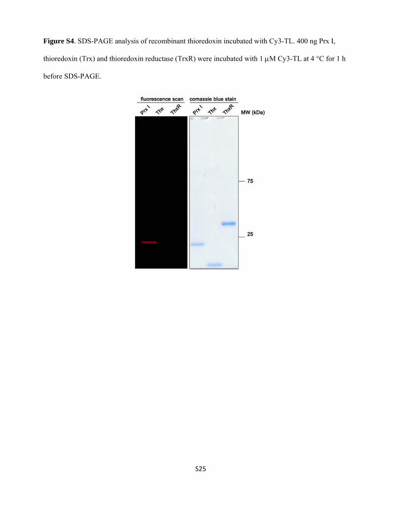
Figure S4. SDS-PAGE analysis of recombinant thioredoxin incubated with Cy3-TL. 400 ng Prx I,
thioredoxin (Trx) and thioredoxin reductase (TrxR) were incubated with 1 M Cy3-TL at 4 °C for 1 h
before SDS-PAGE.

S26
Figure S5. Triplicate western blot analyses of recombinant hPrx I and mutants after incubation with
Biotin-TL.
1st
2nd
3rd
WT C52S C83S C173S C83/173S
Biotin-TL
Prx I
Biotin-TL
Prx I
Biotin-TL
Prx I
S27
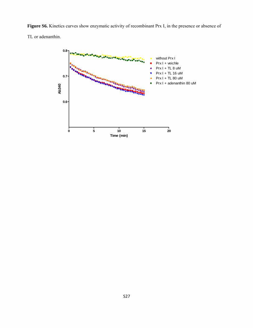
Figure S6. Kinetics curves show enzymatic activity of recombinant Prx I, in the presence or absence of
TL or adenanthin.
Time (min)
Ab
340
0 5 10 15 20
0.6
0.7
0.8
without Prx IPrx I + veichlePrx I + TL 8 uMPrx I + TL 16 uMPrx I + TL 80 uMPrx I + adenanthin 80 uM

S28
Figure S7. Native mass spectrometry analysis on intact wildtype Prx I or mutants in absence or presence
of Cel and WA

S29
Figure S8. LC-MS/MS analysis of tryptic peptide 169HGEVCWAPAGWKPGSDTIKPDVQK190 from
WA-treated Prx I. The ETD fragment ions are manually checked and annotated.
Assignment[M+H]1+
Exact m/z
Experimental m/z
Error (mmu)
z2 259.1527 259.1525 -0.2 z3 358.2211 358.2213 0.2 c4 440.2252 440.2254 0.2 z4 473.2480 473.2478 -0.2 z6 698.3957 698.3949 -0.8 z7 811.4798 811.4764 -3.4 z8 912.5275 912.5261 -1.4 z9 1027.5544 1027.5547 0.3 c9 1424.6919 1424.6920 0.1
[M+3H]3+ 940.4748 940.4810 6.2 [M+4H]4+ 705.6062 705.6129 6.7

S30
Figure S9. Aggregation of MDH in absence of Prx I and in presence of TL, Cel and WA as monitored by
light scattering.

S31
Figure S10. Monitoring aggregation of Prx I in absence of MDH and in presence of TL, Cel and WA by
light scattering.
Table S1. Observed species in native mass spectrometry analysis of intact Prx I monomer and dimer in
absence or presence of TL. hPrx I recombinant protein (10 M) was incubated with DMSO or 40 M TL
at 4 °C overnight. Unbound small molecules were removed using desalting column before MS analysis.
Complex Assignment Measured Mass
Prx I (monomer) 22823.95 ± 3.32 Prx I + TL (monomer) 23183.41 ± 4.93 Prx I + 2TL (monomer) 23544.55 ± 7.55 Prx I (dimer) 45685.39 ± 8.95 Prx I + TL (dimer) 46052.46 ± 4.22 Prx I + 2TL (dimer) 46402.91 ± 6.64

S32
Table S2. HCD MS/MS analysis: a summary of the assigned fragment ion peaks in Figure 2a of a
quaternary charged tryptic peptide, 169HGEVCTLPAGWKPGSDTIKPDVQK190, from the TL-treated Prx
I.
Assignment
[M+H]1+ [M+2H]2+
Exact m/z
Experimental m/z
Error
(mmu)
Exact m/z
Experimental m/z
Error (mmu)
b2 195.0877 195.0880 0.3 b3 324.1302 324.1299 0.3 b4 423.1987 423.1981 -0.6 b5 886.3651 886.3621 -3.0 b6 492.209 492.2197 10.7 b7 1054.4550 1054.4514 -3.6 b8 1111.4765 1111.4723 -4.2 b9 1297.5558 1297.5548 -1.0 b10 1425.6507 1425.6293 -21.4 b14 891.3920 891.3969 5.0
b15-H2O 932.9106 932.9089 -1.6 b16-H2O 989.4526 989.4588 6.3
b16 998.4579 998.4583 0.5 y2 275.1714 275.1701 -1.3 y3 374.2398 374.2397 -0.1 y5 586.3195 586.3205 1.0
y5-H2O 568.3089 568.3098 0.9 y6 714.4145 714.4135 -1.0 y7 827.4985 827.5103 11.8 y8 464.7731 464.776 2.9 y12 1284.6794 1284.6689 -10.5 642.8397 642.8427 3.0
y12-H2O 633.8344 633.8408 6.4 y13 706.8872 706.8895 2.3 y14 799.9269 799.928 1.2 y16 863.9562 863.9618 5.7
y16-NH3 855.4429 855.4129 -29.9 y17 912.4825 912.4859 3.4
[M+4H]4+ 678.0807 678.0865 5.8

S33
Table S3. HCD MS/MS analysis: summary of the assigned fragment ion peaks in Figure 4d of a tryptic
peptides, 79DSHFCWAHLAWVNTPK92, from the WA-treated Prx I.
Assignment
[M+H]1+ [M+2H]2+
Exact m/z
Experimental m/z
Error (mmu)
Exact m/z
Experimental m/z
Error (mmu)
y2 244.1656 244.165543 -0.1 122.5864 y3 345.2132 345.212891 -0.3 173.1102 y4 459.2562 459.256042 -0.2 230.1317 y5 558.3246 558.32489 0.3 279.6659 y6 744.4039 744.403564 -0.3 372.70555 y7 815.4410 815.440308 -0.7 408.2241 y8 928.5251 928.523926 -1.2 464.76615 464.765594 -0.6 b6-H2O 1179.5179 590.2626 590.262329 -0.2 b6 1197.5285 1197.526001 -2.5 599.2679 599.267761 -0.1 b7 1310.6126 655.8099 655.809387 -0.5 b8 1381.6497 691.3285 691.327881 -0.6 y10 1638.8600 819.9336 819.93512 1.5 b12 1881.8880 941.4476 941.444153 -3.4 y12 1922.9873 961.99725 961.994507 -2.7 y13-H2O 1992.0088 1992.011353 2.6 996.508 [ y13]3+ 670.6779 670.677124 -0.8 [M+2H]2+ 1063.0268 1062.969604 -57.1 [M+3H]3+-H2O 703.0167 703.015503 -1.2 [M+3H]3+ 709.02023 709.019836 -0.4
References
1. C. K. Frese, A. F. M. Altelaar, M. L. Hennrich, D. Nolting, M. Zeller, J. Griep-Raming, A. J. R. Heck and S. Mohammed, J. Proteome Res., 2011, 10, 2377-2388.
2. R. J. Rose, A. F. Labrijn, E. T. J. van den Bremer, S. Loverix, I. Lasters, P. H. C. van Berkel, J. G. J. van de Winkel, J. Schuurman, P. W. H. I. Parren and A. J. R. Heck, Structure, 2011, 19, 1274-1282.
3. J.-A. Kim, S. Park, K. Kim, S. G. Rhee and S. W. Kang, Anal. Biochem. , 2005, 338, 216-223. 4. P. Goloubinoff, A. Mogk, A. P. B. Zvi, T. Tomoyasu and B. Bukau, PNAS 1999, 96, 13732-
13737.