ichthyosporidium weissii n. sp. (microsporidia) infecting the arrow goby (clevelandia ios)
TRANSCRIPT

Ichthyosporidium weissii n. sp. (Microsporidia) Infecting the Arrow Goby(Clevelandia ios)
JUSTIN SANDERS,aMARK S. MYERS,
bLARS TOMANEK,
cANN CALI,
dPETER M. TAKVORIAN
d,e
and MICHAEL L. KENTa
aDepartment of Microbiology, Oregon State University, 220 Nash Hall, Corvallis, Oregon, 97331, andbEcotoxicology and Environmental Fish Health Program, Environmental Conservation Division, Northwest Fisheries Science Center,
NOAA Fisheries, 2725 Montlake Boulevard East, Seattle, Washington, 98112, andcEnvironmental Proteomics Laboratory, Center for Coastal Marine Sciences, California Polytechnic State University, San Luis Obispo,
California, 93407-0401, anddDepartment of Biological Sciences, Rutgers University, 195 University Avenue, Newark, New Jersey, 07102, and
eDepartment Pathology, Albert Einstein College of Medicine, Bronx, New York, 10461
ABSTRACT. Gonadal infections by a novel microsporidium were discovered in 34% (13/38) of arrow gobies, Clevelandia ios,sampled over a 3-yr period from Morro Bay Marina in Morro Bay, California. Gonadal tumors had been reported in arrowgobies from this geographic area. The infected gonads, found primarily in females, typically appeared grossly as large, white-grayfirm and lobulated masses. Histological examination revealed large, multilobate xenomas within the ovaries and no evidence ofneoplasia. Typical of the genus Ichthyosporidium, the large xenomas were filled with developmental stages and pleomorphic spores.Wet mount preparations showed two general spore types: microspores with mean length of 6.2 (7.0–4.9, SD = 0.6, N = 20) lmand mean width of 4.3 (5.3–2.9, SD = 0.8) lm; and less numerous macrospores with mean length of 8.5 (10.1–7.1, SD = 1.0,N = 10) lm and mean width of 5.5 (6.2–4.8, SD = 0.5) lm. Transmission electron microscopy demonstrated stages consistent withthe genus and 35–50 turns of the polar filament. Small subunit rDNA gene sequence analysis revealed that the parasite fromarrow gobies was most closely related to, but distinct from Ichthyosporidium sp. based on sequences available in GenBank. Weconclude that this microsporidium represents a new species of Ichthyosporidium, the first species of this genus described from amember of the family Gobiidae and from the Pacific Ocean.
Key Words. Electron microscopy, gonads, neoplasia, new species, parasite, phylogeny.
T HE first description of an Ichthyosporidium species was byThelohan (1895) who described in the corkwing wrasse
Crenilabrus melops, the presence of large parasitic masses thatoccupied most of the abdominal cavity and appeared to origi-nate from the renal connective tissue. Considering the parasiteto belong to the Microsporidia, Thelohan (1895) named theparasite Glugea gigantea but did not describe developmentalstages of the parasite. The genus Ichthyosporidium was latererected by Caullery and Mesnil (1905) who described twonamed species: Ichthyosporidium gasterophilum, which waslater reassigned to the genus Ichthyophonus by Sprague (1965),and Ichthyosporidium phymogenes, with both genera causingparasitic infections in marine fish of the family Labridae. Theynoted the close similarity between I. phymogenes and G. gigan-tea described by Thelohan (1895) and observed that they werefound in the same fish host, C. melops. These parasites werealso considered to be haplosporidians based on the resem-blance of the sporoblasts to developmental stages of haplospo-ridians. Numerous later authors also considered theseparasites to be members of the Haplosporidia (Kudo 1966).
Swellengrebel (1911) described developmental stages of aparasite from C. melops, which he noted as likely being thesame as that described by Thelohan (1895), but placed itwithin the genus Pleistophora due to the formation of a pansp-oroblast. Believing this organism to be a microsporidium, hewas unable to germinate spores to demonstrate the presenceof a polar filament. Later, Sprague and Vernick (1968) demon-strated the presence of polar filaments in spores of this micros-poridium from Swellengrebel’s material by the use of electron
microscopy and the periodic acid-Schiff (PAS) stain by lightmicroscopy, firmly placing these parasites within the Micros-poridia. Sprague and Vernick (1968) also noted that theorganism in question, Pleistophora gigantea, was synonymouswith Ichthyosporidium giganteum.
Infections by members of the genus Ichthyosporidium arecharacterized by the formation of large, multilobate xenomasin infected host tissue. These xenomas are distinguished fromthe cell hypertrophy-type xenoma formed by species in thegenus Glugea in that they appear to be produced by the coa-lescence of multiple infected, hypertrophic cells, forming asyncytial-type xenoma. This type of xenoma does not havehost nuclei in the periphery, is relatively devoid of host organ-elles, and contains a “fibro-granular layer” with no distinctinner boundary. It is limited on the outer surface by microvil-lus-like projections (Canning et al. 1986; Lom and Dykova2005). We found similar xenomas in the gonads of arrowgobies, Clevelandia ios, collected from one site in Morro Bay,California. These xenomas were initially thought to be gona-dal neoplasms. However, we concluded that they are actuallythe result of infection by a novel species of the genus Ichthyos-poridium. This finding represents the first description of Ich-thyosporidium in a member of the family Gobiidae and alsothe first described from the Pacific Ocean.
MATERIALS AND METHODS
Samples, collection, and histological examination. A total of1,115 arrow gobies were collected from Morro Bay, Califor-nia, over a 3-yr period, 2007–2009. Thirty-eight of these fishfrom one site, Morro Bay Marina, were examined by histol-ogy. We also examined 60 arrow gobies collected from thissame site in November 2009. Fish were acclimated in the labo-ratory for 2 wk and held for up to 60 d further at CaliforniaPolytechnic State University, San Luis Obispo. Fish were
Corresponding Author: Justin Sanders, Department of Microbiol-ogy, Oregon State University, 220 Nash Hall, Corvallis, Oregon97331—Telephone number: +1 541 737 1858; FAX number:+1 541 737 0496; e-mail: [email protected]
J. Eukaryot. Microbiol., 0(0), 2012 pp. 1–10© 2012 The Author(s)Journal of Eukaryotic Microbiology © 2012 International Society of ProtistologistsDOI: 10.1111/j.1550-7408.2012.00619.x
1
Published bythe International Society of ProtistologistsEukaryotic Microbiology
The Journal of

euthanized and fixed in 10% (v/v) neutral buffered formalin.After fixation, fish were processed for histology, stained withhematoxylin and eosin (H&E), and examined by light micros-copy.
Three additional female fish were collected from Morro BayMarina in 2011. Ovarian tissue from two infected female fishwas examined by wet mount and 30 microsporidian sporestotal, 20 microspores and 10 macrospores, were measuredusing SPOT Advanced imaging software version 4.0.9 (Diag-nostic Instruments, Sterling Heights, MI). One fish was fixedin Dietrich’s fixative and decalcified using CalExII (Fisher Sci-entific, Fair Lawn, NJ). Fixed and decalcified tissues were pro-cessed for histology. Sections were cut at 5 lm and stainedwith H&E, PAS, and Luna (Luna 1968) stains.
Ultrastructure. Ovarian tissue from two additional infectedfemale arrow gobies with grossly distended abdomens werecut into approximately 2–3 mm sections and placed in fixative;2.5% (v/v) glutaraldehyde in 0.1 M sodium cacodylate bufferat 4 °C overnight. Tissues were removed and post-fixed in 1%(w/v) osmium tetroxide in 0.2 M sodium cacodylate buffer for1 h prior to embedding in epoxy resin. Thick sections werecut at 0.5 lm and stained with toluidine blue. Ultrathin sec-tions (i.e. 30–40 nm) were cut and stained for 1.5 h in 5% (w/v) aqueous uranyl acetate solution and then stained with leadcitrate. Transmission electron microscopy was performedusing either a Philips CM12 scanning transmission electronmicroscope (Philips, Eindhoven, the Netherlands) or a FEITecnai 12 transmission electron microscope (Phillips, Hills-boro, OR).
DNA extraction, PCR amplification, and sequencing of smallsubunit ribosomal RNA (SSU rDNA) gene. Ovarian tissuefrom one female fish displaying gross gonadal lesions andmicrosporidian spores present as seen by wet mount wasremoved and the DNA extracted using the QIAgen Blood andTissue Extraction kit (QIAgen, Valencia, CA) according to themanufacturer’s protocol. A second microsporidium resemblingKabatana sp. was found in the skeletal muscle of 7 of the 60fish collected at the Marina site, held in the laboratory at SanLuis Obispo, and examined by light microscopy. Hence, skele-tal muscle from a separate fish that showed the presence ofthis microspordium was also collected and DNA was extractedusing the above method.
PCR was performed using the general microsporidian ribo-somal primers 18F (5′-GAAAATTACCGGAGCCTGAAGTC-3′) and 580r (5′-GGTCCGTGTTTCAAGACGG-3′) toamplify the 5′-region of the SSU rDNA gene, ITS1 region,and partial large-subunit ribosomal DNA gene segments. Allreactions were performed in 50-ll volumes using the PlatinumPCR Supermix (Invitrogen, Carlsbad, CA), 0.9 mmol of eachprimer, and 5 ll of each DNA extraction. Amplifications wereperformed on a Peltier 200 thermocycler (MJ Research,Watertown, MA) with an initial denaturation at 94 °C for2 min, 40 cycles of 94 °C for 30 s, 55 °C for 30 s, and 72 °Cfor 2 min with a final extension at 72 °C for 10 min. Theresulting PCR product was both sequenced directly andcloned into TOPO TA Cloning vectors (Invitrogen). Threeclones of the gonadal microsporidium and one from the mus-cle parasite were sequenced in both directions using primersflanking the inserted sequence. The primers 530f (5′-GTGCCAGCAGCCGCGG-3′), 1047r (5′-AACGGCCATGCACCAC-3′), and 1061f (5′-CACCAGGTTGATTCTGCC-3′)were used to sequence internal portions of the cloned SSUrDNA gene. All DNA analyzed in the study was sequencedon an ABI Prism® 3730 Genetic Analyzer with the BigDye®
Terminator v.3.1 Cycle Sequencing Kit (Applied Biosystems,Foster City, CA).
Phylogenetic analyses. The sequences were compared withthose in the National Center for Biotechnology Information’sGenBank database using the BLASTN program (Altschulet al. 1990). Multiple sequence alignment was performed usingCLUSTALW in the software package MEGA 5 (Tamuraet al. 2011). Poorly aligned or ambiguous regions of the align-ment were removed using the Gblocks program version 0.91b(Castresana 2000) on the webserver available at http://molevol.cmima.csic.es/castresana/Gblocks_server.html. The results ofthe multiple sequence alignment were analyzed using thejModeltest program version 0.1.1 (Posada 2008) to determinethe most likely model of nucleotide substitution. Based on theresults of the jModeltest analysis, phylogenetic reconstructionusing the maximum likelihood method was performed usingPhyML (Guindon et al. 2010) on the webserver at http://www.atgc-montpellier.fr/phyml/ using the generalized time-reversible model with gamma-distributed rate variation amongsites (GTR+G). The analysis was run using 500 bootstrap rep-licates to test the robustness of the resulting tree. Bayesiananalysis was performed using the software MrBayes version3.1.2 also using the GTR model with a gamma-distributedrate variation across sites. The analysis was run for 1,000,000replicates and sampled every 1,000 generations.
A second multiple sequence alignment was performed usinga region from nucleotides 507 to 867 of the 16S SSU rRNAgene sequence obtained from the arrow goby ovarian tissue,the same region from Ichthyosporidium sp. (GenBank acces-sion number L39110), and sequence from I. giganteum (Gen-Bank accession number L13293). This alignment was used toanalyze the pairwise distance (p-distance) between the threesequences using the MEGA 5 software.
RESULTS
Prevalence. A total of 5.7% (64/1115) of the fish from theoverall collection exhibited prominently distended abdomensat gross examination (Fig. 1A). Upon dissection, large, elon-gate, whitish lesions in the gonadal region were observed(Fig. 1B). Histological examination of 38 fish from a singlesite, Morro Bay Marina, revealed the presence of a microspo-ridian with a prevalence of 34.2% (13/38), with all affectedfish being female. The overall prevalence of infection of a sub-sequent sampling of arrow gobies for a laboratory experimentin 2009 from the same site was 15% (9/60), with one singlemale being affected.
Light microscopy/histology. Refractile spores with indistinctposterior vacuoles were seen in wet mounts prepared frominfected ovarian tissue (Fig. 3–8). Spores were ovoid to pyri-form. They were very pleomorphic, but generally consisted oftwo sizes, with numerous microspores (Fig. 3–8) with a meanlength of 6.2 (7.0–4.9, SD = 0.6, N = 20) lm and a meanwidth of 4.3 (5.3–2.9, SD = 0.8) lm and occasional macrosp-ores (Fig. 6–8) with a mean length of 8.5 (10.1–7.1, SD = 1.0,N = 10) lm and a mean width of 5.5 (6.2–4.8, SD = 0.5) lm.
Histological examination of infected fish (Fig. 2, 9–14)showed the parasite to be confined to cells of the ovarian stro-mal tissue in females and the stromal tissue of the testes in thesingle infected male observed. Parasite-containing lesions oftwo types were observed: large, multilobate xenomas sur-rounded by a common fibrous capsule (Fig. 2) with appar-ently normal ovarian tissue and developing oocytesinterspersed, and smaller rounded cyst-like lesions, appearingto be single, infected and hypertrophic cells with a less definedlaminated capsule (Fig. 9). In some sections, numerous smallerfoci were observed proximal to larger, lobular xenomas, sug-gesting the possibility of autoinfection (Fig. 9).
2 J. EUKARYOT. MICROBIOL., 0, NO. 0, MARCH 2012

Newly developing small xenomas contained only prolifera-tive stages with no apparent spores (Fig. 9), whereas other,more mature xenomas, contained spores dispersed throughoutthe xenoma and intermixed with large numbers of presporog-onic forms (Fig. 10–14). Sections stained with PAS showedxenomas with centrally located spores containing PAS-positive
structures that appeared to be polar filaments and apical polarcaps (Fig. 11, 12). Few, presumably more mature spores,located toward the center of the xenomas, stained brick redwith the Luna stain (Fig. 13, 14).
Ultrastructure. Transmission electron microscopy (TEM) ofinfected ovaries showed that spores (Fig. 15–19) and other
Fig. 1–2. Macroscopic and low-magnification microscopic images of gonadal lesion in female arrow goby, Clevelandia ios. Figure 1 courtesyof Sarah Johnson, California Polytechnic State University, San Luis Obispo, California. 1(A). Female arrow goby, C. ios, with grossly distendedabdomen. Scale bar = 1 mm. 1(B). The same goby with the skin removed from the abdomen. Scale bar = 1 mm. 2. Hematoxylin and eosin(H&E)-stained histological section of an ovary infected with Ichthyosporidium weissii n. sp. Large, multilobate xenomas (X) can be seen withdeveloping ovarian follicles interspersed (arrows) and surrounding the infected area. Scale bar = 100 lm.
SANDERS ET AL.—A NEW SPECIES OF ICHTHYOSPORIDIUM 3

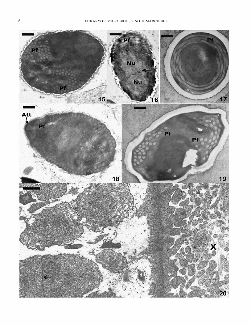
developmental stages (Fig. 20) were diplokaryotic. Sporontswith diplokaryotic nuclei were dispersed throughout the xeno-mas. The xenoma wall was distinct and dense, and the outerlayer consisted of numerous, long, finger-projections (Fig. 20).Mature spores were dense, contained 36–50 turns of the isofi-lar polar filament arranged in four to five rows, and had aprominent endospore wall (Fig. 15–19).
Kabatana sp. Subsequent sampling from the same site of 60arrow gobies, which were held in the laboratory for experi-mentation, revealed the presence of another microsporidium,resembling a Kabatana sp. It was observed in the skeletal mus-cle of seven fish. These parasites did not form xenomas, butrather were limited to development within the sarcoplasm ofskeletal muscle cells (Fig. 21).
Molecular phylogeny. The total length of amplified ribo-somal DNA was 1,839 bp and has been deposited in the NCBIGenBank database as accession number JQ062988. The topthree hits returned by the BLASTN search of the NCBI nonre-dundant database were Ichthyosporidium sp., Pseudoloma neu-rophilia, and Loma embiotocia with percent identities of91.7%, 90.6%, and 89.7%, respectively. Phylogenetic analysis
using both maximum likelihood and Bayesian methods placethe new microsporidium in a clade with Ichthyosporidium sp.,and sister to P. neurophilia (Fig. 22). Pairwise comparison ofthe sequence from arrow goby with the partial sequence(375 bp) obtained from I. giganteum (NCBI accession numberL13293) from Leiostomus xanthurus and that of Ichthyosporidi-um sp. from the same host also showed 10.1% (p-distance) and10.2% nucleotide difference, respectively, within this region ofthe gene. In contrast, the sequence of Ichthyosporidium sp. andI. giganteum had 0.8% (p-distance) nucleotide difference in thisregion of the gene. The sequence obtained from the microspo-ridium from the skeletal muscle has been deposited in theNCBI GenBank database as accession number JQ062989. It issimilar to, but distinct from, other species of the genus Kaba-tana, with 2.8% (p-distance) nucleotide difference between thatand the SSU rDNA gene sequence of K. newberryi.
DISCUSSION
Ichthyosporidium giganteum has been described fromC. melops along the Atlantic coast of France, Crenilabrum
Fig. 3–8. Light micrographs of spores of Ichthyosporidium weissii n. sp. obtained from wet mount preparations of ovarian tissue of aninfected female arrow goby, Clevelandia ios. 3. Spores viewed by Nomarski phase interference. Scale bar = 10 lm. 4. Pleiomorphic spores withan aberrant, triangular-shaped spore (X). Scale bar = 10 lm. 5. Spores containing visible coiled polar filaments (arrows). Scale bar = 10 lm. 6.Numerous microspores with a single macrospore present (M). Nomarski phase interference. Scale bar = 10 lm. 7. Several pleiomorphic sporeswith a single macrospore (M). Nomarski phase interference. Scale bar = 10 lm. 8. One microspore containing a visible coiled polar filament(arrow) with a single macrospore (M). Viewed by Nomarski phase interference. Scale bar = 10 lm.
4 J. EUKARYOT. MICROBIOL., 0, NO. 0, MARCH 2012

Fig. 9–14. Histological sections of gonadal tissue from female arrow gobies, Clevelandia ios, infected with the microsporidium, Ichthyospori-dium weissii n. sp. 9. Hematoxylin and eosin (H&E) stained section with several cyst-like foci of infection (arrows). The margin of a maturexenoma (X) is also visible. Scale bar = 10 lm. 10. H&E stained section of the periphery of a large, multilobate xenoma. Note the fibrouscapsule (arrow) surrounding the xenoma, which is filled with proliferative and spore stages of the parasite. Scale bar = 10 lm. 11. Periodic acid-Schiff (PAS)-stained section of ovarian xenoma with mature spores showing PAS-positive structures. Scale bar = 10 lm. 12. PAS-stained sectionat higher magnification with arrows indicating examples of PAS-positive polar structures. Scale bar = 10 lm. 13. Luna-stained section of thesame xenoma as in Fig. 11. Mature spores stain red while presporogonic stages of the parasite appear blue. Scale bar = 10 lm. 14. Highmagnification of Luna-stained section of xenoma. Mature spores (arrow) stain red. Scale bar = 10 lm.
SANDERS ET AL.—A NEW SPECIES OF ICHTHYOSPORIDIUM 5
6 J. EUKARYOT. MICROBIOL., 0, NO. 0, MARCH 2012

ocellatus in the Black Sea, Ctenolabrus rupestris off the Atlanticcoast of Portugal (Casal and Azevedo 1995), and L. xanthurusalong the Atlantic coast of the United States (Schwartz 1963).The only other described species of Ichthyosporidium,Ichthyosporidium hertwigi, has been described from the gills ofCrenilabrus tinca on the Crimean coast of the Black Sea(Swarczewsky 1914). To our knowledge, this is the first descrip-tion of a species of Ichthyosporidium from the Pacific ocean.
The morphology and histological appearance of the micros-poridium observed in the arrow goby C. ios is consistent withthat previously described for Ichthyosporidium species withregard to the formation of large, irregular, multilobate xeno-mas surrounded by a thick fibrous capsule and the presence ofdiplokaryotic, developmental and mature spore stages. Ichthy-osporidium giganteum infections have been described as beinglocalized to the subcutaneous connective tissue, especiallyfibroblasts, of the anterior abdomen (Sprague and Hussey1980), while the single description of I. hertwigi describes thesite of infection as being in the connective tissue of the gills(Swarczewsky 1914). The microsporidium described hereappears to infect connective tissue cells of the ovigerousstroma, and rarely the intratesticular stroma. The xenomaformed by I. giganteum has been described as having twotypes. (1) A cyst-like structure consisting of a single infected,hypertrophic cell. Proliferating, merogonic stages of the para-site are exclusively present with an absence of spore stages. (2)A lobular, large and irregularly shaped xenoma with prolifer-ating stages and mature spores present. This multicystic lesionmay be formed by the coalescence of several foci of infectionto form a syncytium (Sprague and Hussey 1980). In the pres-ent study, xenomas resembling both of these types wereobserved in arrow gobies infected with Ichthyosporidium weiss-ii n. sp. While the original description of Ichthyosporidium sp.by Schwartz (1963) described the spores as being uniform in
shape and 7 9 4 lm, Sprague (1966) noted the presence of afew larger macrospores in one cyst in a subsequent descriptionfrom the same set of slides. Sprague and Hussey (1980) tenta-tively considered the Ichthyosporidium sp. described by Sch-wartz (1963) and I. giganteum to belong to the same speciesbased on their morphological features. They did not explicitlydescribe the spore morphology of I. giganteum, instead refer-ring to the previous description given by Sprague and Vernick(1974). However, the presence of macrospores was not notedin this latter description. We speculate that this could be dueto the very low number of macrospores produced by I. gigant-eum. The PAS-positive structures within spores of I. weissii,presumably the polar filament and a mass at the anterior endof the spore, seen in several mature spores are also consistentwith the findings of Sprague (1966). Peterson et al. (2011)demonstrated that the Luna stain, which stains chitin, is usefulfor demonstrating mature spores of microsporidia. This stainwas very useful for distinguishing mature spores in histologicalsections of these infected arrow gobies, given that they arequite pleomorphic, in contrast to the uniformly sized sporesdescribed for I. giganteum.
There are two reports describing the ultrastructure of I.giganteum spores (Casal and Azevedo 1995; Sprague and Ver-nick 1974). Spore measurements varied considerably betweenthese two descriptions with Sprague and Vernick (1974)reporting average dimensions of 5.9 9 4 lm and Casal andAzevedo (1995) reporting average dimensions of7.26 9 5.16 lm. In addition, Sprague and Vernick (1974)described the spores as being uniform in size, in contrast tothe spores seen in the present study. The number of coils ofthe polar filament has been used as a criterion in taxonomyfor many microsporidia. However, this character appears tobe quite variable for members of the genus Ichthyosporidium.Sprague et al. (1992) and Casal and Azevedo (1995) reportedapproximately 32 and 43 turns in 4–5 rows, respectively, for I.giganteum, and we found that the number of coils observed inI. weissii n. sp. ranged from 35 to well over 50. The numberof coils of the polar filament is much greater than what hasbeen reported for most other microsporidia, and the variabil-ity observed in the spores in our study and that reported indescriptions of I. giganteum suggests that this characteristic isnot suitable for taxonomic differentiation of species within thegenus Ichthyosporidium. The ultrastructure of I. weissii n. sp.was also consistent with the previous reports on Ichthyospori-dium, with the presence of diplokarya in both spores andpresporogonic stages. Moreover, the xenoma wall was alsosimilar to that of I. giganteum, with distinctive, long finger-likeprojections extending outward. The genus description of Ich-thyosporidium includes tetrasporogonic development. Carefulexamination of wet mount material, histological sections, andimages obtained by electron microscopy did not allow us toconfirm this character. However, the mature spores do notappear to occur in aggregates, particularly well demonstratedwith the Luna stain, which only stains positive with maturespores (Fig. 13, 14).
Phylogenetic analyses of the SSU rDNA genesequence obtained from this organism placed it in the genus
Fig. 21. Hematoxylin and eosin-stained histological section ofskeletal muscle of an arrow goby, Clevelandia ios, infected with Kaba-tana sp., a myocyte-infecting microsporidian. Scale bar = 10 lm.
Fig. 15–20. Transmission electron microscopy of spores and developmental stages of Ichthyosporidium weissii n. sp. 15. Parasagittal viewwith approximately 50 turns of the polar filament (Pf) visible. Scale bar = 0.5 lm. 16. Parasagittal section of a spore with a diplokaryoticnucleus (Nu) visible in the anterior portion (arrow = division between diplokarya). The coiled polar filament (Pf) is visible at one end of thespore. Scale bar = 0.5 lm. 17. Transverse section of a spore with the polar filament (Pf) arranged in four distinct rows. Scale bar = 0.5 lm.18. Mature spore showing the manubroid portion of the polar filament (Pf) and the anterior attachment complex (Att). Scale bar = 0.5 lm.19. Spore with visible cross sections of the isofilar polar filament (Pf). Scale bar = 0.5 lm. 20. Transmission electron microscopy of the marginof a xenoma with sporonts. Arrow = division between diplokarya. X = xenoma wall with numerous projections. Scale bar = 1 lm.
SANDERS ET AL.—A NEW SPECIES OF ICHTHYOSPORIDIUM 7

Ichthyosporidium Caullery & Mesnil, 1905. The most completerDNA sequence of an Ichthyosporidium species available inGenBank before the present study was provided for Ichthyos-poridium sp. (i.e. GenBank accession number L39110) fromthe fish host L. xanthurus (Baker et al. 1995). A partial SSUrDNA gene sequence of I. giganteum (i.e. 404 bp, GenBankaccession number L13293), also obtained from L. xanthurus,is available (Vossbrinck et al. 1993). Pairwise alignment andanalysis of the three putative Ichthyosporidium species suggeststhat the sequences of both Ichthyosporidium sp. and I. giganteumare related to I. weissii n. sp. at the genus level, and clustertogether in a monophyletic clade. The two sequences fromL. xanthurus were almost identical over corresponding regions,and hence presumably both represent I. giganteum. However,it would be very useful to obtain sequence from the type host,C. rupestris. The closest relative to this clade is P. neurophilia,
a microsporidium of the zebrafish Danio rerio. These two gen-era share few similarities in development or spore structure(Cali et al. 2011).
Casal and Azevedo (1995) described C. rupestris infectedwith I. giganteum as having grossly visible abdominal swelling,similar to that seen in C. ios infected with I. weissii n. sp., inthe present study. They noted that these prominent lesionsgrossly resembled tumors and this similarity likely contributedto the initial misdiagnosis of the lesions found in the arrowgobies as resembling ovarian neoplasms. Sprague (1969) usedinfections in L. xanthurus by Ichthyosporidium sp. as an exam-ple to describe the formation of “tumors” by microsporidia.Various other infectious agents, particularly protists, causemacroscopic lesions suggestive of neoplasia (Harshbarger1984), demonstrating that diagnosis of neoplasia in wild fishesmust include proper histological interpretations.
Fig. 22. Phylogenetic tree of microsporidian small subunit (SSU) rRNA gene sequences inferred by Bayesian analysis (BI), showing theplacement of Ichthyosporidium weissii n. sp. (in bold). The placement of Kabatana sp. (in bold) sequenced from the skeletal muscle of an arrowgoby is also shown. Numbers at nodes represent BI posterior probability support and bootstrap values (out of 500 replicates) from maximum-likelihood analysis, respectively.
8 J. EUKARYOT. MICROBIOL., 0, NO. 0, MARCH 2012

The microsporidium observed in the skeletal muscle of sev-eral of these fish appears most closely related to the genusKabatana based on phylogenetic analysis of the SSU rDNAgene sequence. The microsporidium, K. newberryi, infectingthe skeletal muscle of another member of the family Gobiidae,the tidewater goby Eucyclogobius newberryi, has beendescribed from fish collected in Big Lagoon, HumboldtCounty, California (McGourty et al. 2007), located consider-ably north of Morro Bay. The difference in host and SSUrDNA gene sequence between K. newberryi and the microspo-ridium found in the skeletal muscle of arrow gobies describedherein suggests this is a novel species of Kabatana. We electednot to assign a species name to this organism, as we did nothave ultrastructural data.
Phylogenetic analysis, the presence of a diplokaryoticnucleus in both developmental and spore stages of the para-site, and the formation of a multilobate, syncytial xeno-ma support the placement of this microsporidium in thegenus Ichthyosporidium. Based on the geographic location,host species, and differences in the SSU rDNA between thisand the other two sequences of Ichthyosporidium availablein the GenBank database, we conclude that this mi-crosporidium represents a novel species of the genus Ichthy-osporidium.
TAXONOMIC SUMMARY
Phylum Microsporidia Balbiani, 1882Family Ichthyosporidiidae Sprague, Becnel & Hazard, 1992Genus Ichthyosporidium Caullery & Mesnil, 1905Ichthyosporidium weissii n. sp.Diagnosis. With characters of the genus. Meronts, sporonts,
and spores; diplokaryotic, developing within large xenomas.Spores, pleomorphic, ovoid to pyriform. Microspores: length6.2 (7.0–4.9) lm, width 4.3 (5.3–2.9) lm. Macrospores: length8.5 (10.1–7.1) lm, width 5.5 (6.2–4.8) lm.
Type locality. Morro Bay Marina, California (35°20.7′N,120°50.7′W)
Type host. Arrow goby Clevelandia ios (Teleostei, Gobiidae).Site of infection. Cytoplasm of connective tissue cells of the
gonadal stroma in male and female fish.Prevalence. Thirty-four percent of female fish (n = 38) from
one sampling site in type locality were affected.Deposition of type material. Slides of histological sections
from whole fish were deposited in the collections of theQueensland Museum, Brisbane, Australia. (Nos. G465498,G465499, G465500).
Gene sequence. Sequence of the SSU rRNA gene, ITS, andpartial large-subunit rRNA gene was deposited as GenBankAccession JQ062988.
Etymology. The species is named after the prominent pro-tistologist, Dr. Louis Weiss, USA.
ACKNOWLEDGMENTS
This study was supported by grants from the National Insti-tutes of Health (NIH NCRR 5R24RR017386-02 and NIHNCRR P40 RR12546-03S1). We thank Jennifer Oquendo andSarah Johnson for fish collection, Teresa Sawyer, OregonState University Electron Microscopy Facility, for TEM speci-men processing and support, the OSU Veterinary DiagnosticsLaboratory and the UC Davis Veterinary Diagnostics Labora-tory for histological slide preparation, and Dr. T. Peterson,Oregon State University, for helpful comments and review ofthis manuscript.
LITERATURE CITED
Altschul, S. F., Gish, W., Miller, W., Myers, E. W. & Lipman, D. J.1990. Basic local alignment search tool. J. Mol. Biol., 215:403–410.
Baker, M. D., Vossbrinck, C. R., Didier, E. S., Maddox, J. V. &Shadduck, J. A. 1995. Small subunit ribosomal DNA phylogeny ofvarious Microsporidia with emphasis on AIDS related forms. J.Eukaryot. Microbiol., 42:564–570.
Cali, A., Kent, M. L., Sanders, J. L., Pau, C. & Takvorian, P. M.2011. Development, ultrastructural pathology, and taxonomic revi-sion of the microsporidial genus, Pseudoloma and its type speciesPseudoloma neurophilia, in skeletal muscle and nervous tissue ofexperimentally infected zebrafish, Danio rerio. J. Eukaryot. Micro-biol., 59:40–48.
Canning, E. U., Lom, J. & Dykova, I. 1986. The Microsporidia ofVertebrates. Academic Press, London, England.
Casal, G. & Azevedo, C. 1995. New ultrastructural data on themicrosporidian Ichthyosporidium giganteum infecting the marineteleostean fish Ctenolabrus rupestris (L.). J. Fish Dis., 18:191–194.
Castresana, J. 2000. Selection of conserved blocks from multiplealignments for their use in phylogenetic analysis. Mol. Biol. Evol.,17:540–552.
Caullery, M. & Mesnil, F. 1905. Sur des haplosporidies parasites depoissons marins. C. R. Soc. Biol., 58:640–643.
Guindon, S., Dufayard, J. F., Lefort, V., Anisimova, M., Hordijk, W.& Gascuel, O. 2010. New algorithms and methods to estimate max-imum-likelihood phylogenies: assessing the performance of PhyML3.0. Syst. Biol., 59:307–321.
Harshbarger, J. C. 1984. Pseudoneoplasms in ectothermic animals.Natl. Cancer Inst. Monogr., 65:251–273.
Kudo, R. 1966. Protozoology, 5th ed. Charles C. Thomas, Spring-field, Illinois, 762–773 p.
Lom, J. & Dykova, I. 2005. Microsporidian xenomas in fish seen inwider perspective. Folia Parasit., 52:69–81.
Luna, L. G. 1968. Manual of Histologic Staining Methods of theArmed Forces Institute of Pathology, 3rd ed. McGraw-Hill, NewYork, NY. p. 111–112.
McGourty, K. R., Kinziger, A. P., Hendrickson, G. L., Goldsmith,G. H., Casal, G. & Azevedo, C. 2007. A new microsporidian infect-ing the musculature of the endangered tidewater goby (Gobiidae).J. Parasitol., 93:655–660.
Peterson, T. S., Spitsbergen, J. M., Feist, S. W. & Kent, M. L. 2011.The Luna stain, an improved selective stain for detection of micro-sporidian spores in histologic sections. Dis. Aquat. Org., 95:175–180.
Posada, D. 2008. jModelTest: phylogenetic model averaging. Mol.Biol. Evol., 25:1253–1256.
Schwartz, F. J. 1963. A new Ichthyosporidium parasite of the spot(Leiostomus xanthurus)—a possible answer to recent oyster mortali-ties. Progr. Fish Cult., 25:181.
Sprague, V. 1965. Ichthyosporidium Caullery and Mesnil, 1905, thename of a genus of fungi or a genus of sporozoans? Syst. Zool.,14:110–114.
Sprague, V. 1966. Ichthyosporidium sp. Schwartz, 1963, parasite of thefish Leiostomus xanthurus, is a microsporidian. J. Eukaryot. Micro-biol., 13:356–358.
Sprague, V. 1969. Microsporidia and tumors, with particular referenceto the lesion associated with Ichthyosporidium sp. Nat. Cancer.Inst., 31:237–249.
Sprague, V. & Hussey, K. L. 1980. Observations on Ichthyosporidiumgiganteum (Microsporida) with particular reference to the host-par-asite relations during merogony. J. Protozool., 27:169–175.
Sprague, V. & Vernick, S. H. 1968. Observations on the spores ofPleistophora gigantea (Thelohan, 1895) Swellengrebel, 1911, amicrosporidian parasite of the fish Crenilabrus melops. J. Eukaryot.Microbiol., 15:662–665.
Sprague, V. & Vernick, S. H. 1974. Fine structure of the cyst andsome sporulation stages of Ichthyosporidium (Microsporida). J.Eukaryot. Microbiol., 21:667–677.
Sprague, V., Becnel, J. J. & Hazard, E. I. 1992. Taxonomy of phylumMicrospora. Crit. Rev., 18:285–395.
Swarczewsky, B. 1914. Uber den Lebencyclus einiger Haplosporidien.Arch. Protistenk., 33:49–108.
SANDERS ET AL.—A NEW SPECIES OF ICHTHYOSPORIDIUM 9

Swellengrebel, N. H. 1911. The life history of Pleistophora gigantea,Thelohan (Glugea gigantea Thel.). Parasitology, 4:345–363.
Tamura, K., Peterson, D., Peterson, N., Stecher, G., Nei, M. & Ku-mar, S. 2011. MEGA5: molecular evolutionary genetics analysisusing maximum likelihood, evolutionary distance, and maximumparsimony methods. Mol. Biol. Evol., 28:2731–2739.
Thelohan, P. 1895. Recherches sur les myxosporidies. Bull. Sci. FranceBelg., 26:100–394.
Vossbrinck, C. R., Baker, M. D., Didier, E. S., Debrunner-Vossbr-inck, B. A. & Schadduck, J. A. 1993. Ribosomal DNA sequencesof Encephalitozoon hellem and Encephalitozoon cuniculi: speciesidentification and phylogenetic construction. J. Eukaryot. Micro-biol., 40:354–362.
Received: 12/02/11; accepted: 02/23/12
10 J. EUKARYOT. MICROBIOL., 0, NO. 0, MARCH 2012