is the official newsmagazine of the congress … · is the official newsmagazine of the congress of...
TRANSCRIPT
IS THE OFFICIAL NEWSMAGAZINE OF THE CONGRESS OF NEUROLOGICAL SURGEONS
< 4 >
BIG DATA IN NEUROSURGERY
< 21 >
THE GROWTH OF MEDICAL TOURISM
ABROAD
SPRING 2017
CNS_Spring 17_mech.indd 1 3/9/17 11:45 AM

Moore’s Law, developed by Intel co-founder Gordon Moore, suggests that technological advancement occurs at an exponential rate. Neurosurgery is a discipline intimately connected to technology, and over time, Moore’s Law has been proven by this relationship’s explosion of novel instrumentalities aimed at improving and changing our ability to care for patients.
We now ask, how do we effectively evaluate the efficacy of the plethora of burgeoning technologies that are either iterative advancements or completely novel? Additionally, how do we match the pace of training and education to that of technological growth and progress?
Change is constant, and the current issue of the Congress Quarterly (cnsq), is dedicated to this inevitability. As Nelson Mandela said, “Education is the most powerful weapon which you can use to change the world.” CNS President Dr. Alan Scarrow describes the commitment of the CNS to embrace change by informing and educating our membership about the advancements in each subspecialty. By identifying unique and promising technologies, and providing exposure to these innovations through our meetings, the journal Neurosurgery, and the Congress Quarterly (cnsq), we hope our membership will find avenues for progressive advancement and improvement within their respective practices.
In this issue of the cnsq, we begin to examine how “big data” may help individual providers gain more insight into patient care optimization. As Drs. Morrison and Davies suggest, the possibility of harvesting and analyzing large quantities of data is yet unrealized. As a relatively small community of neurosurgeons, the opportunity to jointly collect data to improve care may lead to shorter time horizons in adopting treatments, and significant cost savings in clinical trials.
Simulation and 3D printing is revolutionizing our ability to accelerate the surgical skill sets of our trainees. Can these modalities improve patient safety or allow for uniformity of training standards? In this issue, we explore the utility of endovascular simulation and 3D printing of the neurovasculature.
We believe we are at the forefront of integrating these technologies in the training of future generations of surgeons. As the demographics of neurosurgery residents and attending physicians change, the CNS embraces the fact that the community of neurosurgeons is beginning to more appropriately reflect the diverse population we care for, as described by Dr. Stacey Quintero-Wolfe.
I hope you enjoy the latest installment of the Congress Quarterly.
Elad I. Levy, MD2017 Editor, Congress Quarterly
EDITOR’S NOTESpring 2017Volume 18, Number 2
EDITOR:Elad I. Levy, MDEDITORIAL BOARD:Frederick G. Barker II, MD Nicholas M. Boulis, MDEmad Eskandar, MDGerald A. Grant, MD Kunal Gupta, MD James S. Harrop, MDTodd C. Hollon, MDMichael Lawton, MD Arjun V. Pendharkar, MDJohn Pollina, MDEdward Ringer, MD Edward Smith, MDMartina Stippler, MDRafael Vega, MDMANAGING EDITOR:Antonia D. CallasSTAFF EDITORS:Michele L. LengermanDanielle SloweyDESIGNER:CameronRush, Inc.
CONGRESS OF NEUROLOGICAL SURGEONS2016-2017 OFFICERS
PRESIDENT: Alan M. Scarrow, MD, JDPRESIDENT ELECT: Ashwini D. Sharan, MDVICE-PRESIDENT:Gerald A. Grant, MDSECRETARY:Steven N. Kalkanis, MDTREASURER:Ganesh Rao, MDPAST-PRESIDENT:Russell R. Lonser, MD
CONGRESS OF NEUROLOGICAL SURGEONS MISSION STATEMENT:The Congress of Neurological Surgeons exists to enhance health and improve lives through the advancement of neurosurgical education and scientific exchange.
Congress Quarterly is the official newsmagazine of the Congress of Neurological Surgeons, located at 10 North Martingale Road, Suite 190, Schaumburg, IL 60173. Members of the Congress of Neurological Surgeons may call 847.240.2500 with inquiries regarding their subscription to Congress Quarterly.
© 2017 by the Congress of Neurological Surgeons. No part of this publication may be reproduced in any form or language without written permission from the publisher. Published free of charge for the Con-gress membership with additional distribution. Send address changes to Congress Quarterly, 10 N. Martingale Road, Suite 109, Schaumburg, IL 60173.
CNS_Spring 17_mech.indd 2 3/9/17 11:45 AM
CONTENTS
SPRING 2017 1
2596
Editor’s NoteElad I. Levy
2 President’s MessageAlan M. Scarrow
CHANGE
4 Big Data in Neurosurgery: An Emerging OpportunityJohn F. Morrison; Jason M. Davies
6 Burnout and Renewal: Changing Perspectives in Neurosurgery What I Anticipated and What Actually HappenedJoseph C. Maroon
8 Current Management Strategies for Spinal Cord InjuryAllan D. Levi
12 Revenge of the Nerds: Functional Neurosurgery Past and FutureNicholas M. Boulis
14 Neuroendovascular Simulation and ReplicationHenry H. Woo; Chandramouli Sadasivan; B. Baruch Lieber
18 Targeted Therapy for Brain Tumors: New Approaches to an Old ProblemFred G. Barker II; Daniel P. Cahill
20 WINS: A Changing Landscape in NeurosurgeryStacey Quintero-Wolfe
21 The Advent and Growth of Medical Tourism in India with Reference to NeurosurgeryAlok Ranjan; Rahul Lath; Harinder S. Sidhu
SECTION NEWS
24 Revitalizing the Joint Section on Pain Biannual Meeting: From Didactic to PracticalAndre Machado
25 Image Gently: A Pediatric Neurosurgical PerspectiveSarah J. Gaskill
27 The Evolution of Vascular SubspecializationMary Amann, Steve Napolitan
INSIDE THE CNS
30 Washington Committee Report: New Medicare Quality Payment Program Released John Ratliff; Katie Orrico
CNSQ BACK PAGE
Images in Neurosurgery
CNS_Spring 17_mech.indd 1 3/9/17 11:45 AM

I n 1937, the great American inventor and businessman Charles Kettering said, “It ain’t what you don’t know that gets you into trouble. It’s what you know for sure that just ain’t so.” My hunch is that if Mr.
Kettering were alive today, he would want to double down on that belief.History is full of examples where that which was once universally
accepted as the truth was eventually replaced with equal conviction of the exact opposite. In 1615, Galileo was placed under house arrest for writing that the earth circled around the sun. Today, anyone alleging the opposite with conviction would be considered a lunatic. In 1846, Ignaz Semmelweis, a physician from Vienna, was put in prison and eventually beaten to death after trying to convince other physicians that patients were dying from infections due to physicians not washing their hands (Figure 1). Today, physicians who insist on not washing their hands before and after touching patients would have a hard time find-ing employment anywhere in the world. Up until 1982, every respected physician in the world was absolutely convinced the human stomach had far too much acid for bacteria to survive. That is, until pathologist Robin Warren and gastroenterologist Barry Marshall showed that wasn’t true for the bacteria H. Pylori, which, by the way, also happened to be the cause of stomach ulcers. Warren and Marshall won the Nobel Prize for their discovery and saved millions of people from suffering the pain and disability of stomach ulcers.
For those of you who are old enough to remember a rotary phone, how about these former beliefs: The encyclopedia is the most import-ant and reliable source of knowledge. (True, unless you consider this thing called “the Internet.”) Every major city has one morning and one afternoon newspaper, in addition to radio and television stations. (Raise your hand if you were born after 1980 and have either read a printed newspaper or sat down to watch the evening news in the last six months. I thought so.) High inflation is a permanent part of American economic culture. (Thanks, but we’d prefer a 2 percent mortgage over a 16 percent mortgage.) And finally, only seasoned politicians and military heroes are elected president of the United States. (Hey, my state is fire-engine red and we were just as surprised as you.)
Here are some things I thought were absolutely true until just recently: Only a human being could possibly win a game of Jeopardy! In 2011, IBM’s Watson, a question-answering computer using a clus-ter of 90 servers with 2,880 processors and 16 terabytes of RAM, beat the all-time Jeopardy! winner Ken Jennings (Figure 2). Here’s another—health care providers are the only ones who can accurately diagnose illness. Who else, after all, can talk to patients, examine them, review labs and imaging studies, think about a differential diagnosis, and make
PRESIDENT’S MESSAGE
What You Know Just Ain’t So
2 WWW.CNS.ORG
Alan M. Scarrow, MD, JDPresident, Congress of Neurological Surgeons
Figure 1 – Ignaz Semmelweis, MD
CNS_Spring 17_mech.indd 2 3/9/17 11:45 AM

a treatment plan recommendation? Well, it turns out, the some leading health care organizations are betting that Watson is capable of doing just that. After Watson has a query posed describing a patient’s symp-toms and other related data, it reviews the patient’s health record for pertinent history, labs, images, notes from other care providers, treat-ment guidelines, clinical studies, research materials, and comparisons to other similarly situated patients to come up with a differential diagno-sis and treatment plan.
Now, let’s add this: Today, there are numerous virtual care compa-nies that remotely monitor many patients who have multiple complicated medical conditions (Figure 3). Each of those patients has Bluetooth-enabled monitoring equipment in their home for data like heart rate, temperature, respiratory rate, oxygenation, blood pressure, blood sugar, and weight, which is automatically uploaded into a data cloud and trans-mitted to providers. When their results start to fall out of line, patients receive phone or video calls to put treatment plans in place before an adverse event occurs.
Can you see where this is going? If you, like me, believed the diagno-sis and treatment of human illness was squarely in the hands of other human beings, maybe what we know just ain’t so. We are in a time when many things formerly done by thinking human beings can be reduced to
a computer software algorithm, replaced by a robot, or outsourced to those who can do it better, faster, and cheaper. But before some of you get a sinking feeling in your gut and make predictions about the apoca-lypse, think about these statements. The standard prediction by futurists today is that Artificial Intelligence (AI) will overtake humans in 20 years. What you don’t often read is that this is the same prediction many were making in 1955. Since the 1970s, the biggest increases in the labor force have been in education and health services, which doubled as a percentage of total jobs. During that same time, employment in pro-fessional and business services was up 80 percent and hospitality and leisure services are up 50 percent. This indicates a clear trend toward more employment in industries that value human interaction.
But the trend toward thinking being done by computers is also clear. The analytic skills of math and science are ever more susceptible to low-cost competition and software. College graduates with high cognitive skills like engineers are using those skills less. Since 2000, the amount of brainpower required of college graduates has decreased, and in 2012, it reached the same level as 1980. Cognitive skills are still important, but those who use their cognitive skills in addition to showing an ability to build relationships, persuade, collaborate, and lead are in a superb position to thrive. We have evolved from the industrial era, to the knowl-edge era, to the relationship era. As people who have dedicated our lives to the care of other people based on our ability to use our knowledge to form a caring relationship with them, this should make us feel hopeful.
The fact is, change is inevitable. The way we do things, how we achieve our goals, even where we carry out our service to others is going to change. Those changes don’t make us victims, however. If we accept those changes, adapt our thinking around those changes, create and maintain meaningful relationships with each other, as well as those we serve, we become the masters of our fate. Although there is much we don’t know about our future, when we actively engage in creating that future, there is a lot less to be fearful of, and a lot more to look forward to.
In this edition of the cnsq, you will hear from a number of distin-guished neurosurgeons who offer their perspective on change in our profession. John Morrison explores how big data can impact the future of neurosurgery. Joe Maroon offers a perspective of all that has changed over the course of his career, while Fred Barker describes approaching old problems in new ways. Henry Woo discusses some exciting technol-ogy in aneurysm treatment that could change how we think about that morbid disease. Allan Levi discusses the changing science of spinal cord injury, and we’ll hear from Alok Ranjan about the rapid growth of medical tourism in India.
Thank you for your attention and continued support of the CNS. On behalf of the entire staff and executive committee, we truly appreciate your membership. <
WWW.CNS.ORG 3
Figure 2 – IBM Watson on Jeopardy! ©Sony Pictures Television
Figure 3 – Remote health monitoring.
CNS_Spring 17_mech.indd 3 3/9/17 11:45 AM

4 WWW.CNS.ORG
Neurosurgery evolved over decades as technology enhanced our ability to detect disease, image lesions, pharmacologically manage systems, see pathology, probe genetics, and manipulate
electrochemical environments. Although many exciting areas of innovation promise to continue pushing the field, data science could be the most broadly transformative. Data science revolutionized many aspects of the modern world behind the scenes and can do the same for neurosurgery.
Data science, also colloquially known as “big data,” is an interdis-ciplinary field that seeks to ingest, process, and extract knowledge from various data sources in order to generate actionable insights. It encompasses facets of information management such as collection and storage of large amounts of data, pattern prediction via computational analysis and machine learning, cross-platform integration of varied data sources, and analysis in realtime with high-throughput signal detec-tion. Big data has been embraced in non-medical fields for some time, and business has significantly driven developments within the field, transforming processes from supply chain to marketing to knowledge discovery. For instance, we have come to expect that when we search online, we will find what we are looking for within a few clicks, because search engines know our interests, localities, and tendencies. When we buy a product, we expect it will arrive in short order due to streamlined supply, warehousing, and shipping processes. These modern conve-niences are possible only with nuanced analytics of incredibly complex knowledge webs.
Data science and medicine ought to be a natural fit—the routine course of clinical care generates volumes of data, and our ability to use those data to better understand disease processes, interactions of systems, and efficacy of therapeutic approaches, is really only limited by imagina-tion and reluctance to embrace discovery. In a recent viewpoint, Escobar and colleagues1 identified six use cases with the clearest opportunities for data to impact healthcare, namely high-cost patients, readmissions, triage, decompensation, adverse events, and treatment optimization in complex or multi-system diseases. Despite modest successes in health-care and claims remaining largely unproven, there are several areas of promise.
The high prevalence, profound functional, and financial impacts, and robust trial infrastructure for cardiac disease make it a prime area for data innovation. Pediatric cardiology is one interesting example,
wherein analytics are applied to make patient-specific recommenda-tions for treatment. In order to improve the quality of care, the Pediatric Cardiac Critical Care Consortium (PC4) collects data on clinical practice and outcomes from each patient’s medical record, analyzes the data, and provides timely performance feedback to clinicians. Data analyt-ics feeds into collaborative learning to foster a culture of continuous improvement. In adults, Shah and colleagues2 tackled a heterogeneous syndrome without known treatment—heart failure with preserved ejec-tion fraction—to develop tailored therapeutic strategies. For each patient, the group collected rich phenotyping data, including 46 clinical, labora-tory, ECG, and echocardiographic measures and implemented unbiased machine learning algorithms to cluster patients into groups with more homogenous characteristics, treatment approaches, and outcomes. This study emphasizes how data-driven approaches embracing the complex-ities of heterogeneous clinical phenotypes can transform treatment and decision-making strategies.
Big Data in Neurosurgery:An Emerging Opportunity
Jason M. Davies, MD, PhD1,2 John F. Morrison, MD1
The ongoing “omics” revolution opens a myriad of investigative techniques . . . that allow us to probe specifics of both the individual and the disease.
CNS_Spring 17_mech.indd 4 3/9/17 11:45 AM

WWW.CNS.ORG 5
Radiology and pathology are fields ripe for data-driven innovation. At present, the role of big data in radiology relates to decision support to aid radiologists in reading and interpreting images. A recent survey found 89 percent of radiologists said they always use the clinical deci-sion support software computer-aided diagnosis. Pathology has been even further transformed by data. Tumors are now classified much more meaningfully by clusters of molecular markers rather than microscopic analysis. Further, knowing which mutations are carried by a tumor, and thus its clinical responsiveness to different chemotherapies or its radio-sensitivity, allow for personalized treatment algorithms.
Neurosurgeons, too, are starting to leverage data techniques to per-sonalize management of neurosurgical disease, prevent complications, and improve outcomes. Several areas are currently under investigation, including development of risk models that combine rich clinical and genetic data and real-time analysis of ICU data to avert deterioration and predict outcome.
High-performance models that combine genetic and clinical data will change the way we practice. The ongoing “omics” revolution opens a myriad of investigative techniques, including whole genome sequenc-ing, single nucleotide polymorphism mapping, and high-throughput proteomic assays, that allow us to probe specifics of both the individual and the disease. To this, rich clinical data, augmented by socioeconomic and environmental data, is added to understand how specific biology interacts to produce outcomes, respond to therapies, and predict com-plications. Thus, rather than lumping groups of patients together based on broad demographic information or loosely applied criteria from tradi-tional analysis, each individual’s personal risk profile can be considered with great specificity. Such approaches might allow us to more accu-rately predict who will suffer stroke, develop post-traumatic epilepsy, and recover from infarcts. These insights might help us more intelligently target resources, assign treatments, and counsel patients and families.
Intensive care patients generate continuous data streams, and yet management is typically made based on snapshots of the data with-out consideration for nuances of waveform and temporal variations. Understanding of symbolic relationships between complex physiological signals and creation of predictive models allows for earlier intervention or prevention of adverse events. For instance, the neurosurgeon, aided by analytic algorithms, may avert impending herniation as a result of
early changes in ICP waveform or detect respiratory distress based on changes in ventilator feedback and blood gases. Similarly, acquisition of real-time signals, and integration with other bedside devices, may facil-itate closed loop control that will result in earlier correction of problems and ability to more tightly control important physiologic parameters.
Clinical data registries are a tremendous opportunity for inno-vation, both in terms of how we collect data and how we use data to guide practice. Techniques such as natural language processing and machine learning promise accurate data ingestion minimizing the need for human intervention, and advanced analytic techniques more readily derive insight from large, diverse data sets by considering more broadly the field of potentially contributing variables than traditional regression techniques might allow. These, in combination, open the door for pro-liferation of registry trials. Although randomized controlled trials (RCT) have long been considered the gold standard for data, for many ques-tions, and in particular for fields that are rapidly evolving (for instance, due to device evolution), RCT are not practical or even desirable. Registry trials are emerging as an evidence standard that allows for more rapid, inexpensive, and high-quality evaluation of clinical questions.
Big data’s promise remains largely unrealized, especially within neuro-surgery. We need significant modification in the methods, structures, and institutions of the profession to realize its full potential. Biomedicine—along with other fields—was awakened by major corporations such as Google and Amazon that have revolutionized the Internet roadmap through developing and refining sophisticated data analytics platforms that accurately describe individual human behavior. The reality in biomedicine is there are tremen-dous stockpiles of high-quality data sitting idle. An abundance of knowledge lies hidden within, and yet only a small fraction has been harvested. The future of biomedicine, including neurosurgery, rests on our collective ability to transform big data into intelligible scientific facts and knowledge. <
References:1 Bates DW, Saria S, Ohno-Machado L, Shah A, Escobar G. Big data in health care: using
analytics to identify and manage high-risk and high-cost patients. Health Aff (Millwood).
2014 Jul;33(7):1123-31.
2 Shah SJ, Katz DH, Selvaraj S, et al. Phenomapping for novel classification of heart failure
with preserved ejection fraction. Circulation. 2015; 131:269–279. https://doi: 0.1161/
CIRCULATIONAHA.114.010637.
CNS_Spring 17_mech.indd 5 3/9/17 11:45 AM

6 WWW.CNS.ORG
The Greek philosopher Plutarch said, “The mind is not a vessel to be filled but a fire to be kindled,” and I am forever grateful to my mentors for following this sage advice. Throughout my residencies
at Indiana, Oxford, Georgetown, and Vermont Universities, my instructors lit a fire that continues to burn brightly.1 Indeed, I soared early in my career and accrued numerous marks of success, including publications, international presentations, and technical contributions to our field. However, in my early 40s, I began a frightening descent. Just like another ancient Greek—the mythological Icarus—I flew too high, the sun melted my waxed wings, and I plummeted into a sea of depression. Today, 57 percent of current neurosurgeons report similar “burnout” in their career,2 and we must recognize this as both a very serious issue, and a very preventable one.
For me, it was my unidimensional commitment to become the best neurosurgeon I could be that insidiously led to complete imbalance in my life. Clinical neurosurgery and research had become all consuming, which meant I had neglected my family, my own health, and any deep sense of purpose. So, ten years after completing residency, rather than feeling elated and successful, I had really only succeeded in ruining a marriage, losing any sense of purpose in my work, and becoming physically and emotionally exhausted. I had no idea if I could ever recover, but I did know this was clearly not what I anticipated when I began my career!
British author James Barrie wrote, “Every man’s life is a diary in which he means to write one story but then writes another, and his hum-blest hour is when he compares the story that was written with what he intended to write.” I had reached my humblest hour, but fortunately, I was able to recognize that the adversity I faced was, in fact, a power-ful mentor in another form, and I seized the opportunity to learn from my experience. I eventually recovered and returned to my neurosurgi-cal career, and six years later, was standing in front of the Congress of
Neurological Surgeons to give a presidential address. In my talk, “From Icarus to Aequanimitas,”3 I retold my personal and painful story and described how I discovered the secret to a better, more balanced life.3
My renewal required me to learn how to maintain proper focus not only on my work, but also on the other three “sides” of life: Physical health, a commitment to spirituality, and relationships.
Burnout and Renewal Changing Perspectives in Neurosurgery: What I Anticipated and What Actually Happened
Joseph C. Maroon, MD
Figure 1
CNS_Spring 17_mech.indd 6 3/9/17 11:45 AM

WWW.CNS.ORG 7
Taken together, these four elements make up a balanced and stable square, and represent the critical importance of the mind-body connection. The mind can sicken the body, and an unhealthy body certainly affects the mind, which supports the term psychosomatic (from the Greek psyche, “mind” and somas, “body”). But the field of psychoneuroimmunology and the latest research on exercise and depression prove that the brain and body can also work to heal each other in astounding ways.4
In my latest book, Square One: A Simple Guide to a Balanced Life, (Figure 1),5 I recount my own story of adversity and am humbled to be able to share the stories of three other amazing human beings who represent the best of balanced living. Rajesh Durbal, the only triple amputee to com-plete the Kona Ironman World Championship triathlon, turned to faith to overcome indescribable adversity. Paraphrasing from the Book of Isaiah, Rajesh is now the epitome of someone who “rises up on wings like an eagle, runs without being weary, and walks without getting tired.” Fellow Pittsburgh neurosurgeon Dr. Elizabeth Tyler-Kabara’s story shows how she found ways to combine her passionate work in medicine with a healthy, balanced family life, and poet and professor emeritus Sam Hazo’s story inspires us all to recognize the limitless benefits of forming and maintain-ing the relationships so critical to our health and happiness.
Square One also addresses how to control the stress of our busy careers, how balanced living can help prevent many of the chronic dis-eases of aging, and how creativity, humor, and purpose can affect our health span—not just our life span. My renewal led to a rediscovery of the excitement and the rewards of caring for others, the importance and fun of new research projects, and the undeniable benefits of empathy and stim-ulating friendships. I continue to participate in triathlons (Figure 2) and I am reaping the benefits of better lifestyle choices. With my own “wings like eagles,” I now find neurosurgery, good health, and relationships more fulfilling than ever.
I am certainly deep into the fourth quarter of my life and am cognizant that on an unknown day at an unknown time, all of what I know will come to an end; it’s the moment Stanford neurosurgical resident and author Paul Kalanithi achingly described as “breath becoming air.” Until my story actually ends, however, I live daily with gratitude for everything adversity has taught me and with the deepest respect for those who took the time during my early training to light the fire within me. Now, we must learn to turn and look toward one another to see examples of the resil-iency, compassion, and humility that are needed in times of adversity—both in the O.R. and in life. <
References:1 My mentors include: Robert Campbell, MD (Indiana University); Joe Pennybacker,
MD (the Radcliffe Infirmary, Oxford University); Alfred Luessenhop, MD (Georgetown
University); and RMP Donaghy, MD (University of Vermont).
2 McAbee JH, Ragel BT, McCartney S, Klimo, P. Factors associated with career satisfaction
and burnout among US neurosurgeons: results of a nationwide survey. Jnl Neurosurg.
2015;123(1):1-13.
3 Maroon JC. From Aequanimitas to Icarus. Clinical Neurosurgery, Vol. 34. 1988; 3-15.
4 Cooney GM, Dwan K, Greig CA, et al. Exercise for Depression. Cochrane Database of
Systematic Reviews 9. 2012; CD004366, doi:10.1002/14651858.CD004366.pub.
5 Maroon JC, Kennedy C. Square One—A Simple Guide to a Balanced Life. Bridgeport,
Ohio: Pythia Press; 2016. www.maroonsquareone.com
Figure 2
CNS_Spring 17_mech.indd 7 3/9/17 11:46 AM

8 WWW.CNS.ORG
DemographicsIn spite of many decades of active research, traumatic spinal cord injury (SCI) is a devas-tating disease that still lacks robust treatment options. The knowledge base for the pathophys-iology of SCI has increased substantially, yet translating preclinical success in the laboratory to human patients remains challenging. There are approximately 17,000 new cases of SCI in the United States each year, and 282,000 people currently live with an SCI.1 The average age at time of injury has climbed substantially over the last five decades, from the age of 29 in the 1970s to the age of 42 currently.1 Pedi-atric spinal cord injuries for those 15-years-old or younger are rare (3.5 percent), while injuries in retirees are on the rise, particularly due to falls. Given comorbidities, the mortality in the first year after injury is significantly higher in older (>60 years) patients who sustain a spinal cord injury.
Initial Evaluation In major trauma centers, computer tomo-graphic (CT) images with sagittal and coronal reconstructions have supplanted plain x-rays in evaluating spine fractures for all suspected SCI patients.2 Magnetic resonance imaging (MRI) is crucial in assessing for degree of spinal cord or nerve root compression and any ligamen-tous injury. Optimizing spinal cord perfusion is a critical consideration in the acute manage-ment of traumatic SCI. Recent guidelines make Level III recommendations to avoid episodes of hypotension (defined as SBP < 90 mm Hg) and maintain MAP > 85 to 90 mm Hg for seven days after injury.3 In order to achieve these goals, admission to an intensive care unit and place-ment of appropriate monitoring devices, such as an arterial line, is recommended.
Timing of Decompressive Surgery for SCI There is a growing body of literature to support early surgical intervention in spinal cord injury. The definition of early surgery for traumatic SCI in the past has varied anywhere from 8 to 72 hours, and this should be kept in mind in an eval-uation of the literature. In 2012, Fehlings et al.,4
published a well-designed, prospective cohort study of 313 patients with cervical traumatic SCI comparing early and late decompressive surgery using a 24-hour cutoff. The study was non-ran-domized and the patient selection decision in early versus late group was decided by the sur-geon based on clinical factors. Importantly, the mean time to surgery in the early and late groups was 14.2 and 48.3 hours, respectively. Patients demonstrated a 19.8 percent and 8.8 percent improvement of ≥ 2 AIS grades in the early and late groups, corresponding to 2.8 times higher odds in the early group. Follow-up was conducted at six months after injury. However, this study has several limitations that must be taken into consideration. First and foremost, were the two groups early versus late surgery comparable? In the early surgery group there were 57.7 per-cent of patients with AIS A and B injury versus 38.2 percent in the late surgery group (p <0.01). This can produce a ceiling effect in the degree of improvement patients with AIS C and D type injuries can achieve.
Neuroprotective StrategiesThere are a number of neuroprotective strate-gies in various stages of investigation including steroids, minocycline, riluzole, and spinal cord cooling. Administration of IV methylprednis-olone (MP) is the most highly studied, and perhaps the most controversial therapeutic option, as well as the subject of three National
Acute Spinal Cord Injury Studies (NASCIS). MP was chosen due to effects on reduction of mem-brane lipid peroxidation with possible beneficial effects on blood flow and neuronal excitability.5
A NASCIS II was planned to compare a higher dose MP to naloxone and placebo.6 The primary outcome was the motor and sensory exam at six weeks and six months. Results in NASCIS II showed naloxone and MP given more than eight hours after injury did not lead to neuro-logical improvement, however, when given within eight hours of injury, MP led to increased change in motor (16 vs 11.2 placebo, p=0.03), pinprick (11.4 vs 6.6, p=0.02), and touch (8.9 vs 4.3, p=0.03) scores. Important limitations to interpreting these data were the post-hoc appli-cation of the eight-hour limit, and reporting of only unilateral results. Given modest and ques-tionable benefits from MP in the NASCIS trials, combined with higher rates of adverse events in these and other studies, the most recent AANS/CNS guidelines changed MP from a treatment option to a Level 1 recommendation against utilization.7 The guidelines change was con-troversial, with leading experts arguing there was no new data since the prior guidelines to support the downgraded MP recommenda-tion.8 Other neuroprotective pharmacological strategies for SCI include Riluzole, a sodium channel blocker, which is FDA approved in the use of amyotrophic lateral sclerosis (ALS), and Minocycline, an antibiotic, that is a tetracycline analogue. Both are in phase II/III studies.
Induced local or systemic hypothermia is a treatment option for traumatic spinal cord injury and a current topic of active research. Attempts at local cooling in human SCI patients began in the 1970s. When using an epidural cooling system during the time of surgical decompres-sion for cervical or thoracic ASIA A patients, 65
Current Management Strategies for Spinal Cord Injury
Allan D. Levi, MD, PhD
CNS_Spring 17_mech.indd 8 3/9/17 11:46 AM

WWW.CNS.ORG 9
percent improved at least one ASIA grade. Of 14 patients in the cervical cohort, 5 patients converted to ASIA B, 3 to ASIA C, and 1 to ASIA D. Of 6 patients in the thoracic cohort, 1 con-verted to ASIA B, 2 to ASIA C, and 1 to ASIA D.9
Systemic modest hypothermia, defined as cool-ing to 32–34°C via a central venous catheter, has recently been the focus of several clinical studies in SCI (Figure 1). In 35 neurologically complete, cervical ASIA Impairment Scale (AIS) A, adult patients who received 48 hours of cool-ing starting at mean 5.8 hours after injury, 43 percent improved at least one AIS grade by last follow-up.10 23 percent regained some motor function and 11 percent improved to AIS D or better. A four center, Department of Defense funded, prospective, randomized trial com-paring intravascular mild hypothermia versus normothermia in AIS A, B, and C cervical SCI subjects is underway.
Cell Transplantation Traumatic spinal cord injury results in a dis-ruption and loss of spinal cord tissue, such that cell replacement strategies are important
restorative targets to make new connections and/or remyelinate damaged axons. Schwann cells are the glial cells of the peripheral ner-vous system. Their therapeutic potential is thought to be due to their ability to secrete high levels of neurotrophic growth factors and extracellular matrix molecules that promote axon growth. Schwann cell grafts have been extensively studied in animal models and have been shown to survive post-transplan-tation, decrease the size of the cystic lesion after SCI, and improve locomotor scores.11,12
On the basis of this preclinical data, a phase I clinical trial was recently completed in sub-acute SCI (n=6 patients), and another trial is in progress in chronic SCI patients using autol-ogous Schwann cells at the Miami Project to Cure Paralysis.
Stem cell transplantation for spinal cord injury is another area of ongoing investigation that holds great potential for tissue regenera-tion (Figure 2). Stem cells may mediate repair by secreting growth factors and replacing lost neurons, glial, or other cells. Currently, three main stem cell types are being used in animal
models of SCI: Human embryonic stem cells, neural stem cells, and bone marrow mesen-chymal stem cells.
Embryonic stem cells taken from blastocysts can develop into more than 200 different cell types in the human body with an unrestricted power of self-renewal. They can be directed toward multipotent neural precursors, motor neurons, and oligodendrocyte progenitor cells, and then transplanted. Transplantation of the latter into rats seven days after injury resulted in enhanced myelination and functional recov-ery. These results led to the first approved clinical trial using embryonic stem cells in 2009. The Geron trial involved transplantation of GRNOPC1 (a treatment containing oligo-dendrocyte progenitor cells) into patients with complete thoracic spinal cord injuries. While no safety concerns were reported, in 2011 Geron stopped the trial prematurely largely due to financial reasons. In 2013, Asterias Biotherapeutics acquired GRNOPC1 (now AST-OPC1), and have since initiated a Phase I clinical trial transplanting AST-OPC1 in patients with complete cervical SCI [NCT02302157].
Figure 1 – Hypothermia catheter with several ports. The balloon catheter resides with in the inferior vena cava. A closed loop system exists in which cold saline cools circulates at a rate to achieve the desired systemic temperature by cooling the blood rushing by the catheter.
Figure 2 - Intraoperative photograph of intramedullary injection of human stem cells into the peri-lesional area of a patient with a cervical spinal cord injury.
CNS_Spring 17_mech.indd 9 3/9/17 11:46 AM

10 WWW.CNS.ORG
Neural stem/progenitor cells (NSC) are an alternative pluripotent cell with the potential to differentiate into neurons, oligodendrocytes, and astrocytes in vitro and in vivo. StemCells, Inc. created HuCNS-SC, an adult stem cell from purified human neural stem cells taken from a single fetal brain tissue. A phase I/II trial involv-ing transplantation in HuCNS-SC in 12 patients (AIS categories A and B with chronic paraplegia and an average post-injury time of 11 months) with SCI has recently been completed. Thus far, no safety concerns have been reported, and early results show below injury-level sensory improvements in several patients.
The cervical trial recruited 17 patients who were then transplanted. The dose escalation cohort demonstrated safety and tolerability in perilesional injection up to doses of 40 million cells. In cohort II, randomization was complete in half the anticipated subjects, however, the magnitude of improvement in cohort I at one year, and an interim analysis of cohort 2 at six months, fell below the required clinical effi-cacy threshold set by the sponsor to support further development, resulting in early study termination.
Bone marrow derived mesenchymal stem cells (MSC) display broad potency, with the ability to differentiate not only into multiple mesoder-mal cells such as blood, bone, and muscle, but also CNS cells. Transplantation of MSC confers the advantage of relatively easy procurement from bone marrow aspirate and autologous transplantation, avoiding the need for immuno-suppression. A phase I clinical trial has been completed and establishes safety and potential efficacy of autologous bone marrow MSC trans-plantation at least six months after the procedure in subjects with chronic thoracic and lumbar SCI. However, results regarding efficacy from clinical studies using MSCs for SCI are mixed.
Functional Electrical Stimulation (FES)FES for the upper extremity has the potential to restore important daily hand function to patients with quadriplegia. All of these upper extremity neuroprosthetic devices currently con-
sist of a stimulator with electrodes that activate the muscles of the arm and hand, as well as a controller. There are multiple systems avail-able at this time wherein electrodes are either placed on the surface, within a brace, or per-cutaneously. Robotic training strategies utilize electromechanical, pneumatic, and hydraulic forces to actively move limbs or assist volun-tary movement. Robotic assist devices include driven (i.e., motorized) gait orthoses (DGO) as well as robotic upper extremity assist devices. DGOs such as the Lokomat, generally consist of an exoskeleton that fits over the patient’s legs and assists the physical therapist in stabilizing the lower limbs and gait training.
Epidural stimulation has been used in exper-imental models to increase central pattern generator or lower motor neuron excitability. In one clinical case series a 16-electrode array epidural stimulator is placed over the L1-S1 cord in combination with months of intensive rehabilitation before and after implantation. Four patients with complete motor paralysis (two ASI-B and two ASI-A) were able to execute on-command voluntary movement after implan-tation. With continuous stimulation, all four participants could stand independently with full weight-bearing for several minutes, move their legs in response to cues, and recruit appropri-ate muscles to make specific movements in response to cues. It is thought that epidural stimulation improves lower extremity function by bringing spinal circuits closer to threshold, such that the descending input from the brain or peripheral sensation is sufficient to trigger volitional movement.
Brain computer interfaces are an emerging technology aiming to translate cerebral electrical activity into meaningful commands or move-ments in order to assist patients with SCI and other debilitating neurologic diseases. There are two general forms; invasive and noninvasive. Noninvasive BCI derive the user’s intent from scalp-recorded electroencephalographic (EEG) electrode activity, whereas invasive BCI receives input from surgically placed electrodes directly on the brain’s surface. Several small studies show promise in this arena. <
References1 Spinal Cord Injury: Facts and Figures at a Glance.
National Spinal Cord Injury Statistical Center. https://
www.nscisc.uab.edu/Public/Facts%202016.pdf.
University of Alabama at Birmingham; Published 2016.
2 Panczykowski DM, Tomycz ND, Okonkwo DO.
Comparative effectiveness of using computed
tomography alone to exclude cervical spine injuries
in obtunded or intubated patients: meta-analysis
of 14,327 patients with blunt trauma. Journal of
Neurosurgery. 2011;115(3):541-549.
3 Ryken TC, Hurlbert RJ, Hadley MN, et al. The acute
cardiopulmonary management of patients with cervical
spinal cord injuries. Neurosurgery. 2013;72 Suppl
2:84-92.
4 Consortium for Spinal Cord M. Early acute management
in adults with spinal cord injury: a clinical practice
guideline for health-care professionals. J Spinal Cord
Med. 2008;31(4):403-479.
5 Hall ED, Braughler JM. Glucocorticoid mechanisms
in acute spinal cord injury: a review and therapeutic
rationale. Surg Neurol. 1982;18(5):320-327.
6 Bracken MB, Shepard MJ, Collins WF, et al. A
randomized, controlled trial of methylprednisolone or
naloxone in the treatment of acute spinal-cord injury.
Results of the Second National Acute Spinal Cord Injury
Study. N Engl J Med. 1990;322(20):1405-1411.
7 Hurlbert RJ, Hadley MN, Walters BC, et al.
Pharmacological therapy for acute spinal cord injury.
Neurosurgery. 2013;72 Suppl 2:93-105.
8 Fehlings MG, Wilson JR, Cho N. Methylprednisolone for
the treatment of acute spinal cord injury: counterpoint.
Neurosurgery. 2014;61 Suppl 1:36-42.
9 Hansebout RR, Hansebout CR. Local cooling for
traumatic spinal cord injury: outcomes in 20 patients
and review of the literature. Journal of Neurosurgery.
Spine. 2014;20(5):550-561.
10 Dididze M, Green BA, Dietrich WD, Vanni S, Wang
MY, Levi AD. Systemic hypothermia in acute cervical
spinal cord injury: a case-controlled study. Spinal cord.
2013;51(5):395-400.
11 Guest JD, Rao A, Olson L, Bunge MB, Bunge RP. The
ability of human Schwann cell grafts to promote
regeneration in the transected nude rat spinal cord.
Experimental Neurology. 1997;148(2):502-522.
12 Takami T, Oudega M, Bates ML, Wood PM, Kleitman N,
Bunge MB. Schwann cell but not olfactory ensheathing
glia transplants improve hindlimb locomotor
performance in the moderately contused adult rat
thoracic spinal cord. The Journal of Neuroscience :
the official journal of the Society for Neuroscience.
2002;22(15):6670-6681.
Convenient, Interactive Exam Prep
Oral Board Review Webinar SeriesThese live, interactive reviews on subspecialty topics are held just weeks before the ABNS Oral Board Exam.
Sign up for more than one!
Register at cns.org/education
WEBINARS
Apr 17 Neuro-oncology
Apr 18 Trauma
Apr 19 Functional
Apr 20 Spine
Apr 25 Peripheral Nerve
Apr 26 Cerebrovascular
Apr 27 Pediatric
Webinars are 90 minutes and begin at 7:30 pm EDT. Earn up to 1.5 AMA PRA Category Credits™ per webinar.Rates: Members $75 | Nonmembers $125 CNS Resident members: Free with code (call 847.240.2500)
CNS_Spring 17_mech.indd 10 3/9/17 11:46 AM

Convenient, Interactive Exam Prep
Oral Board Review Webinar SeriesThese live, interactive reviews on subspecialty topics are held just weeks before the ABNS Oral Board Exam.
Sign up for more than one!
Register at cns.org/education
WEBINARS
Apr 17 Neuro-oncology
Apr 18 Trauma
Apr 19 Functional
Apr 20 Spine
Apr 25 Peripheral Nerve
Apr 26 Cerebrovascular
Apr 27 Pediatric
Webinars are 90 minutes and begin at 7:30 pm EDT. Earn up to 1.5 AMA PRA Category Credits™ per webinar.Rates: Members $75 | Nonmembers $125 CNS Resident members: Free with code (call 847.240.2500)
CNS_Spring 17_mech.indd 11 3/9/17 11:46 AM

12 WWW.CNS.ORG
Each subspecialty of neurosurgery has its own cultural archetype. Spine surgeons are jocks, vascular surgeons are fighter
pilots, and functional neurosurgeons are nerds. We always have been, and always will be. What nerds love most is knowing what others don’t. We love it for the sheer enlightenment in it. To many of us, deeper existential questions of identity ultimately spin back to neural function. And in the manipulation of neural function through physical intervention, we confront in its most manifest form that which we are. Functional neurosurgeons love the power in that idea, because viewed from the outside, deep science, that edge where reality meets science fiction, is indistinguishable from magic. In the end, we are more drawn to the power of creation and technology than to poise and virtuosity. Nerds dream of magic.
So where did the magic start? To answer this question, one needs to tell the stories of the methodology and neurobiological under-standing. The notion that the function of the human brain could be manipulated through specific anatomical alterations finds its roots in the emergence of localization. The idea that specific neuroanatomical locations could be correlated to individual elements of human behavior and experience is generally cred-ited to the phrenologists. Franz Joseph Gall introduced the idea of “mental faculties” in 1796. In The Anatomy and Physiology of the Nervous System in General, and of the Brain in Particular, with Observations upon the pos-sibility of ascertaining the several Intellectual and Moral Dispositions of Man and Animal, by the configuration of their Heads, published in 1819, Gall professed several key principles. First, the brain is the organ of mind. Second,
the brain was a collection of anatomically dis-tinct suborgans dedicated to specific functions. While these first principles led to a variety of erroneous conclusions, they also provided the phrenology head which adorns many of our offices and makes a great hat rack. They also influenced early neuroanatomists like Pierre Paul Broca, who published “Sur le principe des localisations cérébrales” in the Bulletin de la Société d”Anthropologie in 1861. The observa-tion the role of Broca’s Area in speech helped to prove Gall’s principles. Interestingly, Broca also concluded the larger size of the male brain proved the intellectual superiority of the sex. The importance of size remains a contentious issue for both genders.
In any case, proof that neural function could be dissected into specific anatomical regions evolved the notion that human expe-rience and identity are fundamentally created by neuroanatomical structures subserving elec-trochemical events, i.e., The Matrix. Nerds love The Matrix, the veracity of which is generally accepted by the Illuminati of the Functional and Stereotactic Section. It wasn’t long until the fairly cool concept of cerebral localization found application in surgery. In 1890, the Swiss psychiatrist Gottlieb Burckhardt attempted cor-tical resections to address various psychiatric symptoms with only a 16.5 percent mortality rate. In the 1930s, notable Yale-Harvard nerd, John Fulton, conducted a series of chimpanzee experiments demonstrating that lesions of the prefrontal cortex could lessen anxiety effects. This work inspired Egaz Moniz to propose the prefrontal leucotomy in 1935. Moniz went on to win the Nobel Prize for this work in functional neurosurgery, an improvement over his earlier cerebrovascular work, for which he was only
nominated for the Nobel. (It is believed that this early “failure,” along with extreme nerd tendencies, led to his career shift.) However, it was Walter Jackson Freeman who had the messianic mission to make transorbital frontal lobotomies accessible across the country. The over-application of the technique, poor external ethical control, and the imprecise nature of the procedure created a backlash from which func-tional neurosurgery is still recovering.
Concurrently, the movement disorder sur-geons were exploring the resection of a variety of targets in the pyramidal system, including the cortex and corticospinal tract. While these approaches reduced tremor, they often created weakness. In 1927, Hugo Spatz proposed a role for extrapyramidal (basal ganglion) systems in motor control. This led to the first lesions of
Revenge of the Nerds: Functional Neurosurgery, Past and Future
Nicholas M. Boulis, MD
John Fulton captured in a particularly nerdish moment… because, let’s face it, functional neurosurgery is not just fun, it’s funny.
CNS_Spring 17_mech.indd 12 3/9/17 11:46 AM

WWW.CNS.ORG 13
the extrapyramidal system by Russell Meyers in the 1940s. These operations largely cen-tered on resection of the caudate head through a transventricular approach, with 62 percent positive results and a 14 percent mortality. At approximately the same time, Irving S. Cooper inadvertently cured tremor through an anterior choroidal artery occlusion that caused a stroke in the globus pallidus. Cooper and colleagues continued to innovate new ways to perform precise lesions. In 1953, Hirotaro Narabayashi pioneered stereotactic pallidotomy. Despite these advances, in the absence of CT or MRI, accuracy was limited. The introduction of L-Dopa in the 1960s dramatically reduced the need for movement disorder surgery, which fell into disuse for several decades.
Throughout the 1970s and ’80s, improved imaging and better stereotactic tools allowed functional neurosurgeons to revisit stereotac-tic lesioning. A better understanding of the circuitry of the basal ganglia, enumerated by Mahlon Delong, and created by the availabil-ity of the MPTP NHP model, added precision to our understanding of how these targets worked. And so the two stories continue, a progressive advance in our understanding of the underlying functional neuroanatomy, and ever-improving tools leading to reproducible, safe procedures.
Nonetheless, all of these circuit manipu-lations depended on lesioning, a crude and irreversible technique. It was the Jedi Benabid who developed the use of adjustable chron-ically implantable electrodes that provided essentially the same effects as lesions with the benefit of not further damaging the ner-vous system, and allowed for removal and adjustment. The added safety provided by this new tool created a liberal environment for test-ing the efficacy of stimulation in a wide range of targets, ushering in a sort of Wild West envi-ronment for exploration, some with limited conceptual basis, and others with extremely well-reasoned targeting. One example is the work of Helen Mayberg, which identified a target in the subgenual cingulate that she
predicted to correct depression states. While the primary RCT failed for this, we continue to see refinements in targeting that promise future success.
Just as there have been an explosion in putative targets for the alteration of functional neural states, so too have the potential tools for intervention expanded. On the engineering side, interventional MRI approaches emerged for DBS implantation, eliminating the need for awake surgery. In addition, a number of tools have emerged for precise lesioning, includ-ing implanted lasers and focused ultrasound. Most impressive is the emergence of the con-cept of the brain-computer interface (BCI), which leverages a human’s innate capacity to adjust the activity of fields of cortical neurons as new motor tasks are learned. By implant-ing an array of electrodes either into the cortex or the epidural space, a human can learn to create specific patterns of activity that can be recognized to encode vectors, thereby allow-ing for the control of computers and robots. We look forward to a future where devices with control implanted efferent electrodes that stimulate movement in the body will restore the ability to stand or walk. To date, the biggest
barrier to BCI has been maintaining stable arrays of recordings over time.
Finally, new techniques have emerged that will allow for a new level of control of neural targets. Lesions and electrical stimulation are fundamentally nonspecific approaches, affecting white matter and gray matter alike. Moreover, they affect all neuronal types in the region of stimulation. The emergence of optoge-netics and chemogenetics will provide this new level of specificity. In the case of optogenetics, the genes encoding photoreceptor membrane proteins (channel rhodopsins) are delivered to neurons in a specific target. Because expres-sion is controlled by cell-specific promoters, only particular cells will bear the photoreceptor. These cells can be activated by specific wave-lengths of light, allowing for differential control of neuronal subpopulations in a given target by expressing different channel rhodopsins under the control of different promoters. If DBS was a snare drum, optogenetics is a symphony. Unfortunately, control still depends on an implanted light source, with many of the disad-vantages of implanted DBS, including infection and device failure.
Chemogenetics, like optogenetics, uses the delivery of the gene for a mutant receptor. These receptors are sensitive to novel ligands. As such, one can activate the neuronal sub-populations expressing the channel/receptors with the administration of a drug. In this sce-nario, patients will require no implanted device while still achieving a whole new level of con-trol and specificity.
So, while what’s going on behind the closed doors of functional neurosurgery may not be everyone’s cup of tea, look forward to some quantum leaps in tools and understanding. It’s been a great ride so far. <
Many of the historical facts discussed in the present piece were taken from a lecture origi-nally prepared by Brian Kopell, MD, who hides his nerd tendencies well, but is, nonetheless, pretty square.
Egaz Moniz, MD (See what I mean?)
CNS_Spring 17_mech.indd 13 3/9/17 11:46 AM

14 WWW.CNS.ORG
The model of “See one, do one, teach one” in medical training, and in particular, neurosurgery, is now obsolete. Advances
in technology and the complexity of pathologies that can be treated effectively transformed the training of medical students, residents, and even experienced practitioners. Furthermore, the evolving economics of health care focus on outcomes and quality. Inexperience resulting in unsatisfactory outcomes are fast becoming unacceptable—especially for complex procedures where errors can result in devastating, life-altering consequences. In neurosurgery, there is a real need to practice specific tasks to avoid the collateral damage to patients when practitioners are in the early stages of the “learning curve.”
The concept of the learning curve was intro-duced by Hermann Ebbinghaus, a German psychologist in the late 1880s, and shortly thereafter was utilized by Theodore Wright to describe its effects on production costs in the airline industry.1 In addition to the learn-ing curve, the concept of “mastery” and the 10,000-hour rule has been popularized more recently by Malcolm Gladwell in his book Outliers.2 The principle holds that 10,000 hours of practice are needed to become world-class in any field.
However, the 10,000-hours concept can further be traced back to a 1993 paper “The Role of Deliberate Practice in the Acquisition of Expert Performance” by K. Anders Ericsson.3
Although the paper studied violinists at the Hochschule der Kuenste, the Music Academy of West Berlin, the idea of deliberate practice clearly correlates to the performance of sur-gical procedures. While the 10,000-hour rule is time-based, deliberate practice involves other conditions that must be met in order to
improve performance. The most important are 1) motivation and concentrated effort by the practitioner, 2) feedback on their performance, and 3) progressively increasing difficulty of the tasks being practiced. Surgical procedures, especially in the practice of neurosurgery, are particularly well-suited to satisfy these conditions. First, most neurosurgeons are self-motivated enough to want to improve their technical skills, as it affects patient outcomes as well as clinical productivity and income. Putting the aura of being a brain surgeon in the lay public’s mind (and the decision-making) aside, the performance of the surgery itself, no matter how complex, is still fundamentally manual labor.
Second, neurosurgeons receive quick and real feedback on their performance. If they did not perform a procedure well, there are radiographic and clinical consequences that manifestly reflect that performance. In neu-rosurgery, the clinical consequence may be death or significant morbidity secondary to neurologic injury. Even if there are no neu-rologic sequelae to the poorly performed procedure, there are radiographic results, and, as one of my colleagues likes to say: “That is a picture you would be embarrassed to show to your grandmother.”
Third, there is a wide variety in the difficulty of cases that are 1) disease specific, i.e., a lumbar disc versus an odontoid screw versus a tumor or AVM resection, and 2) patient specific for anatomic reasons, i.e., a straightforward narrow-necked posterior communicating aneu-rysm versus a giant communicating segment aneurysm involving a large portion of the supr-aclinoid internal carotid artery. Prior to the era of modern simulation, and still existent today, technical mastery requiring those 10,000
hours was obtained in the operating room with the collateral damage being the death or dis-ability those patients suffered.
Medical simulation traces its history back to Galen, Vesalius, and Da Vinci4, but more recently, as technology has changed, the clini-cal practice of medicine has also changed the applicability of simulation. Animal models of simulation such as sidewall and bifurcation aneurysm models for endovascular coiling, typ-ically do not reflect the complexity associated with actual clinical procedures. The aneurysms are relatively uniform in size and configuration, and there is no tortuosity in the proximal vas-culature, which has a profound effect on the performance of endovascular devices. Both of these factors are patient-specific and can-not be easily simulated with animal models. Virtual software-based simulators have also advanced tremendously, but fundamentally they are still highly glorified video games.
The model of virtual simulation has been in the airline industry with flight simulators. In the film Sully, after pilot Chesley Sullenberger landed his debilitated plane on the Hudson River, he was asked in a briefing, “How did you know what to do?” While Sully had never landed a plane with malfunctioning engines on the water prior to this event, he credited his time on flight simulators practicing different disaster/failure scenarios as a major reason why he was able to save those passengers, his crew, and himself. The two major differences between flight simulation and surgical proce-dure simulation are user interface and haptic feedback. While there are some differences in the exact style of the controls for different parts of planes, e.g., the throttle, the yoke, and rudder pedals, what they control is rela-tively standardized. Second, haptic feedback
B. Baruch Lieber, PhDChandramouli Sadasivan, PhD
Henry H. Woo, MD
Neuroendovascular Simulation and Replication
CNS_Spring 17_mech.indd 14 3/9/17 11:46 AM

WWW.CNS.ORG 15
is not required for the technical performance of flying modern planes. If the plane is jerking violently or the plane crashes, the mistakes were made much earlier, and the pilot is mak-ing decisions not based on feedback from the controls, but from data in the cockpit. For surgical procedures, haptic feedback is abso-lutely critical to the technical performance of the procedure. How hard you retract on the brain, the spinal cord, or the carotid artery with your suction device or bipolar is critical to your performance as a surgeon. For this reason, there is no virtual simulator that has adequately recreated the neurosurgical envi-ronment of removing a brain tumor, placing a pedicle screw, or dissecting a sylvian fissure and clipping an aneurysm.
Virtual simulators in neurosurgery have been most prevalent in endoscopic procedures and endovascular neurosurgery. For endo-scopic procedures, haptic feedback, while still important, is not critical as it is for traditional open procedures. Haptic feedback for endo-vascular procedures, however, is essential. The feedback gives you information about the stability of your guide catheter platform, the likelihood of dissection or perforation of an aneurysm, rupture of a catheter, etc. It is real-time feedback with significant consequences for the final outcome of the procedure. For this reason, virtual simulators in endovascular neurosurgery really do nothing more than give
a novice a better understanding of the fun-damental steps of a diagnostic angiogram or straightforward coiling. For patient and device specific procedures, it is impossible to model the performance of the procedure virtually. For example, the exact same aneurysm can be coiled thousands of times with the exact same coils, the exact same guide catheter, and microcatheter, etc., but if you examine how the coils were distributed in the aneurysm, the pattern of that distribution would be different in all of them, making it impossible to virtu-ally model. Furthermore, as new technology is developed, there is no prior behavior of the device allowing you to model it virtually. You would need to rely on a software engineer’s guess as to how that new device will behave, which is certainly inferior to the guess of an experienced interventionalist, even if he or she has never used that device before. In the opinion of the authors, virtual simulation, while improving significantly, is still far from provid-ing clinical applicability except for the most basic of procedures. Complex neurosurgery will need to find a different method of simula-tion if it is going to provide significant value to the experienced practitioner.
Three-dimensional printing has been a revolution across numerous industries and clearly has already significantly impacted our day-to-day lives even beyond medical applica-tions. Three-dimensional printing was invented
by Chuck Hull in 1983, when he realized cur-ing photopolymers with light while he was finishing table tops had a potential beyond that relatively straightforward application. He eventually founded 3D Systems (Rock Hill, South Carolina)5. Fused deposition modeling, another form of three-dimensional printing, was invented by S. Scott Crump while he was making toys for his children. Crump eventually founded Stratasys Ltd. (Eden Prairie, Minnesota)5. Prior to the era of stereolithography, rapid prototyp-ing, also known as three-dimensional printing (all are essentially synonymous terms), the creation of three-dimensional models was pre-dominantly performed through a process of investment casting where an outer shell was machined in metal and filled with a material that was malleable, typically a liquid that eventually hardened, resulting in the three-dimensional model. This process was labor- and time-inten-sive, usually requiring weeks, if not months, to create a single model. Once the outer shell of the investment cast was created, simply dupli-cating the same model was straightforward, but any change in the anatomy of the model itself required creation of a new cast which meant essentially starting from scratch.
As medical imaging has advanced with high-resolution MRI, CT, and cone beam CT, it is now possible to convert the Digital Imaging and Communications in Medicine (DICOM) data-set acquired from MR, CT, and cone beam CT
Figure 1. Three-dimensional medical imaging of a patient’s carotid cavernous aneurysm (A) is pruned and converted to stereolithography file format (B) and 3D-printed to obtain the anatomical model (C). The model can be dip-coated with silicone to obtain a replica of the pathology (D) for neuroendovascular simulation.
CNS_Spring 17_mech.indd 15 3/9/17 11:46 AM

16 WWW.CNS.ORG
(Figure 1A), into a Stereolithography (STL) file for-mat (Figure 1B) native to the stereolithography Computer Aided Design (CAD) software created by 3D systems. In essence, this means data from patient-specific anatomy can be (relatively) easily converted to a language computers and three-dimensional printers understand. From this data, an anatomical model of patient-spe-cific anatomy (Figure 1C) can be created in less than a day. Previously this process would require weeks or months. In the medical realm, the most advanced application of this process is in the recreation of patient-specific anatomy of cerebral, cardiac, and peripheral vasculature. After a three-dimensional model is created, a replica of the vasculature can be recreated in silicone via various methods—dip coating or the core shell methods. Dip coating involves coating the outer wall of the three-dimensional model, then eventually dissolving away the core itself, resulting in a silicone model of the vessel (Figure 1D). The core shell method creates a cast sim-ilar to investment casting, where the silicone can be poured into the cast, and when the cast is removed, the result is a silicone structure or model based on the initial imaging that was acquired. Again, the advantage is that unique
patient-specific anatomy can now be created in a very short timeline compared to prior methods.
Now that patient-specific anatomy can be physically, not virtually, recreated, it provides a platform for replication of procedures that vir-tual simulation cannot recreate. As the friction coefficient of silicone is orders of magnitude higher than the inner wall of blood vessels, a coating to reduce that friction coefficient is required so that catheters, wires, and implant-able devices behave similarly to the clinical conditions. It is now possible to recreate intra-cranial aneurysms based on patient-specific anatomy and perform an endovascular procedure in a silicone model that is indistin-guishable from the clinical procedure itself (Figure 2). Currently, practicing on complex aneurysms is possible prior to the actual pro-cedure itself. This innovative technology was highlighted in a live demo session during the 2016 CNS Annual Meeting.
Following, the physical model is agnostic to the development of new technology because there is no interpretation as to how that novel device will perform when the practitioner is performing the procedure with the actual device. This has tremendous implications, not
just for the resident or fellow in training, but for experienced interventionalists, who can now gain insight into the behavior of devices prior to performing the procedure on the patient in a scenario that is identical to what he or she will eventually encounter. Furthermore, the inter-ventionalist can recreate those failure modes, such as herniation of coils, into the parent vessel that now require removal of coils, a scenario that the interventionalist would pre-fer never to occur clinically. This is akin to Sully landing in the Hudson River! It is a scenario you want to avoid at all costs, but if it occurs, you are ready and familiar with the maneuvers to bail out of that situation without the cost of human life or disability.
In medicine and certainly neurosurgery, good judgment comes from experience, and experience comes from bad clinical judgment or a mistake that has occurred by yourself or someone else. In the past, this frequently resulted in collateral damage to the patient. The era of medical simulation, despite recent advances, is still in its infancy. Medical training is evolving rapidly. There is no doubt that med-ical simulation will be critical to helping practitioners abide by the Hippocratic oath we all took to “First do no harm.” <
References:Sampath SA, Voon SH, Davies H. Factors affecting the
learning curve in Computer Assisted Total Knee
Arthroplasty. Conf Proc IEEE Eng Med Biol Soc. 2008;
2008:3239-40.
Gladwell M. Outliers: the story of success. 2008. New York:
Little, Brown and Co.
Ericsson KA, Krampe RT, Tesch-Romer C. The Role of
Deliberate Practice in the Acquisition of Expert
Performance. Psychological Review: 100 (3) 363-406
Kunkler K. The role of medical simulation: an overview. The
International Journal of Medical Robotics + Computer
Assisted Surgery: MRCAS. 2006; 2(3):203-10.
Gross BC, Erkal JL, Lockwood SY, et al. Evaluation of 3D
printing and its potential impact on biotechnology
and the chemical sciences. Anal Chem. 2014;
86(7):3240-53.
Disclosures: The authors have an interest in Vascular
Simulations, LLC.
Figure 2. The Vascular Simulations Replicator. A replica of the human arterial tree and left side of the human heart. The vasculature can also be customized to be patient-specific based on clinical radiologic imaging such as CT, MRI or cone beam CT.
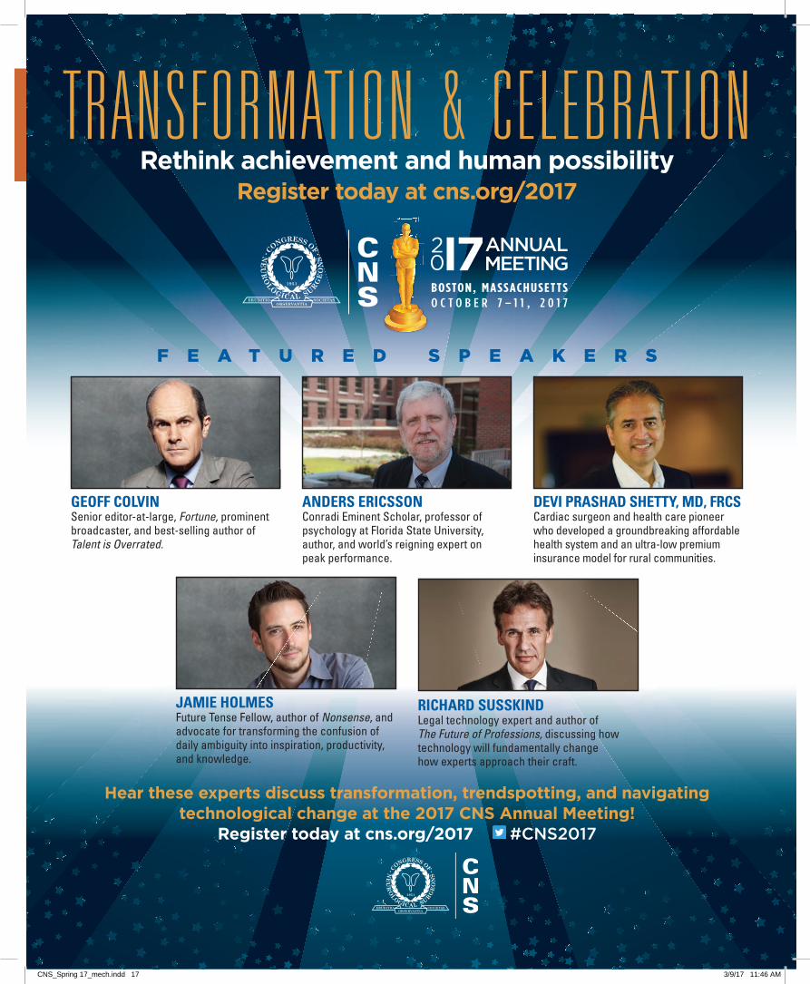
TRANSFORMAT ION & CELEBRAT IONRethink achievement and human possibility
Register today at cns.org/2017
F E A T U R E D S P E A K E R S
BOSTON, MASSACHUSETTSO C T O B E R 7 – 1 1 , 2 0 1 7
BOSTON, MASSACHUSETTSO C T O B E R 7 – 1 1 , 2 0 1 7
Hear these experts discuss transformation, trendspotting, and navigating technological change at the 2017 CNS Annual Meeting!
Register today at cns.org/2017 #CNS2017
GEOFF COLVIN Senior editor-at-large, Fortune, prominent broadcaster, and best-selling author of Talent is Overrated.
ANDERS ERICSSON Conradi Eminent Scholar, professor of psychology at Florida State University, author, and world’s reigning expert on peak performance.
DEVI PRASHAD SHETTY, MD, FRCSCardiac surgeon and health care pioneer who developed a groundbreaking affordable health system and an ultra-low premium insurance model for rural communities.
JAMIE HOLMESFuture Tense Fellow, author of Nonsense, and advocate for transforming the confusion of daily ambiguity into inspiration, productivity, and knowledge.
RICHARD SUSSKIND Legal technology expert and author of The Future of Professions, discussing how technology will fundamentally change how experts approach their craft.
CNS_Spring 17_mech.indd 16 3/9/17 11:46 AM
TRANSFORMAT ION & CELEBRAT IONRethink achievement and human possibility
Register today at cns.org/2017
F E A T U R E D S P E A K E R S
BOSTON, MASSACHUSETTSO C T O B E R 7 – 1 1 , 2 0 1 7
BOSTON, MASSACHUSETTSO C T O B E R 7 – 1 1 , 2 0 1 7
Hear these experts discuss transformation, trendspotting, and navigating technological change at the 2017 CNS Annual Meeting!
Register today at cns.org/2017 #CNS2017
GEOFF COLVIN Senior editor-at-large, Fortune, prominent broadcaster, and best-selling author of Talent is Overrated.
ANDERS ERICSSON Conradi Eminent Scholar, professor of psychology at Florida State University, author, and world’s reigning expert on peak performance.
DEVI PRASHAD SHETTY, MD, FRCSCardiac surgeon and health care pioneer who developed a groundbreaking affordable health system and an ultra-low premium insurance model for rural communities.
JAMIE HOLMESFuture Tense Fellow, author of Nonsense, and advocate for transforming the confusion of daily ambiguity into inspiration, productivity, and knowledge.
RICHARD SUSSKIND Legal technology expert and author of The Future of Professions, discussing how technology will fundamentally change how experts approach their craft.
CNS_Spring 17_mech.indd 17 3/9/17 11:46 AM
18 WWW.CNS.ORG
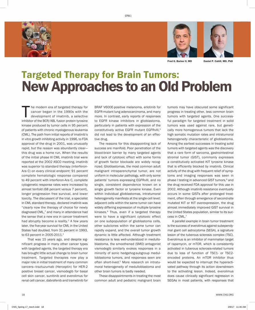
The modern era of targeted therapy for cancer began in the 1990s with the development of imatinib, a selective
inhibitor of the BCR/ABL fusion protein tyrosine kinase produced by tumor cells in 95 percent of patients with chronic myelogenous leukemia (CML). The path from initial reports of imatinib’s in vitro growth inhibiting activity in 1996, to FDA approval of the drug in 2001, was unusually rapid, but the reason was abundantly clear—this drug was a home run. When the results of the initial phase III CML imatinib trial were reported at the 2002 ASCO meeting, imatinib was superior to standard therapy (interferon-Ara C) on every clinical endpoint: 91 percent complete hematologic response compared to 49 percent with interferon-Ara C, complete cytogenetic response rates were increased by almost tenfold (68 percent versus 7 percent), longer progression free survival, and lower toxicity. The discussant of the trial, a specialist in CML standard therapy, declared imatinib was “clearly now the therapy of choice for newly diagnosed CML,” and many in attendance had the sense that a new era in cancer treatment had abruptly become a reality.1 A few years later, the five-year survival for CML in the United States had doubled, from 31 percent in 1993, to 63 percent in 2005-2011.2
That was 15 years ago, and despite sig-nificant progress in many other cancer types with targeted agents, the targeted therapy era has brought little actual change to brain tumor treatment. Targeted therapies now play a major role in initial treatment of many common cancers—traztuzumab (Herceptin) for HER-2 positive breast cancer, vismodegib for basal cell skin cancer, sunitinib and everolimus for renal cell cancer, dabrafenib and trametinib for
BRAF V600E-positive melanoma, erlotinib for EGFR-mutant lung adenocarcinoma, and many more. In contrast, early reports of responses to EGFR kinase inhibitors in glioblastoma, particularly in patients with expression of the constitutively active EGFR mutant EGFRvIII,3
did not lead to the development of an effec-tive drug.
The reasons for this disappointing lack of success are manifold. Poor penetration of the blood-brain barrier by many targeted agents and lack of cytotoxic effect with some forms of growth factor blockade are widely recog-nized. Glioblastomas, the most common adult malignant intraparenchymal tumor, are not uniform in molecular pathology, with only some patients’ tumors expressing EGFRvIII, and no single, consistent dependence known on a single growth factor or tyrosine kinase. Even within individual glioblastomas, intratumoral heterogeneity manifests at the single-cell level; adjacent cells within the same tumor can have widely differing expression of multiple tyrosine kinases.4 Thus, even if a targeted therapy were to have a significant cytotoxic effect on one subpopulation of glioblastoma cells, other subclones within the same tumor can rapidly expand, and the overall tumor growth dynamic is little affected. Although treatment resistance is less well-understood in medullo-blastoma, the smoothened (SMO) antagonist vismodegib similarly evokes responses in a minority of sonic hedgehog-subgroup medul-loblastoma tumors, and responses seen are often short-lived.5 More research on intratu-moral heterogeneity of medulloblastoma and other brain tumors is badly needed.
These disappointments in treating the most common adult and pediatric malignant brain
tumors may have obscured some significant progress in treating other, less common brain tumors with targeted agents. One success-ful paradigm for targeted treatment in solid tumors was used against rare, but geneti-cally more homogenous tumors that lack the high somatic mutation rates and intratumoral heterogeneity characteristic of glioblastoma. Among the earliest successes in treating solid tumors with targeted agents was the discovery that a rare form of sarcoma, gastrointestinal stromal tumor (GIST), commonly expresses a constitutively activated KIT tyrosine kinase that is efficiently blocked by imatinib. Clinical activity of the drug with frequent relief of symp-toms and imaging responses was seen in phase I testing in advanced GIST tumors,6 and the drug received FDA approval for this use in 2002. Although imatinib resistance eventually occurs in some GISTs after prolonged treat-ment, often through emergence of second-site mutated KIT or KIT overexpression, the drug almost immediately improved GIST survival in the United States population, similar to its suc-cess in CML.7
A parallel example in brain tumor treatment is the success of everolimus against subependy-mal giant cell astrocytoma (SEGA), a signature lesion of the tuberous sclerosis complex (TSC). Everolimus is an inhibitor of mammalian target of rapamycin, or mTOR, which is consistently activated in tuberous sclerosis-related tumors due to loss of function of TSC1- or TSC2-encoded proteins. An mTOR inhibitor thus would be expected to interrupt the hyperacti-vated pathway through its action downstream to the activating lesion. Indeed, everolimus does cause clinically significant regression in SEGAs in most patients, with responses that
Targeted Therapy for Brain Tumors: New Approaches to an Old Problem
Daniel P. Cahill, MD, PhDFred G. Barker II, MD
CNS_Spring 17_mech.indd 18 3/9/17 11:46 AM

WWW.CNS.ORG 19
are both rapid and durable.8 In phase III test-ing, everolimus caused regression of SEGAs, TSC-related skin lesions, and renal angiomyo-lipomas9 with little toxicity. Subsequent testing has shown clinical benefits of everolimus in TSC, including reduction in treatment-refractory seizures,10 and perhaps in other associated neurobehavioral problems as well.
Although the three most common intra-cranial tumors—glioblastoma, meningioma, and metastasis—dominate both neurosurgical oncology practice and brain tumor research, the 2016 WHO classification of brain tumors describes 155 distinct brain tumor entities, many of which are rare and poorly character-ized.11 With increasing research into these arcane tumors, an overlapping array of tumor types that harbor treatable mutations or other molecular alterations are being discovered. For example, the highly treatable BRAF V600E mutation discovered in malignant melanoma has now been found in some glioblastomas, in particular those with epithelioid or rhabdoid morphology, some pilocytic astrocytomas, dysembryoplastic neuroepithelial tumors (DNETs), angiocentric gliomas, and pleomor-phic xanthoastrocytomas (PXAs), as well as many gangliogliomas, and nearly all papillary subtype craniopharyngiomas. Improved treat-ments for all of these tumor types are badly needed, and the potential for effective and
safe therapy with targeted agents is an attrac-tive one. Anecdotal reports of significant tumor responses to agents specific for the V600E mutated BRAF protein, as well as to inhibition of the downstream MEK protein, have been reported in many of these tumor types.
Designing and accruing to traditional pro-spective phase II drug trials in many of these rare entities will pose a significant logistic chal-lenge, even in the context of NCI-supported clinical trials groups. The Pediatric Brain Tumor Consortium has recently completed a phase I study of AZD6244, an orally available MEK inhibitor, in pediatric low-grade gliomas (NCT01089101), and a phase II trial is planned. In addition, the Alliance for Clinical Trials in Oncology is developing a phase II pro-tocol of combined oral V600E BRAF and MEK inhibitors in papillary craniopharyngioma. These, and other similar trials, will require the aggressive support of the neurosurgical oncol-ogy community if we hope to see lasting progress in the management of these uncom-mon, but potentially life-threatening conditions. Ultimately, building a firm foundation in molec-ular characterization of all brain tumor types, harnessed to the emerging panoply of novel targeted treatments developed for other tumors, will allow the repurposing of many powerful targeted treatments for tumors affecting the brain and nervous system. <
References1 Delmonte L. Imatinib superior to standard therapy in
newly diagnosed CML. Plenary paper. Oncology Times
June 2002, p. 57.
2 Leukemia—chronic myeloid—CML statistics. http://
www.cancer.net/cancer-types/leukemia-chronic-
myeloid-cml/statistics. Accessed January 26, 2017.
3 Mellinghoff IK, Wang MY, Vivanco I, et al. Molecular
determinants of the response of glioblastomas to
EGFR kinase inhibitors. N Engl J Med. 2005 Nov
10;353(19):2012-24.
4 Snuderl M, Fazlollahi L, Le LP, Nitta M, et al. Mosaic
amplification of multiple receptor tyrosine kinase genes
in glioblastoma. Cancer Cell. 2011 Dec 13;20(6):810-7.
5 Robinson GW, Orr BA, Wu G, et al. Vismodegib
Exerts Targeted Efficacy Against Recurrent Sonic
Hedgehog-Subgroup Medulloblastoma: Results from
Phase II Pediatric Brain Tumor Consortium Studies
PBTC-025B and PBTC-032. J Clin Oncol. 2015 Aug
20;33(24):2646-54.
6 Van Oosterom AT, Judson I, Verweij J, Stroobants S, et
al. European Organisation for Research and Treatment
of Cancer Soft Tissue and Bone Sarcoma Group.
Safety and efficacy of imatinib (STI571) in metastatic
gastrointestinal stromal tumours: a phase I study.
Lancet. 2001 Oct 27;358(9291):1421-3
7 Perez EA, Livingstone AS, Franceschi D, et al.
Current incidence and outcomes of gastrointestinal
mesenchymal tumors including gastrointestinal stromal
tumors. J Am Coll Surg. 2006 Apr;202(4):623-9.
8 Krueger DA, Care MM, Holland K, et al. Everolimus
for subependymal giant-cell astrocytomas in tuberous
sclerosis. N Engl J Med. 2010 Nov 4;363(19):1801-11.
9 Franz DN, Belousova E, Sparagana S, et al. Efficacy
and safety of everolimus for subependymal giant
cell astrocytomas associated with tuberous sclerosis
complex (EXIST-1): a multicentre, randomised,
placebo-controlled phase 3 trial. Lancet. 2013 Jan
12;381(9861):125-32.
10 French JA, Lawson JA, Yapici Z, et al. Adjunctive
everolimus therapy for treatment-resistant focal-onset
seizures associated with tuberous sclerosis (EXIST-3): a
phase 3, randomised, double-blind, placebo-controlled
study. Lancet. 2016 Oct 29;388(10056):2153-2163.
11 Louis DN, Ohgaki H, Wiestler OD, et al. WHO
Classification of Tumours of the Central Nervous
System. Lyon: IARC, 2016
> AMONG THE EARLIEST SUCCESSES IN TREATING SOLID
TUMORS WITH TARGETED AGENTS WAS THE DISCOVERY
THAT A RARE FORM OF SARCOMA, GASTROINTESTINAL
STROMAL TUMOR (GIST), COMMONLY EXPRESSES A
CONSTITUTIVELY ACTIVATED KIT TYROSINE KINASE
THAT IS EFFICIENTLY BLOCKED BY IMATINIB. <
CNS_Spring 17_mech.indd 19 3/9/17 11:46 AM

20 WWW.CNS.ORG
The percentage of women in medicine has increased dramatically, with women comprising the majority of medical
school applicants for the first time in 2003.1
Since that time, women have represented approximately 50 percent of medical students, with equal achievements in GPA, MCAT, and USMLE scores.2 We have seen an increase in women entering all specialties, but with significant variability. The percentage of women in neurosurgery has increased, though slowly when compared with other subspecialties, with just under 8 percent of all neurosurgery residents in 1989, compared to 10 percent in 20083, and now 16.3 percent in the 2016 match (personal communication ABNS). The number of female neurosurgeons in the workforce has remained even more static, with only 5.9 percent of board-certified neurosurgeons being women in 20063 and 6.1 percent in 2016 (personal communication, ABNS).
Recognition of these disparities does seem to be making a difference. There are currently 91 women in academic neurosurgery (unpublished data), up from 25 in 20083. Two female interim department chairs have been named in the past year, after years of only a single female chair of neurosurgery. Additionally, there are currently three female vice-chairs and seven female program directors. This increase in female mentorship is critical for the overall training environment as we work to attract top medical students, 50 percent of whom are women.
Increasing diversity is important in the training and retention of residents, and in the work environment. From 2000-2009, the overall resident attrition rate was 6.7 percent, with a noticeable difference between women and men (17 percent versus 5.3 percent, respectively)4. Leadership must now include an understanding of different learning styles and individual motivations. Addressing differences between generations, cultures, and gender can significantly improve education. Mentorship and promotion are key elements of fostering future generations into faculty and leadership roles. <
References1 U.S. Medical School Applicants and Students 1982-
1983 to 2011-2012.; 2012. Available at: https://www.
aamc.org/download/153708/data/.
2 Cuddy MM, Swanson DB, Clauser BE: A multilevel
analysis of examinee gender and USMLE step 1
performance. Acad Med 83:S58–62, 2008.
3 Benzil DL, Abosch A, Germano I, et al. The future of
neurosurgery: a white paper on the recruitment and
retention of women in neurosurgery. J Neurosurg.
2008;109(3):378-86.
4 Renfrow JJ, Rodriguez A, Liu A, et al. Positive trends in
neurosurgery enrollment and attrition: analysis of the
2000-2009 female neurosurgery resident cohort. J
Neurosurg. 2016;124(3):834-9.
WINS: A Changing Landscape in Neurosurgery
Stacey Quintero-Wolfe, MD
Stacey Quintero-Wolfe, MD, WINS meeting, 2016 CNS Annual Meeting
CNS_Spring 17_mech.indd 20 3/9/17 11:47 AM

WWW.CNS.ORG 21
The pract ice of travel ing across international borders to seek medical services is generally referred to as
“medical tourism.” Though the term “medical tourism” has been used synonymously with “health care tourism,” the two need to be distinguished.1 Health care or wellness tourism includes rejuvenation and alternative therapies like yoga, Ayurveda, etc., in which a country like India has a long tradition of niche market share. Increasingly, the term “medical value travel” (MVT) is being used as it more accurately reflects a patient’s health care seeking behavior; in most part based on the economic impact on the host nation.2 Countries actively promoting medical tourism in Asia include India, Malaysia, Singapore, South Korea, and Thailand.
India has become a popular destination for medical tourism for a variety of reasons. The Medical Tourism Market Report: 2015 stated that India was “one of the lowest cost and high-est quality of all medical tourism destinations, as it offers wide variety of procedures at about one-tenth the cost of similar procedures in the United States.”3 In 2016, India’s medical tourism sector was estimated to be worth $8 billion.4 This is expected to rise at a compound annual growth rate (CAGR) of 15–25 percent until 2020.2
Increasing numbers of foreign tourists are visiting India2,5 over the years. (Figures 1, 2). The number of medical visas issued by India in 2013 was 56,129, in 2014 it was 75,671, and in 2015 it increased to 134,344.6 However, the fact remains that a significant percentage of patients who seek treatment are on a general tourist visa to circumvent the paperwork involved in getting
The Advent and Growth of Medical Tourism in India with Reference to Neurosurgery
Harinder S. Sidhu, MDRahul Lath, MDAlok Ranjan, MD
Figure 2. Percentage of medical tourists from various countries in 2014. (Source: FICCI knowledge paper)
Figure 1. Foreign tourist inflow to India 1999–2015 (Source: Ministry of Tourism website)
CNS_Spring 17_mech.indd 21 3/9/17 11:47 AM
22 WWW.CNS.ORG
medical visas. The majority of patients come from three world regions: The first major block is Asia, and includes Bangladesh (68,034 patients in 2015), Afghanistan (19,644), Bhutan, Sri Lanka, and Nepal, which, as part of the South Asian Association for Regional Cooperation (SAARC) countries, find that India is a natural destination—being in the immediate neighborhood. The second major block of patients come from the Middle East, and include Iran (5,656), Iraq, the central Asian region, and other Middle East regions, including Oman (4,728). The third major block is Africa, with Nigeria (5,765), Kenya, and Tanzania being the major contributors. Other countries with significant medical tourists visiting India included Maldives, Russia, and virtually every major African country other than South Africa.
The vast majority of medical patients are being treated at corporate hospitals which are concentrated in metro cities such as New Delhi, Mumbai, Chennai, Hyderabad, Kolkata, Bengaluru, Pune, and Kochi. The largest chains of corporate hospitals in India in terms of bed strength are the Apollo Group and Fortis Group, with a bed strength of approximately 10,000 beds each.7 The approximate number of beds in various private hospitals treating these patients is about 50,000 to 60,000.
Factors Leading to Medical Tourism in IndiaIndia has become a popular destination for medical tourism for a variety of reasons1,2,9,10
which include: 1. Competent and skilled doctors, nurses, and
allied medical staff. 2. Well-equipped hospitals with national and
international accreditation. 3. Availability of latest technology.4. Significantly lower cost of medical treatment
compared to developed countries.5. English is widely spoken by healthcare
professionals and translators are readily available for other languages.
6. No waiting period for patients to undergo treatment/procedure.
7. General perception of India as being friendly and sensitive to these patients.
8. Medical insurance companies in developed countries are increasingly recognizing India’s potential as a global health care destination.
Neurosurgery in IndiaThe first neurosurgical department in India was set up the Christian Medical College, Vellore, Tamil Nadu in 1949.11 Subsequently, the growth of neurosurgery in India has been phenomenal. Currently there are 75 centers with MCh neu-rosurgery training programs, with 277 seats per year,12 apart from one-tenth of trainees in the DNB Neurosurgery program.13 The current number of neurosurgeons in India serving a population of 1.2 billion people is approximately 1,800, with the number rising annually.14
A large number of neurosurgeons have done fellowships in the US, UK, Europe, and Australia after completing their basic neurosurgical train-ing in India. They have subsequently returned to India and brought back valuable subspe-cialty expertise. The Neurological Society of India (NSI) was founded in 1951 and acts as the parent society. It has affiliated subspecialty societies for cerebrovascular neurosurgery, skull base neurosurgery, stereotactic and functional neurosurgery, pediatric neurosur-gery, and neurotrauma.15 These societies have annual conferences and training courses for residents and young neurosurgeons.
Accredited Neurosurgical Centers in IndiaCurrently there are 30 hospitals in India which are accredited by the Joint Commission Inter-national (JCI), which is the international arm of the Joint Commision Accreditation for Hospital Organizations (JCAHO).16 Of these, 24 hospitals are providing neurosurgical care. This assures that the quality and standard of care provided are up to international standards, and are focused on patient safety. The National Accred-
itation Board for Hospitals and Healthcare providers (NABH) is an institutional member of the International Society for Quality in Health Care (ISQUA). ISQUA is an international body which grants approval to accreditation bodies in the area of health care as mark of equivalence of accreditation program of member countries. Currently there are 425 hospitals in India which are NABH accredited.17
Hospitals providing neurosurgical care are equipped with the latest generation of CT and MRI scan machines, angiography suites, and PET scans. Major neurosurgical operating cen-ters are equipped with the latest operating microscopes, high-speed drills for cranioto-mies, as well as navigation equipment that enables standard neurosurgical procedures to be performed routinely. A few centers in the country also have intra-operative CT/MRI/ultrasound and the Mazor Robotic spinal guid-ance system. There are several centers with comprehensive neurosurgical facilities includ-ing stereotactic radiosurgery (Gamma Knife, Novalis, Cyberknife, TrueBeam, etc.), epilepsy surgery, deep brain stimulation, and other functional procedures, endoscopic brain and spine surgery, endovascular neurosurgery, minimally invasive spine surgery, and robotic surgery.
Costs of Standard Neurosurgical Procedures in IndiaComparative costs of standard therapies across all specialities is shown in Table 1.2 The cost of a diagnostic brain CT scan in India ranges from $40-50; a contrast MRI costs $100-150, and a whole body PET-CT scan costs $300-400. The cost of a single level lumbar fusion could range from $6,000-10,000 depending upon the type of implant used and equipment used such as navigation, robotic assisted, etc.The cost of stereotactic radiosurgery would be $4,000-5,000; bilateral deep brain stimulation (DBS) could cost $15,000-23,000 (depend-ing upon the type of IPG); a typical VP shunt would cost $2,000-3,000 (indigenous versus
CNS_Spring 17_mech.indd 22 3/9/17 11:47 AM

WWW.CNS.ORG 23
variable pressure); a typical craniotomy would cost $6,000-8,500 (depending upon the com-plexity and duration of stay), and a flow diverter intracranial stent would cost $20,000. Scoli-osis surgery using internationally recognized implants such as Medtronics and Globus would cost $15,000. (The costs mentioned above are indicative of the approximate treatment costs at the authors’ hospital and could vary at other centers in India.)
Initiatives by the Government of India to Promote Medical TourismThe Government of India is taking steps to make India stand out in the area of medical tourism.2
Four ministries, including Ministry of Health and Family Welfare, Ministry of Tourism, Ministry of Commerce, and Ministry of AYUSH, along with Services Export Promotion Council (SEPC) and NABH, are involved in promoting India as the preferred destination for medical tourism. The government has also set up a National Medical & Wellness Tourism Promotion Board to look into the various issues such as accreditation and marketing, to give the highest assurance to travelers. A dedicated website has been cre-ated by the government to present India as the
preferred health care destination.5 The SEPC website18 has a detailed listing of accredited hospitals and health care providers with com-parative costs for various medical procedures and other allied facilities. The government has also launched an e-visa application website where tourists from 158 countries may apply safely online for a visa (the visa is valid for med-ical treatment also).
Challenges for the Medical Tourism in IndiaThere is a variable cost structure with wide dif-ferences in the infrastructure and neurosurgical expertise across hospitals. There is a large num-ber of hospitals and smaller nursing homes lacking international accreditation. India has pockets of well-developed health care systems, but the country is still developing, and there are problems with roadways, air connectivity, pollu-tion, cleanliness, etc., which are currently being addressed.1,9,10 There is also a lack of a griev-ance redressal system for medical tourists and there are medicolegal issues which still need to be addressed.10 While large data is available for tourism in general, there is limited data on med-ical tourism specific to neurosurgery. <
References: 1 Sravana Kumar G, Krishan Raj R. Status, Growth and
Impact of Medical Tourism in India. Int J Pharm Sci Rev
Res. 2015:34:284-291.
2 Medical Value Travel in India: Enhancing Medical Value in
India. FICCI (Federation of Indian Chambers of Commerce
and Industry) knowledge paper Oct 2016. Joint Initiative
of FICCI, SEPC, imshealth and Department of Commerce,
Ministry of Commerce and Industry (Govt of India)
3 Medical Tourism Market Report 2015
4 Economic Times : http://economictimes.indiatimes.
com/industry/healthcare/biotech/hyderabad-to-host-
international-medical-tourism-congress-in-september/
articleshow/53831661.cms
5 Ministry of Tourism, India website (http://tourism.
gov.in/wellness-medical-tourism) and http://www.
indiahealthcare tourism.com
6 Economic times: http://economictimes.indiatimes.com/
nri/visa-and-immigration/Medical-tourism-sees-healthy-
growth-13-lakh-visas-in-2015/articleshow/53428421.
cms
7 http://www.india-opportunities.es/archivos/
publicaciones/Healthcare-January-2016.pdf
9 Sharma A. Medical tourism: emerging challenges and
future prospects. International Journal of Business and
Management Invention 2013:2;21-29.
10 Benur S. Medical Tourism: Can it be A New Supernova of
Indian Economy. Asian Journal of Management Sciences
2014:02;10-17.
11 Rajshekhar V. History of neurosurgery at Christian
Medical College, Vellore: A pioneer’s tale. Neurol India.
2016 Mar-Apr;64(2):297-310. doi: 10.4103/0028-
3886.177625.
12 MCI website - http://www.mciindia.org/InformationDesk/
CollegesCoursesSearch.aspx
13 National Board of Examination website -
http://www.natboard.edu.in/
14 Ganapathy K. Neurosurgery in India: An Overview.
World Neurosurgery. DOI: http://dx.doi.org/10.1016/j.
wneu.2013.02.071
15 NSI website - http://www.neurosocietyindia.com/
16 JCI website - http://www.jointcommissioninternational.
org/about-jci/jci-accredited-organizations/?c=India
17 NABH website - http://nabh.co/frmViewAccreditedHosp.
aspx
18 SEPC website - http://www.servicesepc.org/services/
healthcare-service/Table 1. Comparative costs of common procedures in various countries. (Source: FICCI knowledge paper)
CNS_Spring 17_mech.indd 23 3/9/17 11:47 AM

The Biannual Meeting is one of the most important activities of the Joint Section on Pain. It is our opportunity to review
and discuss, in depth, major themes in the neurosurgical management of pain. Past meetings have focused on mechanisms of pain and neuropathy, trigeminal neuralgia, and pain of spinal origin. While these meetings have been enthusiastically received, we have also taken the opportunity to listen to members, feedback.
Among several suggestions, we learned there was a need for more hands-on education to accompany the didactic lectures. Based on this input, we have done a complete overhaul of the meeting, including format, timing, and venue.
We are very excited to invite you to join us for our new Biannual Meeting in Chicago, May 19–20, 2017, at Northwestern University’s NCase facility. Our theme for 2017 is Neuroablation and Neuromodulation for Pain: Expanding the Neurosurgeon’s Toolbox. This exceptional program, organized by our section’s Secretary/Treasurer Bill Rosenberg and hosted by Josh Rosenow, will offer hands-on education on ablative procedures (cordotomy, myelotomy, trigeminal nucleotractotomy, DREZ) as well as neuromodulatory procedures (percutaneous and paddle spinal cord stimulation implantation), along with select didactic lectures on indications, best-practices and complication avoidance.
The course is intended for fully trained colleagues as well as trainees who want to develop or refresh their skills in pain manage-ment. We have a wonderful roster of faculty confirmed, including Ashwin Viswanathan, Parag Patil, Sameer Sheth, Jennifer Sweet, Sean Nagel, Jason Schwalb, Alexandre Francisco, and Steven Falowski. Faculty will be available throughout the symposium to discuss cases and answer questions in an informal and collaborative atmosphere.
We believe our new format will provide great educational value, and we look forward to a productive meeting. Due to the hands-on nature of this activity, we have limited avail-ability, so read the course agenda and sign up today for this unique opportunity to expand your toolbox! <
See you in Chicago!
Rejuvenating the Joint Section on Pain Biannual Meeting:
From Didactic to Practical
Andre Machado, MD
AANS/CNS Joint Section on PainBiannual Meeting
Neuroablation and Neuromodulation for Pain:Expanding the Neurosurgeon’s Toolbox
Join us for this two-day, hands-on course held May 19-20, at the NCase Training Lab at Northwestern University, Chicago, Illinois
REGISTER NOW!cns.org/pain
Neuroablation and Neuromodulation for Pain:
24 WWW.CNS.ORG
SECTION NEWS
Jointly provided by the Congress of Neurological Surgeons and the AANS/CNS Section on Pain.
CNS_Spring 17_mech.indd 24 3/9/17 11:47 AM

In the fall of 2016, the AANS/CNS Section on Pediatric Neurological Surgery became a sponsor of the Image Gently Think-A-Head Campaign. I personally became interested in radiation exposure early in my
career. As a first-year medical student, I participated in an active spina bifida clinic with almost 250 patients. It was not uncommon for a whole trolley to be necessary to carry the x-rays jackets for a single patient to the clinic. I became concerned about the potential overuse of radiology when I tabulated the radiation exposure for the entire clinic population. The data suggested that the radiation burden from diagnostic radiographs (many of which had no medical utility) could contribute to carcinogenesis. The results were alarming and led to the publication of the paper “Radiation exposure in the myelomeningocele population.”1
As a result of my interests, I began to receive frequent calls from my colleagues about adult spina bifida patients with genitourinary cancers, and ultimately published the related article “The development of car-cinoma in the aging myelodysplastic population.”2 These experiences significantly raised my awareness of the diagnostic radiographic studies being ordered on my patients both by myself and others. It is an issue we, as neurosurgeons, need to be aware of, as medical exposure now represents the majority effective dose of radiation exposure in the United States for the first time ever.
As pediatric neurosurgeons, we care for many patients with lifelong, chronic illnesses, including brain and spinal cord tumors, spina bifida, and shunted hydrocephalus (from prematurity, congenital or other causes). These populations are at an increased risk of being exposed to unneces-sary radiographic imaging. While it is easier to justify images on patients with tumors, even these patients have a significant number of unnecessary images performed. Spina bifida patients have a complex medical condition and are often followed by multiple doctors in separate centers, which leads to many repetitious and questionably necessary films. Any child with a shunt is subject to the frequent policy of a CT and shunt series every time they have an office visit in some practices, or every time they show up in an
emergency department. I saw a child once who was not quite three years old, and she had more than 300 CTs and shunt series done at a single insti-tution, often within days of each other.
While I think most pediatric neurosurgeons recognize the need to image more carefully, I believe there continues to be room for improvement. Most of us will replace a CT for the evaluation of a shunt with a rapid sequence MRI, which gives us the advantage of a quick, non-sedated, no radiation evalua-tion of the ventricles. The Lancet article “Radiation exposure from CT scans in childhood and subsequent risk of leukaemia and brain tumours: a retrospec-tive cohort study”3 in June 2012, represented the first direct association with prior CT scans and cancer, and has fueled public and specialty debate about imaging, particularly with CT in the pediatric population. Indeed, 30 percent of patients who undergo a single CT will have at least three CT scans.
Image Gently:
A Pediatric Neurosurgical Perspective
Sarah Gaskill, MD
WWW.CNS.ORG 25
CNS_Spring 17_mech.indd 25 3/9/17 11:47 AM

26 WWW.CNS.ORG
The Image Gently Think-A-Head Campaign rolled out November 2, 2016, as a multispe-cialty campaign under the umbrella of the Image Gently Alliance, which has grown expo-nentially since its inception by the Alliance for Radiation Safety in Pediatric Imaging almost a decade ago. Image Gently is an organization dedicated to education and awareness. Over 100 health care organizations and agencies (>35 international) participate, and there are now over one million professionals worldwide (including radiologists, technologists, medical physicists, and other experts) actively involved in the campaign. It is important to note this is a communication campaign that benefits the market—not the “marketers.” This social mar-keting campaign has the goal of decreasing the
radiation dose in children. We all recognize that children are more radiosensitive than adults, due to their growing tissues and developing central nervous system. As pediatric neurosur-geons, we have an obligation to do our part to reduce the number of unnecessary radiographs performed in children through modification of our own practices, and education of others in the health care profession.
For all neurosurgeons, it is important to remember that no level of radiation exposure is without consequence. If a study is necessary for the care of the patient, it is justified. If the infor-mation gathered from a radiographic study will not change the management, then it likely should not be obtained. I’m sure we can all do better with less imaging. So, Image Gently. <
To learn more about Image Gently and the Think-A-Head Campaign visit imagegently.org.
References:1 Gaskill S, Marlin A. Radiation exposure in the
myelomeningocele population. Pediatr Neurosurg.
1998 Feb;28(2):63-6. DOI:10.1159/000028622
2 Gaskill S. Development of Carcinoma in the Aging
Myelodysplastic Population. Pediatr Neurosurg. 1991–
92;17:34–36 (DOI:10.1159/000120564)
3 Pearce, M, Salotti J, Little M, et al. Radiation exposure
from CT scans in childhood and subsequent risk of
leukaemia and brain tumours: a retrospective cohort
study. Lancet. 2012 Aug 4; 380(9840): 499–505. doi
10.1016/S0140-6736(12)60815-0.
> AS PEDIATRIC NEUROSURGEONS, WE HAVE AN OBLIGATION TO DO OUR PART TO REDUCE THE NUMBER OF UNNECESSARY RADIOGRAPHS PERFORMED IN CHILDREN THROUGH MODIFICATION OF OUR OWN PRACTICES, AND EDUCATION OF OTHERS IN THE HEALTH CARE PROFESSION. <
It’s Back! CNS Trauma: Brain Injury BundleConveniently Earn Trauma CMEThis all-in-one bundle includes the new SANS Neurotrauma Module and five on-demand products.
Earn 16 AMA PRA Category 1 Credits™
Trauma: Brain Injury Bundle• SANS Neurotrauma . . . . . . . . . . . . . . . . . . . . . . . . . . . . . . . . . . . . . . . . . . .10 CME • Multimodality Monitoring: Is There an Effect on Outcome? . . . . . . . . . .1 CME • Trauma Case Vignettes: Open Discussion with the Masters . . . . . . . . . .1 CME • Guidelines Update: Case-Based Update for
Severe Traumatic Brain Injury . . . . . . . . . . . . . . . . . . . . . . . . . . . . . . . . . . .1 CME • Athletic Head Injuries: Return to Play . . . . . . . . . . . . . . . . . . . . . . . . . . . 1.5 CME• Trauma Oral Board Review . . . . . . . . . . . . . . . . . . . . . . . . . . . . . . . . . . . 1.5 CME
CNS Member $400 | Nonmember $650
Visit cns.org/education to purchase this bundle
16 AMA PRA
Category 1 Credits™
CNS_Spring 17_mech.indd 26 3/9/17 11:47 AM

Fifteen years ago, the training path for vascular neurosurgery was paved mainly by residency training, and for
some, augmented with fellowship training. The completion of formal neurovascular training essentially meant it was time for the neurosurgeon to put their training into clinical practice. At that time, vascular neurosurgery was less complex to organize than it is today, as the lines of clinical responsibility with respect to the provider of vascular service were drawn much more clearly.
The substantial evolution of devices coin-ciding with the advancement of clinical treatments in vascular neurosurgery has dras-tically changed the neurovascular field in a relatively short period of time. Endovascular or neuro-interventional techniques have mark-edly increased the “envelope of patients” that can be treated safely.
The literature indicates that the ratio of open surgery procedures compared to those performed endovascularly has shifted signifi-cantly since 2002, primarily due to the results of a now landmark study that focused on intracranial aneurysm outcomes of the two treatments. The International Subarachnoid Aneurysm Trial (ISAT) indicated that a rup-tured aneurysm had better outcomes when
Neurovascular Reformation’s Impact on Training and Business
Steve Napolitan,Chief Department Administrator, Neurosurgery, University of Michigan Health System, NERVES member
Mary Amann,Executive Director, Dept. of Neurosurgery, Washington University School of Medicine, NERVES member
Image 1: Stent Retriever
WWW.CNS.ORG 27
CNS_Spring 17_mech.indd 27 3/9/17 11:47 AM

treated endovascularly. Published data has consistently indicated that 2007 is the year when the two types of procedures crossed paths; at that time, there was an approximate 50 percent split in procedures between open and endovascular—just five years after ISAT. The trend toward endovascular treatment con-tinued, and between 2007 and 2015 data indicated that 70 percent of procedures were being done endovascularly as compared to 30 percent as open surgery. The fallout is the dis-persion of neurovascular care across multiple disciplines. Simply stated, the ability to treat neurovascular patients has expanded signifi-cantly in both technique and sub-specialty.
In February 2015, Nature published the results from five clinical trials that made use of endovascular treatment for stroke patients, and in all five trials, the use of thrombectomy within the first six hours was shown to provide better outcomes. As a result of these trials, the preferred treatment for patients with severe strokes changed, revolutionizing the care pro-vided to stroke patients. Since about 1996, stroke patients have typically been treated first with tPA, a clot-dissolving activator. However, the 2015 studies showed that although tPA is effective for smaller strokes, for large block-ages, it dissolved the clot less than a third of the time. In these studies, a clot-removal device called a stent retriever or “stentriever” (Image 1) was used to go into the brain and grab the clot. Results showed that these devices reopened the artery 80 to 90 percent of the time, which led the American Heart Association to give the treatment its strongest recommen-dation and issue new guidelines in June 2016. This is just one example of how the “tree of
knowledge” has, and is, continuing to grow at an extraordinary pace for neurosurgeons.
This treatment change has significantly impacted who participates in the care of isch-emic stroke patients, giving neurosurgeons access to a whole new patient population. Historically, stroke neurologists have adminis-tered tPA within the first three to four hours of onset. Now, stent retrievers can be adminis-tered by endovascular trained neurosurgeons, interventional neuroradiologists, or inter-ventional neurologists. Up to 20,000 stent thrombectomies are expected to be done this year, which is twice as many as last year. Companies who produce the stent retrievers are projecting that as many as 60,000 acute stroke patients will ultimately receive the pro-cedure. Neurosurgery will be challenged to work collaboratively with radiology and neu-rology, while at the same time expanding the neurosurgical repertoire. Equally important is continuing to lead the management of patients with cerebrovascular disease, despite sharing this responsibility with other specialties.
It is evident that changes in neurovascu-lar treatment have necessitated reflection on sub-specialty education and training require-ments for neurovascular practice. Just this past year, the American Board of Neurological Surgery (ABNS), with support from the Senior Society, recognized the need for a sub-specialty vascular certification. Leaders from neuro-surgery, neurology, and neuroradiology came together to develop a sub-specialty certifica-tion exam. This exam will be administered for the first time in August 2017. The sub-specialty knowledge that is now required in neurosurgical training programs has led residency programs
to reevaluate their curriculums and clinical rotations. Disciplines of training such as inter-ventional and angiographic treatments should now be considered earlier during residency training in order to ensure that the necessary and relevant neurosurgical sub-specialty train-ing occurs at the right time, and adds value to the continuum of neurosurgical training. Residency training programs will need to be flexible, as change is occurring at rapid pace in the neurosurgical field.
From a business perspective, what does the evolution of vascular neurosurgery mean for practice administrators? Historically, vascu-lar neurosurgery patients were sent to large academic centers where they received open or closed treatment for their cerebrovascular dis-order. Today, more of these patients, especially ischemic stroke patients, are being treated at community hospitals. Based on the financial benefits associated with these patients, com-munity hospitals are hiring endovascular trained neurosurgeons, interventional radiolo-gists, and interventional neurologists. Practice administrators will need to identify growth opportunities in key areas and become experts in understanding market trends and revenue streams. Developing strategic initiatives with hospital partners will lead to competitive, suc-cessful centers. Understanding Stroke Designation versus Comprehensive Stroke Center, identifying expansion opportunities in telemedicine, determining faculty recruitment needs, knowing competitors’ strategies, and developing relationships with EMS will all be critical elements for future success. <
Note: The authors acknowledge and appreci-ate the input received for this article from Dr. Ralph G. Dacey, Jr, Dr. Gregory J. Zipfel, Dr. B. Gregory Thompson, and Dr. Aditya Pandey.
NERVES (Neurosurgery Executives’ Resource Value and Education Society) was established with the purpose of helping neurosurgery prac-tice managers and administrators network, combine resources to gather information, and learn from their colleagues about how to build stronger practices.
> THE SUCCESS OF THE STENT RETRIEVER HAS SIGNIFICANTLY IMPACTED WHO PARTICIPATES IN THE CARE OF ISCHEMIC STROKE PATIENTS, GIVING NEUROSURGEONS ACCESS TO A WHOLE NEW PATIENT POPULATION. <
28 WWW.CNS.ORG
CNS_Spring 17_mech.indd 28 3/9/17 11:47 AM

Thank YouIndustry Ambassador
for your valuable support of CNS programs and educational activities
Thank YouIndustry Ambassador
for your valuable support of CNS programs and
Thank YouIndustry Ambassador
for your valuable support of CNS programs and educational activities
Thank YouIndustry Ambassador
for your valuable support of CNS programs and
CNS_Spring 17_mech.indd 29 3/9/17 11:47 AM

30 WWW.CNS.ORG
On October 14, 2016, the Centers for Medicare and Medicaid Services (CMS) released the final rule describing the
new Medicare Quality Payment Program (QPP). This new system was mandated by the Medicare Access and CHIP Reauthorization Act (MACRA). The new QPP rolls many of Medicare’s past quality reporting programs—the Physician Quality Reporting System (PQRS), Electronic Health Records (EHR) Incentive Program and value-based payment modifier—into a new, integrated system.
The MACRA legislation saved practicing neu-rosurgeons significant potential revenue—as much as $100,000—by (1) repealing the sus-tainable growth rate (SGR) and its annual pay cut; (2) preventing the elimination of 10- and 90-day global payments; and (3) streamlin-ing Medicare’s quality programs. This new approach to unified quality reporting is part of CMS’s goal to move away from volume-based, fee-for-service payments to physicians and towards linking reimbursement to the value and quality of care provided.
The QPP offers two pathways for comply-ing with the new quality reporting system. The first option is to participate in an advanced alternative payment model (APM). This option is not widely available, and at the time of this writing, there are only six functioning
advanced APMs—the Pioneer Accountable Care Organization being one example. In the future, bundled payments (such as the Comprehensive Care for Joint Replacement (CJR) program for orthopedics) may qualify as an advanced APM. The American College of Surgeons (ACS) is collaborating with many specialty societies, including the CNS, in developing surgical APMs. However, the 2017 advanced APM options for physicians, particularly surgeons and other spe-cialists, are extremely limited.
The vast majority of neurosurgeons will comply with Medicare’s QPP through the Merit-based Incentive Payment System (MIPS). Performance during 2017 will be used to mod-ify 2019 Medicare payments. Physicians who do not participate in the QPP in 2017 will auto-matically receive the maximum penalty, which for 2019 is a 4 percent Medicare pay cut.
MIPS Reporting
The MIPS system generates a performance score based on 4 different categories:1. Quality Reporting. This category replaces
the former PQRS system, although most of the same quality measures will be used. Initially, this category will account for 60 percent of the 2017 performance score. Ultimately, by 2021, the quality portion of a physician’s MIPS score decreases to 30 percent.
2. Advancing Care Information (ACI). ACI replaces the electronic health record (EHR) meaningful use program and accounts for 25 percent of the 2017 performance score in 2017 and in future years.
3. Clinical Practice Improvement Activities (CPIA). Practice improvement activities, including participating in maintenance of
certification (MOC), reporting to clinical data registries and 24/7 access to care, account for 15 percent of the MIPS score in 2017 and beyond.
4. Resource Use/Cost. Initially worth zero per-cent of the performance score in 2017, this category will compare resources used to treat similar care episodes and clinical condi-tion groups across practices. Over time, this will increase to 30 percent of the MIPS score.
Quality ReportingGenerally speaking, the quality reporting ele-ment of MIPS requires physicians to report six quality measures, including one outcome mea-sure (if available), to achieve full performance. This is a substantial improvement from the ear-lier PQRS system, which required physicians to report a total of nine measures. Measures that are routinely reported by neurosurgeons include:1. PQRS 021: Perioperative Care: Selection of
Prophylactic Antibiotic2. PQRS 022: Perioperative Care:
Discontinuation of Prophylactic Antibiotic3. PQRS 023: Perioperative Care: Venous
Thromboembolism Prophylaxis4. PQRS 130: Documentation of Current
Medications in the Medical Record5. NQF 1789: All Cause Unplanned
Readmissions6. PQRS 046: Medication Reconciliation
Post-discharge7. PQRS 047: Development of an Advanced
Care Plan8. PQRS 128: Preventative Care and
Screening: Body Mass Index Screening and Follow-up
9. PQRS 131: Pain Assessment and Follow-up10. PQRS 226: Preventative Care: Tobacco Use
Screening and Cessation Intervention
INSIDE THE CNS
Washington Committee Report
30 WWW.CNS.ORG
Katie O. Orrico, JDJohn Ratliff, MD, FACS
CNS_Spring 17_mech.indd 30 3/9/17 11:47 AM

WWW.CNS.ORG 31WWW.CNS.ORG 31WWW.CNS.ORG 31
11. PQRS 260: Rates of Carotid Endarterectomy (CEA) for Asymptomatic Patients without Major Complications, Discharged to Home by Post-operative Day Two
12. PQRS 344: Rates of Carotid Artery Stenting (CAS) for Asymptomatic Patients, without Major Complications, Discharged to Home by Post-operative Day Two
13. PQRS 345: Rates of Post-operative Stroke or Death in Asymptomatic Patients Undergoing CAS
14. PQRS 346: Rates of Post-operative Stroke or Death in Asymptomatic Patients Undergoing CEAAll of the quality measures available for MIPS
reporting can be accessed at the CMS QPP Quality Measures website: https://qpp.cms.gov/measures/quality.
Advancing Care InformationThe ACI reporting is a substantial improve-ment versus the previous EHR meaningful use program, only requiring the reporting of five EHR-related measures. Some of these elements merely need attestation and include such things as providing patient access to their medical record, secure messaging, sending care records summaries, e-prescribing, and patient-specific education.
Clinical Practice Improvement ActivitiesClinical practice improvement activities (CPIA) includes a variety of options. Most participants in the system will have to report four activities over a 90-day period. For smaller physician groups or rural physicians, the requirement is two activities over a 90-day reporting period. Unfor-tunately, CPIA options generally focus on primary care, without many options for specialists. Some options that neurosurgeons have for reporting include: Participating in a qualified clinical data registry (QCDR), using Consumer Assessment of Healthcare Providers and Systems (CAHPS®) patient satisfaction questionnaires, participating in MOC Part IV, providing 24/7 access to care, and using patient safety tools. Each of these elements has to be performed for at least 90
consecutive days during the performance period to earn credit. The entire list of CPIA activities is available at the CMS QPP Quality Measures web-site: https://qpp.cms.gov/measures/ia.
Physician Exemptions Certain physicians will be exempt from MIPS and any corresponding pay cuts. Thanks to the advocacy of the CNS and others, more physi-cians will be exempt from the program than was initially proposed by CMS. Three sets of physi-cians will be exempt from 2017 reporting:1. Physicians new to the Medicare program in
20172. Physicians who bill Medicare less than
$30,0003. Physicians who see fewer than 100
Medicare patientsCMS estimates that approximately 1,500
neurosurgeons will be exempt from the program in 2017.
Pick Your PaceOrganized medicine, including the CNS, pushed CMS to phase-in the new QPP program over a few years, rather than implementing it on Jan-uary 1, 2017. Responding to these concerns, CMS adopted the “Pick Your Pace” program, which makes 2017 compliance with the system much easier. Under this approach, physicians have three options for 2017 reporting:1. Report all required elements of MIPS for
either 90 days or the full year to qualify for a bonus payment.
2. Report fewer MIPS measures for less than a full year.
3. Report one measure in the quality perfor-mance category or required measures in the advancing care information (i.e., electronic health record meaningful use) category.
While 2017 can almost be considered a year-long holiday from quality reporting since the burden to avoid any penalties is minimal, the competition for achieving bonus payments in 2019 will likely be relatively low as well—providing neurosurgeons who do full reporting an opportunity to boost their revenues in two years.
Reporting OptionsReporting options for individual physicians include claims, EHR, qualified registry, or QCDR. Many neurosurgeons will report through a group reporting option, either as part of their practice or through their employer for hospital-based phy-sicians. For 2017, the Group Practice Reporting Option (GPRO) under which most employed neurosurgeons will be scored will be more com-plicated than GPRO reporting under previous quality reporting programs. As a result, some hospitals are considering using more than one tax identifier and more than one GPRO for their physicians. Employed neurosurgeons should check with their hospital’s quality department to see what option their facility is using and to make sure that their expectations for quality reporting and MIPS compliance are clear.
More InformationHopefully, this brief overview will provide some background on the MIPS system and how it may affect your practice. Further information and updates may be found on MACRA resource web pages at www.cns.org/MACRA. Additional edu-cational content specific to MIPS and MACRA will be forthcoming from the AANS/CNS Neu-rosurgery Quality Council, the Communications and Public Relations Committee, and the Coun-cil of State Neurosurgical Societies. There are other available resources for MIPS education, including:• Final Rule: http://bit.ly/2fm4oa8• Executive Summary: http://bit.ly/2dh5FfP• Press release: http://bit.ly/2efELa6• Blog post: http://bit.ly/2dBNAtQ• Fact sheet: http://bit.ly/2dpm851• Quality Payment Program website: https://
qpp.cms.gov/
This article originally appeared in the AANS Neurosurgeon. <
Reporting a single quality measure in 2017 is sufficient to avoid a 4% Medicare pay cut in 2019.
CNS_Spring 17_mech.indd 31 3/9/17 11:47 AM

32 WWW.CNS.ORG
WE WELCOME THE 2017 CNS EXECUTIVE COMMITTEE
OfficersPresidentAlan M. Scarrow, MD, JD
President-ElectAshwini D. Sharan, MD
Vice-PresidentGerald A. Grant, MD
Secretary Steven N. Kalkanis, MD
TreasurerGanesh Rao, MD
Past PresidentRussell R. Lonser, MD
Members at largeAlex A. Khalessi, MDClemens M. Schirmer, MD, PhDHenry H. Woo, MDBrian V. Nahed, MDMartina Stippler, MDNader A. Pouratian, MD
Ex-Officio MembersJames S. Harrop, MDBrian L. Hoh, MDElad I. Levy, MDShekhar N. Kurpad, MD, PhDAshok R. Asthagiri, MDNicholas C. Bambakidis, MD
HistorianChristopher Wolfla, MD
CNS ExecutivesCEORegina Shupak
Legal CounselGregory D. Willard, JD
CNS Executive Office10 N. Martingale RoadSuite 190Schaumburg, IL 60173TEL: 847.240.2500FAX: 847.240.0804Toll-free: 877.517.1CNSEmail: [email protected]
www.cns.org
CNS_Spring 17_mech.indd 32 3/9/17 11:47 AM

IMAGES IN NEUROSURGERY
WWW.CNS.ORG
This 56-year-old patient had chronic pain without an injury. He was neurologically intact, apart from moderate restriction of neck movements. He declined an operation.
There are initially five occipital somites, with the most rostral fading away to leave only four. The boundary between the head and neck corresponds to that between the remaining fourth and fifth somite. The four occipital somites fuse to form the clivus; the caudal part of the occipital four somite and rostral cervical somite combine to form the transitional “pro-atlas.” Its rostral part joins the three occipital somites as the anterior foramen magnum and the apex of the dens. Its lateral part forms the occipital condyles. The caudal half of somite five and the cranial half of somite six form the first cervical somite, giving rise to the base of the odontoid. The second cervical somite (from caudal six and cranial seven) form the body of the axis. The current instance is a result of many anomalies in this process. <
Submitted by:Suresh Ramnath, MD, FRCSC; [email protected] Park, MD; [email protected] Megan E. B. Foldenauer, PhD, CMI; [email protected] of Neurosurgery, University of Michigan
Figure 1: CT axial view shows marked narrowing of the foramen magnum, deviating the medullo-spinal junction to the left.
Figure 2: CT axial view with a smaller left posterior fossa.
Figure 3: CT sagittal view shows absent apex of dens, assimilation of atlas into basi-occiput, platybasia, narrowing of the foramen magnum, fusion of atlas and axis spinous processes, and remnant of atlas fused to anterosuperior aspect of odontoid process.
Figure 4: CT coronal view showing the incorporation of the atlas and its condyle into the occiput, particularly on the right.
Figure 5: Sagittal MRI shows stenosis of the foramen magnum and distortion of the medullo-spinal junction.
Figure 6: Coronal T1 MRI scan shows a kinked medulla on the left, a smaller left posterior fossa, and the head tilted to the right.
CNS_Spring 17_mech.indd 33 3/9/17 11:47 AM

Kathryn Ko, MD Instagram @doc_ambidexter; Facebook @art on call
CNS_Spring 17_mech.indd 34 3/9/17 11:47 AM