learning and memory in individuals with agenesis of the ... wms_revision3_full.pdflearning and...
TRANSCRIPT
Learning and Memory in Individuals with Agenesis of the Corpus Callosum
Lynn K. Paula,b, Roger L. Erickson a, Jo Ann Hartman a, and Warren S. Browna
aFuller Graduate School of Psychology, Travis Research Institute, 180 N. Oakland Ave.,
Pasadena, CA 91101, USA
bCalifornia Institute of Technology, Division of Humanities and Social Sciences, 1200 E.
California Blvd., Pasadena, CA 91125, USA
Corresponding Author:
Lynn K. Paul Caltech, HSS 228-77 1200 E. California Blvd. Pasadena, CA 91125 Email: [email protected]

Abstract
Damage to long white matter pathways in the cerebral cortex is known to affect memory
capacity. However, the specific contribution of interhemispheric connectivity in memory
functioning is only beginning to become understood. The present study examined verbal and
visual memory processing in individuals with agenesis of the corpus callosum (AgCC) using the
Wechsler Memory Scale-Third Edition (WMS-III; Wechsler, 1997b). Thirty participants with
AgCC (FSIQ > 78) were compared against 30 healthy age and IQ matched controls on
auditory/verbal (Logical Memory, Verbal Paired Associates) and visual (Visual Reproduction,
Faces) memory subtests. Performance was worse in AgCC than controls on immediate and
delayed verbal recall for rote word pairs and on delayed recall of faces, as well as on percent
recall for these tasks. Immediate recall for thematic information from stories was also worse in
AgCC, but groups did not differ on memory for details from narratives or on recall for thematic
information following a time delay. Groups also did not differ on memory for abstract figures or
immediate recall of faces. On all subtests, individuals with AgCC had greater frequency of
clinically significant impairments than predicted by the normal distribution. Results suggest less
efficient overall verbal and visual learning and memory with relative weaknesses processing
verbal pairs and delayed recall for faces. These findings suggest that the corpus callosum
facilitates more efficient learning and recall for both verbal and visual information, that
individuals with AgCC may benefit from receiving verbal information within semantic context,
and that known deficits in facial processing in individuals with AgCC may contribute to their
impairments in recall for faces.
Keywords: visual memory; verbal memory; corpus callosum; callosal agenesis;
interhemispheric

Section 1: Introduction
Congenital absence of the corpus callosum, also known as agenesis of the corpus
callosum (AgCC), is an anatomically defined neurological defect which occurs in 3-5% of
individuals with neurodevelopmental disorders (Bodensteiner, Schaefer, Breeding, & Cowan,
1994; Jeret, Serur, Wisniewski, & Fisch, 1985) and in approximately 1 out of 4,000 live births
(Glass, Shaw, Ma, & Sherr, 2008). AgCC is also a co-morbid feature present in a wide range of
genetic and prenatal medical conditions (e.g., chromosomal anomalies, toxic syndromes,
metabolic diseases) and thus these individuals present with a highly heterogeneous clinical
presentation (Paul et al., 2007; Siffredi, Anderson, Leventer, & Spencer-Smith, 2013). Callosal
absence may also occur in isolation, with no evidence of other neural malformations or neuro-
developmental syndromes (Paul et al., 2007). Although individuals with isolated AgCC and
normal-range intellectual functioning generally have a more favorable prognosis, they display a
specific pattern of neuropsychological and psychosocial deficits which interfere with daily life
(Paul et al., 2007). The purpose of the present study is to clarify whether verbal and visual
learning and memory impairments are characteristic of high functioning individuals with AgCC,
and to better understand the role of the corpus callosum in verbal and visual memory encoding,
retention, and retrieval.
1.1 Neuropsychological and Social Functioning in Isolated AgCC
Individuals with isolated AgCC and generally intact intellectual functioning present with
a characteristic pattern of neuropsychological and social capacities. Specifically, individuals with
isolated AgCC have been shown to have impairments in the following domains: bimanual
coordination of motor movements (Mueller, Marion, Paul, & Brown, 2009); interhemispheric
transfer of complex sensory information (Brown, Jeeves, Dietrich, & Burnison, 1999); slowed

processing of complex information (Brown et al., 1999; Brown, Thrasher, & Paul, 2001; Hines,
Paul, & Brown, 2002; Marco et al., 2012); comprehension of higher-order aspects of
communication, including language pragmatics and humor (Brown, Paul, Symington, &
Dietrich, 2005; Brown, Symingtion, VanLancker-Sidtis, Dietrich, & Paul, 2005; Paul, Van
Lancker-Sidtis, Schieffer, Dietrich, & Brown, 2003); complex novel problem-solving (Brown &
Paul, 2000; Gott & Saul, 1978; Sauerwein & Lassonde, 1994; Smith, Rourke, & Rourke, 1994;
Solursh, Margulies, Ashem, & Stasiak, 1965); and facial emotion recognition due to atypical
facial scanning (i.e. reduced attention to salient features of the face, such as the eyes, Bridgman
et al., 2014). However, it is still unclear the extent to which verbal and visual memory
impairments are also characteristic impairments in individuals with isolated AgCC.
1.2 The Corpus Callosum & Memory
Disturbance of callosal function has been shown to negatively impact memory in
individuals with a variety of neurological disorders. For example, structural callosal damage in
patients with multiple sclerosis is associated with impaired list learning (Lafosse, Mitchell,
Corboy, & Filley, 2013) and reduced structural integrity of callosal tracts connecting frontal and
temporal regions is associated with diminished verbal and visual memory in Alzheimer’s Disease
and amnestic mild cognitive impairment (Wang et al., 2014).
Much of the current research on relationships between callosal function and cognitive
performance grew out of studies of individuals with intractable epilepsy and who had undergone
resection of their forebrain commissures. Individuals with commissurotomy present with a
disconnection syndrome marked by absence of interhemispheric transfer of sensory information
and deficits in bimanually coordinated motor activity (Sperry, Gazzaniga, Bogen, Vinken, &
Bruyn, 1969). Research with this clinical population also provided information regarding the
role of interhemispheric integration in higher order cognitive functions such as memory. Studies
of memory in commissurotomy patients have produced variable results depending on the level of
observations. Some studies reported intact basic memory functioning, and concluded that an
isolated hemisphere could functionally encode as well as retrieve verbal information (Ledoux,
Risse, Springer, Wilson, & Gazzaniga, 1977; Sperry, 1968). Other studies reported impaired
auditory and visual-spatial memory (D. Zaidel & Sperry, 1974; E. Zaidel, 1990) and concluded
that cerebral commissures are implicated in adequacy of the acquisition, consolidation, and
retrieval of verbal information. However, since commissurotomy involves transection of all
cerebral commissures, including the hippocampal commissure, these studies do not specifically
address the impact of callosal disconnection on memory. Moreover, interpretation of these
findings are complicated by the participants’ prior history of intractable seizures (Clark &
Geffen, 1989; Phelps, Hirst, & Gazzaniga, 1991).
Nevertheless, these investigators posited that the elimination of interhemispheric transfer
impaired performance because visual memory traces in the right hemisphere were inaccessible to
the language dominant left hemisphere for verbal recall (E. Zaidel, 1990). Moreover, they
suggested that performance impairments were potentially related to differences in the respective
ability of the two hemispheres to process linguistic information, with the right hemisphere
having broader semantic processing fields than the left. Reduced interactions between visual and
verbal systems may also have limited the richness of initial encoding for both visual and verbal
tasks. Thus, these studies suggest that the corpus callosum plays an important, but indirect, role
in the facilitation of memory.
1.3 Auditory Learning and Memory in AgCC
Earlier case studies of individuals with AgCC on tests of verbal learning and memory
produced conflicting results. A number of case studies suggested that individuals with AgCC had
relatively intact performance on tests of verbal learning and memory (David, Wacharasindhu, &
Lishman, 1993; Fischer, Ryan, & Dobyns, 1992; Gott & Saul, 1978; Kessler, Huber, Pawlik,
Heiss, & Markowitsch, 1991; Pirozzolo, Pirozzolo, & Ziman, 1979). For two of these studies, the
Wechsler Memory Scale (WMS; Wechsler, 1945) Memory Quotient score, a composite score of
verbal and visual memory subtests, was the only value reported and therefore no information was
available regarding modality specific memory performance. However, several case studies have
described individuals with isolated AgCC who have mild impairments on tests of verbal learning
and recall of word lists (Fischer et al., 1992; Geffen, Forrester, Jones, & Simpson, 1994; Panos,
Porter, Panos, Gaines, & Erdberg, 2001). Fischer et al. (Fischer et al., 1992) administered a
selective reminding paradigm test to two children with AgCC (both age 8) with normal-range IQ.
One individual performed in the 5th percentile and the other in the 16th on long-term retrieval of
verbal information. In another study, the Rey Auditory Verbal Learning Test (Rey, 1958) was
administered to four individuals with AgCC and FSIQ > 80, (Geffen et al., 1994); three
participants (ages 10, 14, 37) had complete AgCC and one participant (age 22) had partial
AgCC. Relative to published norms, the participants with AgCC did not exhibit deficits on some
aspects of learning (i.e., learning slope, proactive and retroactive interference, or metamemory).
However, the two children with complete AgCC had deficient acquisition scores (i.e., poor initial
recall and total recall over trials 1-5). On delayed free recall, all three individuals with complete
AgCC exhibited deficits despite intact recognition memory. This pattern of performance
suggested that they encoded and retained the verbal information, but had difficulty retrieving it
from memory without the help of external cues. Since recall deficits were not evident in the
individual with partial AgCC, the author concluded the remaining portion of the corpus callosum
must play a role in the proper consolidation and retrieval of verbal information (Geffen et al.,
1994). Finally, a case study of an 11-year-old with partial AgCC and FSIQ in the normal range
(Panos et al., 2001) reported impaired recall on the California Verbal Learning Test-Children’s
Version (CVLT-C; Delis, Kaplan, Kramer, & Ober, 1994). Unlike the complete AgCC cases
described above, this child with partial AgCC performed more poorly on the cued recall (two
standard deviations below the mean) than on free recall (one standard deviation below the mean).
The authors suggest that his poor cued memory illuminates a broader impairment in language
processing, characterized by “limited capacity to utilize semantic information to organize his
learning or recall.”
To address the inconsistency across case studies, we recently compared verbal learning
and memory in a relatively large sample of individuals with isolated AgCC (n = 26) against
healthy matched controls (n =26) (Erickson, Paul, & Brown, 2014) using the California Verbal
Learning Test – Second Edition (CVLT-II; Delis, Kaplan, Kramer, & Ober, 2000). Group
comparisons were made on CVLT-II variables as well as on Donders’ four CVLT-II factors (i.e.,
Attention Span, Learning Efficiency, Delayed Memory, and Inaccurate Memory factors;
Donders, 2008). Individuals with AgCC demonstrated significant impairments in list learning
(i.e., combined recall on learning trials 1-5) and on Donders’ Delayed Memory factor (composed
of Short Delay Free Recall; Short Delay Cued Recall; Long Delay Free Recall; Long Delay Cued
Recall; and Recognition). However, the AgCC group did not have impaired scores on the first
learning trial (i.e. memory after a single trial and prior to the repeated learning trials), learning
slope (i.e. increased recall from the first learning trial to the last), or on indices of ability to retain

and retrieve what was actually learned (i.e. amount of information that was learned by the last
learning trial and was also recalled or recognized after the time delay). In this study deficient
recall (i.e., Donders’ Delayed Memory Factor) appeared to be a consequence of poor encoding,
as the AgCC group did not show deficient attention or working memory on the first trial,
diminished capacity to benefit from repetition learning, or impaired recall or recognition relative
to what they originally learned. In general, these findings suggested that callosal absence results
in mild but consistent deficits in encoding on tests of verbal list-learning recall, and implicates
the corpus callosum in facilitating encoding, perhaps through interhemispheric elaboration.
The CVLT provides insight regarding learning and memory of a rote word list, but it
remains to be seen if callosal absence also interferes with aspects of learning and memory
assessed in the WMS, specifically verbal information presented within the context of a narrative,
rote word pairs, faces, and abstract visual-spatial patterns.
1.4 Visual Memory and in AgCC
To date, our knowledge about visual memory in AgCC is informed solely by case studies.
Moreover, generalizations drawn from these case studies are limited by the variety of measures
utilized (e.g., the Rey Complex Figure Test, Benton Visual Retention Test, Corsi Block Tapping
Test, Gollin’s Incomplete Picture Test). Despite methodological variability, most case studies
found that visual memory fell within normal limits (Kessler et al., 1991; Panos et al., 2001;
Sauerwein, Nolin, & Lassonde, 1994). Specifically, a 45-year-old male with complete AgCC had
normal-range visual working memory on the Corsi Block Tapping task and normal visual
perception and memory using the Gollin’s incomplete picture test (Kessler et al., 1991). Using
the Rey-Osterrieth Figure, one study reported normal delayed visuo-spatial memory in an
asymptomatic individual with AgCC and normal-range FSIQ (Sauerwein et al., 1994), while
another study of an 11-year-old with partial AgCC and normal-range FSIQ (Panos et al., 2001)
reported impaired copy and immediate recall, with intact delayed recall. The authors of the latter
study hypothesized that due to this individual’s white-matter deficits he had initial impairments
integrating and organizing the complex figure, but with sufficient time he successfully processed
the information. Finally, two children with AgCC (both age 8) and normal-range FSIQ were
administered the visual memory subtest from the Test of Visual-Perceptual Skills. Visual
memory fell in the mild impairment range for one subject and in the very superior range for the
other (Fischer et al., 1992). In addition to variability of results, generalizability of these findings
is limited by small sample sizes, variability in measures used, and the lack of neurotypical
controls.
1.5 Hypotheses
Based upon our previous findings with the CVLT (Erickson et al., 2014) and examples
from case studies, we predicted the AgCC group would perform more poorly than controls on
immediate and delayed recall for both verbal and visual tasks. Additionally, we hypothesized
that the AgCC group’s pattern of performance on indices of learning in the current study would
be the same as was found with the CVLT (Erickson et al., 2014): no impairment on the first
learning trial or learning slope, despite impaired recall across all learning trials. This pattern
indicates that despite intact attention and working memory on the first trial and the capacity to
benefit from repeated learning trials, the cumulative amount of information acquired during
learning will be below expected for the AgCC group. Finally, as found with the CVLT
(Erickson et al., 2014), we hypothesized that the AgCC group would not differ from controls on
percent retention (an index of ability to retain and retrieve what was actually learned), indicating
that lower performance on delayed recall is a consequence of limitations during encoding and not
retrieval of what they had learned.
Section 2: Materials and Methods
2.1 Research Participants
This study included 30 adolescents and adults with AgCC and 30 heathy control (HC)
participants. AgCC diagnosis was confirmed by brain MRI and background information on each
participant was gathered as part of the authors’ research program. Participants included 21
individuals with complete agenesis (cAgCC) and 9 with partial agenesis (pAgCC). Individuals
with AgCC were included if they had structural findings that commonly co-occur with AgCC:
colpocephaly, Probst bundles, and occasional small heterotopias. Potential participants with
additional neuro-structural abnormalities were not included. Within the AgCC group, we were
able to directly review 25 of the MRI scans, including all 9 partial AgCC participants. The
anterior commissure was visible on all 25 scans we reviewed and posterior was visible on 24.
Probst bundles were visible bilaterally in 18 participants with complete AgCC and 5 with partial
AgCC. Two participants with complete AgCC presented with unilateral Probst bundles (one
with right only and one with left only) and Probst were not visible in one participant with partial
AgCC.
Exclusionary criteria for both groups included English as a second language, history of
moderate-to-severe head injury, major CNS disorder not associated with AgCC, intractable
epilepsy, and drug abuse as assessed by clinical interview. To avoid confounding effects due to
borderline general intellectual function, Full Scale IQ (FSIQ) greater than, or equal to, 78 was
required. Assessment of general intellectual functioning was completed using the Wechsler
Adult Intelligence Scale (WAIS-III; Wechsler, 1997a) for 29 participants with AgCC; the
remaining AgCC participant and all control participants were administered an abbreviated
Wechsler intelligence test (Wechsler, 1999, 2011). 19 participants in the current study (13
complete and 6 partial) were also included in the CVLT-II (Erickson et al., 2014) study. Of these
19 individuals, 14 completed the CVLT-II and WMS-III within the span of 1 year. For the
remaining participants, age at WMS-III and CVLT-II were as follows (WMS: CVLT by
participant): 18: 16, 29:31, 33:36, 22:31, and 18:24). 11 participants in the HC group were also
included in the CVLT-II paper and received the WMS-III and CVLT-II at the same age.
Supplementary section 1.2 and Supplementary Figure 2 present correlations of CVLT-II with
WMS-III scores for participants enrolled in both studies.
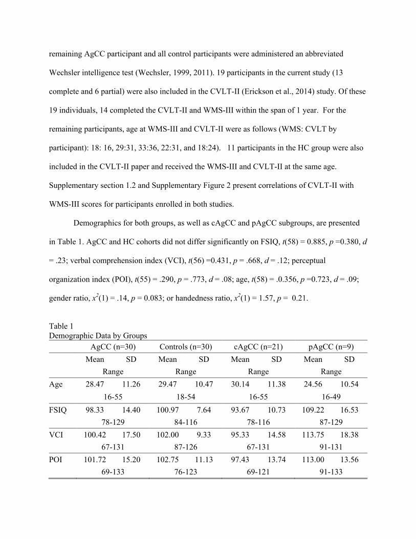
Demographics for both groups, as well as cAgCC and pAgCC subgroups, are presented
in Table 1. AgCC and HC cohorts did not differ significantly on FSIQ, t(58) = 0.885, p =0.380, d
= .23; verbal comprehension index (VCI), t(56) =0.431, p = .668, d = .12; perceptual
organization index (POI), t(55) = .290, p = .773, d = .08; age, t(58) = .0.356, p =0.723, d = .09;
gender ratio, x2(1) = .14, p = 0.083; or handedness ratio, x2(1) = 1.57, p = 0.21.
Table 1 Demographic Data by Groups
AgCC (n=30) Controls (n=30) cAgCC (n=21) pAgCC (n=9) Mean SD Mean SD Mean SD Mean SD Range Range Range Range
Age 28.47 11.26 29.47 10.47 30.14 11.38 24.56 10.54 16-55 18-54 16-55 16-49 FSIQ 98.33 14.40 100.97 7.64 93.67 10.73 109.22 16.53 78-129 84-116 78-116 87-129 VCI 100.42 17.50 102.00 9.33 95.33 14.58 113.75 18.38 67-131 87-126 67-131 91-131 POI 101.72 15.20 102.75 11.13 97.43 13.74 113.00 13.56 69-133 76-123 69-121 91-133
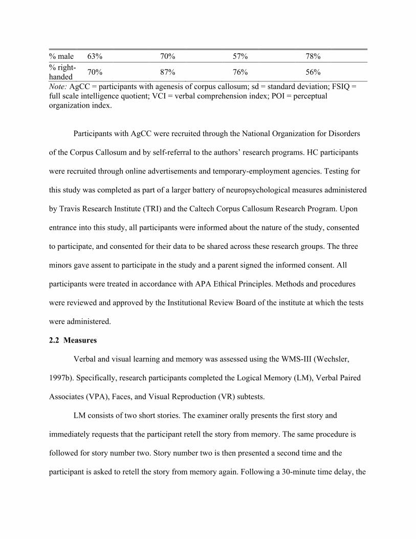
% male 63% 70% 57% 78% % right-handed 70% 87% 76% 56%
Note: AgCC = participants with agenesis of corpus callosum; sd = standard deviation; FSIQ = full scale intelligence quotient; VCI = verbal comprehension index; POI = perceptual organization index.
Participants with AgCC were recruited through the National Organization for Disorders
of the Corpus Callosum and by self-referral to the authors’ research programs. HC participants
were recruited through online advertisements and temporary-employment agencies. Testing for
this study was completed as part of a larger battery of neuropsychological measures administered
by Travis Research Institute (TRI) and the Caltech Corpus Callosum Research Program. Upon
entrance into this study, all participants were informed about the nature of the study, consented
to participate, and consented for their data to be shared across these research groups. The three
minors gave assent to participate in the study and a parent signed the informed consent. All
participants were treated in accordance with APA Ethical Principles. Methods and procedures
were reviewed and approved by the Institutional Review Board of the institute at which the tests
were administered.
2.2 Measures
Verbal and visual learning and memory was assessed using the WMS-III (Wechsler,
1997b). Specifically, research participants completed the Logical Memory (LM), Verbal Paired
Associates (VPA), Faces, and Visual Reproduction (VR) subtests.
LM consists of two short stories. The examiner orally presents the first story and
immediately requests that the participant retell the story from memory. The same procedure is
followed for story number two. Story number two is then presented a second time and the
participant is asked to retell the story from memory again. Following a 30-minute time delay, the

participant is asked to spontaneously recall both stories and then is presented a series of Yes/No
recognition questions about each story. LM scores include total immediate spontaneous recall of
details from all three learning trials (i.e. one trial for story one and two trials for story two; LM
I), spontaneous recall of thematic information from all three learning trials, spontaneous
immediate recall of both stories after only one learning trial (i.e. before story 2 is presented a
second time), learning slope based on change in recall from immediately after the first
presentation of story 2 to immediately after the second presentation, total spontaneous recall of
details from both stories after the time delay (LM II), spontaneous recall of thematic information
after the time delay, and percent retention after the time delay (calculated from the number of
details recalled in the last learning trial for each story – first trial for story one and second trial
for story two – and the number of details recalled after the time delay).
VPA requires learning novel word associations. The individual is orally presented with
eight pairs of unrelated words. The examiner then provides the individual with the first word of
each pair (i.e. a cue) and the participant attempts to respond with the correct corresponding word.
If the participant does not answer or gives an incorrect answer, the examiner provides the correct
response before continuing with the next item. This procedure is repeated three additional times
(four learning trials in total). Presentation order of the eight word pairs varies across learning
trials. Following a 30-minute time delay, the examiner once again provides the first word of each
pair and the participant is asked to provide the corresponding word (no feedback is provided
after the delay). The examiner then reads a list of 24 word-pairs and after each pair the
participant indicates if it was in the original list. VPA scores include total immediate cued recall
across all four learning trials (VPA I), cued immediate recall for only the first learning trial,
learning slope based on change in recall from first to last learning trial, accuracy of spontaneous

recall of word pairs after the time delay, and percent retention after the time delay (calculated
from the number items correctly recalled in last learning trial and the number recalled after the
time delay).
During the learning phase of the Faces subtest, the examiner shows the participant a
series of 24 faces one at a time, at a 2 second interval. The participant is then shown a series of
48 faces including faces from the original series as well as new faces and for each one must
indicate if it was present in the original series. Following a 30-minute delay, another series of 48
faces (the 24 original faces and 24 new faces) is shown and for each one must indicate if it was
present in the original series. Participants receive a score for recognition accuracy immediately
after the learning series (Faces I) and a score for accuracy after the time delay (Faces II). Percent
retention is the ratio of faces recognized after the time delay relative to the number recognized
immediately after first exposure to them.
VR involves 5 abstract figures, each of which is shown to the participant for 10 seconds
and when the image is removed the participant must draw the figure from memory. Following a
30-minute time delay, the participant is asked to draw as many of the figures as possible from
memory. Participants receive a score for accuracy of drawings done immediately after the
learning trials (VR I) and a score for accuracy of drawings completed after the time delay (VR
II), from which percent retention is calculated.
2.3 Procedure
Tasks were administered as part of a multi-day cognitive testing protocol. The WMS-III
tasks were administered in one session, in the following order: LM, VPA, Faces, and VR. If the
following tasks did not use the full 30-minutes between immediate and delayed recall, the
examiner administered another brief unrelated task. All analyses were conducted using age-
corrected scaled scores.
ANOVAs were conducted in SPSS and are reported with two-tailed p-values. T-tests for
independent samples, t-tests for paired samples, unbiased Cohen’s d effect size estimates (dunb),
and 95% CI of dunb, were calculated using Exploratory Software for Confidence Intervals (ESCI;
Cumming, 2012). Independent samples t-tests did not assume population variances are equal.
Analyses comparing the cAgCC and pAgCC groups are presented in Supplementary materials.
Effect sizes were interpreted according to Cohen’s guideline (Cohen, 1988); small d >= .2,
medium d > = .5; large d > = .8.
Section 3: Results
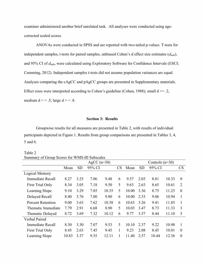
Groupwise results for all measures are presented in Table 2, with results of individual
participants depicted in Figure 1. Results from group comparisons are presented in Tables 3, 4,
5 and 6.
Table 2 Summary of Group Scores for WMS-III Subscales AgCC (n=30) Controls (n=30) Mean SD 95% CI
dafCICI CS Mean SD 95% CI CS
Logical Memory Immediate Recall 8.27 3.25 7.06 9.48 6 9.57 2.03 8.81 10.33 0 First Trial Only 8.34 3.05 7.18 9.50 5 9.63 2.63 8.65 10.61 2 Learning Slope 9.10 3.29 7.85 10.35 5 10.00 3.36 8.75 11.25 0 Delayed Recall 8.40 3.76 7.00 9.80 6 10.00 2.53 9.06 10.94 1 Percent Retention 9.00 3.63 7.62 10.38 6 10.63 3.26 9.41 11.85 1 Thematic Immediate 7.79 2.91 6.68 8.90 5 10.03 3.47 8.73 11.33 3 Thematic Delayed 8.72 3.69 7.32 10.12 6 9.77 3.57 8.44 11.10 5
Verbal Paired Associates
Immediate Recall 8.30 3.30 7.07 9.53 5 10.10 2.37 9.22 10.98 1 First Trial Only 8.45 2.63 7.45 9.45 1 9.23 2.08 8.45 10.01 0 Learning Slope 10.83 3.37 9.55 12.11 1 11.40 2.57 10.44 12.36 0
Delayed Recall 9.03 3.21 7.83 10.23 3 11.13 2.26 10.29 11.97 1 Percent Retention 9.45 3.52 8.11 10.79 5 11.40 1.77 10.74 12.06 1
Faces Immediate Recall 8.67 2.98 7.56 9.78 4 9.37 2.33 8.50 10.24 1 Delayed Recall 8.57 3.00 7.45 9.69 4 10.43 2.36 9.55 11.31 0 Percent Retention 9.43 3.15 8.25 10.61 5 11.07 2.00 10.32 11.82 1
Visual Reproduction Immediate Recall 9.83 3.47 8.53 11.13 5 10.80 2.72 9.78 11.82 0 Delayed Recall 9.53 4.08 8.01 11.05 6 11.00 2.65 10.01 11.99 0 Percent Retention 9.43 3.99 7.94 10.92 7 11.27 2.92 10.18 12.36 1
Note: AgCC = participants with agenesis of corpus callosum; SD = standard deviation; CS = number of participants whose scores were clinically significant (i.e. over 1.5 standard deviations below the normative mean.
(Insert Figure 1)
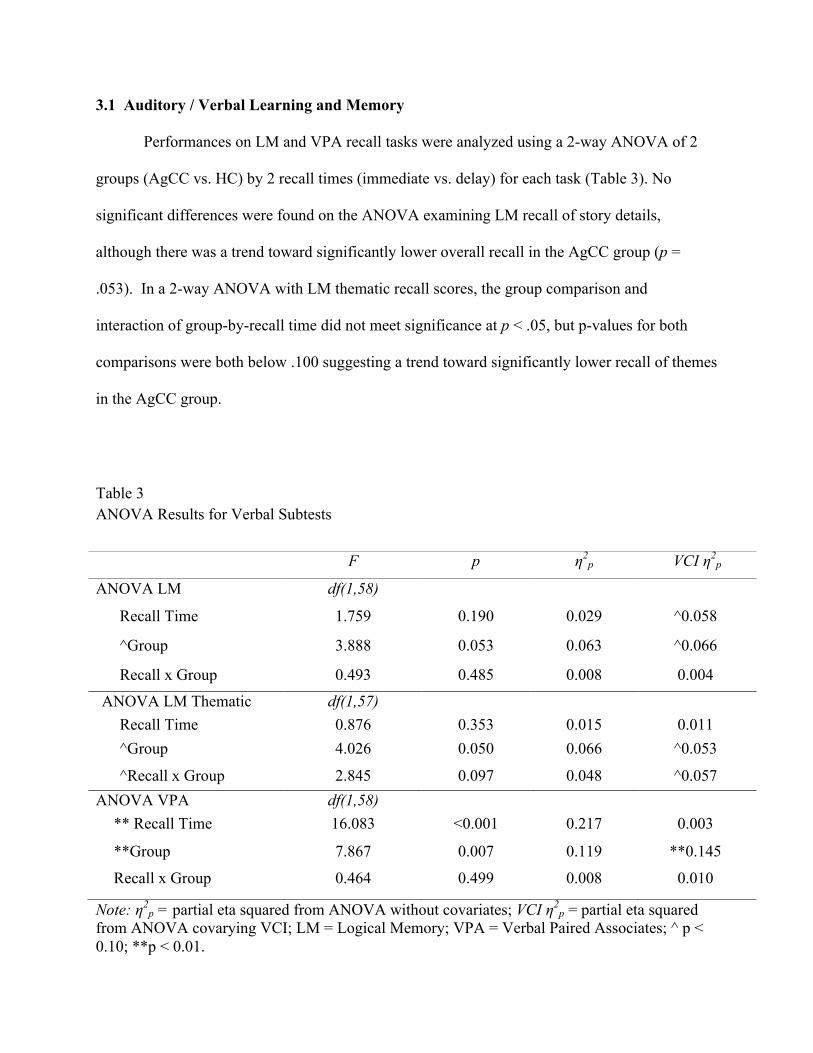
3.1 Auditory / Verbal Learning and Memory
Performances on LM and VPA recall tasks were analyzed using a 2-way ANOVA of 2
groups (AgCC vs. HC) by 2 recall times (immediate vs. delay) for each task (Table 3). No
significant differences were found on the ANOVA examining LM recall of story details,
although there was a trend toward significantly lower overall recall in the AgCC group (p =
.053). In a 2-way ANOVA with LM thematic recall scores, the group comparison and
interaction of group-by-recall time did not meet significance at p < .05, but p-values for both
comparisons were both below .100 suggesting a trend toward significantly lower recall of themes
in the AgCC group.
Table 3 ANOVA Results for Verbal Subtests
Note: η2p = partial eta squared from ANOVA without covariates; VCI η2
p = partial eta squared from ANOVA covarying VCI; LM = Logical Memory; VPA = Verbal Paired Associates; ^ p < 0.10; **p < 0.01.
F p η2p VCI η2
p
ANOVA LM df(1,58)
Recall Time 1.759 0.190 0.029 ^0.058
^Group 3.888 0.053 0.063 ^0.066
Recall x Group 0.493 0.485 0.008 0.004
ANOVA LM Thematic
Thematic
df(1,57) Recall Time 0.876 0.353 0.015 0.011 ^Group 4.026 0.050 0.066 ^0.053
^Recall x Group 2.845 0.097 0.048 ^0.057 ANOVA VPA df(1,58)
** Recall Time 16.083 <0.001 0.217 0.003
**Group 7.867 0.007 0.119 **0.145
Recall x Group 0.464 0.499 0.008 0.010
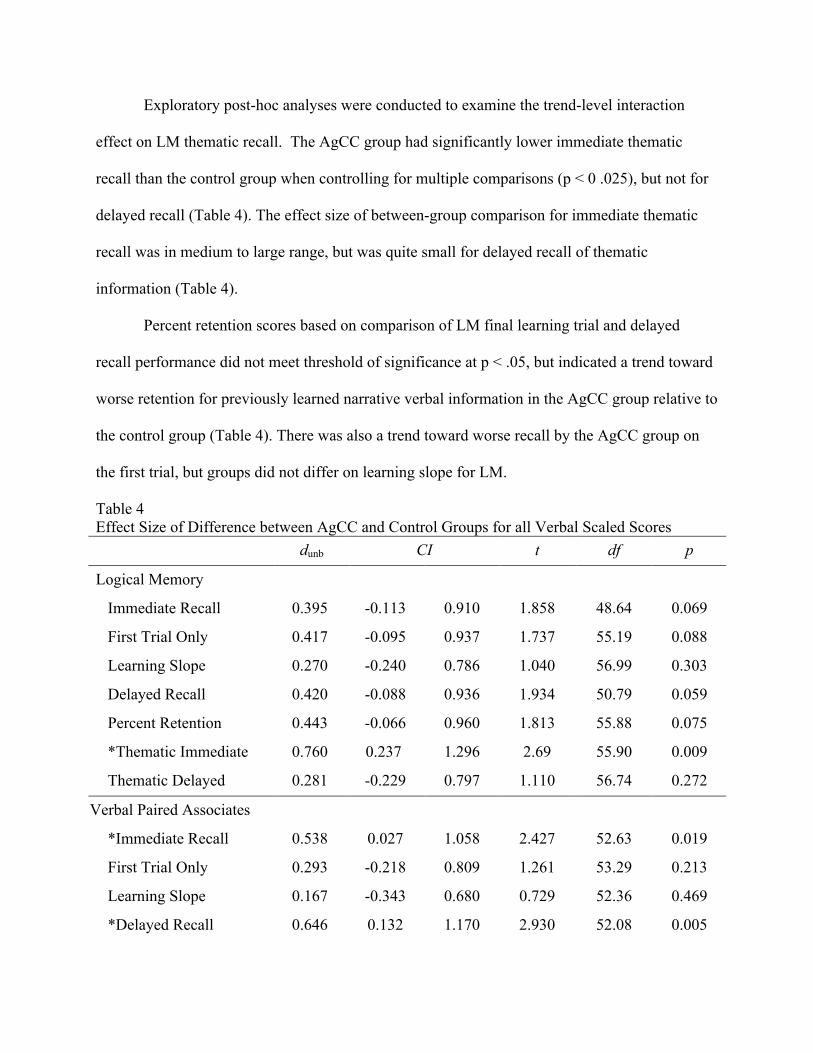
Exploratory post-hoc analyses were conducted to examine the trend-level interaction
effect on LM thematic recall. The AgCC group had significantly lower immediate thematic
recall than the control group when controlling for multiple comparisons (p < 0 .025), but not for
delayed recall (Table 4). The effect size of between-group comparison for immediate thematic
recall was in medium to large range, but was quite small for delayed recall of thematic
information (Table 4).
Percent retention scores based on comparison of LM final learning trial and delayed
recall performance did not meet threshold of significance at p < .05, but indicated a trend toward
worse retention for previously learned narrative verbal information in the AgCC group relative to
the control group (Table 4). There was also a trend toward worse recall by the AgCC group on
the first trial, but groups did not differ on learning slope for LM.
Table 4 Effect Size of Difference between AgCC and Control Groups for all Verbal Scaled Scores
dunb CI t df p
Logical Memory
Immediate Recall 0.395 -0.113 0.910 1.858 48.64 0.069
First Trial Only 0.417 -0.095 0.937 1.737 55.19 0.088
Learning Slope 0.270 -0.240 0.786 1.040 56.99 0.303
Delayed Recall 0.420 -0.088 0.936 1.934 50.79 0.059
Percent Retention 0.443 -0.066 0.960 1.813 55.88 0.075
*Thematic Immediate 0.760 0.237 1.296 2.69 55.90 0.009
Thematic Delayed 0.281 -0.229 0.797 1.110 56.74 0.272
Verbal Paired Associates
*Immediate Recall 0.538 0.027 1.058 2.427 52.63 0.019
First Trial Only 0.293 -0.218 0.809 1.261 53.29 0.213
Learning Slope 0.167 -0.343 0.680 0.729 52.36 0.469
*Delayed Recall 0.646 0.132 1.170 2.930 52.08 0.005
*Percent Retention 0.547 0.031 1.072 2.674 41.00 0.011 Note: dunb = Cohen’s d unbiased; CI = 95% Confidence Interval for dunb; t = independent samples t-test; df = degrees of freedom; p = p-value; * Confidence interval of Cohen’s d unbiased does not contain zero.
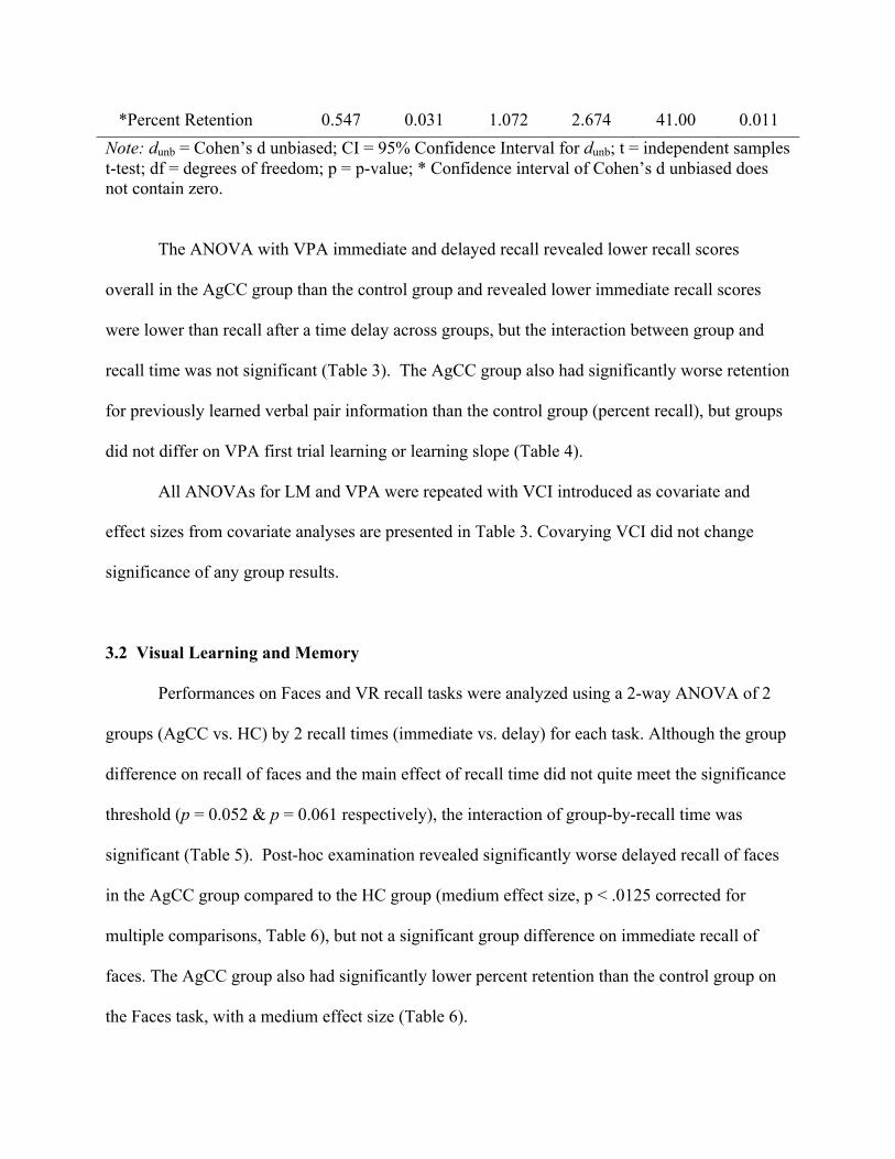
The ANOVA with VPA immediate and delayed recall revealed lower recall scores
overall in the AgCC group than the control group and revealed lower immediate recall scores
were lower than recall after a time delay across groups, but the interaction between group and
recall time was not significant (Table 3). The AgCC group also had significantly worse retention
for previously learned verbal pair information than the control group (percent recall), but groups
did not differ on VPA first trial learning or learning slope (Table 4).
All ANOVAs for LM and VPA were repeated with VCI introduced as covariate and
effect sizes from covariate analyses are presented in Table 3. Covarying VCI did not change
significance of any group results.
3.2 Visual Learning and Memory
Performances on Faces and VR recall tasks were analyzed using a 2-way ANOVA of 2
groups (AgCC vs. HC) by 2 recall times (immediate vs. delay) for each task. Although the group
difference on recall of faces and the main effect of recall time did not quite meet the significance
threshold (p = 0.052 & p = 0.061 respectively), the interaction of group-by-recall time was
significant (Table 5). Post-hoc examination revealed significantly worse delayed recall of faces
in the AgCC group compared to the HC group (medium effect size, p < .0125 corrected for
multiple comparisons, Table 6), but not a significant group difference on immediate recall of
faces. The AgCC group also had significantly lower percent retention than the control group on
the Faces task, with a medium effect size (Table 6).

No significant differences were found on the ANOVA with recall of abstract figures (VR;
Table 5).
Table 5 ANOVA Results for Visual Subtests
Note: η2p = partial eta squared from ANOVA without covariates; POI η2
p = partial eta squared from ANOVA covarying POI; VR = Visual Reproduction; *p < 0.05; ^p < 0.01. Table 6 Effect Size of Difference between AgCC and Control Groups for all Visual Scaled Scores dunb CI t df p
Faces
Immediate Recall 0.232 -0.274 0.742 1.014 54.81 0.315
*Delayed Recall 0.612 0.099 1.136 2.669 54.95 0.010
*Percent Retention 0.514 0.004 1.034 2.407 49.11 0.020
Visual Reproduction
Immediate Recall 0.276 -0.230 0.787 1.205 54.87 0.233
Delayed Recall 0.355 -0.152 0.869 1.655 49.77 0.104
Percent Retention 0.455 -0.054 0.972 2.038 53.14 0.047
F p η2p POI η2
p
ANOVA Faces df(1,58)
^Recall Time 3.65 0.061 0.059 0.049
^Group 3.95 0.052 0.064 ^0.058
*Recall x Group 5.32 0.025 0.084 *0.091
ANOVA VR df(1,58)
Recall Time 0.017 0.897 <0.001 0.015
Group 2.605 0.112 0.043 0.044
Recall x Group 0.419 0.520 0.007 0.004
Note: dunb = Cohen’s d unbiased; CI = Confidence Interval for dunb; t = independent samples t-test; df = degrees of freedom; p = p-value; * Confidence interval of Cohen’s d unbiased does not contain zero.
All ANOVAs involving the Faces and VR subtests were repeated with POI introduced as
covariate and effect sizes from covariate analyses are presented in Table 5. Covarying POI did
not change significance and had minimal impact on effect sizes of group results.
3.3 Clinically Significant Scores
Because the WMS-III is commonly used in clinical assessments, it is important to
identify the likelihood that individuals with AgCC might score outside of normative range. The
frequency of clinically significant scores for each group is reported in Table 2. Using alpha of
.05, only 1 participant in each group of 30 is expected by chance to have a clinically significant
score (> 1.5 sd below the mean). The control group met that expectation for most WMS-III
scores, with the exception of LM 1st trial only and both thematic memory scores. However, in the
AgCC group, the frequency of clinically significant scores was well above expectation for all
scores except VPA 1st trial learning and learning slope.
Section 4: Discussion
This study examined verbal and visual memory in 30 individuals with isolated AgCC and
age-and-IQ matched controls using the WMS-III Logical Memory, Verbal-Paired Associates,
Visual Reproduction, and Faces subtests. Relative to the control group, learning and memory
performance of the AgCC group varied across subtests. The AgCC group exhibited worse recall
than controls both immediately and after a time delay for rote word pairs and worse recall than
controls for faces after a time delay. The AgCC group also exhibited worse recall for thematic
information immediately after learning the stories, but did not differ from the control group on

memory for details from narratives or on recall for thematic information following a time delay.
The AgCC group did not differ from the control group on either immediate or delayed recall of
abstract figures. Contradictory to our prediction, compared to the control group the AgCC group
recalled less of what they had actually learned (i.e. percent retention) on tests with rote word-
pairs and faces. As predicted, groups did not differ on first trial learning or learning slope in
verbal memory tasks.
4.1 Verbal Learning and Memory
Cumulatively across the verbal learning trials with word pairs, the AgCC group had
worse immediate recall than the control group, despite the fact that both groups recalled a similar
amount of information from the first presentation and exhibited a similar degree of incremental
improvement with repetition. Erickson et al. (2014) also found comparable performances in
individuals with AgCC and controls on Donders’ Attention Span factor (which is comprised of
List A Trial 1, Percent Recall from Middle of the List, and List B) and no evidence of significant
group difference in learning slope. Current findings continue to support similarity between
individuals with AgCC and controls on focused auditory attention and acquisition of new rote
verbal information, and indicate that individuals with AgCC are generally less efficient than
controls in encoding the same amount of information overall for efficient later recall.
Although direct comparison within the AgCC group did not reveal differences in
performance on VPA and LM (immediate recall, t(29) = .054, p = .957, CI [-1.099, 1.159];
delayed recall, t(29) = 1.022, p = .315; CI [-0.631, 1.891]), the pattern of results across tests
suggests that weaknesses in AgCC may be more readily apparent in VPA scores during clinical
assessment. On both immediate and delayed recall, as well as on percent retention, effect sizes
for differences between AgCC and HC were larger for VPA than LM. Additionally, null-
hypothesis significant testing revealed worse performance in AgCC than HC groups on VPA
recall and percent retention, but did not find group differences on LM recall of details.
Task-dependent variations on memory performance were previously reported in a smaller
sample of individuals with AgCC who had impaired memory for complex semantic components
such as unrelated sentences, but intact memory for a simpler task (Sauerwein et al., 1994).
Similarly, the verbal information presented in the VPA subtest lacks both the inherent logic and
structure of a story and the categorical structure of lists to be learned in the CVLT-II.
Consequently, VPA places greater demand on the learner to generate a semantic associational
network encompassing the cue and the target words to aid recall. In the absence of a strong self-
generated semantic network, even information which was originally learned may not be readily
recalled. It is notable that the VPA task format provides examinees with cuing on each recall
item (i.e. the examiner reads the first word and participant provides the matching word), but
whatever benefit may have been gleaned from cuing was not sufficient to eliminate the challenge
posed by the task’s lack of inherent semantic context.
Erickson et al. also reported impaired verbal learning and memory in AgCC (Erickson et
al., 2014). Specifically, they reported impaired learning rates and delayed recall for word lists.
However, when recall was examined within the context of only what had been learned (i.e.
differences in performance between recall on the last learning trial and long delay free recall) the
AgCC group performed similarly to the HC group, which suggests the delayed recall scores on
the CVLT-II were primarily limited by the amount of information originally encoded during
learning trials.
To highlight the impact of story logic on recall, we tested recall of thematic information
from LM. Although the AgCC group had weaker immediate recall for thematic information, the

groups did not differ on recall of thematic information after the time delay. This pattern indicates
that individuals with AgCC retain more of the thematic information they initially learned,
perhaps indicating a greater reliance on semantic context provided in the story. The preservation
of these thematic associations may assist in recall of story details.
In order to directly identify factors which may account for lowered VPA performance in
AgCC, future studies should conduct memory tasks which more clearly control factors such as
explicit presentation narrative context, use of recognition cuing vs. free recall, and use of
semantic vs. rote cues.
4.2 Visual Learning and Memory
With regards to visual learning and memory, we found a trend toward significantly worse
performance in the AgCC group than the HC group on Faces but not on the VR task. Although
direct comparison within the AgCC group did not reveal significant differences in performance
on Faces and VR (immediate, t(29) = 1.844, p = .075; CI [ -0.126, 2.446]; delayed t(29) = 1.571,
p = .127; CI [-0.290, 2.210]), the pattern of results across tests suggests that weaknesses in
AgCC may be most readily apparent on clinical assessment of delayed recall for faces. Memory
performance in the AgCC group did not differ from the HC group on VR. Likewise, the groups
did not differ on immediate recall for faces. However, the AgCC group had significantly poorer
delayed recall of faces relative to the HC group and also recalled less of what they had originally
learned.
Difficulty with recalling faces is consistent with previous findings of impairments in
facial processing (Bridgman et al., 2014). Intact learning and memory for abstract figures is

consistent with previously reported case studies which utilized a variety of abstract spatial
patterns to test visual learning and memory in individuals with AgCC (Kessler et al., 1991;
Panos et al., 2001; Sauerwein et al., 1994). There are many factors which might account for
differential performance on the Faces and VR tasks, including meaningfulness vs. abstractness,
social vs. non-social nature of the stimuli, recognition cuing vs. free recall, and oral vs. grapho-
motor response modality. Future studies of visual learning and memory should be designed to
directly control for these factors in order to isolate what may account for performance variations
in AgCC.
4.3 Summary and Interpretations
Individuals with AgCC have the capacity to encode and retain new verbal and visual
information, but as a group they appear to have task-specific limitations in learning and recall of
rote verbal information and in delayed recall/retention of faces. Moreover, the current study
indicates that on the LM, VPA, Faces and VR subtests individuals with AgCC have greater
frequency of clinically significant impairments than predicted by the normal distribution.
Although previous case studies of individuals with AgCC reported overall WMS-III performance
falling within normal limits (Gott & Saul, 1978; Pirozzolo et al., 1979), use of the Wechsler
Memory Quotient (which is a index score comprised of both auditory and visual subtests)
obscured information regarding variations across tasks. In contrast, results from the present study
also suggest that learning and memory in AgCC may be differentially impacted by task-specific
factors other than the general domain (verbal vs. visual). Future studies of memory in AgCC
would benefit from implementation of well-controlled tasks that can isolate relative influences of
factors including explicit narrative context, rote cuing, semantic cuing, graphomotor response
production, and oral response production.
By selecting participants for whom callosal agenesis is the primary neuroanatomical
finding and is the only neuroanatomical malformation these participants have in common, we
can infer that diminished callosal connectivity accounts for the shared profile of cognitive
deficits. Additionally, it would follow that the degree of disconnection would mediate this
cognitive performance. However, groupwise comparisons of participants with complete and
partial AgCC (reported supplementary materials) did not support a pattern of stronger learning
and memory in individuals with some callosal connections (partial AgCC) compared to those
with no connections (complete AgCC).
There is considerable variability in the pattern of interhemispheric connections provided
by remaining callosal fibers in partial AgCC (Wahl et al., 2009). Consequently, it should not be
inferred that the location of the residual callosum correlates with the connectivity pattern and
structure of a similarly located region in an intact corpus callosum. Accurate description of
residual callosal connections in pAgCC requires analysis of diffusion and / or functional MRI
data, which was beyond the scope of this study. However, it may be informative in future studies
to correlate cognitive performance with the area and degree of residual callosal connectivity in
the pAgCC subjects as assessed with MRI techniques.
There are two main perspectives from which to explain the contribution of callosal
connections to learning and memory capacity: hemispheric specialization and processing
resource limitations. These are complimentary perspectives on this relationship, not
contradictory alternatives.
From the perspective of hemispheric specialization, absence of the corpus callosum
disconnects hemispherically lateralized associative networks that aid in memory encoding and
retrieval. This may be particularly important for rote verbal learning. Adequate encoding of
isolated words requires the ability to imagine and generate “meaningful” associations between
the unrelated words, which would be difficult without interaction between visual and
paralinguistic processing systems primarily located in the right hemisphere (Van Lanker Sidtis &
Postman, 2006) and more concrete semantic language systems in the left. Limited integration of
these localized processing systems has been hypothesized to explain several deficits in
individuals with AgCC, such as difficulty generating stories to connect pieces of information
presented in a picture format (Turk, Brown, Symingtion, & Paul, 2010) and deficits in
comprehending the second order meanings of language, such as humor and nonliteral language,
which are largely inferential (Brown, Paul, et al., 2005; Brown, Symingtion, et al., 2005; Paul et
al., 2003). Taken together, these findings suggest that interhemispheric transfer deficits in AgCC
may interfere with the ability to envision, generate, and integrate more complex information into
“meaningful” cognitive associations, as evidenced herein by poor paired associate learning and
recall.
Although memory for faces had not previously been studied in AgCC, Bridgman et al.
(Bridgman et al., 2014) found that individuals with AgCC had impaired recognition of facial
emotion related to specific deficits in processing of the most salient features of the different faces
(i.e., the eyes). They posited that these impairments in face processing might be attributable to
disconnection between face processing in the non-dominant hemisphere and semantic and
conceptual representations in the language-dominant hemisphere. Applying this theory to the
current findings of impaired delayed memory for faces, it is possible that the AgCC group had

increased difficulty associating their visual processing with verbal labels which resulted in less
efficient recall.
In order to apply similar logic to the VR task, we must not only presume that language
and visual-spatial processing are lateralized in opposite hemispheres but we must also identify
the direction of laterality. Presuming directionality of hemispheric specialization is similar in
complete AgCC as in the general population, we would expect the hemisphere dominant for
spatial processing to simultaneously control the writing hand in 3 out of 4 left-handed
participants, but none of the right-handed participants with complete AgCC. Consequently, VR
performance would be better in the left-handed group. However, VR immediate and delayed
recall did not differ between right- and left-handed responders with complete AgCC, η2p = .129,
F(1,18) = 2.667, p = .120. In fact, the left-handed responders’ average performance was below
the average for right-handers. Thus, assuming functional organization of spatial processing is
right-lateralized in individuals with AgCC and memory traces for spatial information are
established primarily in the right hemisphere, poorer performance in the AgCC group cannot be
explained simply by direct disruption of information transfer between the right-hemisphere and
the hand which controlled drawing.
From the perspective of limited overall processing resources, the theory here focuses on
the role of the corpus callosum in marshaling large neural networks to process information of all
sorts. Thus AgCC would result in reduced availability of richer cortical networks to support
processing of particularly complex and novel information (Brown & Paul, 2000). As a
consequence, in the present study individuals with AgCC may have scored lower on the
immediate and delayed recall of verbal pairs as a result of the continued demand to process novel
association, which potentially overloaded their cognitive resources. In contrast, although the

information presented in the story format also demanded processing of novel information, it is
possible that the inclusion of the thematic linkage carried by the narrative reduced the
complexity of the encoding task, allowing the individuals with AgCC to process it more readily
in comparison to the more complex task of encoding unrelated words in the VPA task. Likewise,
greater impairment in memory for faces as compared to design memory could also be explained
in terms of the complexity of the stimulus material. Specifically, individuals with AgCC may
have had difficulty with the recall of faces because the spatial configurations marking differences
in specific faces are generally more novel, complex, and subtle than the stimuli used in the visual
reproduction subtest.
4.4 Limitations and Future Directions
Whatever the nature of the relationship between callosal function and memory encoding,
we presume that the deficits in learning and delayed recall in individuals with AgCC shown in
this study can be attributed to the largest brain abnormality consistently present in this group
(i.e., complete or partial absence of the corpus callosum) and have intentionally selected a
population with few if any other visible brain abnormalities on MRI (other than presence of
Probst bundles or colpocephaly which are structural changes typically accompanying AgCC).
However, it is possible that undetected microscopic abnormalities might be consistently present
and contributing to abnormal learning and memory. For instance, postmortem histological
inspection of two brains with callosal dysgenesis revealed significant differences in the number
of Von Economo neurons (Kaufman et al., 2008). It is also possible that memory disturbance
does not directly result from callosal disconnection, but rather is a by-product of functional
disruption in some other neural system as a result of the acallosal brain’s compensatory
reorganization during development. However, it is most likely the case that compensatory

reorganization would ameliorate the impact of callosal absence on memory and reduce the
impact of AgCC on learning and memory.
Finally, it is noteworthy that the results described above are based on group-wise
analyses. While individuals with AgCC had a greater than expected likelihood of scoring within
the borderline to impaired range on all scores except VPA 1st trial learning and learning slope,
there were also individuals with AgCC who scored in the superior range on some subtests. The
presence of individuals with complete AgCC with superior scores suggests that there may be
intervening factors that modulate the impact of callosal absence on memory encoding and
retrieval. For example, although intelligence scores did not account for differences between
groups, they appear to be uniquely relevant to select subtest performances within the AgCC
group (Supplementary Table 4). In addition, more intense past experience with any elements of a
task would raise scores relative to others with less experience. This seems to be a particularly
important element in domains of above normal capacity occasionally seen in individuals with
AgCC.
4.5 Conclusions
This study supports the hypothesis that callosal absence interferes with the overall
efficiency of auditory and visual learning and memory, with greater impairments noted on paired
associates and delayed memory for faces. The current results from individuals with AgCC
suggest several interpretations of the contribution of interhemispheric interactions via the corpus
callosum to memory. These interpretations are not, however, mutually exclusive, but may reflect
different ways of viewing the impact of reduced hemispheric connectivity. Specifically, the
results could be explained in terms of less efficient processing of information related to reduced
interhemispheric transfer of information or decreased capacity to process the information in a
more richly associative neural network. This study also tentatively suggests that deficits in
individuals with AgCC are related to the demand to imagine and generate semantic linkage of
concepts, and greater complexity of encoding and recalling faces over visual designs. Results
suggest that memory deficits are a characteristic aspect of the neuropsychological profile in
individuals with AgCC.
Acknowledgments
Portions of this paper served as the masters thesis of J.Hartman at the Travis Research Institute,
Fuller Graduate School of Psychology.

References
Bodensteiner, J., Schaefer, G. B., Breeding, L., & Cowan, L. (1994). Hypoplasia of the corpus callosum: a study of 445 consecutive MRI scans. J Child Neurol, 9(1), 47-49.
Bridgman, M. W., Brown, W. S., Spezio, M. L., Leonard, M. K., Adolphs, R., & Paul, L. K. (2014). Facial emotion recognition in agenesis of the corpus callosum. J Neurodev Disord, 6. doi:10.1186/1866-1955-6-32
Brown, W. S., Jeeves, M. A., Dietrich, R., & Burnison, D. S. (1999). Bilateral field advantage and evoked potential interhemispheric transmission in commissurotomy and callosal agenesis. Neuropsychologia, 37(10), 1165-1180.
Brown, W. S., & Paul, L. K. (2000). Cognitive and psychosocial deficits in agenesis of the corpus callosum with normal intelligence. Cognitive neuropsychiatry, 5(2), 135-157.
Brown, W. S., Paul, L. K., Symington, M., & Dietrich, R. (2005). Comprehension of humor in primary agenesis of the corpus callosum. Neuropsychologia, 43(6), 906-916. doi:10.1016/j.neuropsychologia.2004.09.008
Brown, W. S., Symingtion, M., VanLancker-Sidtis, D., Dietrich, R., & Paul, L. K. (2005). Paralinguistic processing in children with callosal agenesis: emergence of neurolinguistic deficits. Brain and Language, 93(2), 135-139. doi:10.1016/j.bandl.2004.09.003
Brown, W. S., Thrasher, E. D., & Paul, L. K. (2001). Interhemispheric Stroop effects in partial and complete agenesis of the corpus callosum. J Int Neuropsychol Soc, 7(3), 302-311.
Cohen, J. (1988). Statistical power analysis for the behavioral sciences (2nd Ed.). Hillsdale, NJ: Lawrence Earlbaum Associates..
Clark, C. R., & Geffen, G. M. (1989). Corpus callosum surgery and recent memory. A review. Brain, 112 ( Pt 1), 165-175.
Cumming, G. (2012). Understanding The New Statistics: Effect Sizes, Confidence Intervals, and Meta-Analysis. . New York: Routledge.
David, A. S., Wacharasindhu, A., & Lishman, W. A. (1993). Severe psychiatric disturbance and abnormalities of the corpus callosum: Review and case series. Journal of Neurology, Neurosurgery, and Psychiatry, 56, 85-93.
Delis, D. C., Kaplan, E., Kramer, J., & Ober, B. (1994). California Verbal Learning Test - Children's Version. San Antonio, TX: The Psychological Corporation.
Delis, D. C., Kaplan, E., Kramer, J., & Ober, B. (2000). California Verbal Learning Test - Second Edition (CVLT-II). San Antonio, TX: The Psychological Corporation.
Donders, J. (2008). A Confirmatory Factor Analysis of the California Verbal Learning Test--Second Edition (CVLT-II) in the Standardization Sample. Assessment, 15.
Erickson, R. L., Paul, L. K., & Brown, W. S. (2014). Verbal learning and memory in agenesis of the corpus callosum. Neuropsychologia, 60, 121-130. doi:10.1016/j.neuropsychologia.2014.06.003
Fischer, M., Ryan, S. B., & Dobyns, W. B. (1992). Mechanisms of interhemispheric transfer and patterns of cognitive function in acallosal patients of normal intelligence. Arch Neurol, 49(3), 271-277.
Geffen, G., Forrester, G., Jones, D., & Simpson, D. (1994). Auditory verbal learning and memory in cases of callosal agenesis. In M. Lassonde & M. Jeeves (Eds.), Callosal Agenesis: A natural split brain? (pp. 247-260). New York: Plenum Press.

Glass, H., Shaw, G., Ma, C., & Sherr, E. H. (2008). Agenesis of the corpus callosum in California 1983-2003: a population-based study. Am J Med Genet A, 146A(19), 2495-2500.
Gott, P. S., & Saul, R. E. (1978). Agenesis of the corpus callosum: limits of functional compensation. Neurology, 28(12), 1272-1279.
Hines, R., Paul, L., & Brown, W. (2002). Spatial attention in agenesis of the corpus callosum: shifting attention between visual fields. Neuropsychologia, 40(11), 1804-1814.
Jeret, J. S., Serur, D., Wisniewski, K., & Fisch, C. (1985). Frequency of agenesis of the corpus callosum in the developmentally disabled population as determined by computerized tomography. Pediatr Neurosci, 12(2), 101-103.
Kaufman, J. A., Paul, L. K., Manaye, K. F., Granstedt, A. E., Hof, P. R., Hakeem, A. Y., & Allman, J. M. (2008). Selective reduction of Von Economo neuron number in agenesis of the corpus callosum. Acta Neuropathol, 116(5), 479-489.
Kessler, J., Huber, M., Pawlik, G., Heiss, W. D., & Markowitsch, H. J. (1991). Complex sensory cross integration deficits in a case of corpus callosum agenesis with bilateral language representation: Positron-Emission-Tomography and neuropsychological findings. International Journal of Neuroscience, 58, 275-282.
Lafosse, J. M., Mitchell, S. M., Corboy, J. R., & Filley, C. M. (2013). The nature of verbal memory impairment in multiple sclerosis: a list-learning and meta-analytic study. J Int Neuropsychol Soc, 19(9), 995-1008. doi:10.1017/S1355617713000957
Ledoux, J. E., Risse, G. L., Springer, S. P., Wilson, D. H., & Gazzaniga, M. S. (1977). Cognition and commissurotomy. Brain, 100 Pt 1, 87-104.
Marco, E. J., Harrell, K. M., Brown, W. S., Hill, S. S., Jeremy, R. J., Kramer, J. H., . . . Paul, L. K. (2012). Processing speed delays contribute to executive function deficits in individuals with agenesis of the corpus callosum. J Int Neuropsychol Soc, 18(3), 521-529. doi:10.1017/S1355617712000045
Mueller, K. L. O., Marion, S. D., Paul, L. K., & Brown, W. S. (2009). Bimanual Motor Coordination in Agenesis of the Corpus Callosum. Behavioral Neuroscience, 123, 1000-1011.
Panos, P. T., Porter, S. S., Panos, A. J., Gaines, R. N., & Erdberg, P. S. (2001). An evaluation of a case of agenesis of the corpus callosum with Rourke's nonverbal learning disorder model. Archives of clinical neuropsychology : the official journal of the National Academy of Neuropsychologists, 16(5), 507-521.
Paul, L. K., Brown, W. S., Adolphs, R., Tyszka, J. M., Richards, L. J., Mukherjee, P., & Sherr, E. H. (2007). Agenesis of the corpus callosum: genetic, developmental and functional aspects of connectivity. Nat Rev Neurosci, 8(4), 287-299. doi:10.1038/nrn2107
Paul, L. K., Van Lancker-Sidtis, D., Schieffer, B., Dietrich, R., & Brown, W. S. (2003). Communicative deficits in agenesis of the corpus callosum: nonliteral language and affective prosody. Brain and Language, 85(2), 313-324.
Phelps, E. A., Hirst, W., & Gazzaniga, M. S. (1991). Deficits in recall following partial and complete commissurotomy. Cereb Cortex, 1(6), 492-498.
Pirozzolo, F. J., Pirozzolo, P. H., & Ziman, R. B. (1979). Neuropsychological assessment of callosal agenesis: Report of a case with normal intelligence and absence of disconnexion syndrome. Clinical Neuropsychology, 13-16.
Rey, A. (1958). L’examen clinique en psychologie. Paris: Presse Univeristaire de France.

Sauerwein, H. C., & Lassonde, M. (1994). Cognitive and sensori-motor functioning in the absence of the corpus callosum: neuropsychological studies in callosal agenesis and callosotomized patients. Behav Brain Res, 64(1-2), 229-240.
Sauerwein, H. C., Nolin, P., & Lassonde, M. (1994). Cognitive functioning in callosal agenesis. In M. Lassonde & M. A. Jeeves (Eds.), Callosal agenesis: A natural split brain? (pp. 221-233). New York: Plenum Press.
Siffredi, V., Anderson, V., Leventer, R. J., & Spencer-Smith, M. M. (2013). Neuropsychological Profile of Agenesis of the Corpus Callosum: A Systematic Review. Developmental neuropsychology, 38. doi:10.1080/87565641.2012.721421
Smith, L., Rourke, B., & Rourke, B. P. (1994). Callosal agenesis: a case study in NLD. Solursh, L. P., Margulies, A. I., Ashem, B., & Stasiak, E. A. (1965). The relationships of
agenesis of the corpus callosum to perception and learning. J Nerv Ment Dis, 141(2), 180-189.
Sperry, R. W. (1968). Hemisphere deconnection and unity in conscious awareness. Am Psychol, 23(10), 723-733.
Sperry, R. W., Gazzaniga, M., Bogen, J., Vinken, P. J., & Bruyn, G. W. (1969). Interhemispheric relationships: the neocortical commissures; sydromes of hemisphere disconnection. Handbook of Clinical Neurology (Vol. 4, pp. 273-290).
Turk, A., Brown, W. S., Symingtion, M., & Paul, L. K. (2010). Social narratives in agenesis of the corpus callosum: Linguistic analysis of the Thematic Apperception Test. Neuropsychologia, 48, 43-50.
Van Lanker Sidtis, D., & Postman, W. A. (2006). Formulaic expressions in spontaneous speech of left hemisphere- and right hemisphere-damaged subjects. Aphasiology, 20, 411 - 426. doi:10.1080/02687030500538148
Wahl, M., Strominger, Z., Jeremy, R. J., Barkovich, A. J., Wakahiro, M., Sherr, E. H., & Mukherjee, P. (2009). Variability of homotopic and heterotopic callosal connectivity in partial agenesis of the corpus callosum: a 3T diffusion tensor imaging and Q-ball tractography study. AJNR Am J Neuroradiol, 30(2), 282-289.
Wang, P. N., Chou, K. H., Chang, N. J., Lin, K. N., Chen, W. T., Lan, G. Y., . . . Lirng, J. F. (2014). Callosal degeneration topographically correlated with cognitive function in amnestic mild cognitive impairment and Alzheimer's disease dementia. Hum Brain Mapp, 35(4), 1529-1543. doi:10.1002/hbm.22271
Wechsler, D. (1945). A standardized memory scale for clinical use. Psychology, 19, 87-95. Wechsler, D. (1997a). Wechsler Adult Intellligence Scale - Third Edition. San Antonio, Tx: The
Psychological Corporation. Wechsler, D. (1997b). Wechsler Memory Scale - Third Edition. San Antonio, TX: The
Psychological Corporation. Wechsler, D. (1999). WASI Administration and Scoring Manual. San Antonio, TX:
Psychological Corporation. Wechsler, D. (2011). Wechsler Abbreviated Scale of Intelligence - Second Edition. San Antonio,
TX: The Psychological Corporation. Zaidel, D., & Sperry, R. W. (1974). Memory impairment after commissurotomy in man. Brain,
97(2), 263-272. Zaidel, E. (1990). Long-term semantic memory in the two cerebral hemispheres. New York:
Cambridge University Press.
Highlights
Lynn K. Paul, Roger Erickson, Jo Ann Hartman, and Warren S. Brown, “Memory in Individuals with Agenesis of the Corpus Callosum”
• Callosal connections play an important role in verbal / visual learning and memory
• Persons with corpus callosum agenesis are likely to show memory impairment on
standardized tests
• Persons with corpus callosum agenesis are likely to have difficulty on delayed
recognition/retention of faces.
• Persons with corpus callosum agenesis are likely to have difficulty on both immediate
and delayed recall of rote word pairs.
• Persons with corpus callosum agenesis are likely to have difficulty recalling thematic
information immediately after hearing new narratives.
Supplemental Results
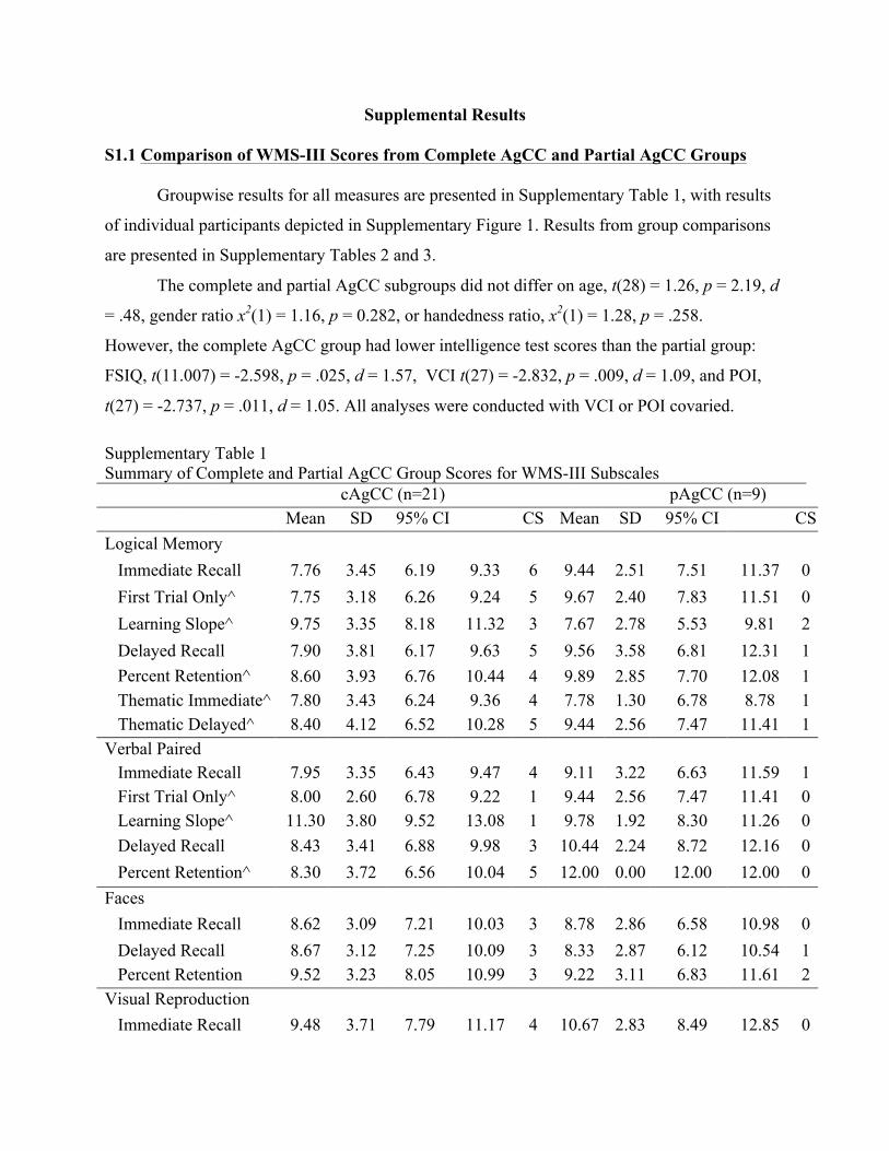
S1.1 Comparison of WMS-III Scores from Complete AgCC and Partial AgCC Groups
Groupwise results for all measures are presented in Supplementary Table 1, with results
of individual participants depicted in Supplementary Figure 1. Results from group comparisons
are presented in Supplementary Tables 2 and 3.
The complete and partial AgCC subgroups did not differ on age, t(28) = 1.26, p = 2.19, d
= .48, gender ratio x2(1) = 1.16, p = 0.282, or handedness ratio, x2(1) = 1.28, p = .258.
However, the complete AgCC group had lower intelligence test scores than the partial group:
FSIQ, t(11.007) = -2.598, p = .025, d = 1.57, VCI t(27) = -2.832, p = .009, d = 1.09, and POI,
t(27) = -2.737, p = .011, d = 1.05. All analyses were conducted with VCI or POI covaried.
Supplementary Table 1 Summary of Complete and Partial AgCC Group Scores for WMS-III Subscales cAgCC (n=21) pAgCC (n=9) Mean SD 95% CI CS Mean SD 95% CI CS Logical Memory
Immediate Recall 7.76 3.45 6.19 9.33 6 9.44 2.51 7.51 11.37 0 First Trial Only^ 7.75 3.18 6.26 9.24 5 9.67 2.40 7.83 11.51 0 Learning Slope^ 9.75 3.35 8.18 11.32 3 7.67 2.78 5.53 9.81 2 Delayed Recall 7.90 3.81 6.17 9.63 5 9.56 3.58 6.81 12.31 1 Percent Retention^ REnRetention+
8.60 3.93 6.76 10.44 4 9.89 2.85 7.70 12.08 1 Thematic Immediate^ 7.80 3.43 6.24 9.36 4 7.78 1.30 6.78 8.78 1 Thematic Delayed^ 8.40 4.12 6.52 10.28 5 9.44 2.56 7.47 11.41 1
Verbal Paired Associates
Immediate Recall 7.95 3.35 6.43 9.47 4 9.11 3.22 6.63 11.59 1 First Trial Only^ 8.00 2.60 6.78 9.22 1 9.44 2.56 7.47 11.41 0 Learning Slope^ 11.30 3.80 9.52 13.08 1 9.78 1.92 8.30 11.26 0 Delayed Recall 8.43 3.41 6.88 9.98 3 10.44 2.24 8.72 12.16 0 Percent Retention^ 8.30 3.72 6.56 10.04 5 12.00 0.00 12.00 12.00 0
Faces Immediate Recall 8.62 3.09 7.21 10.03 3 8.78 2.86 6.58 10.98 0 Delayed Recall 8.67 3.12 7.25 10.09 3 8.33 2.87 6.12 10.54 1 Percent Retention 9.52 3.23 8.05 10.99 3 9.22 3.11 6.83 11.61 2
Visual Reproduction Immediate Recall 9.48 3.71 7.79 11.17 4 10.67 2.83 8.49 12.85 0

Delayed Recall 9.29 3.94 7.50 11.08 5 10.11 4.57 6.60 13.62 1 Percent Retention 9.33 3.88 7.56 11.10 5 9.67 4.47 6.23 13.11 2
Note: AgCC = participants with agenesis of corpus callosum; SD = standard deviation; CS = number of participants whose scores were clinically significant (i.e. over 1.5 standard deviations below the normative mean; ^ = Complete AgCC group n of 20.
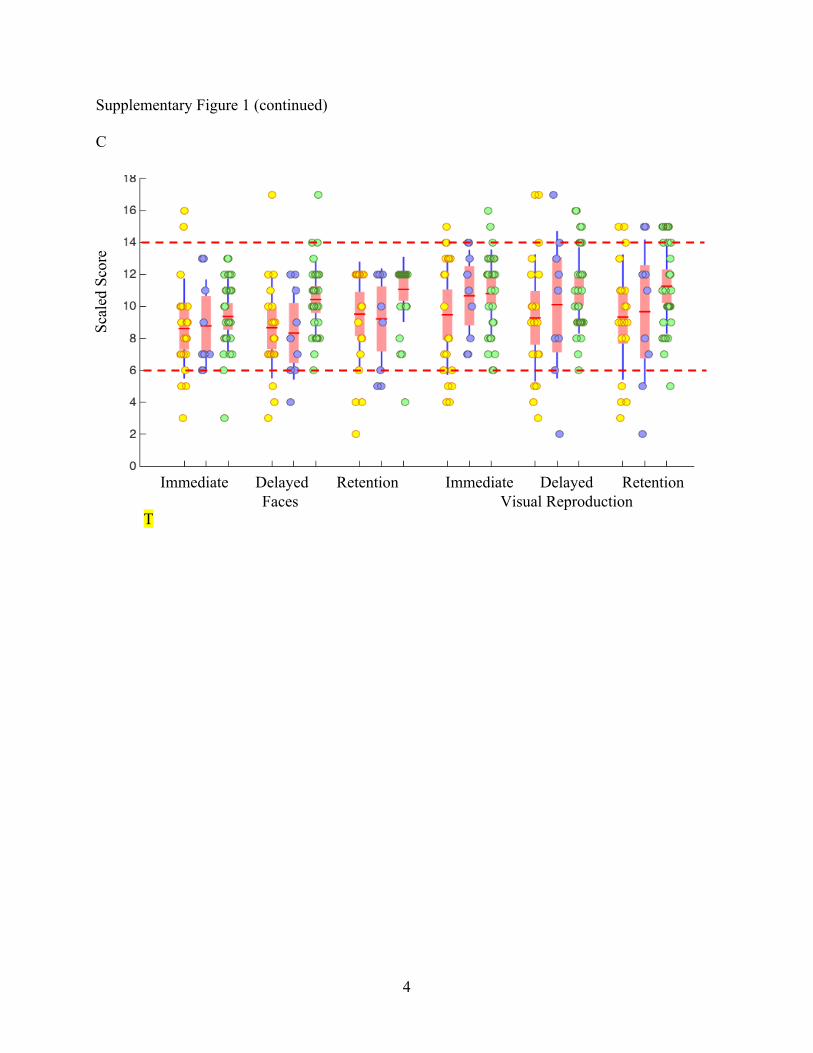
(Insert Supplementary Figure 1)
There were no significant group differences from a 2-way ANOVA of 2 groups
(complete AgCC vs. partial AgCC) by 2 recall times (immediate vs. delay), for either LM or
VPA (with or without VCI included as covariate; Supplementary Table 2). There were also no
significant findings in a 2-way ANOVA for LM thematic recall.
Independent samples t-tests found no differences between complete and partial AgCC
groups for learning slope and first trial recall on LM and VPA, nor for percent retention on LM.
Alhtough percent retention for VPA was significantly higher in the partial AgCC group, t(19) =
4.454, p < .001, covarying VCI reduced the group difference to a trend, F(1,25) = 2.974, p =
0.097, η2p = = 0.106.

Supplementary Table 2 ANOVA Results for Verbal Subtests Comparing Complete and Partial AgCC, with VCI covaried
Note: η2p = partial eta squared; LM = Logical Memory; VPA = Verbal Paired Associates; *p <
0.05.
Faces and VR scaled scores were compared using a 2-way ANOVA (2 groups by 2
recall times) and no significant differences were found with or without POI covaried
(Supplementary Table 3).
Supplementary Table 3
ANOVA Results for Visual Subtests Partial vs. Complete AgCC, with POI covaried
F p η2p VCI η2
p
ANOVA LM df(1,28)
Recall Time 0.140 0.711 0.005 0.091
Group 1.533 0.226 0.052 0.017
Recall x Group 0.002 0.963 <0.001 0.026
ANOVA LM Thematic
Thematic
df(1,27) + Recall Time 3.636 0.067 0.119 <0.001 Group 3.243 0.677 0.007 0.019
Recall x Group 0.805 0.377 0.029 0.027 ANOVA VPA df(1,28)
** Recall Time 7.887 0.009 0.220 0.014
Group 1.632 0.212 0.055 0.006
Recall x Group 1.770 0.194 0.059 0.043
F p η2p POI η2
p
ANOVA Faces df(1,28)
Recall Time 0.226 0.639 0.008 <0.001
Group 0.006 0.939 <0.001 0.015 Recall x Group 0.347 0.561 0.012 0.002
ANOVA VR df(1,28)
Recall Time 0.390 0.537 0.014 0.079
Note: η2
p = partial eta squared; VR = Visual Reproduction; *p < 0.05; **p < 0.01.
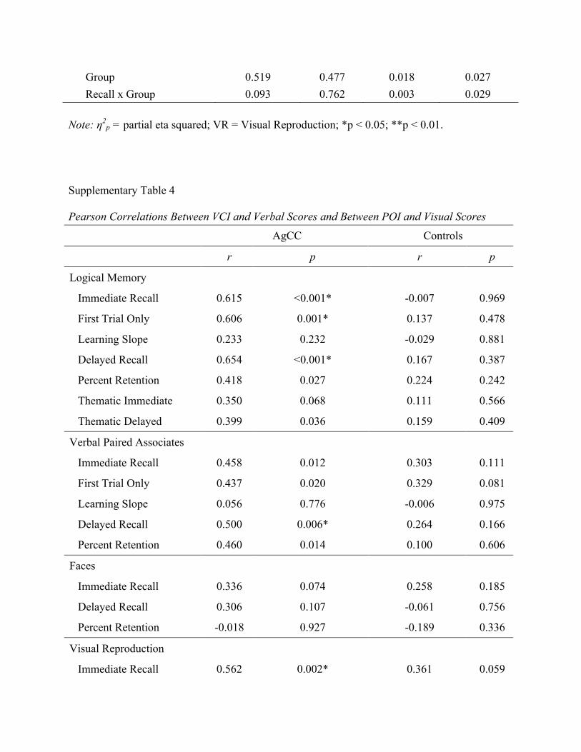
Supplementary Table 4
Pearson Correlations Between VCI and Verbal Scores and Between POI and Visual Scores AgCC Controls r p r p
Logical Memory
Immediate Recall 0.615 <0.001* -0.007 0.969
First Trial Only 0.606 0.001* 0.137 0.478
Learning Slope 0.233 0.232 -0.029 0.881
Delayed Recall 0.654 <0.001* 0.167 0.387
Percent Retention 0.418 0.027 0.224 0.242
Thematic Immediate 0.350 0.068 0.111 0.566
Thematic Delayed 0.399 0.036 0.159 0.409
Verbal Paired Associates
Immediate Recall 0.458 0.012 0.303 0.111
First Trial Only 0.437 0.020 0.329 0.081
Learning Slope 0.056 0.776 -0.006 0.975
Delayed Recall 0.500 0.006* 0.264 0.166
Percent Retention 0.460 0.014 0.100 0.606
Faces
Immediate Recall 0.336 0.074 0.258 0.185
Delayed Recall 0.306 0.107 -0.061 0.756
Percent Retention -0.018 0.927 -0.189 0.336
Visual Reproduction
Immediate Recall 0.562 0.002* 0.361 0.059
Group 0.519 0.477 0.018 0.027 Recall x Group 0.093 0.762 0.003 0.029

Delayed Recall 0.638 <0.001* 0.366 0.055
Percent Retention 0.662 <0.001* 0.287 0.138 *p < 0.05, corrected for multiple comparisons within subtest
S1.2 Correlation of Short and Long Delay Scores on Each WMS-III Subtest with CVLT-II
As indicated in methods, 19 participants with AgCC and 11 HC participants completed
both the WMS-III and CVLT-II. Pearson correlations were conducted within each group,
comparing short and long delay recall and percent retention on each WMS-III subtest with
CVLT-II (on CVLT-II percent retention is called the ‘first rapid forgetting index’). For WMS-III
Logical Memory and Verbal Paired Associates, we also examined correlations with CVLT-II on
single trial learning and learning slope.
While the HC group retained no significant correlations following correction for multiple
comparisons, several correlations within AgCC group remained significant. Single trial learning
on CVLT-II was positively correlated with first trial recall for WMS-III Logical Memory and
Verbal Paired Associates. Both short and long delay free recall on CVLT-II were positively
correlated with short and long delay free recall on all WMS-III tasks. This indicates that there is
an overall pattern of intact initial encoding on learning tasks, paired with limited amounts of
information spontaneously recalled both immediately after learning and after a time delay.
Although AgCC group performance on learning slope did not differ from the HC on either
CVLT-II or WMS-III, these were not strongly correlated. Nor was there a significant correlation
between percent retention on CVLT-II and WMS-III subtests.
(Insert Supplementary Figure 2)

1
Figure 1 A
B
Immediate Delayed Retention Immediate Delayed Retention Logical Memory Verbal Paired Associates
Immediate Delayed Logical Memory Thematic
Fig. 1 Recall and retention scaled scores for Logical Memory and Verbal Paired Associates (A), Logical Memory thematic recall scaled scores (B), and Faces and Visual Reproduction (C) presented for each group as boxplots with individual participant scores overlaid (AgCC = yellow, control = green). Scores above the top dotted line and below the bottom dotted line are greater than 1.5 standard deviations from the normative mean.
Scal
ed S
core
Sc
aled
Sco
re

2
Figure 1 (continued) C
Immediate Delayed Retention Immediate Delayed Retention Faces Visual Reproduction
Scal
ed S
core

3
Supplementary Figure 1
A
B
Immediate Delayed Retention Immediate Delayed Retention Logical Memory Verbal Paired Associates
Immediate Delayed Logical Memory Thematic
Supplementary Fig. 1 Recall and retention scaled scores for Logical Memory and Verbal Paired Associates (A), Logical Memory thematic recall scaled scores (B), and Faces and Visual Reproduction (C) presented for each AgCC subgroup and controls as boxplots with individual participant scores overlaid (complete AgCC = yellow, partial AgCC = blue, control = green). Scores above the top dotted line and below the bottom dotted line are greater than 1.5 standard deviations from the normative mean.
Scal
ed S
core
Sc
aled
Sco
re
4
Supplementary Figure 1 (continued)
C
T
Immediate Delayed Retention Immediate Delayed Retention Faces Visual Reproduction
Scal
ed S
core

5
Supplementary Figure 2
CVLT-II
Single Trial
Learning Slope
SD Free Recall
LD Free Recall
Percent Retention
Logical Memory
Verbal Paired Associates
Faces
Visual Reproduction
Supplementary Fig. 2 Correlations between CVLT-II and WMS-III subtest scaled scores in individuals with AgCC (n = 19). Cells shown in dark blue are not significant at p < 0.05, corrected for multiple comparisons. Color scale indicates Pearson correlation. SD = short delay; LD = long delay.
WM
S-II
I