paths to the pancreas
TRANSCRIPT
news & views
nature genetics • volume 32 • september 2002 85
Deciphering the function of a gene-regulatory factor in development can betricky. Gene expression studies andmutant phenotypes generated by geneinactivation (knockout) can reveal whereand when a factor must function, but maymiss key roles in controlling cell-fate deci-sions. For example, loss of a regulatoryfactor may prevent a cell from adopting aparticular fate. This can result in a cell dif-ferentiating along an alternative routethat is normally adopted by other cells inthe original progenitor population. Thefactor-deficient cells adopting this alterna-tive fate would be difficult to distinguishfrom cells that normally adopt this fate.
The definitive way to assess this is toperform a simultaneous knockout andlineage analysis, where the fate of cellslacking the factor is followed duringdevelopment. In the accompanyingpaper1, Yoshiya Kawaguchi and colleaguespresent such an analysis for the exocrinepancreatic transcription factor subunitPtf1a. The unexpected finding is thatPtf1a has an earlier role in pancreatic fatedecisions than was previously thought,which nicely illustrates how lineage analy-sis can define the functions of transcrip-tion factors in development.
A pancreas primerThe pancreas contains exocrine cells thatsurround ducts and endocrine cells adja-cent to blood vessels. The exocrine,endocrine and duct cells seem to arisefrom common progenitor cells in the dor-sal and ventral regions of the embryonicendoderm2, but much remains to belearned about how the progenitor cells arespecified. Exocrine cells, ultimately themost common pancreatic cell type, formdense epithelial structures called acini andsecrete digestive enzymes into ducts thatdrain into the gut. Endocrine cells resideas islets within the acinar framework; eachof four different endocrine cell typessecretes a unique polypeptide hormoneinto the circulatory system to regulatemetabolism.
The protein Ptf1a, along with p75 andp64, comprises the three subunits of thePtf1 transcription factor; Ptf1 isexpressed in the exocrine cells of the adultpancreas and is a potent activator ofexocrine-specific genes3–5. Althoughexpression of the Ptf1 complex was ini-tially detected by mouse embryonic day 15of gestation (E15), Ptf1a subunit expres-sion was detected by PCR as early as E9(ref. 6). However, it was unclear whether
Ptf1a is exclusive to exocrine progenitorsor is initially activated in exocrine andendocrine progenitors and then silencedin endocrine cells.
Ptf1a and pancreatic originsTo address this issue, Kawaguchi et al.1
replaced the mouse Ptf1a coding regionwith a nuclear Cre recombinase(Ptf1a–cre) and crossed this ‘knock-in’allele to the R26R reporter background7,
Paths to the pancreasRoque Bort & Ken Zaret
Cell and Developmental Biology Program, Fox Chase Cancer Center, Philadelphia, Pennsylvania 19111, USA. e-mail: [email protected]
Understanding how transcription factors control early pancreas development can yield insight into digestive diseases and guideprotocols for therapeutic stem-cell differentiation. A new study, using a sensitive lineage-marking approach, shows that the tran-scription factor Ptf1a is critical to the specification of pancreatic endocrine, exocrine and duct cells in mice. This indicates an ear-lier and more pervasive influence of Ptf1a on organogenesis than was previously revealed by standard gene inactivation.
endodermcell
hepaticlineage
pancreaticlineage
intestinallineage
exocrineprogenitor
endocrineprogenitor
ductprogenitor
Ptf1a+
Ptf1a+
Pdx1+
Ptf1a–
Pdx1+
Ngn3+
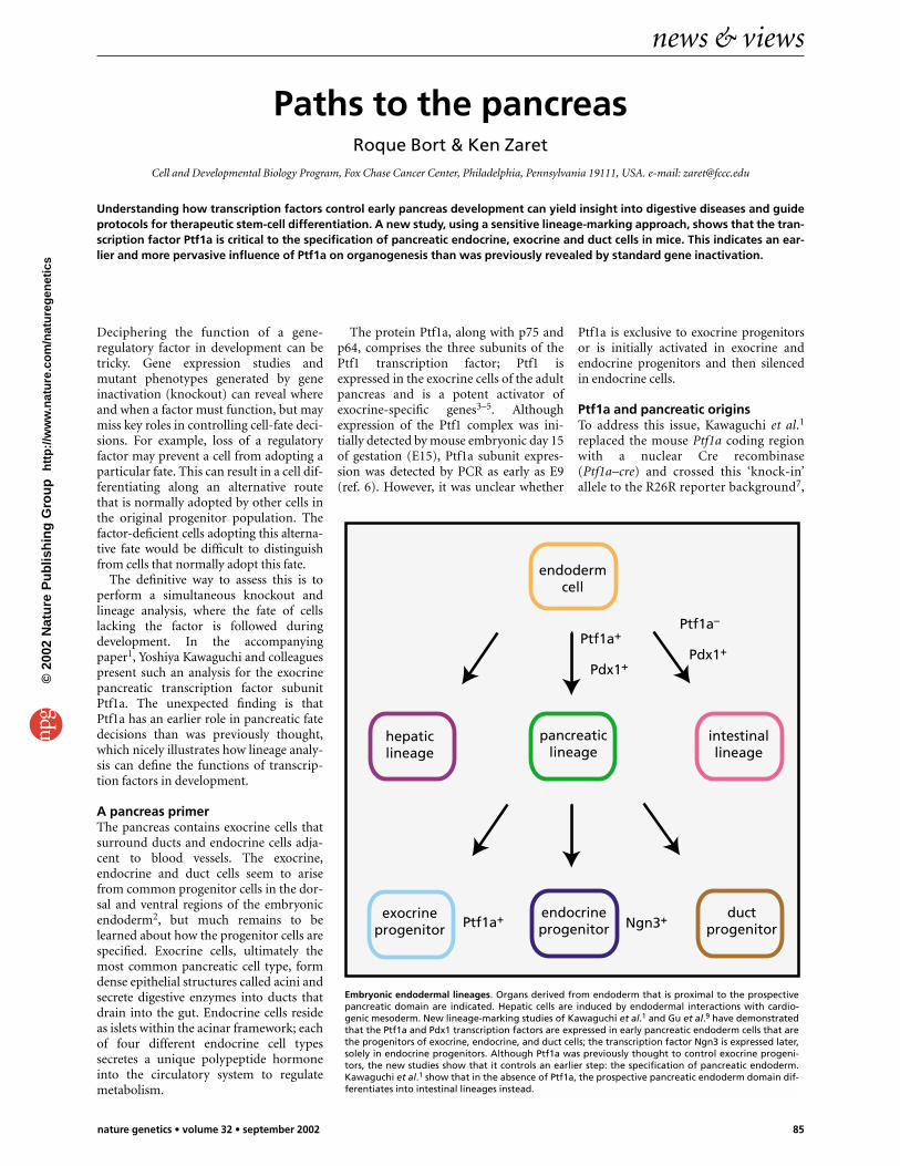
Embryonic endodermal lineages. Organs derived from endoderm that is proximal to the prospectivepancreatic domain are indicated. Hepatic cells are induced by endodermal interactions with cardio-genic mesoderm. New lineage-marking studies of Kawaguchi et al.1 and Gu et al.9 have demonstratedthat the Ptf1a and Pdx1 transcription factors are expressed in early pancreatic endoderm cells that arethe progenitors of exocrine, endocrine, and duct cells; the transcription factor Ngn3 is expressed later,solely in endocrine progenitors. Although Ptf1a was previously thought to control exocrine progeni-tors, the new studies show that it controls an earlier step: the specification of pancreatic endoderm.Kawaguchi et al.1 show that in the absence of Ptf1a, the prospective pancreatic endoderm domain dif-ferentiates into intestinal lineages instead.
©20
02 N
atu
re P
ub
lish
ing
Gro
up
h
ttp
://w
ww
.nat
ure
.co
m/n
atu
reg
enet
ics

news & views
86 nature genetics • volume 32 • september 2002
which requires Cre-mediated recombi-nation in a cell to express β-galactosi-dase. Thus, if the Ptf1a promoterexpresses Cre only transiently in a lin-eage8, all descendant cells will inherit themark of β-galactosidase recombinationand expression.
Gu et al.9 have used a variant of thisapproach10 to definitively identify pancre-atic endocrine progenitors. They createdmouse transgenes where the promoter forthe gene encoding the transcription factorNgn3 drives the expression of a bifunc-tional protein containing Cre fused to aninhibitory segment of the estrogen recep-tor (Cre-ER). The authors showed thatNgn3-cre-ER–positive cells at E8.5–E10.5give rise to all four endocrine cell types,which is consistent with the previousdemonstration that Ngn3 is necessary togenerate all endocrine cells11 (see figure).Although they also provide evidence thatthe Cre-marked progenitor cells do notarise from ducts, it is not clear whethertheir Ngn3-cre-ER transgenes areexpressed as early as endogenous Ngn3,which seems to be expressed initially instructures that may be duct progenitors12.Stepping back further in development, Guet al.9 showed that cre-ER transgenes dri-ven by the promoter for Pdx1, whichencodes a homeobox transcription factorthat helps determine the pancreatic endo-derm2, marks endocrine, exocrine, andduct cells in the pancreas (see figure). Inadult tissues, Pdx1 expression is specific toinsulin-secreting cells13.
If an adult endocrine factor such asPdx1 can be critical for pancreatic deter-mination, what about an adult exocrinefactor, Ptf1a? Kawaguchi et al.1 now findthat early embryonic expression of theirheterozygous Ptf1a–cre knock-in marks allacinar cells and most of the islet and ductcells, thus establishing that Ptf1a isexpressed in progenitors of all pancreaticcell types (see figure). Apparently, Ptf1a issilenced in endocrine and duct cells dur-ing or shortly after the partitioning of theprogenitors into exocrine cells6.
Furthermore, Kawaguchi et al.1 findthat in homozygous Ptf1a–cre/Ptf1a–cremice, acinar cells are completely absent,whereas a small number of endocrineand duct cells differentiate apparently
normally, as reported previously byKrapp et al.6. However, the lineage-marking approach provides insightsbeyond those of the original Ptf1a knock-out. For example, in thePtf1a–cre/Ptf1a–cre embryos, cellsexpressing β-galactosidase descendingfrom the prospective ventral domainbecome incorporated into the gut epithe-lium, where they follow intestinal fatesinstead of those for pancreas. Specifically,the Ptf1a-deficient cells that express β-galactosidase contribute to intestinalcrypts and villi, where they differentiateinto enteroendocrine cells, goblet cells,and other gut cell types. In heterozygousPtf1a–cre embryos, cells expressingβ–galactosidase do not label the gut.
Taken together, the combination oflineage-marking and gene targetingshows that in the absence of Ptf1a, cellsthat normally would become ventralpancreas will assume an alternativeendodermal fate for intestinal epithe-lium, and thus that Ptf1a is necessary tocontrol the initial cell-type decision tomake the pancreas (see figure). Theauthors go on to show that Pdx1expressed from the Ptf1a promoter cancomplement an otherwise Pdx1-nullbackground, further illustrating thatPtf1a is expressed in definitive pancreaticprogenitors.
Fateful decisionsIn the prospective dorsal pancreaticdomain, there are two different outcomesfor the Ptf1a-deficient cells. First, rarecells expressing β-galactosidase becomeincorporated into the gut, as for the ven-tral cells. Second, pancreatic duct-likestructures expressing β-galactosidaseextend initially from the duodenalepithelium to the spleen, and they con-tain rare endocrine cells, of which 50%express β-galactosidase. As Krapp et al.6
observed in the original Ptf1a knockout,endocrine cells of all four types are even-tually found in the spleen, but Kawaguchiet al.1 find that these cells do not expressβ-galactosidase. Thus, a small number ofduct and endocrine cells arise either fromcells that never express Ptf1a or from cellsin which Cre recombinase does not accu-mulate sufficiently.
Regardless of these latter possibilities, thePtf1a null phenotypes and the lineage stud-ies together show that Ptf1a is clearlyexpressed in and required for the majorityof progenitors of all major pancreatic celltypes. They further suggest that the devel-opment of a fraction of duct and endocrineprogenitors is apparently not dependent onPtf1a. Alas, why can’t nature be simple?Well, it does keep us in business.
The differences in dorsal and ventralpancreatic phenotypes seen in the Ptf1a-null embryos may relate to other differencesin how these domains are specified2 andtheir eventual contribution to differentdomains of the mature gland. With regardto the ventral domain, studies of mice14 andzebrafish15 indicate that the endoderm is atleast bipotential. Endoderm proximal to thecardiogenic mesoderm develops into liverand actively excludes the pancreatic fate(see figure), whereas endoderm distal tocardiac develops into the pancreas14. Basedon the studies of Kawaguchi et al.1, we canconclude that there is a third fate availableto such endoderm, as in the following sce-nario: Pdx1 and Ptf1a are first activated inventral foregut endoderm that escapeshepatic induction, and the expression ofPtf1a is necessary for those cells to executeall aspects of the pancreatic program. In theabsence of Ptf1a and in the absence of sig-naling that would induce liver, the endo-derm cells assume a third availablefate—intestinal differentiation. Regardless,it is clear that knockout phenotypes alonecan’t divulge the beginning of the story—but lineage analysis can. �1. Kawaguchi, Y. et al. Nature Genet. 32, 48–54 (2002). 2. Edlund, H. Nature. Rev. Genet. 3, 524–532 (2002).3. Cockell, M., Stevenson, B.J., Strubin, M.,
Hagenbuchle, O. & Wellauer, P.K. Mol. Cell. Biol. 9,2464–2476 (1989).
4. Cockell, M. et al. Mol. Cell. Biol. 15, 1933–1941(1995).
5. Krapp, A. et al. EMBO J. 15, 4317–4329 (1996).6. Krapp, A. et al. Genes Dev. 12, 3752–3763 (1998).7. Soriano, P. Nature Genet. 21, 70–77 (1999).8. Herrera, P.L. Development 127, 2317–2322 (2000).9. Gu, G., Dubauskaite, J. & Melton, D.A.
Development 129, 2447–2457 (2002).10. Danielian, P.S., Muccino, D., Rowitch, D.H., Michael,
S.K. & McMahon, A.P. Curr. Biol. 8, 1323–1326(1998).
11. Gradwohl, G., Dierich, A., LeMeur, M. & Guillemot,F. Proc. Natl Acad. Sci. USA 97, 1607–1611 (2000).
12. Schwitzgebel, V.M. et al. Development 127,3533–3542 (2000).
13. Guz, Y. et al. Development 121, 11–18 (1995).14. Deutsch, G., Jung, J., Zheng, M., Lora, J. & Zaret,
K.S. Development 128, 871–881 (2001).15. Stafford, D. & Prince, V.E. Curr. Biol. 12, 1215–1220
(2002).
©20
02 N
atu
re P
ub
lish
ing
Gro
up
h
ttp
://w
ww
.nat
ure
.co
m/n
atu
reg
enet
ics