haemophilus parasuis and alveolar - uab barcelona · 2014. 12. 17. · insights on the interaction...
TRANSCRIPT

Insights on the interaction between
Haemophilus parasuis and alveolar
macrophages
Mar Costa Hurtado Ph.D. Thesis, 2012

Insights on the interaction between
Haemophilus parasuis and alveolar macrophages
Tesi doctoral presentada por Mar Costa Hurtado per accedir al grau de Doctor dins
del programa de Doctorat en Medicina i Sanitat Animals de la Facultat de
Veterinària de la Universitat Autònoma de Barcelona, sota la direcció de la Dra.
Virginia Aragón Fernández.
Bellaterra, 2012

VIRGINIA ARAGÓN FERNÁNDEZ, investigadora del Centre de Recerca en Sanitat
Animal (CReSA)
Certifica:
Que la memòria titulada, “Insights on the interaction between Haemophilus
parasuis and alveolar macrophages” presentada per Mar Costa Hurtado per a
l’obtenció del grau de Doctor, s’ha realizat sota la seva supervisió en el Centre de
Recerca en Sanitat Animal (CReSA) i que el Dr. Joaquim Segalés i Coma, professor
titular del Departament de Sanitat i d’Anatomia Animals de la Facultat de
Veterinària de la Universitat Autònoma de Barcelona ha actuat com a tutor.
I per tal que consti a efectes oportuns, firmen el present certificat a Bellaterra, a 9
de juliol de 2012.
Dra. Virginia Aragón Fernández Dr. Joaquim Segalés i Coma
Directora Tutor
Mar Costa Hurtado
Doctoranda

Els estudis de doctorat de Mar Costa Hurtado han estat finançats per una beca de
Personal Investigador en Formació, concedida pel Ministerio de Ciencia e
Innovación.
Aquest treball ha estat finançat pels projectes AGL2007‐60432 i AGL2010‐15232
del Ministerio de Ciencia e Innovación.

`
A l’Aleix

Tú no puedes volver atrás porque la vida ya te empuja
como un aullido interminable, interminable.
Te sentirás acorralada,
te sentirás perdida o sola, tal vez querrás no haber nacido,
no haber nacido.
pero tu siempre acuérdate de lo que un día yo escribí
pensando en ti, pensando en ti
como ahora pienso.
La vida es bella, ya verás como a pesar de los pesares
tendrás amigos, tendrás amor, tendrás amigos.
Un hombre solo, una mujer así tomados, de uno en uno
son como polvo, no son nada, no son nada.
Entonces siempre acuérdate de lo que un día yo escribí
pensando en ti, pensando en ti como ahora pienso.
Nunca te entregues ni te apartes
junto al camino, nunca digas no puedo más y aquí me quedo,
aquí me quedo.
La vida es bella, tú verás como a pesar de los pesares
tendrás amigos, tendrás amor, tendrás amigos.
Y siempre, siempre acuérdate
de lo que un día yo escribí pensando en ti como ahora pienso.
Palabras para Júlia, José Agustín Goytisolo, 1979
Música i adaptació Paco Ibánez

ACKNOWLEGMENTS
Gent! Tot comença i tot s’acaba! És ben bé que s’ha de ser tossuda per fer una tesi.... però aquí la que s’endú el primer premi en tenacitat és la Dra. Aragón! Moltes gràcies per aquests 4 anys Vicky! Tot el munt de coses que he aprés i les experiències, tan aquí com a fora, t’ho dec a tu. Una entusiasta de la ciència i del refranyer popular amb qui he tingut el privilegi de compartir la bancada des del primer, al darrer dia de la tesi. Moltes gràcies per les oportunitats que m’has brindat, la paciència, per l’optimisme, per les hores i l’esforç d’ensenyar‐me tot el que ha calgut per a dur a terme aquesta tesi. I sobretot, per creure en mi fins i tot quan ni jo mateixa hi creia. De veritat t’ho dic, no hauria pogut somiar una millor directora! I és clar, aquesta tesi també va dedicada a la Galo, la meva companya de trifulgues! des de la pesca d’arrastre, a la caça del garrí sanguinolent fins a intrèpides aventures i feinades en llocs plens de perills. Amb qui millor em podria haver barallat en un laboratori? Tossuda com una mula, ha sigut tot un plaer treballar amb tu i heretar i/o contradir les teves manies. Evidentment la broma no hagués estat igual, sense la presència de tot un personatge amant de les esquitxades a cop de got, l’Olvera! Sempre posant un poc de pau entre Zipi i Zape, a costa d’un nou sobrenom cada vegada. És clar que tota la feina feta no hagués sigut possible sense tot l’equip del CReSA, la gent de NBS2 com NBS3, la gent d’administració, de neteja, de manteniment, de seguretat...Visca la ferreteria d’en Josep Maria! Moltes gràcies per tot el vostre suport i paciència (paciència! Una paraula clau per treballar amb mi, i amb totes les persones que estem aprenent a investigar). Ja ho diuen que el bosc seria molt trist si només cantessin els ocells que millor ho fan! També vull agrair a les a les meves companyes Haemophiles, la Vero i la Paula, a les bacter‐mosses: la Núria, l’Ana, la Judit, la Tere i com no la Marta Cerdà i en Badiola així com tota la multitud que ronda per bacter i que heu sofert les meves fagocitosis. Evidentment, no puc deixar de mencionar a tot el sector becari, amb qui d’alguna manera hem anat creixent plegats i compartint les frustracions, les anades d’olla, algun que altre moc, els èxits...gràcies per tots els festivals i cafès de màquina! Specially, al Mario, un escorpí en tota regla amb qui vam començar ballant salsa i hem acabat mirant pelis sueques i barallant‐nos per espai a l’estenedor. A vegades t’escanyaria però ja ho diuen que l’amor és irracional!! A la Kate-Tekanagüen, pintes de pollet i ànima de falcó!comando Delta forever! i el Joan calostrales un plaer tenir-vos alrededor!! A la Tufària, la matriarca i ministra, gràcies per la teva pau. A lo Gerard, merci pels teus consells, segur que no vols cigrons? A l’assertivitat del despatx, la Pamela. A la Noelia, la gota malaia. I a la resta del sector l’Aida, la Juliana, la Lacasta, la Lidumila, la Júlia (quin paper tocar presentar ara?), la Meri, la Carolina, l’Alexandra, l’Emma, l’Adriana, la Cris, el Cabezón, l’Elisa, la Paula M., en Max, en Fer petit, en David , en Bernardo i de pas a la Marta! Gràcies també als investigadors del CReSA, un consell de sàvies i savis sempre a punt per resoldre un dubte i aconsellar sobre la dilució d’un sobrenedant. Especialment a la Maria Ballester, moltes gràcies per les esbroncades pedagògiques! quantes hores de confocal amb l’accent de Benicarló! To Ayub for making me laugh, helping with the hybridomas and other tough moments, and to Tuija, Fer, el Quim, alies el tutor (que passava per allà corrent), i cuidaaaaaaao a en Francesc Accensi (¿comprendes la filosofia, no?), a l’Eugènia, en Joan, en Xavi, en José Ignacio, l’Ivan, en Miquel, en Martí, la Roser, et alia.
I would also like to express my gratitude to Dr. Nick Cianciotto, for giving me the opportunity to join your lab at the Microbiology and Immunology Department at Northwestern University. And especially to Meghan, my buddy! To Christa, Catherine, Kassler, Jessica, Denise, Sarah, Felicia and of course, Brendan the tech mas majo ever! I am also very grateful to Dr. Harri Savilati for hosting me and the rest of the Recombination Group members at the University of Turku for being so helpful: Elsi, Saija and Mikko. I evidentment, els efectes col·laterals d’aquesta tesi també han arribat a la meva gent del Vallès! En primer lloc vull agrair a l’Encarna, l’Anton Maria i l’Aleix, de qui he aprés a ser la persona que sóc. Els perfectes corredors de fons, lluitant pel que és de justícia, que m’han donat les bases per ser compromesa. De qui aprenc i seguiré aprenent a ser tossuda i tenaç per aconseguir tot el que em proposi. A la iaia Josefina, ¡A quien quiero un montón! Eres mi fan número uno, esta tesis va directa a tu colección! A la Mireia, que segur que no era una casualitat que ens trobéssim a la vida, però ja se sap “pon alguien de letras en tu vida” i tindràs un paquet de ciències al cotxe. En Serrallonga obsolet ens va unir i Borredà forever. Com unes iaies serenes i satisfetes seguirem tocant les campanades amb cassoles, doncs ja se sap, “no hay que dar pataletas a la vida”. Som del milloret! A l’Adrià, aquesta tesi també és una mica teva. Encara que portis camisa lila t’estic super agraïda per la paciència lloable amb que m’has sofert. Espero que puguem tornar, aviat, a cantar en holandès com ho solíem fer. A la gent del CECA, sobretot a la Neus. Als meus torracollons preferits, els petits saposets, el Roger, la Júlia, el Bernat, la Ceci Suau, a qui li dec aquesta meravellosa coberta! I a l’Arnau per no ser el meu nen preferit i aquí no li diré mai com me l’estimo. Em portes a casa amb cotxe? A les nenes maques al dematí, la Laura i la Maria, s’alcen i reguen i juntament amb les meves bestials companyes de promoció, la Glòria, la Nàdia i la Marisa, sense elles la biblioteca hagués sigut un lloc més tranquil. Al Lluís perquè sense ell no hauria acabat pas al CReSA. I a la Cristina, la Gia i la Natàlia junt amb l’Estel, la tecnològica, simplement per ser‐hi! A les meves Irish princesses, amb qui vam començar a txapurejar l’anglès en el Peadar O’Donells, i ara el pronunciem al més pur Montjoy Terrace style! To all my good friends I met along my way. I know that we’ll be in some sort of touch forever! To Felix, who was my north star, who challenges me, and pushes me to a deeper knowledge of my strength. Thank you for helping my relatives shake off their prejudices. A mis hermanos latinos, German y Janitza, che! cuando es el próximo evento del G‐salsa? To my older sister, Vanya. To Cathy and Amber, Carleton Collage has linked as us in a very peculiar way but I am so glad for such special friendships. To Alessandro (l’únic tiramissú que m’agrada!). To my little sister, Sadie for that picture at Navy Pier, Scyuler, Amada, Sarah, Jon, Taneli, Dustin, and the little ona. Also to Pavel and Karin and Babis, always ready to share some chelas at Bremer and to my Finish friend Anu for our meaningful talks! Perquè aquesta tesi no s’hagués pogut portar a terme sense el suport de totes i tots, sentiu‐vos la vostra. Once again, moltes gràcies!! I com digué en Vincent Vega a Pulp Fiction: “Now, if you'll excuse me, I'm going to go home and have a heart attack.”

I
SUMMARY
Haemophilus parasuis, a member of the family Pasteurellaceae, is a colonizer of the
upper respiratory tract of healthy pigs and the etiological agent of Glässer’s
disease. Differences in virulence among H. parasuis strains have been widely
observed by different tests, including in vivo infections and in vitro phagocytosis
assays with porcine alveolar macrophages (PAMs).
The pathogenicity of the strains has been correlated with resistance to
phagocytosis, but the bacterial factors implicated are not known. To identify
virulence factors involved in phagocytosis resistance, a genomic library of the
virulent reference strain Nagasaki was produced and exposed to PAMs. After
incubation with PAMs, two clones carrying two different virulent‐associated
trimeric autotransporters (VtaA), vtaA8 and vtaA9, were selected. The role of these
molecules was further investigated and a reduction in the interaction of the two
clones with the macrophages was detected by flow cytometry. Monoclonal
antibodies (mAb) produced against the recombinant VtaA8 and VtaA9 proteins
demonstrated the presence of these proteins on the bacterial surface of the
corresponding clone. The same mAb also detected the proteins on the surface of H.
parasuis phagocytosis‐resistant strain PC4‐6P, but not on the non‐virulent strain
F9. The effect of VtaA8 and VtaA9 in the trafficking of the bacteria through the
endocytic pathway was examined by fluorescence microscopy and a delay was
detected in the localization of the vtaA8 and vtaA9 clones in acidic compartments.
Although VtaA8 and VtaA9 delayed phagocytosis, were not sufficient to completely
inhibit the process. These results are compatible with a partial inhibition of the
routing of the bacteria via the degradative phagosome. Finally, antibodies against a
common epitope in VtaA8 and VtaA9 were opsonic and promoted phagocytosis of
the phagocytosis‐resistant strain PC4‐6P by PAMs. Taken together, these results
indicate that VtaA8 and VtaA9 are surface proteins that play a role in phagocytosis
resistance of H. parasuis.
Infection of snatch farrowed, colostrum‐deprived piglets with different strains of
H. parasuis demonstrated differences in the degree of virulence. We used four
strains of H. parasuis: reference virulent strain Nagasaki, reference non‐virulent
strain SW114, and field strains IT29205 (from systemic lesion and virulent in a

II
previous challenge) and F9 (from nasal cavity of a healthy piglet). The infection
was performed intranasally with 107‐108 CFU per animal. Two non‐infected
animals served as controls. At different times after infection (1, 2, 4 and 7 days
post‐infection [dpi]), two animals of each group were euthanized and
bronchoalveolar lavages and sera were collected. Alveolar macrophages were
analyzed for the expression of surface markers CD163, CD172a, SLAI, SLAII,
sialoadhesin, CD14 and SWC8 by flow cytometry. The phenotype of macrophages
changed along with the infection depending on the virulence of the strain. At early
time‐points (1 dpi), non‐virulent strains SW114 and F9 induced higher expression
of CD163, sialoadhesin, SLAII and CD172a than virulent strains Nagasaki and
IT29205. At 2 dpi, the situation switched to a strong induction of expression of
CD172a, CD163 and sialoadhesin by the virulent strains, which was followed by a
steep increase in IL‐8 and soluble CD163 at 3‐4 dpi. The early delay in macrophage
activation by virulent strains may be critical for disease establishment.
The association between the delay produced by VtaA8 and VtaA9 in the endocytic
route and the delay in macrophage activation needs further study.

III
RESUM
Haemophilus parasuis és un bacteri de la família Pasteurellaceae. És un
colonitzador del tracte respiratori superior en animals sans i l’agent etiològic de la
malaltia de Glässer. Les soques de H. parasuis presenten diferències en virulència,
que han estat observades tant en infeccions in vivo com en proves de fagocitosi in
vitro amb macròfags alveolars porcins (PAMs).
La patogenicitat de les soques ha estat correlacionada amb la resistència a la
fagocitosi, però els factors de virulència implicats son desconeguts. Per la
identificació dels factors implicats en la resistència a la fagocitosi, es va generar
una llibreria genòmica de la soca virulenta de referència Nagasaki. De la incubació
d’aquesta llibreria amb PAMs, es varen seleccionar dos clons amb gens codificant
dos autotransportadors trimètrics associats a virulència (VtaA), vtaA8 i vtaA9.
Mitjançant citometria de flux, es va aprofundir en el paper d’aquestes molècules en
ambdós clons, els quals van mostrar una menor interacció amb els PAMs. La
producció d’anticossos monoclonals (mAb) contra les proteïnes recombinants
VtaA8 i VtaA9 van permetre determinar‐ne la localització a la superfície dels clons.
Els mateixos mAb detectaren aquestes proteïnes a la superfície de la soca resistent
a la fagocitosi PC4‐6P, però no a la soca avirulenta F9. Addicionalment, estudis amb
microscòpia de fluorescència varen determinar l’efecte de VtaA8 i VtaA9 en el
transport a la ruta endocítica, tot detectant un retard en la co‐localització dels
clons vtaA8 i vtaA9 amb compartiments àcids. Aquests resultats són compatibles
amb una inhibició parcial del transport del bacteri a través de la degradació per
fagosoma. Finalment, els anticossos contra un epítop comú a VtaA8 i VtaA9 van ser
opsonitzadors i varen promoure la internalització de la soca resistent a la
fagocitosi PC4‐6 pels PAMs. Globalment, aquests resultats indiquen que VtaA8 i
VtaA9 són proteïnes de superfície i juguen un paper en la resistència a la fagocitosi.
La infecció de garrins privats de calostre nascuts de part natural amb soques de H.
parasuis va mostrar diferències en el grau de virulència. Es varen emprar quatre
soques de H. parasuis: les soques de referència virulenta Nagasaki i avirulenta
SW114, la soca de camp IT29205 (obtinguda d’una lesió i virulenta en una infecció
anterior) i la soca F9 (aïllada de la cavitat nasal d’un garrí sa). Els animals es varen
inocular per via intranasal amb 107‐108 CFU per individu. Dos animals no infectats

IV
s’utilitzaren com a controls. A diferents temps (1, 2, 4 i 7 dies post‐infecció [dpi]),
dos animals de cada grup es varen eutanasiar, i es varen prendre mostres de sèrum
i del fluid bronquialveolar. Mitjançant citometria de flux, es varen analitzar els
macròfags alveolars avaluant l’expressió dels marcadors de superfície CD163,
CD172a, SLAI, SLAII, sialoadhesina, CD14 i SWC8. En funció de la virulència de la
soca es varen poder observar canvis en el fenotip dels macròfags. A la fase inicial
de la infecció (1 dpi), les soques no virulentes SW114 i F9 varen induir major
expressió de CD163, sialoadhesina, SLAII i CD172a que les soques virulentes
Nagasaki i IT29205. A 2 dpi, la situació canvià diametralment. Les soques
virulentes generaren una forta inducció de l’expressió de CD172a, CD163 i
sialoadhesina, seguida a continuació d’un sobtat increment d’IL‐8 i CD163 soluble
a 3‐4 dpi. L’activació primerenca dels macròfags per part de les soques virulentes
podria ser crítica per originar malaltia.
L’associació entre el retard produït per les proteïnes VtaA8 i VtaA9 i la ruta
endocítica, així com el retard en l’activació del macròfag, requereix estudis
ulteriors.

V
TABLE OF CONTENTS
INTRODUCTION……………………………………………………………………………… 1
1. Haemophilus parasuis………………………………………………………... 1
1.1. Bacteriological description..…………………………….…… 1
1.2. Molecular methods and strain variability…………….. 2
2. Glässer’s disease …………………………………………………………….. 3
2.1. Pathogenesis……………………………………………………... 5
3. Mechanisms of virulence ………………………………………………… 6
4. Virulence factors of Haemophilus parasuis ………………………... 8
4.1. 6‐phosphogluconate dehydrogenase (6PGD) .………. 10
4.2. Lipooligosaccharide (LOS).…………………………………. 10
4.3. Sialyltransferase LsgB ………………………….……………. 11
4.4. Cytolethal distending toxin (CDT) ..…………………….. 11
4.5. IgA protease activity ………………………………………….. 12
4.6. Capsule ……………………………………………………………... 13
4.7. Fimbria ……………………………………………………………… 13
4.8. Proteins of the porin family……………………………....... 13
4.8.1. OmpP2………………………………………………….. 14
4.8.2. OmpP5 .………………………………………………… 14
4.9. Type V secretion system in H. parasuis…………………………….. 15
4.9.1. Monomeric autotransporters (AT‐1)…………………. 16
4.9.2. Virulence associated trimeric
autotransporters (VtaA) ………………………………… 17
5. Innate immune response in the respiratory tract of pigs …… 17
5.1. The respiratory tract ………………………………………….. 17
5.2. Alveolar macrophages………………………………………... 18
5.3. Macrophages phenotype: activation markers….…… 20

VI
HYPOTHESIS AND OBJECTIVES…………………………………………………… 23
RESULTS……………………………………………………………………………………… 25
Chapter 1: VtaA8 and VtaA9 from Haemophilus parasuis delay phagocytosis by alveolar macrophages…………. 25 Summary..................…...…………………………………………......... 26 Introduction..........…...…………………………………………........... 27 Material and methods.……………………………………………… 28 Results................................................................................................. 34 Discussion.......................................................................................... 46 References.......................................................................................... 48 Chapter 2: Changes in macrophage phenotype after infection of pigs with Haemophilus parasuis strains of different virulence .................................................................................... 53
Summary............................................................................................. 54 Introduction....................................................................................... 55 Material and methods.................................................................... 57 Results.................................................................................................. 61 Discussion........................................................................................... 77
References.......................................................................................... 80
CONCLUSIONS.............................................................................................................. 83
BIBLIOGRAPHY........................................................................................................... 87
ANNEXES
Annex I. ABSTRACT ASM 112th General Meeting ........................... 97
Annex II. FIGURE S1...................................................................................... 99
Annex III. FIGURE S2A AND S2B............................................................ 100

1
INTRODUCTION
1. Haemophilus parasuis
1.1. Bacteriological description
Karl Glässer established an association between fibrinous polyserositis in swine
and a small Gram‐negative rod in 1910. The causative agent was first identified as
Haemophilus influenzae (variety suis) [1] and later on characterized and named as
Haemophilus parasuis [2]. Following the denomination in the Haemophilus genus,
the prefix para was added to indicate that it requires V factor (nicotinamide
adenine dinucleotide, NAD) but not porphyrins, such as hemin (X factor) for
growth. H. parasuis is the aetiological agent of Glässer’s disease.
H. parasuis is a Gram‐negative non‐motile, small pleomorphic rod included in the
genus Haemophilus, within the family Pasteurellaceae of the γ‐proteobacteria.
However, several members of the family are still being reclassified [3] and its
location within this family is still in debate [4]. In fact, H. parasuis does not form a
monophyletic cluster by 16S rRNA gene sequencing, and two main clusters are
defined within the species [5, 6]. In addition to H. parasuis, other NAD‐dependent
Pasteurellaceae can be isolated from swine [7]. Six species of porcine origin have
been defined on the basis of DNA‐DNA hybridization and 16S rRNA gene sequence
[8, 9].
H. parasuis has specific growth requirements and is difficult to culture from clinical
specimens. This bacterium grows on chocolate agar but not on blood agar. It can
also be cultured on the latter with a Staphylococcus nurse streak as a source of V
factor, showing the characteristic satellitic growth. One to three days are required
to produce small brown to gray colonies on chocolate agar plates or small
translucent non‐hemolytic colonies on blood agar. Some strains produce colonies
of different sizes, but the significance of this phenomenon is not known [10]. H.
parasuis grows under normal atmosphere at 37ºC, although added humidity and
5% CO2 may improve growth. When a liquid culture is needed (e.g., for biochemical
tests), it can be cultured in liquid PPLO or BHI broths supplemented with NAD.

2
1.2. Molecular methods and strain variability
Early colonizer agents, such as Actinobacillus suis, Streptococcus suis and H.
parasuis, can emerge as costly and significant pathogens for pig herds, specially in
high health status farms [11]. H. parasuis is an early colonizer and part of the
microbiota of the upper respiratory tract of piglets [12‐14]. H. parasuis strains are
heterogeneous and differences in virulence among strains have been described.
Therefore, strain discrimination is important in H. parasuis diagnosis and control
in order to differentiate between nasal colonizer strains and systemic invasive
strains. The correct diagnosis of this agent is essential to establish the appropriate
control measures.
The slow growth and poor viability of this microbe, together with the lack of a
validated serological test [15], warrant the use of molecular methods to encircle
these limitations. The polymerase chain reaction (PCR) has been a major advance
for the diagnosis of infectious diseases and different tests have been developed for
the detection of H. parasuis, both conventional and real‐time PCR [16‐19]. In
addition, a multiplex PCR test for the simultaneous detection of H. parasuis and the
differentiation of potentially virulent strains has been developed [20, 21].
Fifteen serovars of H. parasuis have been defined based on heat‐stable somatic
antigen and immunodiffusion [22], but some strains are non‐typable by
serotyping, even with the modified serotyping procedure by indirect
hemagglutination [23, 24]. In general, serovars 1, 5, 10, 12, 13 and 14 were defined
as highly virulent; serovars 2, 4 and 15 as moderately virulent and serovars 3, 6, 7,
8, 9 and 11 were considered non‐virulent [22]. However, the correlation between
serovar and virulence is not clear and strains that belong to the same serovar can
exhibit different degrees of virulence [25, 26].
The variability of H. parasuis strains has been studied to great extends by
genotyping, and the high heterogeneity of the strains has been confirmed by
different methods.
Methods based on DNA are superior to serotyping for H. parasuis strain
differentiation. Genotyping is carried out by fingerprinting or sequencing methods.
Fingerprints (or electrophoretic band patterns) can be obtained from whole
bacterial genome or from a single gene, either by digestion or PCR amplification

3
throughout the genome. Sequencing methods can be based on sequences from a
single locus or several loci (multilocus sequence typing or MLST).
Enterobacterial repetitive intergenic consensus (ERIC)‐PCR is based on the
presence of DNA elements that are repeated throughout the genome, which are
targets of the primers used in the PCR. Raffie et al (2000) [27] established the
method for H. parasuis, which was later used for several authors. ERIC‐PCR is
especially suitable for outbreaks studies [15, 28].
PCR‐restriction fragment length polymorphism (PCR‐RFLP) consists in the
digestion with restriction enzymes of specific amplicons. This type of analysis has
been used for H. parasuis typing with the gene of the transferrin binding protein A,
tbpA [29] and the gene of the 5‐enolpyruvylshikimate‐3‐phosphate synthase, aroA
[30]. However, these methods did not provide a correlation between genotype and
serotype or potential virulence of the strains.
Genotyping by sequencing a unique gene fragment of the hsp60 locus confirmed
the heterogeneity of H. parasuis strains and indicated the existence of lateral
transfer of genes between H. parasuis and species of the Actinobacillus genus [6].
To minimize the effect of this horizontal transfer in the classification of the H.
parasuis strains, a MLST, consisting on the partial sequencing of 7 conserved gene,
was established [31]. MLST classifies H. parasuis strains in 3 clusters; one
associated with nasal isolation and another one associated with isolation from
systemic lesions [25]. MSLT can be used for fine epidemiological studies of H.
parasuis strains and allows the comparison of data from different laboratories.
It is worth to highlight in the study of H. parasuis, the publication of two genomic
sequences from the virulent strain SH0165 ([32]; accession number CP001321)
and strain 29755 ([33]; accession PRJNA54869), both from serovar 5.
2. Glässer’s disease
Glässer’s disease is a systemic infection characterized by fibrinous or fibrinous‐
purulent polyserositis, polyarthritis and meningitis. The bacteria replicate in
serosal surfaces causing inflammation and fibrinous exudate lining the membranes
of the body cavities, joints and meninges. In addition, petechiae or ecchymoses in

4
the liver, kidney and meninges can also be found. Fibrinous thrombi can also be
observed in many organs and high levels of endotoxin can be detected in plasma
[34]. LPS is involved in endotoxic shock and can exacerbate clinical signs. The
endotoxic shock is associated with cases of sudden death of piglets with H. parasuis
septicemia [35]. Fibrinous pleuritis may be also found with or without
cranioventral consolidation due to catarrhal‐purulent bronchopneumonia. Animals
with neurological signs may lack gross lesions.
In the past, this disease was of sporadic occurrence and associated to stress
conditions. However, current production techniques and the emergence of
immunosuppressant viruses have generated an increase in the prevalence of
respiratory diseases. Glässer’s disease is present in all major swine‐raising
countries and remains a significant disease in modern age‐segregated production
systems, including high health status systems [15]. Farmers in the United States
have ranked H. parasuis as the second most important health problem in the
nursery herd; and also the 8th and 9th in finishing pigs and sows, respectively [36].
The severity of the disease depends on the virulence of the H. parasuis strain, the
immune status of the piglets, the colonization of the pigs, the genetic resistance of
the host and the presence of other pathogens in the herd (such as porcine
reproductive and respiratory syndrome virus [PRRSV], porcine circovirus type 2
[PCV2] or influenza virus type A) [37‐39]Palzer, 2008 #306; Solano, 1998 #65; Yu,
2012 #481}. Clinical disease in conventional herds is limited to a few individuals.
Specific pathogen free (SPF) herds and some segregated early weaning (SEW)
herds, on the other hand, can suffer devastating outbreaks that affect many
animals [40]. H. parasuis can act as primary or secondary pathogen.
Immunosuppressive events can allow strains usually located in the respiratory
tract to invade systemic sites [39].
Several studies have shown that more than one H. parasuis strain can be isolated in
a herd and even from a single animal at a given point [6, 12, 28, 31, 41]. However, it
is commonly accepted that one single strain is responsible of a disease outbreak.

5
2.1. Pathogenesis
The upper respiratory tract is the natural habitat for many potential pathogens,
including viruses, mycoplasmas, chlamydias, and many other bacteria [42]. On the
other hand, the commensal microbiota has a favorable competitive effect for their
host by outnumbering pathogenic agents and stimulating the proper development
of the immune system [43].
H. parasuis is found exclusively in swine and the initial acquisition of this
bacterium takes place through contact with the sow after birth. H. parasuis is one
of the earliest and most prevalent isolates from nasal swabs of pigs of 15 days of
age [40]. Once it enters the upper respiratory tract, H. parasuis establishes a
colonization of the upper respiratory tract. In some situations, some strains can
spread to the lung, where they cause pneumonia, or invade systemic sites.
H. parasuis is mainly an extracellular pathogen, and therefore, the humoral
response (antibodies) plays an important role in protection and resolution of
disease [10, 44]. Since placentas of pregnant sows are impermeable to
immunoglobulin passage, the neonates are born without antibodies. Their survival
depends on the passive acquisition of maternal immunity, including at least three
components: (1) systemic humoral immunity, transmitted through colostrum; (2)
mucosal humoral immunity, transmitted through milk; and (3) cellular immunity
transmitted via maternal immunocompetent cells present in mammary secretions
[45]. The sow is also the source of the primary infection for these animals.
Circulating antibodies are acquired by the piglet during the first 24 hours after
birth and therefore there is a delicate balance of decreasing passive antibody titer
and mucosal colonization that takes place during the lactation weeks. As the
antibody titer decreases, it must reach a threshold under which there is no longer
protection to mucosal colonization, but there are still enough antibodies to prevent
systemic dissemination of the organism. In this way, piglets switch from passive to
active protection and are commonly able to prevent clinical disease. Thus, piglets
will develop natural immunity to the prevalent strains of H. parasuis on the farm
while they are protected by the maternal immunity. The duration of this passive
protection is highly variable. It is dependent on: immune status of the sow, amount
of colostrum uptake during the first 24 hours and nature of the microorganisms.

6
The entry of a new virulent strain with no cross‐immunity with the prevalent
strains may have a great impact in disease outcome and control [10].
Vahle et al. studied the sequential events of infection in caesarean‐derived,
colostrum‐deprived (CDCD) pigs [46, 47] by intranasal inoculation with a strain
previously isolated from pericardium. The infection resulted in H. parasuis
isolation from nose and trachea after 12 hours, from blood after 36h and from
systemic tissues after 36‐108h. Consequently, lesions progressed from mild and
moderate from 12h to 36h, ending in severe lesions at 96‐108h.
3. Mechanisms of virulence
Bacterial pathogenesis is a multifactorial process and requires different
mechanisms to initially establish and produce disease infection. This process
involves bacterial attachment or other means of gaining entry into the host,
evasion of host defenses, multiplication to significant numbers, production of
damage to the host either directly or indirectly, and conclusion with transmission
of the agent to another susceptible host [48].
H. parasuis strains are heterogeneous in phenotypic and genotypic traits, including
virulence. The comparison of virulent and non‐virulent strains in several
functional assays has allowed the determination of several essential mechanisms
of virulence. As an early colonizer, a common feature of H. parasuis strains is its
ability to produce biofilm in vitro, which is more efficient in non‐virulent nasal
strains [49]. This feature is probably involved in mucosal colonization, but it has
not an essential role in systemic invasion. On the other hand, adhesion to and
invasion of epithelial cells has been described in virulent strains and could be
important in the first steps of infection [50, 51]. Besides, H. parasuis is able to
induce apoptosis of tracheal epithelial cells which may be critical to disrupt the
tracheal mucosa [50].
If the strains reach the lung, they have to confront the host pulmonary defenses. In
the case of non‐virulent strains, they would be eliminated by phagocytosis in an
actin‐dependent mechanism. In contrast, virulent H. parasuis strains are able to
avoid phagocytosis by alveolar macrophages, probably due, at least in part, to

7
capsule production [39]. In the presence of opsonic antibodies, virulent strains
become susceptible to macrophages, which are then able to internalize and destroy
them [39]. Thus, animals with specific antibodies could overcome disease by
efficiently killing of virulent strains by opsonophagocytosis. A significant
implication of nitric oxide by induction of the inducible nitric oxide synthase
(iNOS) in phagocytosis of H. parasuis could not be demonstrated, although low
expression of iNOS transcript was detected [52]. This result may be explained by
the low uptake of virulent H. parasuis by alveolar macrophages, and therefore poor
activation of the cells, or by the intrinsic nature of swine macrophages [53]. Once
virulent H. parasuis reaches the bloodstream, is able to avoid killing by the action
of the complement, in an antibody‐independent manner [54, 55]. The interference
of the capsule in the deposition of the complement can explain this, as this would
explain also the lack promotion of phagocytosis by complement‐opsonization [39].
Therefore, serum resistant H. parasuis strains would survive the bactericidal effect
of the serum and would be able to reach systemic sites. Furthermore, virulent
strains of H. parasuis invade endothelial cells [56, 57] and this may explain the
ability of some strains to cross the blood‐brain barrier and cause meningitis.
Besides, H. parasuis can induce apoptosis and production of pro‐inflammatory
interleukin IL‐6 and IL‐8 in epithelial and endothelial cells [50, 57 , 58] and the role
in cell permeability has been suggested.
All together, H. parasuis has evolved different strategies to avoid the innate
immune system in order to produce disease (Fig. 1). Host‐pathogen interaction
requires further study to determine the mechanisms underling the pathogenicity
of H. parasuis.

8
Serum resistancePhagocytosis
resistance
Adhesion and invasion to
endothelial cells
Adhesion and invasion to epithelial
cells
a
bc
d
Figure 1. Representation of known mechanisms implicated in the pathogenicity of H. parasuis. (a) colonization of the upper respiratory tract by adhesion to epithelial cells, (b) phagocytosis resistance to alveolar macrophages, (c) serum resistance, (d) adhesion and invasion of endothelial cells of internal organs including the brain.
4. Virulence factors of Haemophilus parasuis
Little is know about the virulence of H. parasuis and the majority of potential
virulence factors described so far, require further characterization of function and
regulation.
The search of bacterial virulence factors is usually performed by the construction
of deletion mutants, which in the case of H. parasuis is still hindered by poor
transformation efficiencies. Two different systems were reported for the
transformation of H. parasuis: electroporation with a native plasmid [59] and
natural transformation [60]. This natural‐transformation method has been
recently modified, but it is strain‐dependent and only 1 out of 11 strains were
naturally transformable [61]. In addition, in vitro modification of input plasmid for
electroporation has been reported to increase electroporation efficiency [62].
Recently, we have reported electroporation efficiencies of 103 (for virulent strains)
and 105 (for non‐virulent strains) with the native plasmid pHS‐Tet, when the H.

9
parasuis strains were grown at 30ºC before washing with ice‐cold 10% glycerol
(ANNEX 1; [63]), However, there is still need for a consistent method to obtain
high transformation efficiency to generate H. parasuis mutants. To circumvent this
limitation, the study of differences among virulent and non‐virulent strains has
gained insights into the multifactorial nature of virulence of H. parasuis.
Nonetheless, over the years, several groups have reported genomic and
transcriptomic studies that have detected potential virulence factors of H. parasuis.
Some gene expression studies have attempted to mimic the host conditions. Hill et
al. [64] observed seven up‐regulated genes in response to heat stress using
differential display reverse transcription polymerase chain reaction (DDRT‐PCR),
including genes involved in heat shock response. Since iron resources in the host
are usually restricted, limitation of iron has been also used to mimic in vivo growth
and to identify the bacterial iron‐uptake systems. For instance, ferric hydroxamate
uptake (FhuA) is produced by H. parasuis during infection, but it is constitutive
expressed, and consequently not regulated under iron‐restricted conditions [65].
Melnikow et al. [66] identified genes that were regulated under iron and oxygen
restriction, and acid and heat stress (including numerous genes involved in
metabolic adaptation to the stress conditions, iron adquisition hxuCBA and yfeA
and two proteases). When H. parasuis was grown in cerebrospinal liquid and iron
restriction, differential expression of several genes was observed, but no relation
with virulence could be ascribed [67]. Iron related genes tonB‐exbBD‐tbpAB and
yfeCD were detected under iron‐limitation in an independent study, and
interestingly, pilA was found to be also up‐regulated, suggesting that iron
restriction could be a signal for colonization [68].
Genomic comparison of strains of different virulence has provided some
information, but the role of the specific genes (as examples, hemolysin operon,
hhdBA, the irion adquisition genes cirA, tbpA/B and fhuA, restriction modification
system hsdS or fimbria‐related gene fimB) in H. parasuis virulence has to be
confirmed [69‐71].
More interesting information was obtained in the study of gene expression during
lung infection [72]. Genes involved in metabolism and stress response, cell surface,
transport and regulation were transcribed in the infected lung, and included

10
several genes with homology to putative virulence factors, such as a putative large
adhesin (or vtaA), siaB (involved in sialic acid utilization), a subtilisin‐like
autotransporter protease and several regulators. In addition, genes with putative
function in biofilm formation were detected, supporting a role of biofilm in H.
parasuis infection [72].
At the protein level, different OMP profiles by SDS‐polyacrylamide gel
electrophoresis (PAGE) were detected between isolates recovered from healthy
and sick pigs, suggesting its relation with virulence [73]. More recently,
immunoproteomic approaches have been used to determine protective antigens
from H. parasuis [74‐76], but the role of those antigens in virulence has not been
defined.
4.1. 6phosphogluconate dehydrogenase (6PGD)
Fu et al. [77] recently characterized the cell wall 6‐phosphoglucanate‐
dehydrogenase (6PGD), which has been described in the swine pathogens S. suis as
a protective antigen [78, 79]. This protein seems to be involved in adherence to
swine alveolar epithelial cells (SJPLC), since recombinant 6PGD protein
considerably inhibited the capacity of H. parasuis SH0165 to adhere to SJPLC cells.
Besides, recombinant 6PGD induced IL‐8 and IL‐6 production by those cells.
Immunogenicity and partial protection in mice was also determined, so its role as
potential vaccine was suggested.
4.2. Lipooligosaccharide (LOS)
H. parasuis has a short LPS, by the absence of repeating O‐antigen subunits, which
is denominated lipooligosaccharide or LOS [80]. H. parasuis LOS shows similar
capacity than the LPS from E. coli, Actinobacillus pleuropneumoniae or Pasterurella
multocida to induce TNF‐α and IL‐6 production from RAW 264.7 cells [80]. Later,
Bouchet et al. [50, 58] determined the partial role of LOS (purified from the
virulent Nagasaki strain) in adhesion and induction of inflammation. H. parasuis
LOS was able to induce the release of IL‐8 and IL‐6 by porcine brain microvascular
endothelial cells (PBMEC) and newborn pig tracheal (NPTr) cells. However,

11
competitive assay with LOS did not completely abolish the adhesion of H. parasuis
to PBMEC or NPTr cells, suggesting the involvement of other adhesins.
A monoclonal antibody anti‐LOS showed a protective role in a mouse model
infection [55].
4.3. Sialyltransferase LsgB
Bacterial neuraminidases have been reported as scavengers for sialic acid (N‐
acetylneuraminic acid or Neu5Ac). The sialic acid, once internalized, can be used as
source of carbon and/or nitrogen or can be modified by CMPNeu5Ac synthetases
(such as NeuA and SiaB) to be incorporated into the lipopolysaccharide by the
sialyltransferase LsgB [81]. This modification of the LPS with sialic acid has been
correlated with virulence in other Pasteurellaceae, including Haemophilus
influenzae, specifically with serum resistance [82‐85].
In H. parasuis, a neuraminidase was identified and purified from the outer
membrane [86, 87]. Recently, Martinez‐Moliner et al. [88] evaluated the presence
of neuraminidase activity in H. parasuis strains of different clinical origin. The
presence of the gene nanH (neuraminidase) and the neuraminidase activity was
common in H. parasuis and did not correlate with the clinical origin of the strains.
On the other hand, lsgB was predominantly present in the systemic isolates, and
was not amplified from any of the nasal isolates tested. A correlation between the
possibility to sialylate the LOS molecule and serum resistance was found. In
addition, using the reference strain Nagasaki (virulent, lsgB+) the presence of sialic
acid in the LOS was demonstrated.
The role of sialic acid in H. parasuis pathogenesis has been also suggested by other
authors, who reported the transcription of siaB/neuA during infection [72].
4.4. Cytolethal distending toxin (CDT)
Cytolethal distending toxin (CDT) belongs to a family of bacterial AB2‐type toxins
and generally comprises three subunits CdtA, CdtB and CdtC, in which the CdtB is
the active toxic unit and CdtA and CdtC are required for CDT binding to target cells
ad for delivery of CdtB into the cell [89]. Genes encoding CDT have been found in
12
many Gram‐negative species clinically important mucocutaneous pathogens of
humans and animals [90]. CdtB is a DNAse and is required for the CDT‐mediated
cell cycle arrest at the G2/M phase and eventual death in some cultured
mammalian cell [91‐93].
In H. parasuis, two cdt gene cluster loci have been identified in the genomic
sequence available for H. parasuis strain SH0165 [94]. The recombinant proteins
showed toxic activity and cell cycle arrest in cell culture. CdtB protein was
expressed by 109 clinical isolates and all the 15 reference strains of H. parasuis,
independently of their virulence.
Zhang et al. [95] have recently produced CDT‐deficient mutants of H. parasuis
through natural transformation in the clinical isolate SC096. Surprisingly, those
mutants showed increased sensitivity to serum and reduced adherence and
invasion to porcine umbilicus vein endothelial cells (PUVEC) and porcine kidney
epithelial cells (PK‐15).
4.5. IgA protease activity
In order to colonize the respiratory mucosa, bacteria must overcome the
protective effects of IgA, which participates in host defense by inhibiting microbial
adherence and invasion, inactivating bacterial toxins, and mediating antibody‐
dependent cytotoxicity. The production of bacterial IgA extracellular proteases
results in cleaving and elimination of the agglutination activity of the
immunoglobulin [96].
Mullins et al. [97] demonstrated swine IgA protease activity in culture
supernatants of H. parasuis. However, no homologue the Haemophilus influenzae
iga or igaB was detected in the genome of the strains.
Recently, an espP2 gene homologue (extracellular putative serine protease) has
been detected in H. parasuis, which provided partial protection against a
homologous challenge in guinea pigs [98]. However, the activity of this protein was
not determined. This protein is a monomeric autotransporter (AT) and
corresponds to the BmaA5 and BmaA6 reported by Pina‐Pedrero et al. [99]. The

13
correlation between the IgA protease activity described in the supernatant and the
espP2 gene has to be evaluated.
4.6. Capsule
Morozumi and Nicolet [100] demonstrated capsular material in several H. parasuis
strains and the acidic polysaccharide nature was suggested. Even thought the
production of capsule has not been clearly associated with virulence [101], Olvera
et al. [39] found that after incubation with PAMs, virulent strains showed distinct
capsule, and the role of this surface structure in phagocytosis resistance was
suggested.
4.7. Fimbria
Fimbria‐like structures were observed in H. parasuis when grown in embryonated
eggs [102]. H. parasuis SH0165 possesses four type IV fimbrial genes encoding the
major structural unit PilA (HAPS2013) and three biogenesis proteins PilBCD
(HAPS2011–2009) [103]. The implication of these molecules in bacterial
adherence is expected, but it has not been demonstrated yet.
4.8. Proteins of the porin family
Porins are proteins that form water‐filled channels across the outer membranes of
Gram‐negative bacteria and thus make this membrane semipermeable. There are
four types of porins: general/non‐specific porins, substrate‐specific porins, gated
porins, and efflux porins (also called channel‐tunnels).
In the case of H. parasuis, outer membrane protein P2 (OmpP2) and OmpP5 have
been studied by different groups. Mullins et al. showed that the predicted amino
acid sequences for both P2 and P5 proteins were considerable heterogeneous,
particularly the predicted extracellular loops [104].
14
4.8.1. OmpP2
OmpP2 is the most abundant protein in the outer membrane of H. parasuis [75].
Omp P2 is highly conserved in H. parasuis, but some differences in sequence were
found, including insertion sequences that were found preferently in non‐virulent
strains [104, 105].
The functional role of OmpP2 has been recently studied using knockout mutants. A
deletion mutant of the SC096 strain [61] was produced and the loss of OmpP2
resulted in increased sensitivity to complement killing, indicating the role in serum
resistance of this protein. However, defective mutants showed growth defects and
further alterations at protein composition level of the outer membrane, which
could result in instability of the outer membrane. Thus, the defect in serum
susceptibility of the OmpP2 mutant could be an indirect effect and not due directly
from the functionality of P2.
But somehow, when ompP2 from different strains was studied, some virulent
strains (including Nagasaki, 84‐17975 and SC096) showed shorter sequences than
non‐virulent strains (including SW114, C5 or SC003) [61]. It was previously
described that the longer sequences would include an extra loop in the predicted
protein [104], which might contribute to serum susceptibility in H. parasuis.
OmpP2 has also been implicated in adherence to porcine alveolar macrophages
(3D4/21 cell line) and resistance to phagocytosis. Mutant ∆ompP2 showed
reduced adherence to 3D4/21 cells and pre‐incubation of macrophages with
purified P2 resulted in an increase survival of wild–type SC096 [106].
4.8.2. OmpP5
A homologous to H. influenzae P5 was purified and shown to have different
adhesion attributes in H. parasuis (H. parasuis P5, or OmpA, did not bind
carcinoembryonic antigen) [107]. Later, the corresponding gene was cloned [108]
and the analysis of the sequences from different strains showed certain variability,
with 4 hypervariable domains encoding the 4 putative surface‐exposed loops [104,
109]. Although P5 has been shown to be involved in various pathogenic processes,
including serum resistance and cell adhesion and invasion [110, 111], a ∆ompP5

15
mutant did not show a defect in serum susceptibility or in adhesion and invasion to
epithelial and endothelial cells [112]. However, as it was observed with the
∆ompP2 mutant, the ∆ompP5 mutant showed growth defects and alterations in
protein expression.
At the same time, the immunogenicity of P5 was confirmed and its potential used
as vaccine was suggested [74]
4.9. Type V secretion system in H. parasuis
The type V secretion system consists of proteins whose structure is composed of
an amino‐terminal leader peptide (for secretion across the inner membrane), a
passenger domain (which gives the function), and a C‐terminal domain that forms
a pore in the outer membrane through which the passenger domain passes to the
cell surface (Henderson & Nataro 2001). Type V secretion is an energy‐
independent process. Once across the inner membrane, the fate of the translocated
proteins diverges. This family of secreted proteins includes those secreted via the
autotransporter system (type Va or AT‐1), the two partner secretion pathway
(type Vb) and the type Vc system (also termed AT‐2) (Fig. 2. [113, 114]. These
surface exposed proteins seem to participate in diverse host‐pathogen interactions
associated with virulence; e.g. adhesion, invasion, autoagglutination, inhibition of
the complement activation or IgA1 protease [115‐117]. Furthermore, they can
induce a good antibody bactericidal response [118].
Analysis of the SH0165 and Nagasaki sequence and proteomic studies determined
the presence of autotransporters in H. parasuis [20, 32, 75] and up‐regulation
during infection in lungs has been reported [72].

16
Figure 2. Schematic representation of the type V secretion system. Cyto: cytoplasm; IM: inner membrane; Peri: periplasma; OM: outer membrane; EM: extracellular milieu. From: Henderson et al., 2004 [114].
4.9.1. Monomeric autotransporters (AT1)
Pina‐Pedrero et al. (2012) described the presence of six β‐barrel monomeric
autotransporters (Bma/AT‐1) in H. parasuis. Comparative genomic analysis of the
AT‐1 coding loci and their neighboring genes from three H. parasuis strains serovar
5 was performed. Using the recombinant passenger domains of bmaA1, bmaA4,
bmaA5 and bmaA6 (bmaA2 and bmaA3 were predicted to be pseudogenes in at
least one of the three H. parasuis strains), their in vivo expression and antigenicity
was demonstrated [99].
The putative extracellular serine protease (ESP), which corresponds to BmaA5/6,
has been confirmed as an antigen expressed during infection in pigs [98]. In
addition, the same report showed partially protection in guinea pig after
vaccination with recombinant EspP. However, the functionality of EspP has not yet
been determined; although as mentioned before, it may correspond to the IgA
protease activity found by Mullins et al. [97].

17
4.9.2. Virulence associated trimeric autotransporters (VtaA)
VtaA constitute a multigene family of about 10 copies per genome in H. parasuis,
subdivided into three groups (group 1, 2 and 3) by sequence similarity in the
translocator domain [20]. These vtaA genes encode for putative outer membrane
proteins with characteristic adhesion domains. As several vtaA copies per genome
have been detected, it has been interpreted as a strategy to escape the immune
system by antigenic switching. The presence of vtaA from group 3 is highly
conserved in H. parasuis, while vtaA from group 1 and 2 vtaA is detected mainly in
virulent strains. This differential presence of the vtaA genes was used for the
identification of potentially virulent isolates by PCR [21]. Ten paralog genes vtaA
encoding for VtaA were also found in SH0165.
In addition, the antigenicity of VtaA was examined using sera from deprived‐
colostrum pigs challenged with a sub‐lethal dose of Nagasaki [119]. This study
revealed that VtaA1, 5, 6, 8, 9 and 10 are antigenic and expressed in vivo, but
poorly expressed in in vitro growth conditions. The mixture of the six
immunogenic passenger domains of VtaA1, 5, 6, 8, 9 and 10 were found to partially
protect against a lethal challenge with the Nagasaki virulent strain [120].
5. Innate immune response in the respiratory tract of pigs
5.1. The respiratory tract
The respiratory tract is a critical interface between the pig and the environment. It
is lined by a mucosal surface, which provides a specialized defense system. After
filtration of big particles in the nasal turbinates, particles trapped in the
muccociliary system are cleared by ciliary movement, giving a continuous flow of
mucus toward the pharynx; this system is also known as the mucociliary escalator.
In the mucus, pathogenic microorganisms are neutralized with the aid of
secretions, such as lysozyme, defensins, interferons, protelytics enzymes and
enzymes inhibitors, opsonins, lactoferrins, complement factors, oxygen radicals
and free radicals scavengers, and specific immunoglobulins [42].
The production of specific antibodies is crucial in the respiratory immune defense.
Immunoglobulin A (IgA) is the predominant antibody in the mucus of the

18
conducting airways. IgM antibodies are potent proteins released in the early
immune response, particularly in the newborn pig. IgG antibodies are the
predominant immunoglobulin in the mucus of the lower respiratory tract, near the
alveoli. Immunoglobulins in the mucosa act primarily to prevent the establishment
and penetration of pathogens. In healthy pigs, the normal ratio of cells in the
broncho‐alveolar mucus is 70‐90% alveolar macrophages, 5‐18% lymphocytes, 4‐
12% neutrophils, and up to 5% eosinophilic granulocytes [42, 121].
The upper respiratory tract of healthy pig harbours a wide spectrum of V factor‐
dependent Pasteurellaceae, including non‐virulent strains and virulent strains of H.
parasuis that are controlled by the immune system [14, 15]. Several strains of H.
parasuis can colonize a single animal and the dynamics of colonization is affected
by the levels of specific antibodies [12]. The immune status of the piglets is a key
feature to control Glässer’s disease.
5.2. Alveolar macrophages
Innate immunity is the first line of defense against microbial infection and it is
mediated by leucocytes, such as macrophages, neutrophils and dendritic cells
(DCs) [122].
The predominant cells involved in the innate defense in the lungs against bacteria
are the alveolar macrophages. They remove foreign material that escapes the
mucociliary defense mechanism by phagocytosis [123]. If the invading agents are
not neutralized, the activity of these phagocytes is highly accelerated and
inflammation or tissue damage can result. Pro‐inflammatory cytokines produced
by macrophages play an important role in porcine respiratory disease by
coordinating and activating the adaptive immune response, which enables the host
to eliminate pathogens [124].
Phagocytosis includes the internalization of particles (>0.5 µm), through
cytoskeletal rearrangements, which enclose the particle into an intracellular
compartment. To initiate this process is essential a receptor‐mediated recognition.
On one hand, opsonin‐dependent phagocytosis involves either Fcγ receptors
(FcγR) or complement receptors (CR1, CR3 and CR4), which bind particles that
have either immunoglubulin or complement bound to their surface, respectively

19
[125]. On the other hand, opsonin‐independent phagocytosis is triggered by
engagement of a variety of cellular receptors (pathogen recognition receptors or
PRR) capable of recognizing and binding molecular motifs directly on the surface
of microbial pathogens (pathogen‐associated molecular patterns, or PAMPs).
Phagocytic pathways are diverse and extremely complex. Uptake usually results in
a respiratory burst and an inflammatory response in macrophages [126]. Uptake
process can be facilitated by opsonization of the bacteria with antibodies, known
as antibody‐mediated phagocytosis (type I). Type I phagocytosis is efficient for H.
parasuis strains independently of their virulence, but virulent H. parasuis prevents
complement‐mediated (type II) phagocytosis [39]. Many successful bacterial
pathogens can escape macrophages surveillance, either by modifying their surface
(including capsule production) to prevent detection and attachment or by
engaging alternative receptors to alter their uptake outcome [127‐129].
Pathogens avoid killing and actively modify the cytoesqueletal elements that
mediate ingestion, alter the maturation of phagosomes and interfere with
macrophage signaling and immflamation [125, 130]
The innate immune system can be activated by PRRs binding to PAMPs [131] and
the recognition of these foreign structures culminates in various antimicrobial
responses [132, 133]. Toll‐like receptors (TLRs) are one of the best PRRs
characterized. Interestingly, in the mucosal surfaces recognition of pathogenic
microbes occurs while preserving tolerance to the commensal microbiota [134].
Binding of PAMPs to TLRs initiates signaling, which ultimately triggers two signal
transduction pathways: the nuclear factor κB (NFκB) and mitogen activated
protein (MAP) kinase cascades, which leads to transcription of genes encoding
inflammatory cytokines [135]. Inhibition of the release of these cytokines, involved
in recruitment cells to the site of infection, can facilitate initial colonization of the
lung. Habitually, the pathogen is internalized, localized in the phagosome, which
later through a series of fusion and fission events matures to become a
phagolysosome by its final fusion with lysosomes. In the phagolysosome, the
internalized pathogen is killed by a variety of microbicidal mechanisms. The
degradation of the internalized microbe, mainly through the action of hydrolases,
produces small peptides, which reach the major histocompatibility complex (MHC)

20
class II molecules through a complex route of membrane trafficking. Some peptides
bind to the MHC molecule, and the complexes are transported to the cell surface,
where the peptide/MHC class II complex binds to its cognate T cell receptor for
antigen presentation to specific T cell activation [122, 136].
Some pathogenic bacteria can alter the trafficking to phagolysosomes at different
levels. To the best of our knowledge, in vitro assays have shown that H. parasuis
prevents phagocytosis but it does not incapacitate macrophages to phagocyte
other susceptible bacteria [39]. Thus, H. parasuis strategy seems to be the
modification of its surface either by the addition of sialic acid to the
lipooligosaccharide (LOS) [88] or by production of capsule [39], rather than
affecting the macrophage per se. However, the direct implication of both strategies
in phagocytosis resistance has not been demonstrated.
5.3. Macrophage phenotype: activation markers
When stimulated, macrophages adopt context‐dependent phenotypes that
promote or inhibit host antimicrobial defense, inflammatory and immune
responses [137, 138]. The study of myeloid markers is a relevant tool to determine
the dynamics of maturation and differentiation of macrophages after a bacterial
challenge [138, 139].
Several markers are known to be expressed during maturation of porcine myeloid
cells. CD172a (SWC3) has been suggested as an indicator of proliferation,
differentiation and activation into more mature stages of tissue macrophages and
blood granulocytes [138]. Additionally, SWC8 marker can be used to discriminate
monocytic cells (SWC3+ SWC8‐) from granulocytes (SWC3+ SWC8+) [140].
Silaoadhesin is an endocytic receptor involved in cell‐cell, cell‐matrix and cell‐
pathogen interactions through interactions of sialic acid. It can act as an effector of
T cell responses and uptake of pathogens [141]. CD163 is another endocytic
receptor whose expression is restricted to monocytes and macrophages [138].
CD163 has been proposed to operate as a sensor for bacterial infections capture
and is an indicator of the capacity of the monocytic cells to present antigens to
lymphocytes [142]. Signalling through CD163 leads to the production of pro‐
and anti‐ inflammatory cytokines. Interestingly, the extracellular portion of

21
CD163 can be shed from the cell surface, in response to a variety of stimuli, by a
protease‐dependent mechanism. When soluble (sCD163), it can be detected in
serum and other fluids as an indicator of macrophage activation. Soluble CD163
has a good predictive value in sepsis, morbidity and mortality [143, 144]. Finally,
CD14 is a PRR, expressed on monocytes, tissue macrophages and, at lower levels,
granulocytes. CD14 can bind bacterial ligands, including LPS [145], and can
mediate phagocytosis of bacteria [146] and clearance of apoptotic cells. Its
activation promotes the secretion of pro‐inflammatory cytokines and chemokines
[138].
Besides, surface proteins belonging to the major histocompatibility complex
(MHC), or the so‐called swine leukocyte antigen (SLA) [147], play a significant role
in the cellular and humoral immune response to the gene complex. Up‐regulation
of these receptors has a significant impact in the capacity of macrophages to
present antigen to T and B cells [148, 149]. Reduction of expression of SLA I and
SLA II has been observed in pigs susceptible to Glässer’s disease [150].
Immunological studies of H. parasuis infection are scarce. Analysis of gene
expression in PAMs after H. parasuis infection showed up‐regulation of genes
involved in the inflammatory response, as well as genes involved in cell adhesion,
cytokine‐cytokine receptor interaction, complement and coagulation cascade, toll‐
like receptors and MAPK signaling [151]. In agreement, during in vivo infection,
Chen et al. observed up‐regulation of genes of the inflamosome, adhesion, acute‐
phases and complement cascade [152]. In addition, an imbalance between pro and
anti‐inflamatory cytokines and an increased expression of genes involved in
biological processes associated with inflammation were observed during H.
parasuis infection, including acute phase proteins [153, 154].
It is also worth mentioning some in vitro experiments performed with different cell
types reporting the release of IL‐8 and other proinflammatory cytokines by
epithelial and endothelial cells [50, 58, 77, 155].

22

23
HYPOTHESIS AND OBJECTIVES
H. parasuis comprises virulent and non‐virulent strains. The determination of the
virulence factors is important for understanding the pathogenesis of Glässer’s
disease and its control. One of the early steps in the pathogenesis of H. parasuis is
the bacterial survival from the host pulmonary defences, which would precede the
subsequent systemic dissemination. In the lung, one of the first lines of defence is
constituted by alveolar macrophages. Virulent strains of H. parasuis are known to
be resistant to phagocytosis by alveolar macrophages, but the specific bacterial
factors involved in this virulence mechanism are not defined. On the other hand,
the response of alveolar macrophages to H. parasuis infection is also not well
characterized.
The main goal of this work was to study the elements involved in the interaction
between H. parasuis and porcine alveolar macrophages, since it seems to be
determinant for the final outcome of Glässer’s disease. Specifically we aimed to:
1. Determine virulence factors from H. parasuis involved in phagocytosis
resistance.
2. Evaluate phenotypical changes in alveolar macrophages in response to infection
by H. parasuis.

24

25
RESULTS
CHAPTER 1.
VtaA8 and VtaA9 from Haemophilus parasuis delay phagocytosis
by alveolar macrophages
Accepted in Veterinary Research, 2012

26
Summary
Haemophilus parasuis, a member of the family Pasteurellaceae, is a common
inhabitant of the upper respiratory tract of healthy pigs and the etiological agent of
Glässer’s disease. As other virulent Pasteurellaceae, H. parasuis can prevent
phagocytosis, but the bacterial factors involved in this virulence mechanism are
not known. In order to identify genes involved in phagocytosis resistance, we
constructed a genomic library of the highly virulent reference strain Nagasaki and
clones were selected by increased survival after incubation with porcine alveolar
macrophages (PAMs). Two clones containing two virulent‐associated trimeric
autotransporters (VtaA) genes, vtaA8 and vtaA9, respectively, were selected by this
method. A reduction in the interaction of the two clones with the macrophages was
detected by flow cytometry. Monoclonal antibodies were produced and used to
demonstrate the presence of these proteins on the bacterial surface of the
corresponding clone, and on the H. parasuis phagocytosis‐resistant strain PC4‐6P.
The effect of VtaA8 and VtaA9 in the trafficking of the bacteria through the
endocytic pathway was examined by fluorescence microscopy and a delay was
detected in the localization of the vtaA8 and vtaA9 clones in acidic compartments.
These results are compatible with a partial inhibition of the routing of the bacteria
via the degradative phagosome. Finally, antibodies against a common epitope in
VtaA8 and VtaA9 were opsonic and promoted phagocytosis of the phagocytosis‐
resistant strain PC4‐6P by PAMs. Taken together, these results indicate that VtaA8
and VtaA9 are surface proteins that play a role in phagocytosis resistance of H.
parasuis.

27
Introduction Haemophilusparasuis is a member of the family Pasteurellaceaeand a common
inhabitant of the upper respiratory tract of healthy pigs. It is also known as the
etiological agent of Glässer’s disease in swine, a systemic disease characterized by
fibrinouspolyserosytis, which causes high morbidity and mortality in piglets. H.
parasuis can also produce pneumonia and sudden death [1]. Glässer’s disease has
gained considerable importance in recent years and it is recognized as one of the
main causes of economic loss in the pig industry [2]. Little is known about the
pathogenesis and the virulence factors of H. parasuis. Some putative virulence
factors have been reported [3‐8], including a family of trimeric autotransporters,
designated virulence‐associated trimeric autotansporters (VtaA) [9]. Trimeric
autotransporters are present in Gram‐negative bacteria and they have been widely
confirmed as virulence factors in other bacteria [10, 11]. Vahle et al. 1995 [12]
determined the dynamics of infection with H. parasuis after intranasal inoculation
with a systemic isolate, showing that H. parasuis has to survive host pulmonary
defences in order to produce systemic disease. In the lung, the first line of defense
is composed of alveolar macrophages, whose main role is the elimination of
airborne pathogens and other environmental particles [13, 14]. The phagocytosed
particles are subsequently destroyed as they progress along the
degradativeendocytic pathway, culminating in the formation of the mature
phagolysosome. [15]. Like other virulent Pasteurellaceae [16‐19], H. parasuis has
evolved mechanisms to prevent phagocytosis as part of its pathogenic profile, as
demonstrated in a previous study [20]. These mechanisms allow microorganisms
to avoid destruction via the degradativeendocytic pathway and in some cases
prevent phagocytosis [21].
In order to identify the genes involved in this virulence mechanism of H. parasuis,
we constructed a genomic library of the highly virulent reference strain Nagasaki
and clones from the library were selected by incubation with porcine alveolar
macrophages (PAMs). Two vtaA, vtaA8 and vtaA9 were identified and their role in
phagocytosis resistance was explored, demonstrating for the first time, the
involvement of these two proteins in resistance to phagocytosis in H. parasuis.

28
Materials and methods Bacterial strains and plasmids
Bacterial strains and plasmids used in this study are listed in Table 1. Escherichia
coli EPI300 was used as host for recombinant plasmids and was grown on Luria‐
Bertani (LB) agar or in LB broth, supplemented with 100 µg/mL ampicillin, 12.5
µg/mL (forpCC1FOS ) or 30 µg/mL (for pACYC184) of chloramphenicol, as
appropriate. H. parasuis strains were grown on chocolate agar.
Table 1 Bacterial strains used in this study.
Description Reference
H. parasuis Nagasaki virulent reference strain,
serovar 5 Kielstein & Rapp‐Gabrielson,
1992 [33] PC4‐6P virulent field strain,
serovar 12 Olvera et al., 2009 [20] SW114 non‐virulent reference
strain, serovar 3 Kielstein & Rapp‐Gabrielson,
1992 [33] F9 non‐virulent strain,
serovar 6 Olvera et al., 2009 [20] E. coli EPI300 Phage T1‐resistant Epicentre Biotechnologies
‐ Kielstein P, Rapp‐Gabrielson VJ. J Clin Microbiol 1992, 30:862‐865. ‐ Olvera A, Ballester M, Nofrarias M, Sibila M, Aragon V. Vet Res 2009, 40:24.
Genomic library production
A genomic library derived from the H. parasuis virulent strain Nagasaki was
produced with the CopyControlTMFosmid Library Production kit
(EpicentreBiotechnologies, USA) with pCC1FOS™, following manufacturer’s
instructions. Genomic DNA from the Nagasaki strain was purified with a
Nucleospin blood kit (Macherey‐Nagel, Germany) and fragments of approximately
40kb were used for library construction. The genomic library consisted of 300
fosmid clones, to ensure a complete library with a 99% probability.

29
Sequencing, PCR and cloning
To identify the genomic sequence included in selected fosmids, those clones were
induced to high copy number and pCC1/pEpiFOS forward and reverse primers
(Epicentre Biotechnologies) were used in sequencing reactions using a BigDye
Terminator v.3.1 kit and an ABI 3100 DNA sequencer (Applied Biosystems). The
complete sequence included in each fosmid was deduced by comparison with the
Nagasaki genome sequence [9].
Since we identified two genes of interest, vtaA8 and vtaA9, in the clones, those
genes were PCR‐amplified from the corresponding fosmid clones with primers
GCGCGGATCCTCTTAGTTTTGTGTAACTCTT and
GCGCGGATCCTTCTAATTTATAGGTGCTAGATTAC (BamHI site in primer sequence
is underlined) and AccuprimeTMTaq DNA polymerase high fidelity (Invitrogen,
Spain). The amplicons were then digested with BamHI and cloned into the
BamHIsite of pACYC184 to yield pMCH‐vtaA8 and pMCH‐vtaA9 (Table 2) for
further study.
Table 2 Plasmids used in this study.
Description Reference pCC1FOS inducible copy, CmR Epicentre Biotechnologies pACYC184 low copy, CmR, TetR ATCC number 37033 pEGFP gfp, AmR Clontech pCC1FOS‐8 pCC1FOS with an insert,
including vtaA8 this study
pCC1FOS‐9 pCC1FOS with an insert, including vtaA9
this study
pMCH‐vtaA8 vtaA8 cloned in the BamHI site of pACYC184
this study
pMCH‐vtaA9 vtaA9 cloned in the BamHI site of pACYC184
this study
Cm: Chloramphenicol Tet: Tetracycline Am: Ampicillin

30
Phagocytosis assay
118 Phagocytosis assays were performed as described before [20]. Briefly, porcine
alveolar macrophages (PAMs) were seeded in 6‐well plates at a concentration of 5
x 105 cells in 3 mL per well of Dulbecco’s modified Eagle’s medium (DMEM)
supplemented with 10% fetal bovine serum (FBS) and 1% L‐glutamine, complete
DMEM (CDMEM). Plates were incubated at 37ºC with 5% CO2, and after
attachment of the cells to the wells (for a minimum of 1h up to overnight
incubation), wells were inoculated with bacteria at a multiplicity of infection (MOI)
of 200. Selected E. coli clones were previously transformed with pEGFP (plasmid
carrying the green fluorescent protein [GFP] gene) for this assay, and fluorescein
isothiocyanate (FITC)‐labeled H. parasuis strains were used as controls. After
incubation at 37ºC for different times, wells were washed to eliminate unbound
bacteria and PAMs with associated bacteria were detected by flow cytometry in an
EPICS XL‐MCLTM flow cytometer (Beckman Coulter, Spain). Assays were
performed in duplicate and were repeated using PAMs from different animals.
In some experiments, pMCH‐vtaA8 and pMCH‐vtaA9 were incubated in the same
well with PAMs to examine their interaction.
Bacterial survival after incubation with macrophages
For survival studies, an MOI of 1 was used in the phagocytosis assay. After 1h, 2h,
3h and 5h PAMs were lysed with 0.1% saponin and pippeting. Live bacteria in the
wells were quantified by dilution and plating. Duplicates wells were used and the
assay was repeated four times.
Monoclonal antibody production
Monoclonal antibodies (mAb) were produced against VtaA8 and VtaA9 by
immunizing BALB/c mice with their recombinant passenger domains. All
procedures involving animals were performed in accordance with the regulations
required by the Ethics Commission in Animal Experimentation of the Generalitat
de Catalunya (Approved Protocol Number 5767). Passenger domains of VtaA8 and

31
9 were produced and purified as recombinant proteins (rVtaA8 and rVtaA9)
following the protocol of Olvera et al 2010 [22].
Mice were subcutaneously immunized with 50 µg of purified rVtaA8 or rVtaA9 with
complete Freund’s adjuvant, followed by a second dose of protein with incomplete
Freund’s adjuvant 2 weeks later. Two weeks after the second immunization and
one day before the sacrifice, animals were boosted with 10 µg of protein in saline
solution.
Hybridomas were produced by fusion of lymphoid cells with X63AG8 myeloma
cells following standard methods. Undiluted supernatants from growing
hybridomas were screened by indirect ELISA, using 100 ng/well of rVtaA8 or
VtaA9. Positive hybridomas were sub‐cloned and further characterized by western
blotting after purification using a protein A‐Sepharose column (GE Healthcare,
Spain). Western blotting was performed following standard methods in a SDS‐
PAGE system with 10% polyacrylamide gels and nitrocellulose membranes.
Isotyping of selected mAb was performed with a mouse monoclonal antibody
isotyping test kit (AbDSerotec, UK) following manufacturer’s instructions.
Bacterial VtaA8 and VtaA9 localization
MAb were used to detect VtaA8 and VtaA9 on the bacterial surface by flow
cytometry. EPI300 (pACYC184), (pMCH‐vtaA8) and (pMCH‐vtaA9) were
resuspended to an OD660 of 1. Monoclonal antibodies were used at 50 ng/µl, and
incubated with the bacterial suspensions overnight on ice. After 3 washes with 1%
bovine serum albumin (BSA) in phosphate buffered saline (PBS) to eliminate
unbound antibodies, samples were incubated with a FITC‐conjugated goat anti‐
mouse IgG (Jackson ImmunoResearch Europe Ltd, UK). After washes to eliminate
unbound conjugate, bacterial suspensions were analysedby flow cytometry in an
EPICS XL‐MCLTM flow cytometer. H. parasuis PC4‐6P was also processed in the
same manner, with strain F9 as negative control.

32
Opsonophagocytosis
The opsonic capacity of antibodies against VtaA8 and VtaA9 was tested in an
opsonophagocytosis assay. Phagocytosis‐resistant strain PC4‐6P was used in these
assays and was opsonized with monoclonal antibodies. A hyperimmune rabbit
serum produced against strain Nagasaki was used as positive control.
Opsonization of PC4‐6P was performed overnight on ice using 200 ng/µl of each
monoclonal antibody. After washes to eliminate unbound antibodies, bacterial
suspensions were labeled with FITC and used in phagocytosis assays in triplicate
wells as described above. F9 and SW114 were also included as control of
phagocytosis.
Immunofluorescence
Bacterial intracellular localization was determined by immunofluorescence
following a previously published protocol with modifications [23]. PAMs were
seeded on glass coverslips in 6‐well plates with CDMEM and phagocytosis assays
were performed as described above. Early endosomes were detected with
antibodies against early endosome antigen 1 (EEA‐1), purchased from Santa Cruz
Biotechnologies (USA) and acidic compartments were detected with Lysotracker
Red DND‐99 (Invitrogen). H. parasuis strains PC4‐6P (virulent) and F9 (non‐
virulent) were used to examine intracellular localization after 1h of incubation at
37ºC with PAMs. For E. coli clones (pACYC184, pMCH‐vtaA8 and pMCH‐vtaA9; all
with pEGFP), 1h of pre‐incubation on ice was performed to allow bacterial
attachment to macrophages. Then, plates were incubated for 30 min and 1h at
37ºC. For the detection of acidic compartments, Lysotracker was added to the
wells at a 1:2000 dilution at the same time as the bacterial inoculum. After the
corresponding incubation, coverslips were washed with PBS and immediately
fixed with 4% paraformaldehyde (PFA) in PBS for 15 min at room temperature
(RT). After fixing, samples were permeabilized with 0.5% Triton X‐100 for 15 min
at RT. Samples were then blocked with donkey serum (Jackson ImmunoResearch)
for 1h at RT and then incubated at 4ºC overnight with goat anti‐EEA1 diluted 1:20
in 3% BSA‐PBS. H. parasuis strains were detected inside the PAMs with a 1:100
dilution of a mix of hyperimmune rabbit sera produced against strains Nagasaki

33
and SW114. After three washes with PBS, coverslips were incubated with Cy3‐
conjugated donkey anti‐goat IgG (H+L) or FITC‐conjugated antirabbit IgG (whole
molecule) for 1h in 3% BSA‐PBS at RT. E. coli clones were detected by GFP
expression. Finally, nuclei were counterstained with 4',6‐diamidino2‐phenylindole
(DAPI) at 1 µg/ml and coverslips were mounted with Vectashield. Fluorescent
images were viewed on a Nikon eclipse 90i epifluorescence microscope equipped
with a DXM 1200F camera (Nikon Corporation, Japan). Image stacks were captured
using OLYMPUS FluoView FV1000 confocal microscope (x60/NA 1.35 objective). Z
stack images were acquired at intervals of 1 µm. Images were processed by using
FV10‐ASW 1.7 Viewer software from Olympus and Image J v1.46d software
(http://rsb.info.nih.gov/ij).
To determine the percentage of bacteria that co‐localized with each marker,
approximately 100 cells were analyzed in each experiment. Results were
calculated from 2 independent experiments as the percentage of cells with bacteria
co‐localizing with each marker within the group of infected PAMs and statistical
differences were determined by a Chi‐squared test using a significance threshold
of p < 0.05.
Catepsine antibodies (cathepsin D, G‐19, Santa Cruz Biotechnology) could not be
used with GFP and anti‐tubuline (P‐16, Santa Cruz Biotechnology) did not work in
the conditions tested.
Apoptosis assay
Apoptosis was detected by immunofluorescence assay using caspase‐3 antibodies
(Asp175, Cell Signaling technology). Apoptotic cells reacting with the antibody
were observed after 1h at RT incubation with a DyLight 549 goat anti‐rabbit IgG
(H+L) (Jackson ImmunoResearch) under a fluorescence microscope.

34
Results Selection of phagocytosisresistant clones
In order to identify genes involved in phagocytosis resistance, a complete pool of a
genomic library derived from the Nagasaki strain was screened by sequential
incubations with PAMs (Fig. 1). Based on increased survival as compared to E. coli
EPI300 (pCC1FOS), several clones were selected.
PAMs
PAMs
Genoteca Nagasaki
1.5h incubation
10µl
Dilutions
Bacterial suspension
(PBS)
PoolStore at -80ºC
o/n incubation
PAMs
10µl
PAMs1h incubation
Selected clonesPAMsPAMs
PAMsPAMs
Genoteca Nagasaki
1.5h incubation
10µl
Dilutions
Bacterial suspension
(PBS)
Bacterial suspension
(PBS)
PoolStore at -80ºC
o/n incubation
PAMsPAMs
10µl
PAMsPAMs1h incubation
Selected clones Figure 1. Schematic representation of the selection of pahogocytosisresistant clones
Twenty clones were partially sequenced and their complete sequences were
deduced by comparison with the Nagasaki genomic sequence. Two of those fosmid
clones (pCC1FOS‐8 and pCC1FOS‐9) contained genes encoding 2 different trimeric
autotransporters genes (vtaA8 and vtaA9, respectively) and were selected for
further study (Table 2). Insert size was 47,195 bp in pCC1FOS‐8 (corresponding to
nucleotides 912,650 to 959,845 of the SH0165 genome; accession number
NC_011852) and 38,659 bp in pCC1FOS‐9 (corresponding to nucleotides 676,395
to 700,323 of the SH0165 genome).
E. coli EPI300 (pCC1FOS‐8) and EPI300 (pCC1FOS‐9) were transformed with
pEGFP and analysed in phagocytosis assays by flow cytometry. Both clones
showed a reduced interaction with PAMs as compared to E. coli EPI300 (pCC1FOS
pEGFP) (Fig.2). A randomly selected fosmid clone (pCC1FOS‐H) was used as

35
control and it showed the same interaction with PAMs as the control with the
empty vector.
pCC1FOS pCC1FOS-8
pCC1FOS- HpCC1FOS-9
FL1
(GFP
)FL
1 (G
FP)
SS SS
20
40
60
80
100
% G
FP-P
AM
s
no ba
cteria
pCC1F
OS-8pC
C1FOS-9
pCC1F
OS- H
pCC1F
OS
A
B
pCC1FOS pCC1FOS-8
pCC1FOS- HpCC1FOS-9
FL1
(GFP
)FL
1 (G
FP)
SS SS
pCC1FOS pCC1FOS-8
pCC1FOS- HpCC1FOS-9
FL1
(GFP
)FL
1 (G
FP)
SS SS
20
40
60
80
100
% G
FP-P
AM
s
no ba
cteria
pCC1F
OS-8pC
C1FOS-9
pCC1F
OS- H
pCC1F
OS
20
40
60
80
100
% G
FP-P
AM
s
no ba
cteria
pCC1F
OS-8pC
C1FOS-9
pCC1F
OS- H
pCC1F
OS
A
B
Figure 2. Fosmid clones pCC1FOS8 and pCC1FOS8 showed a reduced interaction with porcine alveolar macrophages (PAMs). (A) Flow cytometry plots of PAMs after 1h incubation with E. coli carrying the empty vector pCC1FOS, or clones pCC1FOS‐8 and pCC1FOS‐8. Clone pCC1FOS‐H served as control. (B) Graph showing the percentage of PAMs with associated bacteria.

36
To determine the role of vtaA8 and vtaA9, the genes were PCR‐amplified and
cloned into pACYC184 to give pMCH‐vtaA8 and pMCH‐vtaA9, and were introduced
into E. coli EPI300 together with pEGFP. Compared to EPI300 (pACYC184 pEGFP),
EPI300 (pMCH‐vtaA8 pEGFP) and EPI300 (pMCH‐vtaA9 pEGFP) showed a reduced
interaction with PAMs in phagocytosis assays (Fig. 3).
Figure 3 VtaA8 and VtaA9 reduced the bacterial interaction with porcine alveolar macrophages (PAMs). E. coli EPI300 clones pMCH‐vtaA8 (vtaA8) and pMCH‐vtaA9 (vtaA9), also carrying pEGFP, were incubated for 1h or 3h with PAMs and the interaction with the macrophages was analysed by flow cytometry. Results show the macrophages with fluorescent bacteria given by GFP and measured in FL1 (gates B and C, for 1h and 3h, respectively). E. coli with the empty vector pACYC184 and pEGFP was used as control. Percentages of macrophages in gates A, B and C are included in the panels.

37
After 1h of incubation at 37ºC, 29% of PAMs incubated with EPI300 (pACYC184
pEGFP) had associated bacteria (gate B of control panel) and at 3h almost all
bacteria were degraded, as shown by a reduction in fluorescence intensity (xmean
of 40, gate C of control panel). In contrast, PAMs incubated 1h with EPI300 (pMCH‐
vtaA8 pEGFP) or EPI300 (pMCH‐vtaA9 pEGFP) showed lower percentage of
macrophages with fluorescent bacteria than the control with the empty vector
(around 10%; Fig. 3, gate B of panels vtaA8 and vtaA9). After 3h of incubation, a
high percentage of PAMs had associated EPI300 (pMCH‐vtaA8 pEGFP) and EPI300
(pMCH‐vtaA9 pEGFP) (Fig. 3, gate C of panels vtaA8 and vtaA9). However, the
mean fluorescence intensity of these macrophages was higher than the
macrophages with control bacteria (Fig. 3; gate C xmean of 50 and 95.5 for vtaA8
and vtaA9, respectively), indicating that those clones required longer periods of
incubation for being finally phagocyted and for complete bacterial destruction.
Simultaneous incubation of both the clones had no synergic effect in phagocytosis.
As control, growth curves of the clones in this study were evaluated and no
differences were observed (data not shown), indicating that differences in
phagocytosis susceptibility were not due to differences in growth.
Attachment of the clones was also examined by incubating the clones with PAMs
on ice. EPI300 (pMCH‐vtaA8 pEGFP) and EPI300 (pMCH‐vtaA9 pEGFP) showed no
reduction in adhesion to the surface of the PAMs, as compared to EPI300
(pACYC184 pEGFP) (Fig. 4), indicating that the differences observed in
phagocytosis assays were not due to differences in adhesion ability. In fact, a slight
increase in attachment was observed in EPI300 (pMCH‐vtaA8 pEGFP).

38
Figure 4 VtaA8 and VtaA9 did not reduce bacterial attachment to PAMs.
Flow cytometry of PAMs after 1h incubation at 0ºC with EPI300 (pACYC184 pEGFP) (gray histogram), EPI300 (pMCH‐vtaA8 pEGFP) (solid line) and EPI300 (pMCH‐vtaA9 pEGFP) (dotted line).
Bacterial survival in the presence of PAMs
Survival kinetics of EPI300 (pMCH‐vtaA8) and EPI300 (pMCH‐vtaA9) was
examined after 1h, 2h, 3h and 5h of incubation with PAMs. Although EPI300
(pMCH‐vtaA8) showed better survival after 1 h of incubation and EPI300 (pMCH‐
vtaA9) after 2 h or longer incubations times, no significant differences with EPI300
(pACYC184) were observed. This may indicate that there is a small difference in
survival (not detected by this test) that can be discerned only after successive
passes with PAMs (as the selection of the clones was performed).

39
Surface detection of VtaA8 and VtaA9
Preliminary observations of different auto‐agglutination patterns were detected in
EPI300 (pMCH‐vtaA8) and EPI300 (pMCH‐vtaA9) with respect to the control,
pointing out differences on the surface of the clones (Figure 5).
Figure 5. Autoagglutination patterns of EPI300 (pMCH‐vtaA8) (centre) and EPI300 (pMCH‐vtaA9) (left) as compared to the control EPI300 (pACYC184) (right).
To confirm the surface expression of VtaA8 and VtaA9 in the clones, mAb were
produced against the proteins and were used in flow cytometry. MAb 69C6, which
demonstrated by ELISA and western blot reaction against both VtaA8 and VtaA9,
also showed a positive reaction with the clones EPI300 (pMCH‐vtaA8) and EPI300
(pMCH‐vtaA9) in flow cytometry (Fig. 6). These results indicate that effectively the
proteins VtaA8 and VtaA9 are expressed by the corresponding clone.
In addition, mAb 69C6 also detected an epitope on the surface of strain PC4‐6P,
which was not detected in nasal strain F9 (Fig. 7).

40
Figure 6 . Detection of VtaA8 and VtaA9 on the surface of the corresponding clone. E. coli EPI300 (pMCH‐vtaA8) (bold line) and EPI300 (pMCH‐vtaA9) (fine line) were incubated with monoclonal antibody 69C6 directed against VtaA8 and VtaA9. Reaction was then detected with an anti‐mouse antibody conjugated with FITC and analyzed by flow cytometry. E. coli EPI300 (pACYC184) served as negative control (shown in gray).
Figure 7 Detection of VtaA8 and VtaA9 on the surface of H. parasuis. Phagocytosis resistant strain PC4‐6P was incubated with monoclonal antibody 69C6 directed against VtaA8 and VtaA9. Reaction was detected with a FITC‐conjugated anti‐mouse antibody and analyzed by flow cytometry (bold line). Phagocytosis susceptible strain F9 was included as negative control (shown in gray). As an additional control, PC4‐6P incubated only with the secondary antibody is also shown (fine line). Intracellular localization of phagocyted bacteria

41
To explore the role of VtaA8 and VtaA9 in phagocytosis resistance, we examined
the bacterial trafficking within the endosomal network. Initially, experiments with
the phagocytosis‐resistant strain PC4‐6P and the phagocytosis‐susceptible strain
F9 of H. parasuis were performed. As previously described [20], PC4‐6P, which is a
systemic strain, showed a lower level of association with the macrophages than the
nasal strain F9. After incubation of the H. parasuis strains for 1 h with PAMs, we
labelled EEA1 (as early endosome marker) and the bacteria. Bacterial co‐
localization with this marker was quantified as the percentage of infected
macrophages with co‐localizing bacteria marker. No differences were detected in
the co‐localization of H. parasuis F9 and PC4‐6P with EEA1, which was observed in
a low percentage of infected macrophages (Fig. 8). Since endosomal compartments
acidify during the maturation process, LysoTracker Red DND‐99 was used to
monitor the maturation of endocytic compartment. With this marker, clear
differences were observed in co‐localization of strains PC4‐6P and F9 (p=0.001).
Strain F9 was found in acidic compartments (co‐localizing with LysoTracker Red
DND‐99) approximately two times more frequently than strain PC4‐6P (Fig.8).
These results suggest that F9 bacteria are properly internalized and degraded in
acidic compartments while phagocytosis of the few internalized PC4‐6P bacteria is
postponed.

42
Figure 8 Intracellular localization of two different H. parasuis strains: F9 (nonvirulent strain) and PC46P (virulent strain) within the endosomal network of PAMs. After 1 h of incubation with PAMs, bacteria were labelled with a rabbit anti‐H. parasuishyperimmune serum followed by an anti‐rabbit‐FITC (green signal) and nuclei were counterstained with DAPI (blue signal). Upper panels show the co‐localization between F9 (left panel) or PC4‐6P (right panel) and the early endosomal marker EEA1. EEA1 was stained with goat anti‐EEA1 and anti‐goat‐TRITC (red) antibodies. Lower panels show the co‐localization between F9 (arrow in the left panel) or PC4‐6P (detail in the inset in the right panel) and the acidic lysosomes stained with LysoTracker Red DND‐99 (red signal). Percentages of co‐localization are indicated in each panel. Scale bars, 5 µm. The images showing the individual fluorescence are presented in supplementary figure S1 (ANNEX 2).
In order to examine whether VtaA8 and VtaA9 play a role in altering the
endocytosis route, co‐location of EPI300 (pMCH‐vtaA8 pEGFP) and (pMCH‐vtaA9
pEGFP) with EEA1 and lysotracker was studied. After 1h of incubation on ice to
synchronize attachment, subsequent time points (30 min and 1h) at 37ºC were
analyzed. After 30 min of phagocytosis, a small percentage of macrophages showed
the control EPI300 (pACYC184 pEGFP) in EEA1‐positive compartments, while the
majority had the E. coli control in lysotracker‐positive compartments, indicating its
main localization in acidic compartments (Fig. 9). In contrast, EPI300 (pMCH‐

43
vtaA8 pEGFP) and EPI 300 (pMCH‐vtaA9 pEGFP) were detected in EEA1‐positive
compartments more frequently than the control (20% and 33% respectively vs.
14%) at 30 min, but these differences were not statistically significant (p>0.05). In
addition, EPI300 (pMCH‐vtaA8 pEGFP) and EPI300 (pMCH‐vtaA9 pEGFP) showed
a reduced co‐localization with lysotracker after 30 min of phagocytosis as
compared to the control (11% and 21% vs. 70%, p<0.001; Fig. 9).
After 1h of phagocytosis, the percentage of macrophages with EPI300 (pACYC184
pGFP) in EEA1‐positive compartments increased as compared to 30 min (although
no statistically significative), while the co‐localization with lysotracker showed a
reduction (p=0.02; Fig. 9). These results are compatible with a second wave of
phagocytosis of these bacteria. Co‐localization of EPI300 (pMCH‐vtaA8 pEGFP)
with EEA‐1 and lysotracker was found in higher percentage of macrophages after
1h than at 30 min, indicating a progressive trafficking within the endocytic
pathway. This progression was more clearly observed in EPI300 (pMCH‐vtaA9
pEGFP) infected macrophages, which showed a high percentage of co‐localization
with lysotracker after 1h of phagocytosis (p=0.004; Fig.9).
Taken together, these results support a delay in the processing by macrophages of
the E. coli clones carrying the VtaA8 or VtaA9 genes with respect to the control
carrying the empty vector.
No apoptosis, as determined by caspase‐3 detection, was observed after incubation
of PAMs with H. parasuis strains PC4‐6P or F9, or after incubation with the vtaA8
and vtaA9 clones (not shown).

44
Figure 9 Intracellular localization of the E. coli clones carrying vtaA8 and vtaA9 from H. parasuis within the endosomal network of PAMs at different times postinfection (30 min and 1h). E. coli (pACYC184 pEGFP) (Control, first column) and the clones with vtaA8 (pMCH‐vtaA8 pEGFP; vtaA8, second column) or vtaA9 (pMCH‐vtaA9 pEGFP; vtaA9, third column) were expressing GFP (green signal). Nuclei were counterstained with DAPI (blue signal). Upper panels show the co‐localization (arrows) between control, vtaA8 or vtaA9 and the early endosomal marker EEA1 (red signal). Lower panels show the co‐localization (arrows) between control, vtaA8 or vtaA9 and the acidic lysosomes stained with LysoTracker Red DND‐99 (red signal). Percentages of co‐localization are indicated in each panel. Scale bars, 5 µm. Individual images for each fluorescence are presented in the supplementary figures S2A and S2B (ANNEX 3).
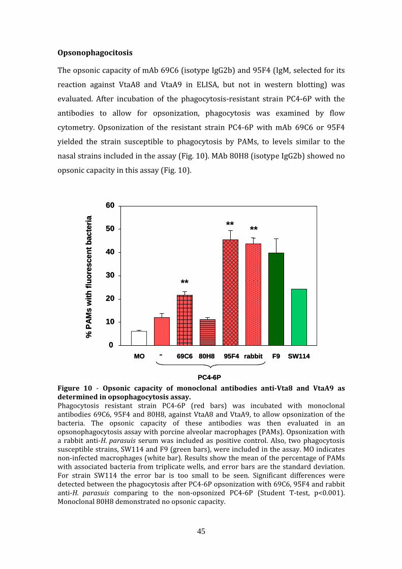
45
Opsonophagocitosis
The opsonic capacity of mAb 69C6 (isotype IgG2b) and 95F4 (IgM, selected for its
reaction against VtaA8 and VtaA9 in ELISA, but not in western blotting) was
evaluated. After incubation of the phagocytosis‐resistant strain PC4‐6P with the
antibodies to allow for opsonization, phagocytosis was examined by flow
cytometry. Opsonization of the resistant strain PC4‐6P with mAb 69C6 or 95F4
yielded the strain susceptible to phagocytosis by PAMs, to levels similar to the
nasal strains included in the assay (Fig. 10). MAb 80H8 (isotype IgG2b) showed no
opsonic capacity in this assay (Fig. 10).
MO 69C6 80H8 95F4 rabbit F9 SW114 -
PC4-6P
% P
AMs
with
fluor
esce
ntba
cter
ia
0
10
20
30
40
50
60
**
** **
MO 69C6 80H8 95F4 rabbit F9 SW114 -
PC4-6P
% P
AMs
with
fluor
esce
ntba
cter
ia
0
10
20
30
40
50
60
**
** **
Figure 10 ‐ Opsonic capacity of monoclonal antibodies antiVta8 and VtaA9 as determined in opsophagocytosis assay. Phagocytosis resistant strain PC4‐6P (red bars) was incubated with monoclonal antibodies 69C6, 95F4 and 80H8, against VtaA8 and VtaA9, to allow opsonization of the bacteria. The opsonic capacity of these antibodies was then evaluated in an opsonophagocytosis assay with porcine alveolar macrophages (PAMs). Opsonization with a rabbit anti‐H. parasuis serum was included as positive control. Also, two phagocytosis susceptible strains, SW114 and F9 (green bars), were included in the assay. MO indicates non‐infected macrophages (white bar). Results show the mean of the percentage of PAMs with associated bacteria from triplicate wells, and error bars are the standard deviation. For strain SW114 the error bar is too small to be seen. Significant differences were detected between the phagocytosis after PC4‐6P opsonization with 69C6, 95F4 and rabbit anti‐H. parasuis comparing to the non‐opsonized PC4‐6P (Student T‐test, p<0.001). Monoclonal 80H8 demonstrated no opsonic capacity.
46
Discussion In this study, we provide evidence showing that two trimeric autotransporters of
H. parasuis, VtaA8 and VtaA9, are surface‐exposed proteins that are involved in
resistance to phagocytosis. Since the production of mutants in H. parasuis is
hindered by low and strain‐dependent transformation efficiency [7, 24], we
decided to use the strategy of studying the function of specific genes in the
heterologous host E. coli. Thus, the expression of the individual proteins in E. coli,
although was not enough to prevent phagocytosis, produced a delay in the
phagocytosis process. This strategy allowed us also to circumvent the problem
derived from the existence of several vtaA in H. parasuis [9], which suggests a
functional redundancy.
Trimeric autotransporters mediate virulence mechanisms in other Gram‐negative
bacteria [25‐27] and their involvement in phagocytosis resistance has been
previously demonstrated in Neisseria meningitiditis, a Gram‐negative bacterium
capable to produce systemic disease [28]. Recently, VtaA genes of group 1 (in
which vtaA8 and vtaA9 are included) have been found widely represented in
virulent strains of H. parasuis [9, 29]. When expressed in E.coli, VtaA8 and VtaA9
promoted resistance to phagocytosis by PAMs in an attachment‐independent way.
Both, flow cytometry and fluorescent microscopy support the role of these
proteins in phagocytosis, but the proteins were unable to block the process and the
bacteria eventually proceeded into the endocytic network. Our results indicate that
VtaA8 and VtaA9 alone are not sufficient to avoid phagocytosis, and other factors
may be involved, such as capsule, as it was previously suggested [20].
The N‐terminal domain of trimeric autotransporters is exposed to the external
media and to the host immune response [11]. The exposure of VtaA8 and VtaA9 on
the bacterial surface was demonstrated with monoclonal antibodies. The presence
of these proteins on the surface of virulent strain PC4‐6P, while absent on the non‐
virulent strain F9, support the association of these trimeric autotransporters with
virulence. In addition, the antibodies produced against the VtaA8 and VtaA9 of the
Nagassaki strain were able to recognize the heterologous virulent strain PC4‐6P.
Although a mixture of VtaAs was not able to fully protect against a severe H.
parasuis challenge [32], this cross‐reactivity supports the potential of these
47
proteins as candidates in vaccine formulations against H. parasuis. VtaA8 and
VtaA9 probably are redundant proteins affecting the PAMs through the same
mechanism, since co‐incubation with both proteins (both clones) did not increase
synergistically the effect against bacterial phagocytosis. Directing the immune
response to common epitopes from both proteins can constitute a rational to
improve the probability of vaccine success. Nevertheless, the choice of adjuvant is
also important for the induction of the right antibody subclass, since not all the
IgGisotypes have the same capacity to promote opsonophagocytosis [30, 31].
In conclusion, VtaA8 and VtaA9 are surface exposed proteins that play a role in
phagocytosis resistance in virulent strains of H. parasuis. VtaA8 and VtaA9 share
epitopes, which are also present on the surface of heterologous virulent strains.
These properties make these proteins promising vaccine candidates against
Glässer’s disease.

48
References [1]. Rapp‐Gabrielson V, Oliveira S, Pijoan C: Haemophilus parasuis. In Diseases of
swine, 9th edition. Edited by Straw BE, Zimmerman JJ, D'Allaire S, Taylor DJ.
Iowa: Blackwell publishing; 2006, 681‐690.
[2]. Holtkamp D, Rotto H, Garcia R: Economic Cost of Major Health Challenges in
Large US Swine Production Systems. Swine News newsletter2007, 30.
[3]. Bouchet B, Vanier G, Jacques M, Gottschalk M: Interactions of Haemophilus
parasuis and its LOS with Porcine Brain Microvascular Endothelial Cells.
Vet Res 2008, 39:42.
[4]. Fu S, Yuan F, Zhang 424 M, Tan C, Chen H, Bei W: Cloning, expression and
characterization of a cell wall surface protein, 6
phosphogluconatedehydrogenase, of Haemophilus parasuis. Res Vet
Sci2012, in press.
[5]. Lichtensteiger CA, Vimr ER: Purification and renaturation of membrane
neuraminidase from Haemophilus parasuis. Vet Microbiol2003, 93:79‐87.
[6]. Sack M, Baltes N: Identification of novel potential virulenceassociated
factors in Haemophilus parasuis. Vet Microbiol2009, 136:382‐386.
[7]. Zhang B, Feng S, Xu C, Zhou S, He Y, Zhang L, Zhang J, Guo L, Liao M: Serum
resistance in Haemophilus parasuis SC096 strain requires outer
membrane protein P2 expression. FEMS Microbiol Lett 2012, 326:109‐115.
[8]. Zhang B, He Y, Xu C, Xu L, Feng S, Liao M, Ren T: Cytolethal distending toxin
(CDT) of the Haemophilus parasuis SC096 strain contributes to serum
resistance and adherence to and invasion of PK15 and PUVEC cells. Vet
Microbiol 2012.
[9]. Pina S, Olvera A, Barcelo A, Bensaid A: Trimeric autotransporters of
Haemophilus parasuis: generation of an extensive passenger domain
repertoire specific for pathogenic strains. J Bacteriol2009, 191:576‐587.
[10]. Cotter SE, Surana NK, St Geme JW, 3rd: Trimeric autotransporters: a
distinct subfamily of autotransporter proteins. Trends Microbiol2005,
13:199‐205.
[11]. Henderson IR, Nataro JP: Virulence functions of autotransporter proteins.
Infect Immun 2001, 69:1231‐1243.

49
[12]. Vahle JL, Haynes JS, Andrews JJ: Experimental reproduction of
Haemophilus parasuis infection in swine: clinical, bacteriological, and
morphologic findings. J Vet Diagn Invest 1995, 7:476‐480.
[13]. Arredouani MS, Palecanda A, Koziel H, Huang YC, Imrich A, Sulahian TH,Ning
YY, Yang Z, Pikkarainen T, Sankala M, et al: MARCO is the major binding
receptor for unopsonized particles and bacteria on human alveolar
macrophages. J Immunol 2005, 175:6058‐6064.
[14]. McCusker K, Hoidal J: Characterization of scavenger receptor activity in
resident human lung macrophages. Exp Lung Res 1989, 15:651‐661.
[15]. Aderem A, Underhill DM: Mechanisms of phagocytosis in macrophages.
Annu Rev Immunol1999, 17:593‐623.
[16]. Fives‐Taylor PM, Meyer DH, Mintz KP, Brissette C: Virulence factors of
Actinobacillus actinomycetemcomitans. Periodontol 2000 1999, 20:136‐
167.
[17]. Harper M, Boyce JD, Adler B: Pasteurella multocida pathogenesis: 125
years after Pasteur. FEMS MicrobiolLett2006, 265:1‐10.
[18]. Noel GJ, Hoiseth SK, Edelson PJ: Type b capsule inhibits ingestion of
Haemophilus influenzae by murine macrophages: studies with isogenic
encapsulated and unencapsulated strains. J Infect Dis 1992, 166:178‐182.
[19]. Wood GE, Dutro SM, Totten PA: Haemophilus ducreyi inhibits phagocytosis
by U937 cells, a human macrophagelike cell line. InfectImmun2001,
69:4726‐4733.
[20]. Olvera A, Ballester M, Nofrarias M, Sibila M, Aragon V: Differences in
phagocytosis susceptibility in Haemophilus parasuis strains. Vet Res
2009, 40:24.
[21]. Celli J, Finlay BB: Bacterial avoidance of phagocytosis. Trends Microbiol
2002, 10:232‐237.
[22]. Olvera A, Pina S, Perez‐Simo M, Oliveira S, Bensaid A: Virulence‐associated
trimeric autotransporters of Haemophilus parasuis are antigenic
proteins expressed in vivo. Vet Res 2010, 41:26.
[23]. Morey P, Cano V, Marti‐Lliteras P, Lopez‐Gomez A, Regueiro V, Saus C,
Bengoechea JA, Garmendia J: Evidence for a nonreplicative intracellular

50
stage of nontypable Haemophilus influenzae in epithelial cells.
Microbiology 2011, 157:234‐250.
[24]. Chen L, Wu D, Cai X, Guo F, Blackall PJ, Xu X, Chen H: Electrotransformation
of Haemophilus parasuis with in vitro modified DNA based on a novel
shuttle vector. Vet Microbiol 2012, 155:310‐316.
[25]. Leduc I, Olsen B, Elkins C: Localization of the domains of the Haemophilus
ducreyi trimeric autotransporter DsrA involved in serum resistance and
binding to the extracellular matrix proteins fibronectin and vitronectin.
Infect Immun 2009, 77:657‐666.
[26]. Scarselli M, Serruto D, Montanari P, Capecchi B, Adu‐Bobie J, Veggi D,Rappuoli
R, Pizza M, Arico B: Neisseria meningitides NhhA is a multifunctional
trimeric autotransporter adhesin. Mol Microbiol 2006, 61:631‐644.
[27]. Serruto D, Spadafina T, Scarselli M, Bambini S, Comanducci M, Hohle S,Kilian
M, Veiga E, Cossart P, Oggioni MR, et al: HadA is an atypical new
multifunctional trimeric coiledcoil adhesin of Haemophilus influenzae
biogroup aegyptius, which promotes entry into host cells. Cell Microbiol
2009, 11:1044‐1063.
[28]. Sjolinder H, Eriksson J, Maudsdotter L, Aro H, Jonsson AB:
Meningococcalouter membrane protein NhhA is essential for
colonization and disease by preventing phagocytosis and complement
attack. Infect Immun 2008, 76:5412‐5420.
[29]. Olvera A, Pina S, Macedo N, Oliveira S, Aragon V, Bensaid A: Identification of
potentially virulent strains of Haemophilus parasuis using a multiplex
PCR for virulenceassociated autotransporters (vtaA). Vet J 2012,
191:213‐218.
[30]. Li Y, Gottschalk M, Esgleas M, Lacouture S, Dubreuil JD, Willson P, Harel J:
Immunization with recombinant Sao protein confers protection against
Streptococcus suis infection. Clin Vaccine Immunol 2007, 14:937‐943.
[31]. Michaelsen TE, Ihle O, Beckstrom KJ, Herstad TK, Sandin RH, Kolberg J,Aase A:
Binding properties and antibacterial activities of Vregion identical,
human IgG and IgM antibodies, against group B Neisseria meningitidis.
BiochemSoc Trans 2003, 31:1032‐1035.

51
[32]. Olvera A, Pina S, Perez‐Simo M, Aragon V, Segales J, Bensaid A:
Immunogenicity and protection against Haemophilus parasuis infection
after vaccination with recombinant virulence associated trimeric
autotransporters (VtaA). Vaccine 2011, 29:2797‐2802.
[33]. Kielstein P, Rapp‐Gabrielson VJ: Designation of 15 serovars of
Haemophilus parasuis on the basis of immunodiffusion using heatstable
antigen extracts. J ClinMicrobiol1992, 30:862‐865.

52

53
RESULTS
CHAPTER 2.
Changes in macrophage phenotype after infection of pigs with
Haemophilus parasuis strains of different virulence

54
Summary
Haemophilus parasuis is a colonizer of healthy piglets and the etiological agent of
Glässer’s disease. Differences in virulence among strains of H. parasuis have been
widely observed by different tests, including experimental infections and in vitro
assays. Here, we performed an infection via intranasal of snatch farrowed,
colostrum‐deprived piglets with 4 strains of H. parasuis: reference virulent strain
Nagasaki, reference non‐virulent strain SW114, and field strains IT29205 (from
systemic lesion and virulent in a previous challenge) and F9 (from nasal cavity of
healthy piglet). Two non‐infected animals served as controls. At different times
after infection (1, 2, 4 and 7 days) two animals of each group were euthanized and
alveolar macrophages were analysed by flow cytometry with specific antibodies to
CD163, CD172a, SLAI, SLAII, Sialoadhesin, CD14 and SWC8. At 1dpi, non‐virulent
SW114 and F9 strains induced higher expression of CD163, sialoadhesin, SLAII and
CD172a in the surface of the macrophages than virulent strains Nagasaki and
IT29205. At 2dpi, the situation switched into a strong expression of CD172a,
CD163 and sialoadhesin by the virulent strains, which was followed by a steep
increase in IL‐8 and soluble CD163 at 3‐4 dpi. The early increase of surface‐
expression of CD163 and sialoadhesin in macrophages induced by non‐virulent
strains went along with higher level of IL‐8 in serum than the level induced by
virulent strains in the first 2 days of infection, and interestingly by INF‐α, which
was not induced by virulent strains. Overall, virulent strains delay the activation of
macrophages as compared with non‐virulent strains, down‐regulating the levels of
CD163, sialoadhesin, SLA II and CD172a. This delay in macrophage activation may
be critical for disease production.
55
Introduction
Haemophilus parasuis is the etiological agent of Glässer’s disease, a systemic
disease characterized by fibrinous polyserosytis [1]. Infection by H. parasuis is
common in commercial farms and Glässer’s disease is recognized as one of the
main causes of economic loss in the pig industry [2]. H. parasuis contains also non‐
virulent strains, which are considered part of the respiratory microbiota. However,
the differences between virulent and non‐virulent strains are not well defined.
During the first hours of infection H. parasuis can be isolated from the lungs of
animals experimentally infected via intranasal [3]. In this location, H. parasuis
comes into contact with cells of the immune system, such as the alveolar
macrophages. Virulent strains of H. parasuis have been shown to be resistant to
phagocytosis by alveolar macrophages by avoiding uptake [4]. In addition to
evading direct killing by macrophages, phagocytosis resistance may imply a delay
in the interaction with the immune system, preventing the induction of a proper
response by the host. Macrophages are common target for bacterial pathogens that
benefit from avoiding an encounter with the immune system, as well as those that
are aiming to secure systemic spread [5]. In addition, macrophages have important
regulatory and effector functions in the immune response. Several molecules in the
surface of macrophages are regulated during their maturation and differentiation
process and these surface changes might account for their heterogeneity and
functional plasticity, which is associated with different capacity to process and
present antigens, release cytokines and other mediators, recruit other cells to the
site of infection and coordinate their responses to clear the microbe [6, 7]. When
stimulated, macrophages adopt context‐dependent phenotypes that either
promote or inhibit host antimicrobial defense, inflammatory and immune
responses [7, 8].
Recently, Ondrackova et al. [9] investigated changes in macrophage populations
following Actinobacillus pleuroneumoniae infection using a combination of myeloid
markers. In that study macrophages from non‐infected piglets were described as
CD14+, CD163+, CD172ahi, and SLAII+ (SLA, Swine histocopatibility leucocyte
antigen), SWC8‐, discerning between FSClo and FSChi (for mature and less mature
macrophages).
56
A description of transcriptional changes in porcine alveolar macrophages after
virulent H. parasuis infection has been recently reported [10]. After 6 day of
infection, alveolar macrophages showed up‐regulation of genes involved in
immune and inflammatory response through regulation of certain pathways like
cell adhesion, cytokine‐cytokine receptor interaction, complement and coagulation
cascades, toll‐like receptors and MAPK signalling [10]. Other studies have
described differences in host susceptibility to infection with virulent H. parasuis
[11], which were confirmed at transcriptional level, finding a correlation between
the animal susceptibility and a reduction in the expression of genes involved in the
antigen presentation pathway and increase in the expression of genes involved in
inflammation [12].
Here, using flow cytometry and monoclonal antibodies (mAb) against surface
markers, we have studied the lung macrophages after H. parasuis infection.
Changes in the expression of CD163, sialoadhesin, CD172a, SLA I, SLA II and CD14
of BALF cells from animals infected with virulent and non‐virulent strains were
determined and their role in tuning the immune response during H. parasuis
infection and influence in the final outcome of the disease are discussed.

57
Materials and methods
Animal infection
Animal care and all procedures described were performed in accordance with the
regulations required by the Ethics Commission in Animal Experimentation of the
Generalitat de Catalunya (Approved Protocol Number 5796). H. parasuis infection
was performed as previously described [13] with 33 snatch‐farrowed colostrum‐
deprived piglets. The animals were obtained from a conventional farm with a
standard health status. Briefly, sows were seropositive against porcine
reproductive and respiratory virus (PRRSV) and swine influenza, and seronegative
against Aujeszky’s disease virus. Piglets were seronegative for porcine circovirus 2
(PCV2), but positive for PRRSV. The outline of the experiment is presented in Table
1.
Table 1. Scheme of the animal experiment, including the Haemophilus parasuis strains used and their main characteristics
Number of animals necropsied at different times after inoculation
Strain Strain characteristics
Bacterial inoculum §
Number of animals
1 day
2 days
3 days
4 days
5 days
7 days
Nagasaki Reference, virulent, serovar 5
4 x 10⁶ 8 2 2 3 1 ‐ ‐
IT29205 Isolated from lesion, virulent, serovar 4
8 x 10⁷ 8 2 2 ‐ 2 1 1
SW114 Reference, non‐virulent, serovar 3
8 x 10⁸ 7 2 2 ‐ 2 ‐ 1
F9 Isolated from nasal cavity, non‐virulent*, serovar 6
5 x 10⁸ 8 2 2 ‐ 2 ‐ 2
* Non‐virulent in in vitro assays (phagocytosis and serum sensitive, non‐invasive in endothelial cells. § Colony forming units (CFU) per animal

58
Bacterial suspensions for inoculation were made in phosphate buffered saline
(PBS) from overnight cultures of the four H. parasuis strains on chocolate agar
plates. Suspensions were adjusted to get the desired bacterial concentration (Table
1). The real number of bacteria in the suspensions was confirmed by serial dilution
and plating on chocolate agar. At 1 month of age, groups of 8 piglets were
intranasally inoculated with 1.5 ml (0.75 ml per nostril) of the corresponding
bacterial suspension using a plastic syringe. For strain SW114 only 7 piglets were
available.
After 1, 2, 4 and 7 days (or at other times if the animals showed signs of suffering),
two piglets from each group were euthanized. Lesions were assessed and samples
for bacterial culture were taken. In addition, brochoalveolar lavage fluid (BALF)
was performed with sterile PBS. Cells in the BALF were pelleted at 300 x g for 10
min and stored at ‐80ºC until analysed. Analysis of the cells was performed within
one month of necropsy.
H. parasuis isolation
Swabs from peritoneal, thoracic, pericardial and nasal cavity, meninges, lung and
joints were taken and immediately transferred to the laboratory for bacterial
isolation on chocolate agar plates. Also, BALF and blood samples were examined
for H. parasuis presence. After 24‐48 h of incubation at 37ºC, H. parasuis growth
was semi‐quantified as previously described [13]: a score of 3 was assigned to
samples with confluent growth, 2 to samples yielding isolated colonies (down to
20 colonies), 1 to samples yielding 1 to 19 colonies and 0 to samples yielding no
bacterial growth. Two animals served as non‐infected controls and were processed
as the rest.
Analysis of alveolar macrophages
Macrophages recovered from the infected pigs at different times after infection
were processed for the detection of surface CD163, CD172a, SLAII, SLAI, CD14,
SWC8 and sialoadhesin with specific monoclonal antibodies (mAb) [7]. An
irrelevant mAb was used as negative control. Macrophages were incubated on ice

59
for 40 min with each of the hybridoma supernatant, washed three times in 1%
fetal bovine serum (FBS)‐PBS and the bound antibody was detected with a FITC‐
conjugated goat anti‐mouse IgG (Jackson ImmunoResearch Europe Ltd, UK) for 30
min, except for CD14 that was detected directly with a FITC‐conjugated mAb α‐pig
CD14 (clone MIL‐2, AbD Serotec, UK). Label intensity in the samples (identified by
FL1 x‐mean in the gated population) was determined by flow cytometry in a BD
FACSAria I Flow cytometer (Becton Dickinson, Madrid, Spain).
Nonspecific phagocytosis assay
Formaline‐killed Escherichia coli strain ATCC 25922 was used as particles for
phagocytosis. Overnight bacterial growth was resuspended in PBS and formalin
was added to a final concentration of 1%. This suspension was incubated during
1h at room temperature to kill the bacteria. After washing with PBS, fluorescein
isothiocyanate (FITC) was added to a final concentration of 100 μg/ml and
incubated 1h at 37ºC. Unbound FITC was eliminated by washing with PBS, and the
labelled E. coli was stored at 4ºC until used.
For phagocytosis assays, macrophages recovered from the pigs were counted and
seeded in 6 well‐plates at a concentration of 5 x 10⁵ cells per well in 3 ml of
Dulbecco’s modified Eagle’s medium (DMEM) supplemented with 10% FBS and
1% L‐glutamine (CDMEM). Plates were incubated at 37ºC with 5% CO2, and after
attachment of the cells to the wells for 1h. FITC‐labelled E. coli was added to each
well and incubated for 30 min. PAMs from non‐infected animals were pre‐treated
with 100 pg/ml of IL‐8 to evaluate their role in phagocytosis. After 2 washes,
macrophages with associated bacteria were analysed in a BD FACSAria I Flow
cytometer (Becton Dickinson). Phagocytosis experiments were performed one
time in duplicate wells.

60
In vitro infection of alveolar macrophages with H. parasuis
Porcine alveolar macrophages (PAMs) from healthy animals were seeded in 6‐well
plates at a concentration of 5 x 10⁵ cells in 3 mL per well of CDMEM. After
overnight incubation at 37ºC with 5% CO2, wells were inoculated with H. parasuis
at a multiplicity of infection (MOI) of 1 or 200 and incubated during 20h or 1h,
respectively. After washing with PBS, PAMs were scrapped and surface expression
of CD172a, CD163, Sialoadhesin, SLAII and SLAI was analyzed as described above.
Soluble CD163, INFα and IL8 detection
Soluble CD163 (sCD163) was quantified in sera by a previously described ELISA
[14]. IL‐8 was detected in sera by ELISA using a swine IL‐8 kit (KingFisher Biotech,
INC. St Paul MN, USA) following manufacturer’s instructions. INF‐α was
determined in sera by ELISA using anti‐α‐IFN monoclonal antibodies (K9 and F17)
and recombinant IFN‐α (PBL Biomedical Laboratories, Piscataway, NJ, USA) as
described before [15, 16] with some modifications. Briefly, ELISA plates were
coated with anti‐pig IFN‐α mAb K9 (0.5 μg ⁄ml in carbonate‐bicarbonate buffer
[pH 9.4]). After blocking with PBS containing 0.05% Tween‐20 and 0.5% bovine
serum albumin, serum samples were added to the wells and incubated for 1h at
37ºC. After washing, biotinylated pig IFN‐α mAb F17 was added to the wells and
incubated for 1h at 37ºC. Finally, peroxidase‐conjugated streptavidin was added
and incubated 30 min at 37ºC. Reactions were developed with 3,3’,5,5’‐
tetramethylbenzidine (TMB). Non‐infected animals provided basal levels of
cytokine expression. INF‐α was also determined in supernatants of in vitro infected
PAMs following the above described protocol.

61
Results
Clinicopathological findings and bacterial isolation
Pulmonary and systemic infections in the animals at time of necropsy are
summarized in Table 2. As expected, animals infected with virulent strains
Nagasaki or IT29205 showed severe lesions of Glässer’s disease, while animals
infected with the non‐virulent strains SW114 or F9 continued healthy during the
experiment (with the exception of one animal infected with SW114, which showed
systemic lesions).
Table 2. Lesions and bacterial recovery from pigs infected with virulent strains Nagasaki and IT29205 or nonvirulent strains SW114 and F9. The animals were sacrificed at different time points and necropsies were performed.
Pulmonary infection Systemic infection
Strain Time of necropsy Bacterial
score# CPBN* Bacterial score$
Bacteria in blood
Fibrinous poliserositis
Fibrinous arthritis
Nagasaki 1 dpi 2/2 ‐/‐ 0/0 0/0 ‐/‐ ‐/‐ 2 dpi 0/1 ‐/‐ 0/0 0/0 ‐/‐ ‐/‐ 3 dpi 3/3/3 ‐/‐ 14/13/11 3/3/3 +/+/+ +/‐/‐ 4 dpi 3 ‐ 11 3 + ‐ IT29205 1 dpi 0/3 ‐/‐ 0/1 0/1 ‐/‐ +b/‐ 2 dpi 1/1 ‐/‐ 0/1 0/1 ‐/‐ ‐/‐ 4‐5§dpi 1/3/3 ‐/+ 7/17/15 1/3/3 ‐/+/‐ +/‐/+ 7 dpi 1 ‐ 0 1 ‐ +b SW114 1 dpi 0/2 ‐/‐ 0/0 0/0 ‐/‐ ‐/‐ 2 dpi 0/1 ‐/‐ 0/0 0/0 ‐/‐ ‐/‐ 4 dpi 0/1 ‐/‐ 0/9 0/1 ‐/+ ‐/+ 7 dpi 0 ‐ 0 0 ‐/‐ ‐/‐
F9 1 dpi 0/1 ‐/‐ 0/0 0/0 ‐/‐ ‐/‐ 2 dpi 0/0 ‐/‐ 0/0 0/0 ‐/‐ ‐/‐ 4 dpi 0/2 ‐/‐ 0/1 0/1 ‐/‐ ‐/‐ 7 dpi 0/1 ‐/‐ 0/3 0/0 ‐/‐ ‐/+
# bacterial score of each animal in pulmonary samples
* Catarral‐purulent bronchopneumonia
$ Bacterial score of each animal calculated as the sum of the score in blood, abdominal cavity, thorax, pericardium, meninges and the maximum score found in four joint samples.
b mild lesion
§ 2 animals were sacrificed at day 4 and a third one on day 5

62
Although the dose of Nagasaki was reduced to avoid excessive mortality, 3 animals
infected with this strain had to be euthanized at day 3 and the last animal was
euthanized at 4 days post‐infection (dpi); no animal survived till day 7. H. parasuis
Nagasaki was found in lung at 1 and 2 dpi. As infection progressed, Nagasaki was
recovered in high quantities from systemic organs, including blood, pericardium,
joints and brain, at 3 dpi and 4 dpi (Table 2). In the case of strain IT29205,
pulmonary infection was detected in the infected animals at 1 dpi and 2 dpi,
followed by systemic infection at 4 dpi with severe Glässer´s disease lesions. After
5 dpi, one animal died from sepsis and arthritis, while the other one survived until
day 7 displaying no clinical signs.
Animals infected with SW114 and F9 exhibit large amounts of bacteria in nasal
cavities at all time points. While in general piglets infected with the non‐virulent
strains stayed healthy, one piglet infected with SW114 showed arthritis and
polyserositis at 4 dpi, with systemic isolation of the bacteria. In the case of F9, one
animal showed arthritis in the right elbow at 7 dpi, with recovery of the bacteria.
No lesions were determined at any time point in the rest of the animals infected
with SW114 or F9 strains. Nonetheless, bacteria were recovered in low quantities
from the BALF at 1 dpi and 2 dpi.
Changes in BALF cell population
Cells recovered from BALF were analysed with specific mAb by flow cytometry.
Macrophages were identified according to their light scatter properties (FSC and
SS) and the expression of the pan‐myelomonocytic marker CD172a (Fig. 1).

63
93,6%
4,4%
P2
P2
A
B
93,6%
4,4%
P2
P2
A
B
A
B
Figure 1. Analysis of the BALF cells by flow cytometry. Gate for analysis of macrophages (P2) was selected by forward and side scatter characteristics (FS‐SS) and positivity to CD172a (Panel A). Panel B shows the results of the negative control for the labelling. Gate P2 was used in further analysis of macrophages markers.
At early time points (1dpi), the gated population from animals infected with
virulent strains showed SWC8+ cells, which were not detected in animals infected
with non‐virulent strains or non‐infected animals. After 2 days, the SWC8+ cells
were not detected.
Surface markers were analysed taking together the data from animals infected
with virulent strains versus non‐virulent strains at each time point.
CD172a
Differences of CD172a expression were found between animals infected with
virulent or non‐virulent strains, with the exception of the animals examined at 7
dpi (Fig. 2). At 1 dpi, macrophages from animals infected with virulent strains
Nagasaki and IT29205 showed lower intensity of labelling (as FL1 x‐mean) with
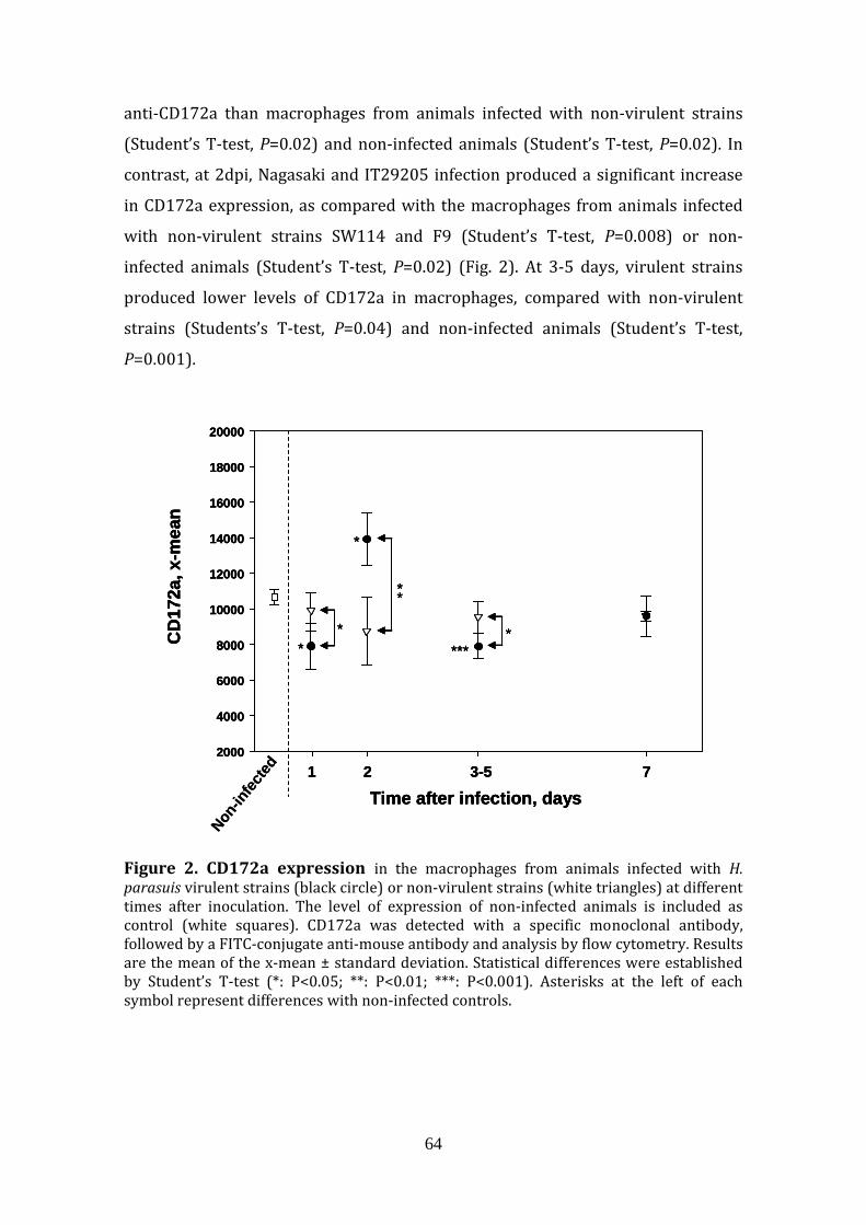
64
anti‐CD172a than macrophages from animals infected with non‐virulent strains
(Student’s T‐test, P=0.02) and non‐infected animals (Student’s T‐test, P=0.02). In
contrast, at 2dpi, Nagasaki and IT29205 infection produced a significant increase
in CD172a expression, as compared with the macrophages from animals infected
with non‐virulent strains SW114 and F9 (Student’s T‐test, P=0.008) or non‐
infected animals (Student’s T‐test, P=0.02) (Fig. 2). At 3‐5 days, virulent strains
produced lower levels of CD172a in macrophages, compared with non‐virulent
strains (Students’s T‐test, P=0.04) and non‐infected animals (Student’s T‐test,
P=0.001).
Non-in
fected
Time after infection, days1 2 3-5 7
CD
172a
, x-m
ean
2000
4000
6000
8000
10000
12000
14000
16000
18000
20000
*
**
****
*
*
Non-in
fected
Time after infection, days1 2 3-5 7
CD
172a
, x-m
ean
2000
4000
6000
8000
10000
12000
14000
16000
18000
20000
*
**
****
*
*
Time after infection, days1 2 3-5 7
CD
172a
, x-m
ean
2000
4000
6000
8000
10000
12000
14000
16000
18000
20000
*
**
****
*
*
Figure 2. CD172a expression in the macrophages from animals infected with H. parasuis virulent strains (black circle) or non‐virulent strains (white triangles) at different times after inoculation. The level of expression of non‐infected animals is included as control (white squares). CD172a was detected with a specific monoclonal antibody, followed by a FITC‐conjugate anti‐mouse antibody and analysis by flow cytometry. Results are the mean of the x‐mean ± standard deviation. Statistical differences were established by Student’s T‐test (*: P<0.05; **: P<0.01; ***: P<0.001). Asterisks at the left of each symbol represent differences with non‐infected controls.

65
CD163
Regarding CD163 expression, significant differences were detected when animals
infected with virulent strains were compared with those infected with non‐
virulent strains at 1 and 2 dpi (Fig. 3).
Time after infection, days
1 2 3-5 7
CD
163,
x-m
ean
0
20000
40000
60000
80000
Non-in
fected
***
*
**
**
***
Time after infection, days
1 2 3-5 7
CD
163,
x-m
ean
0
20000
40000
60000
80000
Non-in
fected
***
*
**
**
***
Figure 3. CD163 expression in the macrophages from animals infected with H. parasuis virulent strains (black circle) or non‐virulent strains (white triangles) at different times after inoculation. The level of expression of non‐infected animals is included as control (white squares). CD163 was detected with a specific monoclonal antibody, followed by a FITC‐conjugate anti‐mouse antibody and analysis by flow cytometry. Results are the mean of the x‐mean ± standard deviation. Statistical differences were established by Student’s T‐test (*: P<0.05; **: P<0.01; ***: P<0.001). Asterisks at the left of each symbol represent differences with non‐infected controls.
At 1dpi, Nagasaki and IT29205 strains down‐regulated the amount of CD163 on
the surface of macrophages as compared with the levels in macrophages of non‐
infected piglets and piglets infected with non‐virulent strains (Student’s T‐test,
P=0.009 and P=0.00001, respectively). SW114 and F9 strains caused an increase in
CD163 expression as compared with control piglets (Student’s T‐test, P=0.04) after
1 day of infection. After 2 days, the tendency switched and virulent strains

66
produced a sharp increase in the CD163 expression as compared with non‐infected
control (Student’s T‐test, P=0.0009), indicating a massive but probably late
activation of macrophages, while expression in the macrophages from animals
infected with non-virulent strains came down to control levels. At later time points,
virulent and non‐virulent strains did not modify the levels of CD163 in the surface
of macrophages, which were similar as non‐infected animals (Fig. 3).
Sialoadhesin
When expression of sialoadhesin was examined, macrophages from animals
infected with virulent strains presented lower sialoadhesin on their surfaces than
macrophages from animals infected with non‐virulent strains at 1dpi (Student’s T‐
test, P=0.02; Fig 4). This tendency shown at 1dpi, switched at 2 dpi, when detection
of sialoadhesin was higher in macrophages from animals infected with virulent
strains than in macrophages from animals infected with non‐virulent strains
(Student’s T‐test, P=0.005) or from non‐infected animals (Student’s T‐test,
P=0.000004; Fig. 4). After 3‐5 dpi, macrophages from all H. parasuis infected
animals decreased their levels of sialoadhesin expression as compared with the
non‐infected animal (Student’s T‐test, P=0.0007; Fig. 4). Interestingly, at later time
points (7 dpi), infection by non‐virulent strains significantly increased the
expression of this marker compared with non‐infected animals (Student’s T‐test,
P=0.01; Fig 4). The only remaining animal infected with a virulent strain at this
time point exhibited lower levels of sialoadhesin on the surface of macrophages
than non‐infected animals (Student’s T‐test, P=0.0007) or animals infected with
non‐virulent strains (Student’s T‐test, P=0,04).

67
Time after infection, days1 2 3-5 7
Sial
oadh
esin
, xm
ean
0
5000
10000
15000
20000
25000
Non-in
fected
*
**
***
***
***
*
**
Time after infection, days1 2 3-5 7
Sial
oadh
esin
, xm
ean
0
5000
10000
15000
20000
25000
Non-in
fected
*
**
***
***
***
*
**
Figure 4. Sialoadhesin expression in the macrophages from animals infected with H. parasuis virulent strains (black circle) or non‐virulent strains (white triangles) at different times after inoculation. The level of expression of non‐infected animals is included as control (white squares). Sialoadhesin was detected with a specific monoclonal antibody, followed by a FITC‐conjugate anti‐mouse antibody and analysis by flow cytometry. Results are the mean of the x‐mean ± standard deviation. Statistical differences were established by Student’s T‐test (*: P<0.05; **: P<0.01; ***: P<0.001). Asterisks at the left of each symbol represent differences with non‐infected controls.
SLAII
As observed with other markers, virulent strains down‐regulated the expression of
SLA II on the macrophages at 1 dpi as compared with non‐infected control
(Student’s T‐test, P=0.008) and non‐virulent strains (Student’s T‐test, P=0.02; Fig.
5). This effect switched at 2 dpi, when macrophages from animals infected with
virulent strains showed higher x‐mean as compared with animals infected with
non‐virulent strains and non‐infected animals, but these differences were not
significant (Fig.5). At 3‐5 dpi, animals infected with virulent strains showed lower
levels of SLAII on their macrophages than those infected with non‐virulents strains
(Student’s T‐test, P=0.001) and than the non‐infected controls (Student’s T‐test,

68
P=0.03) (Fig. 5). Later on, at 7 dpi, no significant differences in SLAII expression
were found.
Time after infection, days1 2 3-5 7
SLA
II, x
mea
n
20000
40000
60000
80000
10000
12000
14000
16000
***
Non-in
fected
*
**
Time after infection, days1 2 3-5 7
SLA
II, x
mea
n
20000
40000
60000
80000
10000
12000
14000
16000
****
Non-in
fected
*
****
Figure 5. SLAII expression in the macrophages from animals infected with H. parasuis virulent strains (black circle) or non‐virulent strains (white triangles) at different times after inoculation. The level of expression of non‐infected animals is included as control (white squares). SLAII was detected with a specific monoclonal antibody, followed by a FITC‐conjugate anti‐mouse antibody and analysis by flow cytometry. Results are the mean of the x‐mean ± standard deviation. Statistical differences were established by Student’s T‐test (*: P<0.05; **: P<0.01; ***: P<0.001). Asterisks at the left of each symbol represent differences with non‐infected controls.
SLAI
No major differences were found when the intensity of SLA I was examined (Fig.
6), only at 3‐5 dpi, SLAI showed lower levels in macrophages from animals infected
with virulent strains than those infected with non‐virulent strains (Student’s T‐
test, P=0.014; Fig. 6. No differences with control animals were found in any case.

69
Time after infection, days1 2 3-5 7
SLA
I, x
-mea
n
0
20000
40000
60000
80000Non
-infec
ted
**
Time after infection, days1 2 3-5 7
SLA
I, x
-mea
n
0
20000
40000
60000
80000Non
-infec
ted
**
Figure 6. SLAI expression in the macrophages from animals infected with H. parasuis virulent strains (black circle) or non‐virulent strains (white triangles) at different times after inoculation. The level of expression of non‐infected animals is included as control (white squares). SLAI was detected with a specific monoclonal antibody, followed by a FITC‐conjugate anti‐mouse antibody and analysis by flow cytometry. Results are the mean of the x‐mean ± standard deviation. Statistical differences were established by Student’s T‐test (*: P<0.05; **: P<0.01; ***: P<0.001). Asterisks at the left of each symbol represent differences with non‐infected controls.
CD14
Differences in CD14 surface expression were found at 2 dpi between macrophages
from animals infected with virulent strains and those infected with non‐virulent
strains (Student’s T‐test, P=0.005; Fig. 7). At that time, macrophages from animals
infected with non‐virulent strains showed a decrease in surface CD14 as compared
with non‐infected controls (Student’s T‐test, P=0.03; Fig. 7).

70
Time after infection, days1 2 3-5 7
CD
14, x
mea
n
2000
4000
6000
8000
10000
12000
14000
16000
18000
20000
Non-in
fected
**
*
Time after infection, days1 2 3-5 7
CD
14, x
mea
n
2000
4000
6000
8000
10000
12000
14000
16000
18000
20000
Non-in
fected
**
*
Figure 7. CD14 expression in the macrophages from animals infected with H. parasuis virulent strains (black circle) or non‐virulent strains (white triangles) at different times after inoculation. The level of expression of non‐infected animals is included as control (white squares). CD14 was detected with a specific FITC‐antibody and analysis by flow cytometry. Results are the mean of the x‐mean ± standard deviation. Statistical differences were established by Student’s T‐test (*: P<0.05; **: P<0.01). Asterisks at the left of each symbol represent differences with non‐infected controls.
Soluble CD163 (sCD163) detection
Figure 8 shows the results of the detection of sCD163 in serum.

71
1 2 3-4 7
Abs
0
2
4
6
8
10
12
**
1 2 3-4 7
Abs
Time after infection, days
Non-in
fected
0,5
1,0
1,5
8
12
1,8
1 2 3-4 7
Abs
0
2
4
6
8
10
12
**
1 2 3-4 7
Abs
0
2
4
6
8
10
12
**
1 2 3-4 7
Abs
Time after infection, days
Non-in
fected
0,5
1,0
1,5
8
12
1,8
Figure 8. Soluble CD163 (sCD163) in serum of animals infected with different strains of H. parasuis. The results are shown as sCD163 (mean absorbance in ELISA ± standard deviation) in sera of animals infected with virulent strains (black circles) or with non‐virulent strains (white triangles) at different times after inoculation. The level of sCD163 found in non‐infected animals (white square) is also indicated as control. Bottom panel shows the framed area from the top panel in expanded scale.
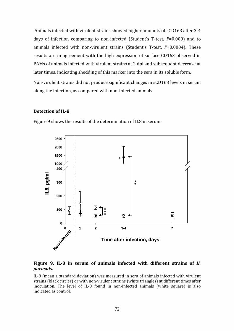
72
Animals infected with virulent strains showed higher amounts of sCD163 after 3‐4
days of infection comparing to non‐infected (Student’s T‐test, P=0.009) and to
animals infected with non‐virulent strains (Student’s T‐test, P=0.0004). These
results are in agreement with the high expression of surface CD163 observed in
PAMs of animals infected with virulent strains at 2 dpi and subsequent decrease at
later times, indicating shedding of this marker into the sera in its soluble form.
Non‐virulent strains did not produce significant changes in sCD163 levels in serum
along the infection, as compared with non‐infected animals.
Detection of IL8
Figure 9 shows the results of the determination of IL8 in serum.
0 2 3-41 7
IL8,
pg/
ml
0
100
200
300
4001000
1500
2000
2500
***
**
*
Time after infection, days
Non-in
fected
0 2 3-41 7
IL8,
pg/
ml
0
100
200
300
4001000
1500
2000
2500
***
**
*
Time after infection, days
Non-in
fected
Figure 9. IL8 in serum of animals infected with different strains of H. parasuis. IL‐8 (mean ± standard deviation) was measured in sera of animals infected with virulent strains (black circles) or with non‐virulent strains (white triangles) at different times after inoculation. The level of IL‐8 found in non‐infected animals (white square) is also indicated as control.

73
At 2 dpi, higher levels of IL8 were found in animals infected with non‐virulent
strains when compared the level in animals infected with virulent strains
(Student’s T‐test, P=0.0002). At day 3‐4 dpi, the tendency switched, and the
animals infected with virulent strains showed higher IL‐8 in serum than the
animals infected with non‐virulent strains (Student’s T‐test, P=0.006) and non‐
infected animals (Student’s T‐test, P=0.05). Similar levels of IL‐8 were observed
between animals infected with virulent and non‐virulent strains at 7 dpi.
Detection of IFNα
At 2dpi, higher levels of IFN‐α were detected in sera of animals infected with non‐
virulent strains than in sera of the animals infected with virulent strains (Student’s
T‐test, P=0.0004; Fig. 10). At day 3‐5, animals infected with virulent strains
remained at same levels of non‐infected animals, and non‐virulent strains infected
animals returned to those levels, which were maintained until the end of the
experiment.
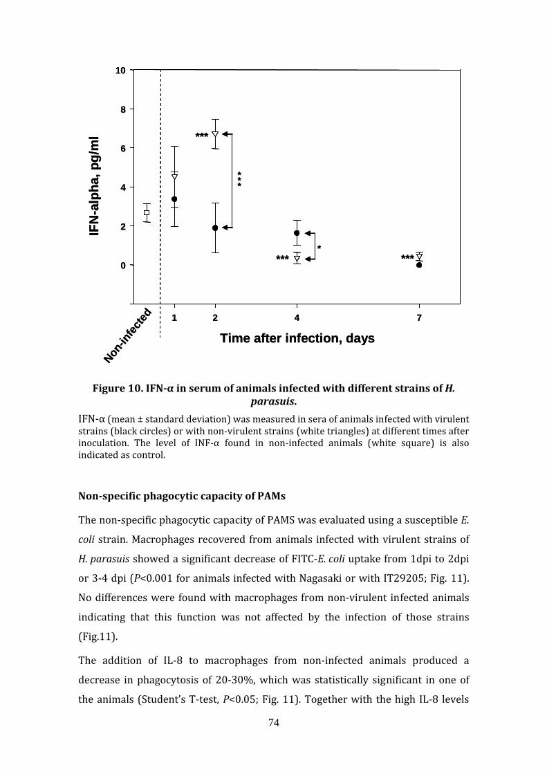
74
1 2 4 7
IFN
-alp
ha, p
g/m
l
0
2
4
6
8
10
Non-in
fected
Time after infection, days
***
*
***
*** ***
1 2 4 7
IFN
-alp
ha, p
g/m
l
0
2
4
6
8
10
Non-in
fected
Time after infection, days
***
*
***
*** ***
Figure 10. IFNα in serum of animals infected with different strains of H.
parasuis. IFN‐α (mean ± standard deviation) was measured in sera of animals infected with virulent strains (black circles) or with non‐virulent strains (white triangles) at different times after inoculation. The level of INF‐α found in non‐infected animals (white square) is also indicated as control.
Nonspecific phagocytic capacity of PAMs
The non‐specific phagocytic capacity of PAMS was evaluated using a susceptible E.
coli strain. Macrophages recovered from animals infected with virulent strains of
H. parasuis showed a significant decrease of FITC‐E. coli uptake from 1dpi to 2dpi
or 3‐4 dpi (P<0.001 for animals infected with Nagasaki or with IT29205; Fig. 11).
No differences were found with macrophages from non‐virulent infected animals
indicating that this function was not affected by the infection of those strains
(Fig.11).
The addition of IL‐8 to macrophages from non‐infected animals produced a
decrease in phagocytosis of 20‐30%, which was statistically significant in one of
the animals (Student’s T‐test, P<0.05; Fig. 11). Together with the high IL‐8 levels

75
found at 2dpi in animals infected with virulent strains, indicates how H. parasuis
may modulate the host in order to produce disease.
- -+ +IL8:
Non-infected
*
% F
ITC
-PAM
20
40
60
80
1 dpi 3 dpi
Nagasaki
2dpi 1 dpi 4 dpi
IT29205
2dpi 1 dpi 4 dpi
SW114
2dpi 1 dpi 4 dpi
F9
2dpi
*****
****
- -+ +IL8:
Non-infected
**
% F
ITC
-PAM
20
40
60
80
1 dpi 3 dpi
Nagasaki
2dpi 1 dpi 4 dpi
IT29205
2dpi 1 dpi 4 dpi
SW114
2dpi 1 dpi 4 dpi
F9
2dpi
*****
****
****
Figure 11. Phagocytic capacity of alveolar macrophages from animals infected with different strains of H. parasuis.
Alveolar macrophages from animals infected with virulent strains Nagasaki and IT29205 or with non‐virulent strains SW114 and F9 at different times post‐infection were infected ex vivo with a susceptible FITC‐E. coli. After 30 min of incubation, macrophages were recovered and analysed by flow cytometry to determine the percentage of cells with associated bacteria. Macrophages recovered from non‐infected pigs were used as control. In addition, macrophages from non‐infected were processed in the phagocytosis assay in the presence of 100 pg/ml of IL‐8. Results were compared by Student’s T‐test and differences are indicated by asterisks (*: P<0.05; **: P<0.01; ***: P<0.001)
In vitro infection of PAMs
In vitro infection of PAMs with H. parasuis strains did not reproduce the effects
seen in the in vivo infection with the same strains.
When MOI 1 and 20 h of incubation were used, PAMs infected with all H. parasuis
strains expressed lower levels of surface CD172a and CD163 (Fig. 12) compared
with non‐infected control PAMs (P< 0.05 and P< 0.01 for CD172a and CD163,
respectively). The rest of markers, SLAI, SLAII and sialoadhesin, did not changed
after infection. The experiments performed at MOI 200 for 1h, showed high
variability, and no differences could be determined.

76
SWC3
0
500
1000
1500
2000
2500
3000
Non in
fect
edNag
asak
iIT2
9205
SW11
4 F9
SWC
3, x
-mea
n
* * * *
Non in
fect
edNag
asak
i
CD
163,
x-m
ean
0
1000
2000
3000
4000
5000
6000
7000
IT292
05
SW11
4 F9
CD163
** ** ** **
SWC3
0
500
1000
1500
2000
2500
3000
Non in
fect
edNag
asak
iIT2
9205
SW11
4 F9
SWC
3, x
-mea
n
* * * *
Non in
fect
edNag
asak
i
CD
163,
x-m
ean
0
1000
2000
3000
4000
5000
6000
7000
IT292
05
SW11
4 F9
CD163
** ** ** **
Figure 12. Expression of surface CD172a and CD163 markers by alveolar macrophages after in vitro infection with different H. parasuis strains.
Alveolar macrophages from healthy animals were infected in vitro with H. parasuis virulent strains Nagasaki and IT29205 or non‐virulent strains SW114 and F9 at a MOI of 1. Incubation was performed for 20h and CD172a and CD163 were detected with specific antibodies and flow cytometry.
IFN‐α was not detected in the supernatants from the in vitro infection of PAMs,
independently of the strain used for the infection. INF‐α was neither detected in
non‐infected wells.

77
Discussion
In this study, we describe the progression of infection of four different strains of H.
parasuis in snatch‐farrowed colostrum‐deprived piglets. Our results demonstrate
differences in the interaction between the host and the H. parasuis strains
depending on the bacterial virulence.
The expected virulence of the strains used in this study was confirmed during the
infection. According to a previous infection [13], strain IT29205 is less virulent
than strain Nagasaki, but both virulent strains caused systemic infection after an
interval of 2 days of pulmonary infection. In contrast, the 2 non‐virulent strains,
F9 and SW114, did not produce severe clinical signs and most animals remained
healthy throughout the experiment.
After the first day of infection, differences in the macrophage population were
observed between animals infected with virulent or non‐virulent strains. At this
early time point, non‐virulent strains induced higher level of CD172a, CD163,
sialoadhesin and SLAII in the surface of the alveolar macrophages, which may
reflect an activation of these cells [7]. This is in agreement with the susceptibility
of these strains to the phagocytosis and efficient killing by alveolar macrophages
[4]. On the other hand, this activation was not observed with virulent strains,
which caused a reduction of the expression of CD172a, CD163 and SLAII. The
ability of these strains to prevent the activation of the macrophages, together with
the resistance of these virulent strains to phagocytosis by alveolar macrophages
[4] would allow the establishment of infection in the lungs. Moreover, the
reduction in SLAII could reflect a diminished ability for antigen presentation, with
the subsequent delay in specific antibody production. Antibody‐opsonization
would render virulent H. parasuis susceptible to killing by phagocytosis [4] and
this delay in antibody production could be essential for the success of the infection.
At this time, a population of SWC8+ was observed in the BALF of animals infected
with virulent strains, possibly due to the infiltration of neutrophils, as it has been
observed before [17, 18].
Bacterial pathogens have evolved mechanisms to interfere with phagocytosis in
order to enhance their extracellular survival and impair the development of
cellular immunity [19]. Virulent H. parasuis has different strategies to evade the
innate immune system, such as production of capsule or sialylation of LPS [4, 20].

78
In agreement, our results confirm the host inability to respond properly against
Nagasaki and IT29205 strains, which delayed the macrophage activation
compared with SW144 and F9 infected animals, leading into the development of
Glässer’s disease. This inhibition of the early response has been observed in other
bacterial pathogens, such as Salmonella typhi [21].
After 2 days, the situation changed dramatically and the tendency observed at 1
day switched into a strong up‐regulation of CD172a, CD163 and sialoadhesin by
the virulent strains. This up‐regulation of surface markers was followed by a steep
increase in IL‐8 at 3‐4 dpi. The activation of CD172a expression in PBMC after a H.
parasuis challenge and its role in inflammation has been already suggested [22].
The increase in IL‐8 can be explained by the systemic spread of the virulent strains
at this time of infection, which was also accompanied by high levels of sCD163. In
agreement, sCD163 has been described as a good marker for sepsis, morbidity and
mortality [23, 24] and, although its origin can also be resident macrophages of
other organs, our results are compatible with the shedding of sCD163 by alveolar
macrophages (high level of surface CD163/low level of sCD163 at 2 dpi, followed
by low level of surface CD163/high level of sCD163 at 3‐4 dpi). This inverse
relationship between surface and soluble CD163 has been already described [25‐
27]. The early immune activation induced by non‐virulent strains, suggested by
increase of surface‐expression of CD163 and sialoadhesin in macrophages, went
along with higher level of IL‐8 in serum than the level induced by virulent strains
in the first 2 days of infection. However, IL‐8 levels never reached those observed
at 3‐4 dpi with virulent strains, probably due to the lack of systemic spread of non‐
virulent strains. The activation of macrophages and the moderate levels of IL‐8
indicate an initial response to non‐virulent strains that seems to be adequate for
bacterial clearance.
When the functionality of the alveolar macrophages in phagocytosis was studied,
no major differences were observed. However, a slight decrease in the phagocytic
capacity was found in the macrophages after 2‐4 days of infection with virulent
strains. Interestingly, inhibition of non‐specific phagocytosis was reproduced with
IL‐8 treatment of PAMs from non‐infected animals and therefore high IL‐8 levels
could at least partially explain the inhibition of phagocytosis observed at days 3‐4.
Although the serum levels of IL‐8 observed at day 2 does not agree with this

79
hypothesis, levels of IL‐8 in the local milieu of the macrophages or the contribution
of other signal/mediators are not known.
An unexpected result was the detection of high levels of INF‐α induced by the
infection with non‐virulent strains. Usually, INF‐α is produced after a viral
infection, but the type 1 response evoked by this cytokine may be also important in
the proper response to bacterial infection. In an in vitro infection of dendritic cells
with H. parasuis, only the non‐virulent strain was able to induce the secretion of
INF‐α [28].
The effects seen in the in vivo infection were not reproduced in in vitro infection of
PAMs with the same strains, indicating that other cellular or soluble effectors are
involved in the response by PAMs to this infection. This is evident in the case for
INF‐α, which was not produced by PAMs but by dendritic cells in vitro.
In conclusion, the initial response to non‐virulent strains seems to be essential for
bacterial clearance and maintenance of pulmonary health, while the initial
inhibition of the inflammatory response by virulent strains could lead to
establishment and spread of infection.

80
References
[1]. Aragon V, Segales J, Oliveira S: Glässer's disease. In Diseases of swine, 10th
edition. Edited by Zimmerman JJ, Karriker LA, Ramirez A, Schwartz KJ,
Stevenson GW. Iowa: Blackwell publishing; 2012, 760‐769.
[2]. Holtkamp D, Rotto H, Garcia R: Economic Cost of Major Health Challenges
in Large US Swine Production Systems. Swine News newsletter 2007, 30.
[3]. Vahle JL, Haynes JS, Andrews JJ: Experimental reproduction of
Haemophilus parasuis infection in swine: clinical, bacteriological, and
morphologic findings. J Vet Diagn Invest 1995, 7:476-480.
[4]. Olvera A, Ballester M, Nofrarias M, Sibila M, Aragon V: Differences in
phagocytosis susceptibility in Haemophilus parasuis strains. Vet Res 2009,
40:24.
[5]. Rosenberger CM, Finlay BB: Phagocyte sabotage: disruption of macrophage
signalling by bacterial pathogens. Nat Rev Mol Cell Biol 2003, 4:385-396.
[6]. Chamorro S, Revilla C, Alvarez B, Lopez-Fuertes L, Ezquerra A, Dominguez J:
Phenotypic characterization of monocyte subpopulations in the pig.
Immunobiology 2000, 202:82-93.
[7]. Ezquerra A, Revilla C, Alvarez B, Perez C, Alonso F, Dominguez J: Porcine
myelomonocytic markers and cell populations. Dev Comp Immunol 2009,
33:284-298.
[8]. Rutherford MS, Witsell A, Schook LB: Mechanisms generating functionally
heterogeneous macrophages: chaos revisited. J Leukoc Biol 1993, 53:602-
618.
[9]. Ondrackova P, Nechvatalova K, Kucerova Z, Leva L, Dominguez J, Faldyna M:
Porcine mononuclear phagocyte subpopulations in the lung, blood and bone
marrow: dynamics during inflammation induced by Actinobacillus
pleuropneumoniae. Vet Res 2010, 41:64.
[10]. Wang Y, Liu C, Fang Y, Liu X, Li W, Liu S, Liu Y, Liu Y, Charreyre C,
Audonnet JC, et al: Transcription analysis on response of porcine alveolar
macrophages to Haemophilus parasuis. BMC Genomics 2012, 13:68.

81
[11]. Blanco I, Canals A, Evans G, Mellencamp MA, Cia C, Deeb N, Wang L,
Galina-Pantoja L: Differences in susceptibility to Haemophilus parasuis
infection in pigs. Can J Vet Res 2008, 72:228-235.
[12]. Wilkinson JM, Sargent CA, Galina-Pantoja L, Tucker AW: Gene expression
profiling in the lungs of pigs with different susceptibilities to Glasser's
disease. BMC Genomics 2010, 11:455.
[13]. Aragon V, Cerda-Cuellar M, Fraile L, Mombarg M, Nofrarias M, Olvera A,
Sibila M, Solanes D, Segales J: Correlation between clinico-pathological
outcome and typing of Haemophilus parasuis field strains. Vet Microbiol
2010, 142:387-393.
[14]. Perez C, Ortuño E, Gomez N, García-Briones M, Alvarez B, Martínez de la
Riva P, Alonso F, Revilla C, Domínguez J, Ezquerra A: Cloning and
expression of porcine CD163: Its use for characterization of monoclonal
antibodies to porcine CD163 and development of an ELISA to measure.
Spanish Journal Agricultural Research 2008, 6:59-72.
[15]. Diaz de Arce H, Artursson K, L'Haridon R, Perers A, La Bonnardiere C, Alm
GV: A sensitive immunoassay for porcine interferon-alpha. Vet Immunol
Immunopathol 1992, 30:319-327.
[16]. Guzylack-Piriou L, Balmelli C, McCullough KC, Summerfield A: Type-A CpG
oligonucleotides activate exclusively porcine natural interferon-producing
cells to secrete interferon-alpha, tumour necrosis factor-alpha and
interleukin-12. Immunology 2004, 112:28-37.
[17]. Jung K, Chae C: In-situ hybridization for the detection of Haemophilus
parasuis in naturally infected pigs. J Comp Pathol 2004, 130:294-298.
[18]. Segales J, Domingo M, Solano GI, Pijoan C: Porcine reproductive and
respiratory syndrome virus and Haemophilus parasuis antigen distribution
in dually infected pigs. Vet Microbiol 1999, 64:287-297.
[19]. Celli J, Finlay BB: Bacterial avoidance of phagocytosis. Trends Microbiol
2002, 10:232-237.
[20]. Martinez-Moliner V, Soler-Llorens P, Moleres J, Garmendia J, Aragon V:
Distribution of genes involved in sialic acid utilization in strains of
Haemophilus parasuis. Microbiology 2012.

82
[21]. Sharma A, Qadri A: Vi polysaccharide of Salmonella typhi targets the
prohibitin family of molecules in intestinal epithelial cells and suppresses
early inflammatory responses. Proc Natl Acad Sci U S A 2004, 101:17492-
17497.
[22]. Martín de la Fuente AJ, Gutierrez-Martin CB, Rodriguez-Barbosa JI, Martinez-
Martinez S, Frandoloso R, Tejerina F, Rodriguez-Ferri EF: Blood cellular
immune response in pigs immunized and challenged with Haemophilus
parasuis. Res Vet Sci 2009, 86:230-234.
[23]. Fabriek BO, van Bruggen R, Deng DM, Ligtenberg AJ, Nazmi K, Schornagel K,
Vloet RP, Dijkstra CD, van den Berg TK: The macrophage scavenger
receptor CD163 functions as an innate immune sensor for bacteria. Blood
2009, 113:887-892.
[24]. Moller HJ: Soluble CD163. Scand J Clin Lab Invest 2012, 72:1-13.
[25]. Davis BH, Zarev PV: Human monocyte CD163 expression inversely
correlates with soluble CD163 plasma levels. Cytometry B Clin Cytom 2005,
63:16-22.
[26]. Hintz KA, Rassias AJ, Wardwell K, Moss ML, Morganelli PM, Pioli PA, Givan
AL, Wallace PK, Yeager MP, Guyre PM: Endotoxin induces rapid
metalloproteinase-mediated shedding followed by up-regulation of the
monocyte hemoglobin scavenger receptor CD163. J Leukoc Biol 2002,
72:711-717.
[27]. Weaver LK, Hintz-Goldstein KA, Pioli PA, Wardwell K, Qureshi N, Vogel SN,
Guyre PM: Pivotal advance: activation of cell surface Toll-like receptors
causes shedding of the hemoglobin scavenger receptor CD163. J Leukoc Biol
2006, 80:26-35.
[28]. Mussá T, Rodríguez‐Cariño C, Sánchez‐Chardic A, Baratelli M, Costa‐Hurtado
M, Fraile L, Dominguez J, Aragon V and Montoya M. 2012. Differential
dendritic cell interaction with virulent and non‐virulent Haemophilus
parasuis strains is immunomodulated by a pre‐infection with H3N2 swine
influenza virus. Unpublished

83
CONCLUSIONS

84

85
CONCLUSIONS
1. The production of a genomic library of the virulent strains of Haemophilus parasuis
provides a tool to study virulence factors in bacteria with limited mutant production
systems.
2. VtaA8 and VtaA9 are surface exposed proteins of Haemophilus parasuis.
3. VtaA8 and VtaA9 delay phagocytosis by alveolar macrophages, but are not sufficient to
completely inhibit the process.
4. VtaA8 and VtaA9 have at least one common epitope, which is present in strains of
different serotype and is protective in a phagocytosis assay.
5. The response elicited by Haemophilus parasuis infection on alveolar macrophages
depends on the virulence of the strain. Non‐virulent strains produced an adequate
activation of macrophages. On the other hand, virulent strains prevented this initial
activation of macrophages (reduction of the expression of CD172a, CD163 and SLAII),
which probably lead to development of disease.
6. The strong, but late, activation of macrophages after infection with virulent strains of
Haemophilus parasuis is coupled with a subsequent release of high levels of sCD163 and
IL‐8, which is concurrent to disease outcome.
7. Infection by non‐virulent strains of Haemophilus parasuis induces INF‐α production at
early time points. This effect is not seen in the infection with virulent strains.
8. Macrophages in vitro do not show comparable phenotype as in vivo experiments
indicating that other effectors are involved in their activation.

86

87
BIBLIOGRAPHY [1]. Lewis PA, Shope RE: Swine Influenza: Ii. a Hemophilic Bacillus from the
Respiratory Tract of Infected Swine. J Exp Med 1931, 54:361‐371. [2]. Biberstein EL, White DC: A proposal for the establishment of two new
Haemophilus species. J Med Microbiol 1969, 2:75‐78. [3]. Christensen HC, Kuhnert PS: Minutes of the meeting of the International
Committee on Systematics of Prokaryotes, Subcommittee on the taxonomy of Pasteurellaceae. International Journal of Systematic and Evolutionary Microbiology 2009, 59:202–203.
[4]. Dewhirst FE, Paster BJ, Olsen I, Fraser GJ: Phylogeny of 54 representative strains of species in the family Pasteurellaceae as determined by comparison of 16S rRNA sequences. J Bacteriol 1992, 174:2002‐2013.
[5]. Angen Ø, Oliveira S, Ahrens P, Svensmark B, Leser TD: Development of an improved species specific PCR test for detection of Haemophilus parasuis. Vet Microbiol 2007, 119:266‐276.
[6]. Olvera A, Calsamiglia M, Aragon V: Genotypic Diversity of Haemophilus parasuis Field Strains. Appl Environ Microbiol 2006, 72:3984‐3992.
[7]. Kielstein P, Wuthe H, Angen O, Mutters R, Ahrens P: Phenotypic and genetic characterization of NADdependent Pasteurellaceae from the respiratory tract of pigs and their possible pathogenetic importance. Vet Microbiol 2001, 81:243‐255.
[8]. Møller K, Fussing V, Grimont PA, Paster BJ, Dewhirst FE, Kilian M: Actinobacillus minor sp. nov., Actinobacillus porcinus sp. nov., and Actinobacillus indolicus sp. nov., three new V factordependent species from the respiratory tract of pigs. Int J Syst Bacteriol 1996, 46:951‐956.
[9]. Morozumi T, Pauli U, Braun R, Nicolet J: Deoxyribonucleic Acid Relateness among Strains of Haemophilus parasuis and other Haemophilus spp. of swine origin. Int J Syst Bacteriol 1986, 36:17‐19.
[10]. Aragon V, Segales J, Oliveira S: Glässer's disease. In Diseases of swine, 10th edition. Edited by Zimmerman JJ, Karriker LA, Ramirez A, Schwartz KJ, Stevenson GW. Iowa: Blackwell publishing; 2012: 760‐769.
[11]. MacInnes JI, Desrosiers R: Agents of the "suiside diseases" of swine: Actinobacillus suis, Haemophilus parasuis, and Streptococcus suis. Can J Vet Res 1999, 63:83‐89.
[12]. Cerdá‐Cuéllar M, Naranjo JF, Verge A, Nofrarias M, Cortey M, Olvera A, Segales J, Aragon V: Sow vaccination modulates the colonization of piglets by Haemophilus parasuis. Vet Microbiol 2010.
[13]. Harris DL, Ross RF, Switzer WP: Incidence of certain microorganisms in nasal cavities of swine in Iowa. Am J Vet Res 1969, 30:1621‐1624.
[14]. Møller K, Kilian M: V factordependent members of the family Pasteurellaceae in the porcine upper respiratory tract. J Clin Microbiol 1990, 28:2711‐2716.
[15]. Oliveira S, Pijoan C: Haemophilus parasuis: new trends on diagnosis, epidemiology and control. Vet Microbiol 2004, 99:1‐12.
[16]. Jung K, Ha Y, Kim SH, Chae C: Development of polymerase chain reaction and comparison with in situ hybridization for the detection of Haemophilus

88
parasuis in formalinfixed, paraffinembedded tissues. J Vet Med Sci 2004, 66:841‐845.
[17]. Oliveira S, Galina L, Pijoan C: Development of a PCR test to diagnose Haemophilus parasuis infections. J Vet Diagn Invest 2001, 13:495‐501.
[18]. Turni C, Pyke M, Blackall PJ: Validation of a realtime PCR for Haemophilus parasuis. J Appl Microbiol 2010, 108:1323‐1331.
[19]. Yu J, Wu J, Zhang Y, Guo L, Cong X, Du Y, Li J, Sun W, Shi J, Peng J, et al: Concurrent highly pathogenic porcine reproductive and respiratory syndrome virus infection accelerates Haemophilus parasuis infection in conventional pigs. Vet Microbiol 2012, 158:316‐321.
[20]. Pina S, Olvera A, Barcelo A, Bensaid A: Trimeric autotransporters of Haemophilus parasuis: generation of an extensive passenger domain repertoire specific for pathogenic strains. J Bacteriol 2009, 191:576‐587.
[21]. Olvera A, Pina S, Macedo N, Oliveira S, Aragon V, Bensaid A: Identification of potentially virulent strains of Haemophilus parasuis using a multiplex PCR for virulenceassociated autotransporters (vtaA). Vet J 2012, 191:213‐218.
[22]. Kielstein P, Rapp‐Gabrielson VJ: Designation of 15 serovars of Haemophilus parasuis on the basis of immunodiffusion using heatstable antigen extracts. J Clin Microbiol 1992, 30:862‐865.
[23]. Del Rio ML, Gutierrez CB, Rodriguez Ferri EF: Value of indirect hemagglutination and coagglutination tests for serotyping Haemophilus parasuis. J Clin Microbiol 2003, 41:880‐882.
[24]. Tadjine M, Mittal KR, Bourdon S, Gottschalk M: Development of a new serological test for serotyping Haemophilus parasuis isolates and determination of their prevalence in North America. J Clin Microbiol 2004, 42:839‐840.
[25]. Aragon V, Cerda‐Cuellar M, Fraile L, Mombarg M, Nofrarias M, Olvera A, Sibila M, Solanes D, Segales J: Correlation between clinicopathological outcome and typing of Haemophilus parasuis field strains. Vet Microbiol 2010, 142:387‐393.
[26]. Morris S, Carrington L, Guttierez‐Martin C, Jackson G, Tucker A: Characteristion of field isolates of Haemophilus parasuis from the UK. In 19th IPVS Congress; Copenhagen. 2006: 201.
[27]. Rafiee M, Bara M, Stephens CP, Blackall PJ: Application of ERICPCR for the comparison of isolates of Haemophilus parasuis. Aust Vet J 2000, 78:846‐849.
[28]. Olvera A, Cerda‐Cuellar M, Nofrarias M, Revilla E, Segales J, Aragon V: Dynamics of Haemophilus parasuis genotypes in a farm recovered from an outbreak of Glasser's disease. Vet Microbiol 2007, 123:230‐237.
[29]. de la Puente Redondo VA, Navas Mendez J, Garcia del Blanco N, Ladron Boronat N, Gutierrez Martin CB, Rodriguez Ferri EF: Typing of Haemophilus parasuis strains by PCRRFLP analysis of the tbpA gene. Vet Microbiol 2003, 92:253‐262.
[30]. del Rio ML, Martin CB, Navas J, Gutierrez‐Muniz B, Rodriguez‐Barbosa JI, Rodriguez Ferri EF: aroA gene PCRRFLP diversity patterns in Haemophilus parasuis and Actinobacillus species. Res Vet Sci 2006, 80:55‐61.
[31]. Olvera A, Cerda‐Cuellar M, Aragon V: Study of the population structure of Haemophilus parasuis by multilocus sequence typing. Microbiology 2006, 152:3683‐3690.

89
[32]. Yue M, Yang F, Yang J, Bei W, Cai X, Chen L, Dong J, Zhou R, Jin M, Jin Q, Chen H: Complete Genome Sequence of Haemophilus parasuis SH0165. J Bacteriol 2008.
[33]. Mullins MA, Register KB, Bayles DO, Dyer DW, Kuehn JS, Phillips GJ: Genome sequence of Haemophilus parasuis strain 29755. Stand Genomic Sci 2011, 5:61‐68.
[34]. Amano H, Shibata M, Kajio N, Morozumi T: Pathologic observations of pigs intranasally inoculated with serovar 1, 4 and 5 of Haemophilus parasuis using immunoperoxidase method. J Vet Med Sci 1994, 56:639‐644.
[35]. Amano H, Shibata M, Takahashi K, Sasaki Y: Effects on endotoxin pathogenicity in pigs with acute septicemia of Haemophilus parasuis infection. J Vet Med Sci 1997, 59:451‐455.
[36]. Holtkamp D, Rotto H, Garcia R: Economic Cost of Major Health Challenges in Large US Swine Production Systems. Swine News newsletter 2007, 30.
[37]. Blanco I, Canals A, Evans G, Mellencamp MA, Cia C, Deeb N, Wang L, Galina‐Pantoja L: Differences in susceptibility to Haemophilus parasuis infection in pigs. Can J Vet Res 2008, 72:228‐235.
[38]. Li JX, Jiang P, Wang Y, Li YF, Chen W, Wang XW, Li P: Genotyping of Haemophilus parasuis from diseased pigs in China and prevalence of two coexisting virus pathogens. Prev Vet Med 2009, 91:274‐279.
[39]. Olvera A, Ballester M, Nofrarias M, Sibila M, Aragon V: Differences in phagocytosis susceptibility in Haemophilus parasuis strains. Vet Res 2009, 40:24.
[40]. Pijoan C: Disease of highhealth pigs:some ideas on pathogenesis. In Allen D Leman Swine Conference. 1995: 16.
[41]. Oliveira S, Blackall PJ, Pijoan C: Characterization of the diversity of Haemophilus parasuis field isolates by use of serotyping and genotyping. Am J Vet Res 2003, 64:435‐442.
[42]. VanAlstine WG: Respiratory System. In Diseases of swine, 10th edition. Edited by Zimmerman JJ, Karriker LA, Ramirez A, Schwartz KJ, Stevenson GW. Iowa: Blackwell publishing; 2012:348‐362.
[43]. Arrieta MC, Finlay BB: The commensal microbiota drives immune homeostasis. Front Immunol 2012, 3:33.
[44]. Martin de la Fuente AJ, Rodriguez‐Ferri EF, Frandoloso R, Martinez S, Tejerina F, Gutierrez‐Martin CB: Systemic antibody response in colostrumdeprived pigs experimentally infected with Haemophilus parasuis. Res Vet Sci 2009, 86:248‐253.
[45]. Salmon H: Mammary gland immunology and neonate protection in pigs. Homing of lymphocytes into the MG. Adv Exp Med Biol 2000, 480:279‐286.
[46]. Vahle JL, Haynes JS, Andrews JJ: Experimental reproduction of Haemophilus parasuis infection in swine: clinical, bacteriological, and morphologic findings. J Vet Diagn Invest 1995, 7:476‐480.
[47]. Vahle JL, Haynes JS, Andrews JJ: Interaction of Haemophilus parasuis with nasal and tracheal mucosa following intranasal inoculation of cesarean derived colostrum deprived (CDCD) swine. Can J Vet Res 1997, 61:200‐206.
[48]. Post KW: Overview of Bacteria. In Diseases of swine, 10th edition. Edited by Zimmerman JJ, Karriker LA, Ramirez A, Schwartz KJ, Stevenson GW. Iowa: Blackwell publishing; 2012:649‐652.

90
[49]. Jin H, Zhou R, Kang M, Luo R, Cai X, Chen H: Biofilm formation by field isolates and reference strains of Haemophilus parasuis. Vet Microbiol 2006.
[50]. Bouchet B, Vanier G, Jacques M, Auger E, Gottschalk M: Studies on the interactions of Haemophilus parasuis with porcine epithelial tracheal cells: limited role of LOS in apoptosis and proinflammatory cytokine release. Microb Pathog 2009, 46:108‐113.
[51]. Frandoloso R, Martinez‐Martinez S, Gutierrez‐Martin CB, Rodriguez‐Ferri EF: Haemophilus parasuis serovar 5 Nagasaki strain adheres and invades PK15 cells. Vet Microbiol 2012, 154:347‐352.
[52]. Pampusch MS, Bennaars AM, Harsch S, Murtaugh MP: Inducible nitric oxide synthase expression in porcine immune cells. Vet Immunol Immunopathol 1998, 61:279‐289.
[53]. Jungi TW, Adler H, Adler B, Thony M, Krampe M, Peterhans E: Inducible nitric oxide synthase of macrophages. Present knowledge and evidence for speciesspecific regulation. Vet Immunol Immunopathol 1996, 54:323‐330.
[54]. Cerdà‐Cuéllar M, Aragon V: Serumresistance in Haemophilus parasuis is associated with systemic disease in swine. Vet J 2008, 175:384‐389.
[55]. Tadjine M, Mittal KR, Bourdon S, Gottschalk M: Production and characterization of murine monoclonal antibodies against Haemophilus parasuis and study of their protective role in mice. Microbiology 2004, 150:3935‐3945.
[56]. Aragon V, Bouchet B, Gottschalk M: Invasion of endothelial cells by systemic and nasal strains of Haemophilus parasuis. Vet J 2009, 186:264‐267.
[57]. Vanier G, Szczotka A, Friedl P, Lacouture S, Jacques M, Gottschalk M: Haemophilus parasuis invades porcine brain microvascular endothelial cells. Microbiology 2006, 152:135‐142.
[58]. Bouchet B, Vanier G, Jacques M, Gottschalk M: Interactions of Haemophilus parasuis and its LOS with Porcine Brain Microvascular Endothelial Cells. Vet Res 2008, 39:42.
[59]. Lancashire JF, Terry TD, Blackall PJ, Jennings MP: Plasmidencoded Tet B tetracycline resistance in Haemophilus parasuis. Antimicrob Agents Chemother 2005, 49:1927‐1931.
[60]. Bigas A, Garrido ME, de Rozas AM, Badiola I, Barbe J, Llagostera M: Development of a genetic manipulation system for Haemophilus parasuis. Vet Microbiol 2005, 105:223‐228.
[61]. Zhang B, Feng S, Xu C, Zhou S, He Y, Zhang L, Zhang J, Guo L, Liao M: Serum resistance in Haemophilus parasuis SC096 strain requires outer membrane protein P2 expression. FEMS Microbiol Lett 2012, 326:109‐115.
[62]. Chen L, Wu D, Cai X, Guo F, Blackall PJ, Xu X, Chen H: Electrotransformation of Haemophilus parasuis with in vitro modified DNA based on a novel shuttle vector. Vet Microbiol 2012, 155:310‐316.
[63]. Costa‐Hurtado M, Galofré‐Milà N, Martinez‐Moliner V, Kiljunen S, Savilahti H, Aragon V: Enhancement of ElectroTransformation Efficiency of Haemophilus parasuis In 112th General Meeting; San Francisco. Edited by ASM. 2012
[64]. Hill CE, Metcalf DS, MacInnes JI: A search for virulence genes of Haemophilus parasuis using differential display RTPCR. Vet Microbiol 2003, 96:189‐202.
[65]. del Rio ML, Navas J, Martin AJ, Gutierrez CB, Rodriguez‐Barbosa JI, Rodriguez Ferri EF: Molecular characterization of Haemophilus parasuis ferric hydroxamate

91
uptake (fhu) genes and constitutive expression of the FhuA receptor. Vet Res 2006, 37:49‐59.
[66]. Melnikow E, Dornan S, Sargent C, Duszenko M, Evans G, Gunkel N, Selzer PM, Ullrich HJ: Microarray analysis of Haemophilus parasuis gene expression under in vitro growth conditions mimicking the in vivo environment. Vet Microbiol 2005, 110:255‐263.
[67]. Metcalf DS, MacInnes JI: Differential expression of Haemophilus parasuis genes in response to iron restriction and cerebrospinal fluid. Can J Vet Res 2007, 71:181‐188.
[68]. Xie Q, Jin H, Luo R, Wan Y, Chu J, Zhou H, Shi B, Chen H, Zhou R: Transcriptional responses of Haemophilus parasuis to ironrestriction stress in vitro. Biometals 2009.
[69]. Sack M, Baltes N: Identification of novel potential virulenceassociated factors in Haemophilus parasuis. Vet Microbiol 2009, 136:382‐386.
[70]. Wang X, Xu X, Zhang S, Guo F, Cai X, Chen H: Identification and analysis of potential virulenceassociated genes in Haemophilus parasuis based on genomic subtraction. Microb Pathog 2011, 51:291‐296.
[71]. Zhou H, Yang B, Xu F, Chen X, Wang J, Blackall PJ, Zhang P, Xia Y, Zhang J, Ma R: Identification of putative virulenceassociated genes of Haemophilus parasuis through suppression subtractive hybridization. Vet Microbiol 2010.
[72]. Jin H, Wan Y, Zhou R, Li L, Luo R, Zhang S, Hu J, Langford PR, Chen H: Identification of gene transcribed by Haemophilus parasuis in necrotic porcine lung through the selective capture of transcribed sequences (SCOTS). Environ Microbiol 2008, 10:3326‐3336.
[73]. Ruiz A, Oliveira S, Torremorell M, Pijoan C: Outer membrane proteins and DNA profiles in strains of Haemophilus parasuis recovered from systemic and respiratory sites. J Clin Microbiol 2001, 39:1757‐1762.
[74]. Tian H, Fu F, Li X, Chen X, Wang W, Lang Y, Cong F, Liu C, Tong G, Li X: Identification of the immunogenic outer membrane protein A antigen of Haemophilus parasuis by a proteomics approach and passive immunization with monoclonal antibodies in mice. Clin Vaccine Immunol 2011, 18:1695‐1701.
[75]. Zhou M, Zhang A, Guo Y, Liao Y, Chen H, Jin M: A comprehensive proteome map of the Haemophilus parasuis serovar 5. Proteomics 2009, 9:2722‐2739.
[76]. Zhou M, Guo Y, Zhao J, Hu Q, Hu Y, Zhang A, Chen H, Jin M: Identification and characterization of novel immunogenic outer membrane proteins of Haemophilus parasuis serovar 5. Vaccine 2009, 27:5271‐5277.
[77]. Fu S, Yuan F, Zhang M, Tan C, Chen H, Bei W: Cloning, expression and characterization of a cell wall surface protein, 6phosphogluconate dehydrogenase, of Haemophilus parasuis. Res Vet Sci 2012, in press.
[78]. Tan C, Fu S, Liu M, Jin M, Liu J, Bei W, Chen H: Cloning, expression and characterization of a cell wall surface protein, 6phosphogluconatedehydrogenase, of Streptococcus suis serotype 2. Vet Microbiol 2008, 130:363‐370.
[79]. Tan C, Liu M, Liu J, Yuan F, Fu S, Liu Y, Jin M, Bei W, Chen H: Vaccination with Streptococcus suis serotype 2 recombinant 6PGD protein provides protection against S. suis infection in swine. FEMS Microbiol Lett 2009, 296:78‐83.

92
[80]. Ogikubo Y, Norimatsu M, Kojima A, Sasaki Y, Tamura Y: Biological activities of lipopolysaccharides extracted from porcine vaccine strains. J Vet Med Sci 1999, 61:1265‐1269.
[81]. Steenbergen SM, Lichtensteiger CA, Caughlan R, Garfinkle J, Fuller TE, Vimr ER: Sialic Acid metabolism and systemic pasteurellosis. Infect Immun 2005, 73:1284‐1294.
[82]. Hood DW, Makepeace K, Deadman ME, Rest RF, Thibault P, Martin A, Richards JC, Moxon ER: Sialic acid in the lipopolysaccharide of Haemophilus influenzae: strain distribution, influence on serum resistance and structural characterization. Mol Microbiol 1999, 33:679‐692.
[83]. Inzana TJ, Glindemann G, Cox AD, Wakarchuk W, Howard MD: Incorporation of Nacetylneuraminic acid into Haemophilus somnus lipooligosaccharide (LOS): enhancement of resistance to serum and reduction of LOS antibody binding. Infect Immun 2002, 70:4870‐4879.
[84]. Marti‐Lliteras P, Lopez‐Gomez A, Mauro S, Hood DW, Viadas C, Calatayud L, Morey P, Servin A, Linares J, Oliver A, et al: Nontypable Haemophilus influenzae displays a prevalent surface structure molecular pattern in clinical isolates. PLoS One 2011, 6:e21133.
[85]. Nakamura S, Shchepetov M, Dalia AB, Clark SE, Murphy TF, Sethi S, Gilsdorf JR, Smith AL, Weiser JN: Molecular basis of increased serum resistance among pulmonary isolates of nontypeable Haemophilus influenzae. PLoS Pathog 2011, 7:e1001247.
[86]. Lichtensteiger CA, Vimr ER: Neuraminidase (sialidase) activity of Haemophilus parasuis. FEMS Microbiol Lett 1997, 152:269‐274.
[87]. Lichtensteiger CA, Vimr ER: Purification and renaturation of membrane neuraminidase from Haemophilus parasuis. Vet Microbiol 2003, 93:79‐87.
[88]. Martinez‐Moliner V, Soler‐Llorens P, Moleres J, Garmendia J, Aragon V: Distribution of genes involved in sialic acid utilization in strains of Haemophilus parasuis. Microbiology 2012.
[89]. Lara‐Tejero M, Galan JE: Cytolethal distending toxin: limited damage as a strategy to modulate cellular functions. Trends Microbiol 2002, 10:147‐152.
[90]. Jinadasa RN, Bloom SE, Weiss RS, Duhamel GE: Cytolethal distending toxin: a conserved bacterial genotoxin that blocks cell cycle progression, leading to apoptosis of a broad range of mammalian cell lineages. Microbiology 2011, 157:1851‐1875.
[91]. Elwell CA, Dreyfus LA: DNase I homologous residues in CdtB are critical for cytolethal distending toxinmediated cell cycle arrest. Mol Microbiol 2000, 37:952‐963.
[92]. Peres SY, Marches O, Daigle F, Nougayrede JP, Herault F, Tasca C, De Rycke J, Oswald E: A new cytolethal distending toxin (CDT) from Escherichia coli producing CNF2 blocks HeLa cell division in G2/M phase. Mol Microbiol 1997, 24:1095‐1107.
[93]. Smith JL, Bayles DO: The contribution of cytolethal distending toxin to bacterial pathogenesis. Crit Rev Microbiol 2006, 32:227‐248.
[94]. Zhou M, Zhang Q, Zhao J, Jin M: Haemophilus parasuis encodes two functional cytolethal distending toxins: CdtC contains an atypical cholesterol recognition/interaction region. PLoS One 2012, 7:e32580.

93
[95]. Zhang B, He Y, Xu C, Xu L, Feng S, Liao M, Ren T: Cytolethal distending toxin (CDT) of the Haemophilus parasuis SC096 strain contributes to serum resistance and adherence to and invasion of PK15 and PUVEC cells. Vet Microbiol 2012.
[96]. St. Geme III JW: The pathogenesis of nontypable Haemophilus influenzae otitis media. Vaccine 2001, 19 S41–S50.
[97]. Mullins MA, Register KB, Bayles DO, Butler JE: Haemophilus parasuis exhibits IgA protease activity but lacks homologs of the IgA protease genes of Haemophilus influenzae. Vet Microbiol 2011, 153:407‐412.
[98]. Zhang NZ, Chu YF, Gao PC, Zhao P, He Y, Lu ZX: Immunological Identification and Characterization of Extracellular Serine ProteaseLike Protein Encoded in a Putative espP2 Gene of Haemophilus parasuis. J Vet Med Sci 2012.
[99]. Pina‐Pedrero S, Olvera A, Perez‐Simo M, Bensaid A: Genomic and antigenic characterization of monomeric autotransporters of Haemophilus parasuis: an ongoing process of reductive evolution. Microbiology 2012, 158:436‐447.
[100]. Morozumi T, Nicolet J: Morphological variations of Haemophilus parasuis strains. J Clin Microbiol 1986, 23:138‐142.
[101]. Rapp‐Gabrielson VJ, Gabrielson DA, Schamber GJ: Comparative virulence of Haemophilus parasuis serovars 1 to 7 in guinea pigs. Am J Vet Res 1992, 53:987‐994.
[102]. Munch S, Grund S, Kruger M: Fimbriae and membranes on Haemophilus parasuis. Zentralbl Veterinarmed B 1992, 39:59‐64.
[103]. Xu Z, Yue M, Zhou R, Jin Q, Fan Y, Bei W, Chen H: Genomic characterization of Haemophilus parasuis SH0165, a highly virulent strain of serovar 5 prevalent in China. PLoS One 2011, 6:e19631.
[104]. Mullins MA, Register KB, Bayles DO, Loving CL, Nicholson TL, Brockmeier SL, Dyer DW, Phillips GJ: Characterization and comparative analysis of the genes encoding Haemophilus parasuis outer membrane proteins P2 and P5. J Bacteriol 2009, 191:5988‐6002.
[105]. Li P, Bai J, Li JX, Zhang GL, Song YH, Li YF, Wang XW, Jiang P: Molecular cloning, sequencing, and expression of the outer membrane protein P2 gene of Haemophilus parasuis. Res Vet Sci 2012, 93:736‐742.
[106]. Zhang B, Xu C, Liao M: Outer membrane protein P2 of the Haemophilus parasuis SC096 strain contributes to adherence to porcine alveolar macrophages cells. Vet Microbiol 2012, 158:226‐227.
[107]. McVicker JK, Tabatabai LB: Isolation and characterization of the P5 adhesin protein of Haemophilus parasuis serotype 5. Prep Biochem Biotechnol 2006, 36:363‐374.
[108]. Zhang B, Tang C, Yang FL, Yue H: Molecular cloning, sequencing and expression of the outer membrane protein A gene from Haemophilus parasuis. Vet Microbiol 2009, 136:408‐410.
[109]. Tang C, Zhang B, Yue H, Yang F, Shao G, Hai Q, Chen X, Guo D: Characteristics of the molecular diversity of the outer membrane protein A gene of Haemophilus parasuis. Can J Vet Res 2010, 74:233‐236.
[110]. Dabo SM, Confer AW, Quijano‐Blas RA: Molecular and immunological characterization of Pasteurella multocida serotype A:3 OmpA: evidence of its role in P. multocida interaction with extracellular matrix molecules. Microb Pathog 2003, 35:147‐157.

94
[111]. Ojha S, Lacouture S, Gottschalk M, MacInnes JI: Characterization of colonizationdeficient mutants of Actinobacillus suis. Vet Microbiol 2010, 140:122‐130.
[112]. Zhang B, Xu C, Zhou S, Feng S, Zhang L, He Y, Liao M: Comparative proteomic analysis of a Haemophilus parasuis SC096 mutant deficient in the outer membrane protein P5. Microb Pathog 2012, 52:117‐124.
[113]. Desvaux M, Parham NJ, Henderson IR: The autotransporter secretion system. Res Microbiol 2004, 155:53‐60.
[114]. Henderson IR, Navarro‐Garcia F, Desvaux M, Fernandez RC, Ala'Aldeen D: Type V protein secretion pathway: the autotransporter story. Microbiol Mol Biol Rev 2004, 68:692‐744.
[115]. Cotter SE, Surana NK, St Geme JW, 3rd: Trimeric autotransporters: a distinct subfamily of autotransporter proteins. Trends Microbiol 2005, 13:199‐205.
[116]. Linke D, Riess T, Autenrieth IB, Lupas A, Kempf VA: Trimeric autotransporter adhesins: variable structure, common function. Trends Microbiol 2006, 14:264‐270.
[117]. Mistry D, Stockley RA: IgA1 protease. Int J Biochem Cell Biol 2006, 38:1244‐1248. [118]. Giuliani MM, Adu‐Bobie J, Comanducci M, Arico B, Savino S, Santini L, Brunelli B,
Bambini S, Biolchi A, Capecchi B, et al: A universal vaccine for serogroup B meningococcus. Proc Natl Acad Sci U S A 2006, 103:10834‐10839.
[119]. Olvera A, Pina S, Perez‐Simo M, Oliveira S, Bensaid A: Virulenceassociated trimeric autotransporters of Haemophilus parasuis are antigenic proteins expressed in vivo. Vet Res 2010, 41:26.
[120]. Olvera A, Pina S, Perez‐Simo M, Aragon V, Segales J, Bensaid A: Immunogenicity and protection against Haemophilus parasuis infection after vaccination with recombinant virulence associated trimeric autotransporters (VtaA). Vaccine 2011, 29:2797‐2802.
[121]. Jolie R, Olson L, Backstrom L: Bronchoalveolar lavage cytology and hematology: a comparison between high and low health status pigs at three different ages. J Vet Diagn Invest 2000, 12:438‐443.
[122]. Aderem A: Phagocytosis and the inflammatory response. J Infect Dis 2003, 187 Suppl 2:S340‐345.
[123]. Schneberger D, Aharonson‐Raz K, Singh B: Monocyte and macrophage heterogeneity and Tolllike receptors in the lung. Cell Tissue Res 2011, 343:97‐106.
[124]. Thanawongnuwech R, Thacker B, Halbur P, Thacker EL: Increased production of proinflammatory cytokines following infection with porcine reproductive and respiratory syndrome virus and Mycoplasma hyopneumoniae. Clin Diagn Lab Immunol 2004, 11:901‐908.
[125]. Aderem A, Underhill DM: Mechanisms of phagocytosis in macrophages. Annu Rev Immunol 1999, 17:593‐623.
[126]. Caron E, Hall A: Identification of two distinct mechanisms of phagocytosis controlled by different Rho GTPases. Science 1998, 282:1717‐1721.
[127]. Celli J, Finlay BB: Bacterial avoidance of phagocytosis. Trends Microbiol 2002, 10:232‐237.
[128]. Finlay BB, McFadden G: Antiimmunology: evasion of the host immune system by bacterial and viral pathogens. Cell 2006, 124:767‐782.
95
[129]. Rosenberger CM, Finlay BB: Phagocyte sabotage: disruption of macrophage signalling by bacterial pathogens. Nat Rev Mol Cell Biol 2003, 4:385‐396.
[130]. Underhill DM, Ozinsky A: Phagocytosis of microbes: complexity in action. Annu Rev Immunol 2002, 20:825‐852.
[131]. Janeway CA, Jr.: Approaching the asymptote? Evolution and revolution in immunology. Cold Spring Harb Symp Quant Biol 1989, 54 Pt 1:1‐13.
[132]. Iwasaki A, Medzhitov R: Regulation of adaptive immunity by the innate immune system. Science 2010, 327:291‐295.
[133]. Janeway CA, Jr., Medzhitov R: Innate immune recognition. Annu Rev Immunol 2002, 20:197‐216.
[134]. Alexopoulou L, Kontoyiannis D: Contribution of microbialassociated molecules in innate mucosal responses. Cell Mol Life Sci 2005, 62:1349‐1358.
[135]. Shames SR, Auweter SD, Finlay BB: Coevolution and exploitation of host cell signaling pathways by bacterial pathogens. Int J Biochem Cell Biol 2009, 41:380‐389.
[136]. Fairn GD, Gershenzon E, Grinstein S: Membrane trafficking during phagosome formation and maturation. In Phagocytepathogen interactions: macrophages and the host response to infection. Edited by Russell DG, Gordon S. Washington, DC: ASM Press; 2009: 209‐223.
[137]. Chamorro S, Revilla C, Alvarez B, Alonso F, Ezquerra A, Dominguez J: Phenotypic and functional heterogeneity of porcine blood monocytes and its relation with maturation. Immunology 2005, 114:63‐71.
[138]. Ezquerra A, Revilla C, Alvarez B, Perez C, Alonso F, Dominguez J: Porcine myelomonocytic markers and cell populations. Dev Comp Immunol 2009, 33:284‐298.
[139]. Ondrackova P, Nechvatalova K, Kucerova Z, Leva L, Dominguez J, Faldyna M: Porcine mononuclear phagocyte subpopulations in the lung, blood and bone marrow: dynamics during inflammation induced by Actinobacillus pleuropneumoniae. Vet Res 2010, 41:64.
[140]. Summerfield A, McCullough KC: Porcine bone marrow myeloid cells: phenotype and adhesion molecule expression. J Leukoc Biol 1997, 62:176‐185.
[141]. Revilla C, Poderoso T, Martinez P, Alvarez B, Lopez‐Fuertes L, Alonso F, Ezquerra A, Dominguez J: Targeting to porcine sialoadhesin receptor improves antigen presentation to T cells. Vet Res 2009, 40:14.
[142]. Poderoso T, Martinez P, Alvarez B, Handler A, Moreno S, Alonso F, Ezquerra A, Dominguez J, Revilla C: Delivery of antigen to sialoadhesin or CD163 improves the specific immune response in pigs. Vaccine 2011, 29:4813‐4820.
[143]. Fabriek BO, van Bruggen R, Deng DM, Ligtenberg AJ, Nazmi K, Schornagel K, Vloet RP, Dijkstra CD, van den Berg TK: The macrophage scavenger receptor CD163 functions as an innate immune sensor for bacteria. Blood 2009, 113:887‐892.
[144]. Møller HJ: Soluble CD163. Scand J Clin Lab Invest 2012, 72:1‐13. [145]. Antal‐Szalmas P: Evaluation of CD14 in host defence. Eur J Clin Invest 2000,
30:167‐179. [146]. Grunwald U, Fan X, Jack RS, Workalemahu G, Kallies A, Stelter F, Schutt C:
Monocytes can phagocytose Gramnegative bacteria by a CD14dependent mechanism. J Immunol 1996, 157:4119‐4125.

96
[147]. Lunney JK, Ho CS, Wysocki M, Smith DM: Molecular genetics of the swine major histocompatibility complex, the SLA complex. Dev Comp Immunol 2009, 33:362‐374.
[148]. Akira S, Takeda K, Kaisho T: Tolllike receptors: critical proteins linking innate and acquired immunity. Nat Immunol 2001, 2:675‐680.
[149]. Krieg AM: CpG motifs in bacterial DNA and their immune effects. Annu Rev Immunol 2002, 20:709‐760.
[150]. Wilkinson JM, Sargent CA, Galina‐Pantoja L, Tucker AW: Gene expression profiling in the lungs of pigs with different susceptibilities to Glasser's disease. BMC Genomics 2010, 11:455.
[151]. Wang Y, Liu C, Fang Y, Liu X, Li W, Liu S, Liu Y, Liu Y, Charreyre C, Audonnet JC, et al: Transcription analysis on response of porcine alveolar macrophages to Haemophilus parasuis. BMC Genomics 2012, 13:68.
[152]. Chen H, Li C, Fang M, Zhu M, Li X, Zhou R, Li K, Zhao S: Understanding Haemophilus parasuis infection in porcine spleen through a transcriptomics approach. BMC Genomics 2009, 10:64.
[153]. Martin de la Fuente AJ, Carpintero R, Rodriguez Ferri EF, Alava MA, Lampreave F, Gutierrez Martin CB: Acutephase protein response in pigs experimentally infected with Haemophilus parasuis. Comp Immunol Microbiol Infect Dis 2008.
[154]. Martín de la Fuente AJ, Ferri EF, Tejerina F, Frandoloso R, Martinez SM, Martin CB: Cytokine expression in colostrumdeprived pigs immunized and challenged with Haemophilus parasuis. Res Vet Sci 2009, 87:47‐52.
[155]. Chen Y, Jin H, Chen P, Li Z, Meng X, Liu M, Li S, Shi D, Xiao Y, Wang X, et al: Haemophilus parasuis infection activates the NFkappaB pathway in PK15 cells through IkappaB degradation. Vet Microbiol 2012.

97
ANNEX I ASM2012 General Meeting, in San Francisco, California June 16‐19, 2012.
Enhancement of electrotransformation efficiency of Haemophilus parasuis
M. Costa‐Hurtado*1, N. Galofré‐Milà1, V. Martinez‐Moliner1, S. Kiljunen2, H.
Savilhathi2, V. Aragon1,3
1Ctr. de Recerca en Sanitat Animal (CReSA), Bellaterra, SPAIN 2Division of Genetics and Physiology, Department of Biology, University of Turku,
FINLAND 3Inst. de Recerca i Tecnologia Agroalimentàries (IRTA), Barcelona, SPAIN.
Haemophilus parasuis is a gram‐negative bacterium, member of the
Pasteurellaceae family. It is a common inhabitant of the upper respiratory tract of
healthy pigs but it is also the etiological agent of Glasser’s disease, a systemic
infection characterized by fibrinous polyserositis and arthritis. H. parasuis strains
are heterogeneous and show wide variability in virulence, with strains ranging
from non‐virulent to highly virulent. However, the virulence factors and
mechanisms of this bacterium are barely known.
The study of H. parasuis pathogenesis is hampered by the difficulty in the
production of knout‐out mutants for the evaluation of the role in virulence of
specific genes. Low transformation efficiency is a major limiting step. Thus,
virulent and non‐virulent strains of H. parasuis were assessed for electro‐
transformation efficiency and different conditions were tested in the production of
electrocompetent cells. Variables like growth, cell wall/membrane fragility, host
for plasmid preparation, restriction system activity, electroporation conditions and
recovery of the recipient cells were tested. The native plasmid of H. parasuis pHS‐
Tet was used in the assays. The best conditions for transformation were obtained
when bacterial cells were washed with ice‐cold 10% glycerol after growth on
chocolate agar at 30ºC for 1 to 2 days, depending on the strain. With these
conditions, we reached efficiencies of 104 to 105 CFU/µg of DNA for non‐virulent
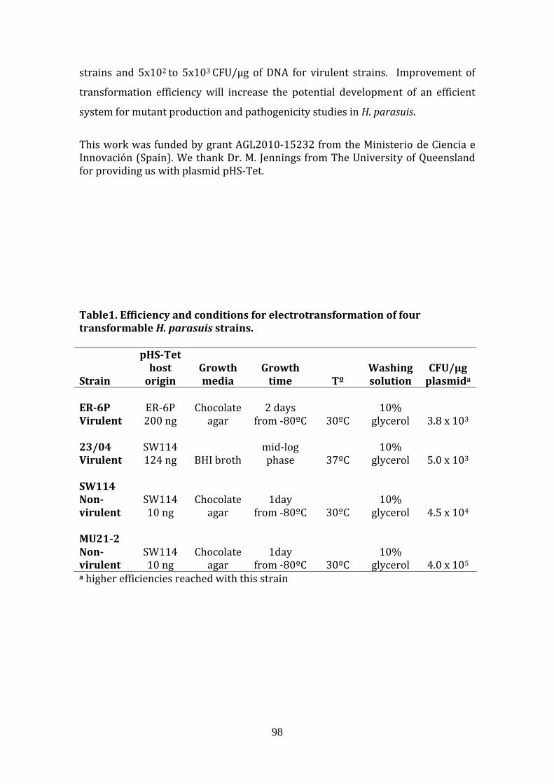
98
strains and 5x10² to 5x10³ CFU/µg of DNA for virulent strains. Improvement of
transformation efficiency will increase the potential development of an efficient
system for mutant production and pathogenicity studies in H. parasuis.
This work was funded by grant AGL2010‐15232 from the Ministerio de Ciencia e Innovación (Spain). We thank Dr. M. Jennings from The University of Queensland for providing us with plasmid pHS‐Tet. Table1. Efficiency and conditions for electrotransformation of four transformable H. parasuis strains.
Strain
pHSTet host origin
Growth media
Growth time Tº
Washing solution
CFU/µg plasmida
ER6P Virulent
ER‐6P 200 ng
Chocolate agar
2 days from ‐80ºC 30ºC
10% glycerol 3.8 x 10³
23/04 Virulent
SW114 124 ng BHI broth
mid‐log phase 37ºC
10% glycerol 5.0 x 10³
SW114 Nonvirulent
SW114 10 ng
Chocolate agar
1day from ‐80ºC 30ºC
10% glycerol 4.5 x 10⁴
MU212 Nonvirulent
SW114 10 ng
Chocolate agar
1day from ‐80ºC 30ºC
10% glycerol 4.0 x 10⁵
a higher efficiencies reached with this strain
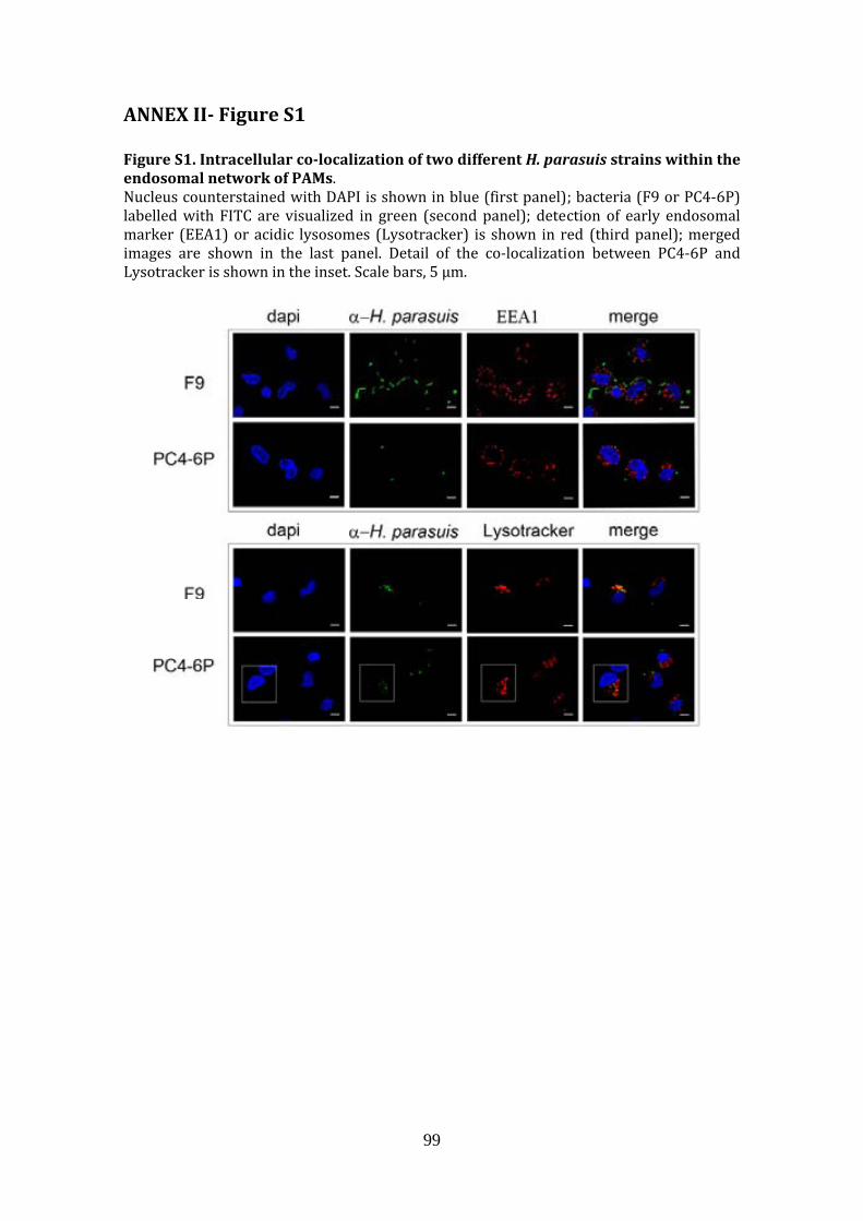
99
ANNEX II Figure S1 Figure S1. Intracellular colocalization of two different H. parasuis strains within the endosomal network of PAMs. Nucleus counterstained with DAPI is shown in blue (first panel); bacteria (F9 or PC4‐6P) labelled with FITC are visualized in green (second panel); detection of early endosomal marker (EEA1) or acidic lysosomes (Lysotracker) is shown in red (third panel); merged images are shown in the last panel. Detail of the co‐localization between PC4‐6P and Lysotracker is shown in the inset. Scale bars, 5 µm.

100
ANNEX III Figures S2A and S2B Figure S2A. Intracellular colocalization of EPI300pACYC184 pEGFP (Control), EPI300pMCHvtaA8 pEGFP (vtaA8) and EPI300pMCHvtaA9pEGFP (vtaA9) bacteria with early endosomal antigen 1 (EEA1) at different times postinfection (30 min and 1h). Nucleus counterstained with DAPI is shown in blue (first panel); bacteria (Control, vtaA8 and vtaA9) expressing GFP are visualized in green (second panel); detection of EEA1 is shown in red (third panel); merged images are shown in the last panel. Scale bars, 5 µm.

101
Figure S2B. Intracellular localization of EPI300pACYC184 pEGFP (Control), EPI300pMCHvtaA8 pEGFP (vtaA8) and EPI300pMCHvtaA9pEGFP (vtaA9) bacteria in acidic compartments at different times postinfection (30 min and 1h). Nucleus counterstained with DAPI is shown in blue (first panel); bacteria (Control, vtaA8 and vtaA9) expressing GFP are visualized in green (second panel); detection of acidic lysosomes (as detected with Lysotracker) is shown in red (third panel); merged images are shown in the last panel. Scale bars, 5 µm.